Note: Descriptions are shown in the official language in which they were submitted.
CA 02308492 2008-01-30
COMPUTER-AIDED DIAGNOSIS SYSTEM AND METHOD
Field and Background of the Invention
This invention relates to displaying radiological images and other information
in a manner
which is believed to assist users such as physicians in reading such images
and other
information. More specifically, the invention relates to a computer-aided
diagnosis ("CAD")
system and method for detection and identification of abnormalities from
radiological images
which can be viewed in a conventional format but in conjunction with viewing
an annotated
road map of the location and/or the -identification of suspected abnormalities
found in
accordance -Aith the invention through computer processing of the
conventionally obtained
radiological image. The annotated map highlights and/or identifies suspected
abnormalities
to help the reader better assess the presence and/or meaning and simnificance
of
abnorrnalities in the conventionally obtained radiological imaee.
The detection of abnormal anatomic regions in radiological images using a
computer system
comprising specialized software and possibly specialized hardware has been
reported. For
examplc, i:. tl:e r.r=a of _^lamsr_egraphy, representative reoorts are: Gager
et al in the May
1993 issue of RadioGraphics, pages 647-656; Giger et al in Proceedings of
SPIE, Volume
1445 (1991), pages 101-103; Doi et al in U.S. Patent No. 4,907,156: and Giger
et al in U.S.
Patent No. 5.133.020. See, also, the disclosure of and in prior art cited in
said parent
applications. In panicular, in the area of detecting spiculated or stellate
lesions in
CA 02308492 2008-01-30
applications. In particular, in the area of detecting spiculated or stelliate
lesions in
mammograms using covergent line detectors as the principal abnormal feature
detection
algorithm, representative reports are: N. Karssemeijer in the book entitled
"Digital
Mammography", edited by A. G. Gale et al, published by Elsevier in 1994, pages
211-219;
and Kegelmeyer et al in Volume 191 (1994) of Radiology, pages 331-337. In the
area of
detecting clusters of microcalcifications in mammograms using thresholding and
a
clustering kernel as the principal abnormal feature detection algorithm,
representative
report are: Nishikawa et al in Volume 20 (1993) of Medical Physics, pages 1661-
1666; and
Feig et al in Volume 33 (1995) of Radiological Clinics of North America, pages
1205-30.
See also US Patents Nos. 5,815,591 entitled "Method and apparatus for fast
detection of
spiculated lesions in digital mammograms," and 6,014,452 entitled "Method and
system for
using local attenuation in the detection of abnormalities in digitized medical
images."
These systems are generally referred to as Computer-Aided Diagnosis ("CAD")
systems,
and are believed to be particularly useful to radiologists in the diagnostic
process and
particularly in screening radiological procedures.
In a screening radiological procedure, such as screening mammography, the
patients
typically are asymptomatic and true abnormalities (e.g. cancers) are said to
occur at a typical
rate of about one case per one hundred patient examinations. Reading of the
mammograms.
when most of them are negative, can be a tedious task that can make it
difficult to maintain a
constantly high attention level. Some detectable abnormalities can be missed
or
misdiagnosed, which can result in delayed or more costly treatment, and can
even result in a
reduction of patient's longevity or chance of survival. According to an
article in the May 26,
1993 issue of JAMA, pages 2616-2617, the misdiagnosis rate in mammograms can
be in the
range of 15 to 63%. The CAD system, serving as an electronic reminder or
second reader, as
a spell-checker can be in a word processor, can assist radiologists in
attaining higher
detection rate (:u&.er sensitivi:}=) for abnc.^.7ali*.ies or reducing the
misdiagnosis rate
(lowering the false-negative rate).
Applicant understands that a current procedure using a CAD mammographic system
proceeds
CA 02308492 2000-05-04
WO 99/28857 PCTIUS98/25357
as follows. The physician views a radiological image, reaches a preliminary
diagnostic
decision, and then views a separate second image displayed on a CAD system.
This second
image is marked or annotated with a localized identification of the
abnormalities that the
CAD system has detected through computer analysis of a digitized version of
the
conventionally obtained radiological image. After a reexamination the area of
the
radiological image that corresponds to the position of the detected
abnormalities displayed on
the CAD system, the physician makes the final diagnostic decision. This final
diagnostic
decision may or may not be the same as the preliminary decision, depending on
whether the
physician found the additional diagnostic infonnation provided by the CAD
system to be
significant and, if so, what significance the physician ascribed to it.
Following the final
diagnostic decision, and perhaps depending on the degree of suspicion for
malignancy, the
physician can recommend a course of further action, which can include no
further action or
further follow-up examinations or biopsy.
In the process of detecting abnormal anatomic features in radiological images
using a CAD
system as described in the above cited references, the radiological film image
of a patient is
processed through a film digitizer to generate a digitized image which is
input as such into
the system. The digitized image is then analyzed by a digital image processing
computer
with specialized software and perhaps also specialized hardware for abnormal
anatomic
feature detection. If abnormalities are detected, an annotated radiological
image is displayed
on a special TV monitor, with markers placed around or adjacent the detected
abnormalities.
This TV monitor typically has a large dimension (typically a screen diagonal
of 12 inches or
larger) and a high spatial resolution (typically more than 1000 x 1000
pixels). Because of the
large dimension and high spatial resolution, this TV monitor typically is
positioned at some
distances away from the film. Typically the center of the monitor is more than
12 inches
from the center of the film on the conventional film illumination box. In
addition, this
special TV monitor typically has a low brightness and a high cost.
While the above described CAD system can point out the CAD-detected
abnormalities to the
physician, it is believed that the display method that it utilizes. using a
high-resolution TV
monitor. has certain shortcomings which make the process of using it
inconvenient and
inefficient. The high-resolution TV monitor is expensive, i:s spatial
resolution although high
3
CA 02308492 2008-01-30
for monitors is still much less than that of the original x-ray film, and its
brightness and
dynamic range are also very much inferior to those of an x-ray film viewed on
a light box.
Therefore, it is believed that a physician would not rely solely on the image
displayed on the
TV monitor to make diagnosis. but typically would repeatedly go back to the
conventional
film illurnination box to view the original film image. This can lead to the
loss of valuable
time and can be uncomfottable at least because of the different brightness
levels and spatial
resolution levels of the two images. In addition, it is believed that
diagnostic errors can arise
from the need for the physician to shuttle back and forth between two
different displayed
images. Even when a potentially true abnonnality (cancer) is detected and
pointed out by the
CAD svstem to the phvsician, the fatigue and eve discomfort and other effects
due to viewing
two images of such different characteristics may still cause the physician to
miss the
significance of the corresponding area on the original x-ray film and to fail
to notice or
appreciate the abnormal features of the detected abnormality and decide to
ignore the
detected abnormality.
Summary of the Invention
In one aspect, the present invention provides a system which includes a
mammography unit
acquiring a film image having a relatively low contrast and relatively wide
exposure range
and converting the film image to a low-contrast, wide latitude digital image.
A
programmed computer is provided for processing the low-contrast, wide-latitude
digital
image to automatically identify suspected abnormalities therein and to
automatically find at
least one relatively narrow exposure range that includes an exposure range
related to at
least one of the abnormalities and converting the low-contrast wide latitude
digital image to
a display image that is high-contrast in at least one narrow latitude related
to the at least
one narrow exposure range. The system also includes a display device for
displaying the
display image.
4
CA 02308492 2008-01-30
It would be desirable to provide an improved combined display of an x-ray
radiological
image and CAD detected abnormalities from the x-ray image. A more specific
object is to
provide the CAD user with further processed, annotated and enhanced image
tiles of the
regions around the CAD detected abnormalities for the purpose of emphasizing
the
abnormal image features of these detected abnormalities so that the user
(physician) can
better assess the type and degree of abnormality of these detected
abnormalities in the
radiological image.
Another objective is to present the further processed image of the area around
the CAD
detected abnormalities on a small TV monitor located in such close proximity
to the x-ray
film during viewing of the x-ray film that eye and other discomfort due to
viewing two
different images alternately would be reduced. Still another object of the
invention is to
print the annotated road map and/or the further processed image tiles of the
area around the
CAD detected abnormalities on the same sheet of photographic film that
contains a printout
of the radiological image.
The further processed image of the area around the CAD detected abnormalities
from a
radiological film may be presented on a small TV monitor, located in close
proximity to the
radiological film being viewed at the light box. The display of this further
processed image
shares (e.g., is toggled on) the small TV monitor with the display of a
miniaturized
annotated road map. On demand by the CAD user, e.g. the physician using a
toggle switch,
the miniaturized annotated road map image and the further processed image
tiles of the
areas around the CAD detected abnormalities are displayed alternatively on the
small TV
monitor.
In another embodiment the miniaturized annotated road map image is presented
on a small
TV monitor and the further processed image tiles of the areas around the CAD
detected
abnormalities from a radiological film are presented on a second and separate
small TV
monitor, both located in close proximity to the radiological film being viewed
at the light
box.
CA 02308492 2008-01-30
In the case of a radiological image that has been acquired through digital
means, and thus is
in digital form initially, the final image is frequently printed on a sheet of
photographic
film for later viewing on a light box. In an exemplary and non-limiting third
embodiment
of the invention, both a miniaturized annotation road map and the further
processed image
of the area around the CAD detected abnormalities from a radiological film are
printed on
the same sheet of photographic film that contains a print of the x-ray
radiological image.
In an exemplary embodiment a particularly simple digital radiography system
comprises a
CAD system and an analog acquisition system.
In an exemplary embodiment a CAD system is used to decide how the relatively
wide
latitude digitally acquired image should be displayed on a display medium such
as film
having a narrower latitude.
Brief Description of the Drawings
Figure 1 is a block diagram illustrating a CAD system and its output display
according to a
5a
CA 02308492 2000-05-04
WO 99/28857 PCT/US98/25357
first embodiment of the invention.
Figure 2 is a block diagram illustrating a CAD system and its output display
according to a
second embodiment of the invention.
Figure 3 is a block diagram illustrating a CAD system and a first method of
output displaNaccording to the third embodiment of the invention.
Figure 4 is a block diagram illustrating a CAD system and a second method of
output displav
according to the third embodiment of the invention
Figure 5 is a block diagram illustrating a CAD system and an output display
according to a
fourth embodiment of the invention.
Figure 6 illustrates displaying an output according to a fifth embodiment of
the invention.
Detailed Descrintion
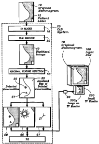
Referring to Figure 1, a preferred but non-limiting example, according to a
first embodiment
of the invention, generates an annotated road map of CAD-detected abnormality
and a further
processed image of the area around the CAD detected abnormalities from a
radiological film.
Both the annotated road map and the further processed image are displayed on a
small TV
monitor located in close proximity to the radiological film being viewed at a
light box. In
this example, the radiological film is in the form of a mammographic x-ray
film, which is
acquired with a conventional mammographic film-screen imaging system. The
original
analog two-dimensional mammographic x-ray film 10, w-ith a patient information
label 12
printed on the edge of the film, is sent through a film digitizer 30 of a CAD
(computer-aided
diagnosis) system 20 (such as that disclosed in said U.S. patent applications
which are
incorporated by reference hereir.) to obtain a digitized two-dimensional
mammographic
image 40. Preferably, the film digitizer 30 should be a laser film digitizer
or a high
performance CCD based film digitizer and should have a dynamic range and a
spatial
resolution comparable to those of the original mammographic film which
typically has a
6
CA 02308492 2000-05-04
WO 99/28857 PCT/US98/25357
dvnamic range of 10,000:1 and spatial resolution of approximately 50 microns
per pixel (or
about 4,000 x 5,000 pixels for a 8-inch x 10-inch film). The identity of the
original
manuaographic image 10 is entered into the CAD system at this point to
identify the digitized
mammographic image 40 and thus the original film 10. An useful option at this
point is to
automatically input the identity of this original mammographic image 10 into
the CAD
machine. This can be accomplished, for example, by labeling the mammographic
film 10
with a code such as a bar code next to the a patient information label 12
printed on the edge
of the film, or by incorporating the bar code into the patient information
label 12. and then
reading the label into the CAD system 20 with an optional ID bar code reader
15 as the
mammographic film 10 is being fed into the film digitizer 30.
The digitized mammographic image 40 is then sent through an abnormal feature
detection
stage 50 of the CAD system, or CAD machine, 20. The findings or results,
positive or
negative in nature, from the abnormal feature detection stage 50 are in the
form of a
two-dimensional annotation map 55, or x-y coordinate information, of the
locations and types
of the CAD-detected abnoanalities 56 and 57 (in this illustrative example)
present in the
original film image 10. For the purpose of illustration, let the abnormality
56 be a spiculated
lesion and let its location on the annotated map be marked with a star-shaped
marker. Let the
abnormality 57 be a cluster of microcalcifications and let its location on the
annotated map be
marked with a triangular shaped marker. Thus, the markers identify not only
the detected
location but also the detected nature of the suspected abnormality identified
at this stage. The
annotation map 55 can be scaled down to the same size of a sub-sampled image,
say 512 x
512 pixel in size and 8-bit in gray scale, of the digitized image 40, and the
two superimposed
images in registration with each other forms a miniaturized annotated road map
image 58.
Enhanced image tiles 66 and 67, centered respectively around the CAD-detected
abnormalities 56 and 57, say 512 x 512 pixel in size and 8-bit in gray scale,
are generated by
further image processing the regions in the digitized image 40 which
correspond to the CAD
detected abnormalities 56 and 57. The CAD-generated annotation map 55, the
miniaturized
annotated road map image 58, the enhanced image tiles 66 and 67, together with
the digitizcu
image 40 and its corresponding identification, may be stored for later use in
an optional
memory storage unit 70.
7
CA 02308492 2000-05-04
WO 99/28857 PCT/US98/25357
The annotation road map 58 and the enhanced image tiles 66 and 67 are
transferred to an
output display section of the system for display. The output display section
of the CAD
svstem can be a part of the total CAD system, and in which case the data
transfer is
conducted through a
dedicated shielded cable. Or, the output display section can be a separate
system. in which
case an additional data storage memory can be added to the unit to store the
transferred
interim data and the data transfer can be through a dedicated shielded cable
or an existing
network where the equipment is installed.
It is imporcant to point out and emphasize the abnormal features of the CAD
detected
abnormalities to the physician, because it is believed that the physician,
even after seeing the
location of the CAD detected abnormalities on the miniature road map 58, can
fail to notice
or appreciate these abnormal features on the original the x-ray film. By
pointing these
abnormal features out, with further emphasis, to the phvsician, it is believed
that the
physician would be in better position to assess the level of abnormality of
these CAD
detected abnormalities. The principal abnormal feature detection algorithms
used in the
abnormal feature detection stage 50 to detect the abnormalities can be used to
further
emphasize the abnormal features of the CAD detected abnormalities. For
example, in the
case of the abnormality 56, the principal abnormal feature used to detect the
spiculated lesion
can be a set of convergent lines. Since the presence of the convergent lines
around a lesion
raises the probability of malignancy, it is believed that there would be less
a chance that the
physician could ignore the lesion if the convergent lines were made more
noticeable.
Therefore, this set of convergent lines around the spiculated lesion could be
contrast and edge
enhanced to form the image tile 66. For example, Figure 3(B) of Karssemeijer
article cited
shows a set of detected pixels pointing to the center of a suspected
spiculated lesion.
Superimposing these detected pixels on the image can forrn the image tile 66.
In the case of
the abnormality 57, the principal abnormal feature used to detect the cluster
of
microcalcifications would be the small clustering of three or more high
contrast spots. This
small ciustering of 1-,igh cc;itrst spots could be e^.hanced in brig-htness to
form the imaize tile
67. The formation of this small clustering of bright spots can be of great
interest to the
physician. This is because the probability of malignancy is higher for a
linear or branching
formation. Therefore, this small clustering of high contrast spots should also
be magnified in
8
CA 02308492 2000-05-04
WO 99/28857 PCT/US98/25357
size, for example by a factor of 2 or more, to form the image tile 67 in order
to help the
physician see the formation of these bright spots clearly in the image tile
67. In this manner,
it is believed that the CAD user, the physician, after seeing the enhanced
abnormal image
features of these detected abnormalities and reexamining the original x-ray
image, can better
assess the level of abnormality of these detected abnormalities in the x-ray
image.
Also shown in Figure 1 is an illustration of a CAD output display consisting
of a
conventional film illuminator, commonly called a light box, 100 and a small TV
monitor 200
according to the first embodiment of the invention. In this exemplary
embodiment, the
miniaturized annotated road map 58 and the enhanced image tiles 66 and 67 are
altematively
or sequentially displayed as images 3001. by operating a toggle switch 90 to
display one
image at a time (where x= a, b and c) on the small TV monitor 200 located in
close
proximity to the original film 10. Respectively, the image 300a represents the
miniaturized
annotated road map 58, the image 300b represents the enhanced image tile 66
around the
CAD detected spiculated lesion, and the image 300c represents the enhanced
image tile 67
around the CAD detected cluster of microcalcifications. If more abnormalities
are detected,
there would be images 300d, 300e, etc. to be toggled through the small TV
monitor.
The dimension of the display screen of the small TV monitor 200 in this
example of the
invention are of the order of 1/4 to 1/2 of the dimension of original film 10.
In addition, the
small TV monitor 200 should be located as close as practical to the light box
100 displaying
the original film 10. Preferably the center of the small TV monitor 200 should
be less than
12 inches from the center of the original film 10 on the conventional film
illumination light
box 100. The preferred position, as shown in Figure 1, for mounting the small
TV monitor
200 is just beneath the light box 100 which displays the original image 10. It
is also
convenient to display a pair of images on each TV monitor, since frequently a
pair of the
original mammographic films 10, such as the mammograms of the left and right
breasts, are
displayed and viewed next to each other. In this manner, the physician still
has to minimally
move his or her eyes back and forth between the or.ginal radiologic film image
10 orl thE filtn
illuminator 100 and the images 300x displayed on the small TV monitor 200. The
spatial
resolution of the small TV monitor 200 can be in the range of 500 TV lines, or
comparable to
that of NTSC or PAL. The brightness level of the small TV monitor 200 should
be similar to
9
CA 02308492 2008-01-30
that of the averaee brightness transmitted througlt the original film 10. so
that the observer
would not be bothered by a change in brightness. In using the CAD system as a
second
reader in a screening situation, it is sometimes preferred that the display on
the small TV
monitor 200 can be easily toggled on-off with a switch 90 by the observer.
Figure 2 is similar to Figure 1 in many respects, and similarly labeled
components serve a
similar function and therefore will not be described again in detail. Figure 2
shows a CAD
output display comprising a conventional film illuminator 100 and, in this
case. two small TV
monitors 200 and 250, according to the second embodiment of the invention. In
this
exemplary embodiment, the annotated information 58 is presented as a
miniaturized
annotated road map image 300a on the first small TV monitor 200, located in
close proximit<-
to the original film 10. The enhanced image tiles 66 and 67 are alternativelv
or sequentially
presented, by operating a toggie switch 90, as images 300b and 300c on the
second small TV
monitor 250. located next to the first small TV monitor 200 and in close
proximity to the
original film 10. It is sometimes preferred that two or more small TV monitors
are used, in
place of monitor 250, to display the further processed image tiles 66 and 67
such that each
detected abnormality is displayed on a separate small TV monitor at the same
time. The small
TV monitors 200 and 250 can be placed at other positions relative to the light
box 100, e.g. to
the side or above light box 100.
Referring to Figure 3, a preferred but non-limiting example according to the
third
embodiment of the invention receives radiological images which already are in
the digital
format, detects abnormalities on these radiological images with a CAD system,
and prints out
these radiological
images together with CAD results on photographic film. Again, components
labeled the
same as in Figures 1 and 2 serve similar functions and therefore will not be
described again in
detail. Digital imaging systems, such as magnetic resonance imaging ("MRI")
systems,
computed tomography ("CT") systems, ultrasound imaging systems, scintillation
cameras.
computed radiography ("CR" ) systenis (such as Fuji's CR sys-em based on
stimulated
emission phosphor detector), and recently reported digital radiography and
digital
mammoeraphy systems (for example, see Feig article cited earlier; using CCDs
or
amorphous silicon array detectors), provide radiological images in the digital
format. In this
CA 02308492 2000-05-04
WO 99/28857 PCTIUS98/25357
example, the radiological image is in the form of a digital mammogram. which
is acquired
with a digital mammography system. This digital mammogram 14, already having
properly
encoded identification and patient information 12, is reformatted into the
digitized
mamrnographic image 40 and is sent through the abnormal feature detection
stage 50 of the
CAD machine 20. If the information is already properly formatted for the CAD
machine 20.
it is sent directly to and through the abnormal feature detection stage 50 of
the CAD machine
20 without reformatting. The initial film digitization step used in the first
and second
embodiments for analog mammograms is not needed in this case. As in the first
and second
embodiments, the findings or results, positive or negative in nature, from the
abnormal
feature detection stage 50 are in the form of a two-dimensional annotation map
55, or x-y
coordinate information, of the locations and types of the CAD-detected
abnormalities 56 and
57 from the original film image 10. For the purpose of illustration, let the
abnormality 56 be
a spiculated lesion and let its location on the annotated map be marked by a
star shaped
marker. Let the abnormality 57 be a cluster of microcalcifications and let its
location on the
annotated map be marked by a triangular shaped marker. The annotation map 55
can be
scaled down to the same size of a sub-sampled image, say 512 x 512 pixel in
size and 8-bit in
gray scale, of the digitized image 40, and the two superimposed images in
registration with
each other form a miniaturized annotated road map image 58. Enhanced image
tiles 66 and
67, centered respectively around the CAD-detected abnormalities 56 and 57, say
512 x 512
pixel in size and 8-bit in gray scale, are generated by further image
processing the regions in
the digitized image 40 which correspond to the CAD detected abnormalities 56
and 57. The
CAD-generated annotation map 55, the miniaturized annotated road map image 58,
the
enhanced image tiles 66 and 67, and together with the digitized image 40 and
its
corresponding identification, can be stored for later use in an optional
memory storage unit
70.
The annotation road map 58 and the enhanced image tiles 66 and 67 are
transferred to the
output display section of the system for display. There are several methods to
display the
CAD results and the digitally acquired mammogram. Since the digital system
produces iiu
film to start with at the acquisition, the first method is a totally filmless
display by using a
high resolution TV monitor 400. The resolution should be at least 1000 x 1000
pixels. In this
method the annotation road map 58, the enhanced image tiles 66 and 67, and the
digital
11
CA 02308492 2000-05-04
WO 99/28857 PCTIUS98/25357
mammogram 40 are all displayed on the same TV monitor as a combined digital
image 450
as shown in Figure 3. The annotation road map 58 and the enhanced image tiles
66 and 67
are shown placed at the edge or margin of the combined digital image 450.
Patient
information 12 can also be displayed at the same edge or margin of the
combined digital
image 450. The annotation road map 58 may alteraatively be displayed by
overlaying it on
top of the digital mammogram 40. This overlay may be toggled (switch not
shown) on and
off so that the digital mammogram 40 can be examined without obstruction. The
enhanced
image tiles 66 and 67 can also be toggled (switch not shown) on and ofl:
The second method of display, shown in Figure 4, where the same reference
numerals have
the same significance as in the earlier Figures, makes a photographic film
printout of the
digital mammogram 40, the miniaturized annotation road map 58 and the enhanced
image
tiles 66 and 67 all on a same sheet of film 500. The printout film 500 is
viewed on a light
box 550. Since, at the present time, physicians usually are more accustomed to
a
photographic film, which conveys information with much higher resolution and
gray scale
than a high resolution TV monitor, the second method of display can be
preferred over the
first method of display. The annotation road map 58 and the enhanced image
tiles 66 and 67
are shown in Figure 4 placed at the same edge or margin of the printout film
as the patient
information label 12. The photographic film printout, typically having a
resolution of 4000 x
5000 pixels, can be made with a high resolution laser film printer 580. Such
high resolution,
4000 x 5000 pixels, laser film printers are commercially available with a
resolution of 40
microns per pixel for 8 inch x 10 inch size films and 100 microns per pixel
for 14 inch x 17
inch size films. It is sometimes preferred that only the miniaturized
annotation road map 58
be printed at the edge of the printout film 500.
Digital radiological images, such as in the case of images from magnetic
resonance imaging
computed tomography ("CT"), digital fluorography ("DF"), and computed
radiography ("CR"), are sometimes printed out on sheets of 4000 x 5000 pixels
photographic
film for later viewing on a light box. Since MR1 images are typically
formatted ir,to 2-55 6 x
256 pixels. CT images into 512 x 512 pixels, DF images into 1024 x 1024
pixels, and CR
images into 2048 x 2048 pixels. many images from MRI or CT or CR modalities
can be
printed as small tiles in an array on one sheet of 4000 x 5000 pixels
photographic film.
12
CA 02308492 2000-05-04
WO 99/28857 PCT/US98/25357
Therefore, the CAD findings from these images can be printed as one or several
of the tiles.
A further refinement on the use of CAD is to reduce the number of images to be
presented to
the physician, for example by not presenting radiological images on which no
suspected
abnormalities are found by the CAD system, or by not presenting such
radiological images
for a second opinion or review by another professional.
Although the embodiments discussed above have been described in terms of
preferred
structures involving a single sheet of film, it should be apparent to those
skilled in the art that
this invention applies to viewing of multiple films on a multiple-film viewing
station or an
alternator (a multiviewer having pre-loaded films and a transport belt), to
allow several x-ra,,films 10 or printout films 500 to be viewed at the same
time or different times, with or
without their respective annotation maps 58 and the enhanced image tiles 66
and 67.
Figure 5 illustrates a particularly efficient digital radiography system
according to the fourth
embodiment of the invention. Again, the same reference numerals have the same
meanings
as in the earlier Figures. The Figure 5 system comprises an analog film-screen
acquisition
system and a CAD system similar to the third embodiment. In this example, the
radiological
image is in form of a low contrast and wide latitude mammographic film 600.
This
embodiment and the third embodiment differ in the source of the digitized
image 40. The
acquisition system, in this case, is an inexpensive conventional mammographic
intensifying
screen cassette 620. The cost ratio between this screen cassette 620 and a
typical current
digital mammographic system is several order of magnitude, e.g., approximately
1000 times.
The standard screen-film acquisition and exposure technique and the
conventional
mammographic x-ray system (not shown) will be unchanged for use in this
embodiment of
the invention. By reducing the contrast gradient G of the mammographic film
600 say from
about 3.0 to about 2.0 to 1.5, we obtain a film with a factor of 5 to 25 more
in latitude. That
is, the exposure range expands from about 25 to about 125 to 630. The contrast
gradient G is
generally defined as the slope of the H & D [or film characteristics] curve
where the optical
density [ar the iog (basc 10) of the reciprocal of the f lm tr3.+ismission,
the y-axis] is plotted
against the log (base 10) of x-ray exposure [or light exposure. the x-axis].
Because of the low
contrast, the physician cannot be expected to make diagnosis on this low
contrast and wide
latitude mammogram 600. However, this mammographic film 600 can be digitized
through
13
CA 02308492 2000-05-04
WO 99/28857 PCT/US98/25357
the film digitizer 30 and reformatted into a digitized image 40, and can be
put through the
CAD abnormal feature detection stage 50. The reformaned version of the wide
latitude
image 600 is fed through a high resolution film printer 580 and printed out as
a conventional
high contrast mammogram 650. Its respective annotated road maps 58 and the
enhanced
image tiles 66 and 67 are also printed at the edge or margin of the mammogram
650. The
physician, as in the third embodiment, finally reads the mammogram on the
light box 550.
Using the miniaturized road map 58 as a guide, the physician reexamines the
mammogram
650 to see if any of the CAD detected abnormalities suggest further action.
Ihe enhanced
image tiles 66 and 67 provide the physician with further information by
emphasizing the
abnormal features of the CAD detected abnorrnalities. It is sometimes
preferred that only
the miniaturized annotation road map 58 be printed at the margin of the
mammogram 650.
Figure 6 illustrates how a CAD system in accordance with a fifth embodiment of
the
invention can be used to decide how a wide latitude digitally acquired image
should be
displayed on a display medium having a narrower latitude. One of the major
problems in
digital imaging is how to display all the information contained in the
digitally acquired
images. Typically, according to Feig et al in Volume 33 (1995) of Radiologic
Clinics of
North America, pages 1205-30, and other sources, the exposure range of the
latitude (the
ratio of the exposure at the highest signal region to that at the lowest
signal region) of the
digitally acquired image is of the order of 100 to over 1000, which is much
broader than that
of the display media. As a comparison, if a radiogtam is to be displayed on a
photographic
film, the exposure range of the latitude (the ration of exposure at the
highest signal region to
that at the lowest signal region where the display gradient is significant) is
of the order of 25
and the exposure range of the latitude of a TV monitor is less than 10. As
illustrated in
Figure 6, a CAD system 20 is used to guide into two different display media
the display of a
wide latitude digitally acquired image 800 formatted for example such that the
log of its
output signal (with an exposure range of over 1000) is encoded into 12 bits
(4096 levels of
gray). In this example, the CAD system decides how the digitally acquired wide
latitude
radiogram 800 should be printed on a photographic film 830 (having a display
latitude o: fcr
example, 1-5) and dispiayed on a high resolution TV or computer monitor 860
(having a
display latitude of, for example. 10). As illustrated in Figure 6, this is
done by centering the
display latitude of the display medium (photographic film 830 or monitor 860)
around the
14
CA 02308492 2000-05-04
WO 99/28857 PCT/US98/25357
region of the CAD detected abnormality identified at 890 on the wide latitude
digitally
acquired image 800. That is, only the range of say the relative exposure 10 to
100, around
the CAD detected abnormality 890, say around relative exposure 30, is provided
at the
optimum contrast gradient (say G=3.0) while other image content, above or
below the
exposure range of the CAD detected abnormality 890, are compressed in display
latitude and
are displayed with reduced contrast gradient on film 830 and/or monitor 860.
Although the invention has been described in terms of preferred structures,
methods and
processes, it should be apparent to those skilled in the art that various
alterations and
modifications can be made without departing from the invention and that such
modifications
and alterations are intended to be considered to be within the spirit and
scope of the
invention as defined by the appended claims.
1~