Note: Descriptions are shown in the official language in which they were submitted.
CA 02309151 2006-05-05
51744-1
VIDEO RECTOSCOPE
FIELD OF THE INVENTION
The present invention relates generally to medical probes, and specifically to
endoscopes.
BACKGROUND OF TIHE INVENTION
Various types of endoscopes are known in the art for visual inspection and
diagnosis of
the rectum and sigmoid of the large intestine. Rigid rectoscopes and
sigmoidoscopes are most
commonly used for this purpose, since they are durable, easy to use and
relatively inexpensive.
To perform an examination using the rigid rectoscope (or simoidoscope), the
physician
first inserts a speculum through the anus. An obturator, having a blunt,
rounded distal end, is
fitted inside the speculum and protrudes distally therefrom to ease the
insertion. Once the
speculum is in place, the physician withdraws the obturator and closes the
proximal end of the
speculum with a plug. A special telescope is fitted into the plug, which
enables the physician to
view the inside of the rectum and sigmoid at the distal end of the speculum;
either by direct
vision or using a video camera coupled proximally to the telescope. In order
to illuminate the
area under view, a light source in an external console is coupled proximaIly
to the speculum by
fiberoptics. Typically, the plug also includes one or more working channels,
which are used for
irrigation, insufflation or insertion of surgical tools through the speculum.
Various rectoscopes and sigmoidoscopes of this type are available
commercially. For
example, Karl Storz GmbFL of Tuttlingen, Gerrnany, offers the model 24911S
rectoscope,
which may be used with a rod lens telescope (for example, model 24946B) to
view the rectum
either under direct view or using a Karl Storz Endovision video camera. The
rectoscope and
accessories are made from stainless steel to allow disinfection and
autoclaving between uses.
Rectoscopes and sigmoidoscopes having a disposable speculum and obturator are
also
available, such as the KleenSpec disposable sigmoidoscope produced by Welch
Allyn Inc., of
Skaneateles FaIls, New York. The speculum and obturator are made of plastic.
The speculum
clips or screws onto a multi-use handle, to which a light source, telescope,
video camera and
instruments may be attached.
Flexible sigmoidoscopes and colonoscopes are also known in the art. Most such
scopes
include a fiberoptic bundle for conveying images from their distal end, within
the intestine, to an
eyepiece or video camera fixed to the proximal end. Some such scopes, such as
the Pentax ES-
I
CA 02309151 2006-05-05
51744-1
3801 PVE Video Sigmoidoscope;' made by Asahi Optical Company of Tokyo, have a
CCD
detector with an objective lens at its distal end. By comparison with rigid
rectoscopes and
sigmoidoscopes, however, flexible scopes are costly, fragile and ditTicult to
clean.
SUMMARY OF TFI.E INVEN'ITON
It is an object of some aspects of the present invention to provide an
improved
endoscope, and particularly an improved rectoscope.
It is an object of some aspects of the present invention to provide a video
rectoscope
that gives improved visibility of anatomical features within the rectum.
It is another object of some aspects of the present invention to provide a
rectoscope
that is easy and convenient for medical personnel to use.
It is a further object of some aspects of the present invention to provide a
rectoscope
including an easily-replaceable disposable portion, thus minimizing or
eliminating the need for
disinfection and steriiization between uses.
It is yet another object of some aspects of the present invention to provide
methods and
apparatus that facilitate insertion of an endoscope, particularly a rectoscope
or sigmoidoscope,
into a body cavity.
It is still another object of some aspects of the present invention to provide
an improved
steering mechanism for an endoscope.
3n preferred embodiments of the present invention, a rectoscope comprises an
elongate,
generally rigid insertion member having a video camera head at its distal end.
The insertion
member and camera head are covered by a disposable sheath, which includes a
transparent
distal portion, preferably including an optical window, covering the camera
head. The camera.
head includes an image detector array, preferably a CCD array, as is known in
the art, along
with a light source for illuminating an area of the intestine viewed by the
camera head and an
objective lens, which focuses an image of the area onto the array. The sheath,
which is
preferably made of a biocompatible plastic material, is removed and discarded
after use. The
rectoscope is used to examine and treat areas of the colon of a patient,
specifically the rectal
and sigmoid portions of the colon.
By comparison with rigid rectoscopes and sigmoidoscopes known in the art, the
present
invention can provide substantially improved image quality, because the video
camera head is
placed at the distal end of the rectoscope, near the area under view. The
distal positioning of
the camera head enables the present rectoscope to achieve both higher
mamnification and a
I
CA 02309151 2000-05-03
WO 99/23812 PCT/IL98/00529
wider field of view than video rectoscopes and sigmoidoscopes known in the
art, in which the
camera head views the intestine through a telescope from the proximal end of
the scope.
In preferred embodiments of the present invention, the sheath comprises a
narrowed,
generally bluntly pointed tip, to ease insertion of the rectoscope through the
anus. The
narrowed tip thus performs the function of the obturator used in insertion of
rectoscopes and
sigmoidoscopes known in the art. Elimination of the obturator relieves a
physician using the
rectoscope of unpleasant odors and possible exposure to infection that are
commonly
associated with removal of the obturator.
In some preferred embodiments of the present invention, the optical axis of
the camera
head is angled relative to the longitudinal axis of the insertion member, and
the distal end of the
sheath is likewise angled, thus defining the narrowed tip. Preferably, the
sheath has a bulbous
protrusion at the tip, which further eases insertion of the rectoscope through
the anus.
In one such preferred embodiment, a mirror is fixed to the bulbous protrusion,
which is
shaped so that the mirror is positioned within the field of view of the camera
head and is
oriented obliquely relative to the optical axis thereof. An image of the inner
wall of the rectum
or sigmoid is reflected from the mirror and captured by the camera head. The
mirror thus
enables the physician to see an image from an alternative view angle, which
may be useful
particularly in observing and controlling the movement of tools that the
physician inserts into
the rectum using the rectoscope, as described below.
In other preferred embodiments of the present invention, the narrowed tip of
the sheath
is formed by a plurality of leaves, formed of a resilient material, which butt
together tightly in a
closed configuration to cover the transparent portion of the sheath and the
camera head.
During insertion through the anus, the closed leaves protect the transparent
portion from
fouling by fecal matter, lubricants or other substances. After the rectoscope
has been inserted,
the leaves are drawn apart, preferably by pulling the sheath back proximally,
so as to expose the
transparent portion of the sheath and allow the camera head to receive images
of the area of the
intestine under view.
In some preferred embodiments of the present invention, the sheath includes
one or
more working channels, which may be used for suction, irrigation,
insufflation, passage of
surgical tools or other functions known in the art. The rectoscope includes a
handle, to which
the sheath is attached, preferably by a quick connect/disconnect fitting.
Preferably, the handle
includes user controls for regulating such functions as suction, irrigation
and insufflation
3
CA 02309151 2000-05-03
WO 99/23812 PCT/IL98/00529
through the working channels. Further preferably, one or more of the working
channels inciude
a one-way valve, to prevent back-flow of fluid (gas or liquid) used in
irrigation or insu$lation
through the channels.
Although rectoscopes in accordance with the present invention are described
herein as
being generally rigid, in some preferred embodiments the insertion member may
include a
flexible, resilient longitudinal section. This section allows the rectoscope
to bend during
insertion into the rectum, to make the insertion easier and less painful for
the patient. Such
bending is not possible using rigid endoscopes known in the art, and it
difffers from the bending
of flexible endoscopes, which do not have the rigidity to maintain a
particular shape and require
complex steering mechanisms to control their angle and direction of bend.
In one such preferred embodiment, the rectoscope sheath comprises one or more
flexible, longitudinally- disposed inflatable bladders, preferably at least
two such bladders
disposed radially within the sheath on different sides of the flexible section
of the insertion
member. Preferably, the bladders are placed on opposing sides of the sheath.
The bladders are
connected by respective valves to a suitable pressure source. When the
pressure in one of the
bladders is increased, the bladder lengthens, whereas when the pressure is
decreased, the
bladder tends to shorten. Thus, the bend angle of the sheath and of the
insertion member
therein is controlled by varying the pressure in the one or more bladders.
In another preferred embodiment of the present invention, a generally rigid
rectal insert
having a curved channel therethrough is inserted into the rectum, up to the
sigmoid flexure.
The insert is turned within the rectum so that the exit from the channel at
the sigmoid flexure
opens into the intestine in a direction generally along the axis of the
sigmoid. The rectoscope
including the flexible section, as described hereinabove, is then inserted
through the channel and
is forced by the rigidity of the insert to bend toward the sigmoid axis. The
combination of the
insert and rectoscope, in accordance with this preferred embodiment, thus
allows endoscopic
inspection of a portion of the intestine that can generally be reached only
using a more costly
flexible endoscope. The rectal insert described herein can also be used in
conjunction with
flexible endoscopes known in the art, making it easier to pass the endoscope
through the
sigmoid and reducing pain and the possibility of accidental perforation of the
intestine.
Although preferred embodiments are described herein with reference to
inspection and
treatment of the rectum, and endoscopes in accordance with such preferred
embodiments are
referred to herein generaUy as rectoscopes, it will be appreciated that such
endoscopes may
4
*rB
CA 02309151 2000-05-03
WO 99/23812 PCT/IL98/00529
similarly be adapted and applied to serve as sigmoidoscopes. The principles of
the present
invention may similarly be applied to produce rigid endoscopes for insertion
into other body
cavities, such as the throat, the vagina, or any other cavity large enough to
receive an
endoscope having a video camera head at its distal end, as described herein.
There is therefore provided, in accordance with a preferred embodiment of the
present
invention, a video endoscope, including:
a generally rigid, elongate insertion member, having distal and proximal ends;
a video camera head fixed at the distal end of the insertion member; and
a sheath, which fits over and covers the insertion member, the sheath having a
distal
portion covering the video camera head, at least a section of which distal
portion is
substantially transparent.
Preferably, the camera head includes a detector array and an objective lens,
which forms
an image on the detector array.
In a preferred embodiment, the camera head is inclined obliquely relative to a
longitudinal axis of the insertion member.
Preferably, the camera head includes a light source, most preferably including
one or
more LEDs.
Preferably, the substantially transparent section of the distal portion of the
sheath
includes an optical window.
Preferably, the distal portion of the sheath includes a narrowed tip, which
facilitates
insertion of the endoscope through the anus. Further preferably, the narrowed
tip includes a
bulbous protrusion.
Alternatively or additionally, the narrowed tip includes two or more leaves,
which are
closed during insertion through the anus and open apart after insertion to
expose the
transparent section of the distal portion of the sheath. Preferably, to open
the leaves, the sheath
is shifted in a proximal direction drawn relative to the insertion member.
Preferably, the sheath contains a working channel passing longitudinally
therealong.
Further preferably, the sheath includes a one-way valve within the working
channel, for
preventing back-flow of fluid therethrough.
Preferably, the endoscope includes a handle, fixed to the proximal end of the
insertion
member, and the handle includes user controls for controlling the passage of
fluid through the
working channel.
5
CA 02309151 2000-05-03
WO 99/23812 PCT/IL98/00529
In a preferred embodiment, at least a longitudinal section of the insertion
member is
flexible and resilient. Preferably, the sheath includes an inflatable bladder
disposed
longitudinally within the sheath, such that inflation of the bladder causes
its length to increase
so that the insertion member bends.
In another preferred embodiment, a mirror is fixed in a position distal to the
camera
head within a field of view thereof and oriented obliquely relative to an
optical axis of the
camera head, so that the camera head captures an image reflected from the
mirror. Preferably,
the image reflected from the mirror includes an area of a physiological
structure in which a
surgical procedure is to be performed using the endoscope.
There is further provided, in accordance with a preferred embodiment of the
present
invention, a steering mechanism for an elongate medical probe, including at
least one
controllably inflatable element fixed to the probe, such that a longitudinal
dimension of the
element changes responsive to inflation thereof, thereby causing the probe to
bend.
Preferably, the at least one inflatable element includes two elements disposed
radially on
different sides of the probe, so as to bend the probe in different, respective
directions. Further
preferably, the two elements include first and second elements on opposing
sides of the probe,
such that the first element is inflated and the second element is deflated so
as to bend the probe
toward the side on which the second element is fixed.
Preferably, the at least one inflatable element includes an expandable wall
section on an
outer surface thereof, which expands and contracts responsive to changes in
the inflation of the
element.
Preferably, the mechanism includes a pressure source which is controllably
applied to
the inflatable element so as control the bending of the probe.
In a preferred embodiment, the probe includes a generally rigid endoscope,
having a
flexible section which is bent by the mechanism, wherein the endoscope
preferably includes a
disposable sheath, which includes the at least one inflatable element.
There is moreover provided, in accordance with a preferred embodiment of the
present
invention, a generally rigid rectal insert, having a distal end which is
inserted into the rectum of
a patient, the insert having a curved channel therethrough through which an
endoscope is
inserted into the sigmoid portion of the colon of a patient.
Preferably, the distal end of the insert is brought into proximity with the
patient's
sigmoid flexure, and the channel is curved so as to open laterally out of the
distal end into the
6
CA 02309151 2000-05-03
WO 99/23812 PCT/IL98/00529
flexure. Further preferably, the insert is rotatable within the rectum so as
to align the opening
of the channel at the distal end thereof with the intestine.
There is also provided, in accordance with a preferred embodiment of the
present
invention, a method of endoscopic inspection, including:
providing an elongate, generally rigid insertion member having a video camera
head at a
distal end of the member;
fitting a disposable sheath over the member, such that the camera head is
covered by a
substantially transparent portion of the sheath;
inserting the insertion member, covered by the sheath, into a body cavity of a
subject;
and
receiving video images within the body cavity from the camera head.
Preferably, fitting the sheath includes fitting a sheath having a narrowed
tip, and
inserting the member includes first inserting the narrowed tip into the
cavity.
In a preferred embodiment, the narrowed tip is opened after insertion to
expose the
substantially transparent portion of the sheath covering the camera head,
wherein opening the
narrowed tip preferably includes shifting at least a portion of the sheath in
a proximat direction
over the insertion member so as to separate two or more leaves that meet to
form the narrowed
tip.
Preferably, receiving the video images includes illuminating the inside of the
body cavity
using a light source in the video camera head.
Further preferably, fitting the disposable sheath includes fitting a sheath
having at least
one working channel passing therethrough. In a preferred embodiment, the body
cavity is
insufflated by passing a pressurized fluid through the working channel.
In a preferred embodiment, providing the insertion member includes providing a
member at least a longitudinal portion of which is flexible and resilient, and
inserting the
member includes flexing the flexible, resilient portion. Preferably, inserting
the member
includes inserting an insert into the body cavity having a curved channel
therethrough, and
passing the member through the channel. Alternatively or additionally, flexing
the flexible,
resilient portion includes inflating an inflatable bladder disposed within the
sheath so that the
length of the bladder increases, thereby bending the portion.
7
CA 02309151 2007-02-05
51744-1
Preferably, inserting the insertion member
includes inserting the member through the anus of the
subject, and receiving the images includes receiving images
within the rectum or, alternatively or additi_onally,
receiving images within the sigmoid.
There is further provided, in accordance with a
preferred embodiment of the present inventiori, a me-:hod for
steering a medical probe, including:
fixing an inflatable element to the probe; and
controllably inflating the element so as to change
the length thereof, thus causing the probe to bend.
Preferably, fixing the inflatable element includes
fixing a plurality of elements on different sides of the
probe, so that each element causes the probe to bend in a
different, respective direction.
In a preferred embodiment, the medical probe
includes a generally rigid endoscope at least a section of
which is flexible, and wherein controllably inflating the
element causes the flexible section of the endoscope to
bend.
There is additionally provided, in accordance with
a preferred embodiment of the present invention, a method
for inserting an endoscope into the colon of a patient,
including:
providing a generally rigid insert having proxima:L
and distal ends and having a curved channel passing
therethrough with a lateral opening at the distal end;
8
CA 02309151 2007-02-05
51744-1
inserting the insert into the patient's rectum so
that the distal end is in proximity to the sigmoid flexure
of the colon; and
passing the endoscope through the channel and intc
the sigmoid colon.
Preferably, the insert is rotated to align the
opening at the distal end of the insert with the colon
before passing the endoscope therethrough.
Preferably, passing the endoscope includes passing
a flexible endoscope through the channel or, alternatively,
passing a generally rigid endoscope including a flexible
portion through the channel, such that the flexible portion
is bent by the curve of the channel.
According to one aspect of the present invention,
there is provided an endoscope, comprising: a generally
rigid, elongate insertion member, having distal and proximal.
ends; and a sheath, which comprises inner and outer layers
which fit over and cover the insertion member, with the
outer layer having a distal portion comprising a narrowed
tip, which facilitates insertion of the endoscope and which
comprises two or more leaves, which are closed during
insertion and open apart after insertion to expose the
transparent section of the distal portion of the sheath,
wherein the endoscope is for insertion in an anus; a video
camera head is fixed at the distal end of the insertion
member; the inner layer of the sheath has a distal portion
covering the video camera head, at least a section of which
distal portion is substantially transparent; and in that
each leaf comprises an inner protrusion which engages the
surface of the inner layer of the sheath and forces the leaf
8a
CA 02309151 2007-02-05
51744-1
outward as the outer layer is moved relative to the inner
layer.
The present invention will be more fully
understood from the following detailed description of the
preferred embodiments thereof, taken together with the
drawings in which:
8b
CA 02309151 2000-05-03
WO 99/23812 PCT/IL98100529
BRIEF DESCRIPTION OF THE DRAWINGS
Fig. 1 is a schematic, partly sectional illustration of a rectoscope, in
accordance with a
preferred embodiment of the present invention;
Fig. 2 is a sectional, detail view of a distal portion of the rectoscope of
Fig. 1;
Figs. 3A and 3B are schematic, sectional illustrations of a rectoscope covered
by a
sheath in closed and open configurations thereof, respectively, in accordance
with another
preferred embodiment of the present invention;
Fig. 4A is a schematic, sectional view of the distal portion of a rectoscope,
in
accordance with another preferred embodiment of the present invention;
Fig. 4B is a schematic illustration of a video image captured by the
rectoscope of Fig.
4A;
Fig. 5A is a schematic, sectional view of the distal portion of a rectoscope,
in
accordance with a preferred embodiment of the present invention;
Fig. 5B is a schematic, sectional illustration of a rectal insert, useful
particularly in
conjunction with the rectoscope of Fig. 5A, in accordance with a preferred
embodiment of the
present invention; and
Fig. 6 is a schematic, sectional illustration of a rectoscope, in accordance
with still
another preferred embodiment of the present invention.
9
CA 02309151 2006-05-05
51744-1
DETAII..ED DESCRIPTION OF PREFERRED EMBODIlYIENTS
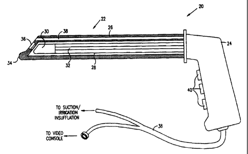
Reference is now made to Fig. 1, which is a schematic, partly sectional
illustration of a
video rectoscope 20, in accordance with a preferred embodiment of the present
invention.
Rectoscope 20 comprises an insertion section 22, which is inserted into the
rectum of a subject,
and a handle 24, which is Lxrasped by a physician making the examination.
Insertion section 22
includes an insertion member 28, fixed to handle 24, and a disposable sheath
26, covering the
insertion member. Member 28 includes a video camera head 30 at the member's
distal end,
connected by wires 32 to handle 24. Preferably, sheath 26 comprises a
biocompatible plastic
materzal, such as polycarbonate, and is attached to handle 24 using a quick-
connect fitting, as is
known in the art. After use, sheath 26 is removed and disposed of.
Fig. 2 is a schematic, sectional illustration showing details of the distal
end of insertion
section 22, inciuding camera head 30. The camera head comprises a detector
array chip 46,
preferably comprising a color CCD arrav 50, such as a Sony ICX087AKB CCD chip,
with a
suitable optical flter 48 affixed thereto. One or more lamps 42 illuminate an
area of the
intestine under view by the camera head. An image of the area is received
through an optical
window 36 in sheath 26 and focused onto array 50 by an objective lens 44.
Signals generated by chip 46 are conveyed by wires 32 to handle 24, and from
there via
a cable 99 (Fig. 1) to a video console, where the simals are processed and
used to drive a video
display. T'ne video console and dispiay (not shown in the figures) use
standard video
electronics, well known in the art. Alternatively, handle 24 may include a
power source, such
as one or more batteries, processing electronics and a video transmitter, and
convey processed
signals from chip 46 to the video console over a wireless link.
Lamps 42 preferably comprLse wliite-light LEDs, such as T 1 White L3-W31N LEDs
produced by Sloan AG of Basel, Switzerland The lamps are powered by eiectrical
current
conveyed over wires 32 and cable 99 from the console. Alternatively, the lamps
may be
powered by batteries in handle 24, as described above. In either case, the use
of lamps 42
alleviates the complication and expense of using a high-power fiberoptic light
source to
illuminate the area under view, as in endoscopes known in the art.
Window 36 is shown in Fig. 2 as being relatively small, in order to simplify
production
and reduce the cost of sheath 26. Sheath 26 also includes a transparent area
surrounding the
window to allow light from lamps 42 to reach the area under view, but this
area need not be of
optical quality. In fact, all of sheath 26 may be made of transparent plastic
if desired.
CA 02309151 2000-05-03
WO 99/23812 PCT/IL98/00529
It wiil be observed that the optical axis of lens 44 and array 50 is angled
obliquely
relative to the longitudinal axis of member 28. Window 36 and the distal end
of sheath 26 are
similarly angled. This angling allows camera head 30 to view an area of the
intestinal wall
relatively near the distal end of section 22, rather than pointing straight
down the intestine as in
rigid rectoscopes and sigmoidoscopes known in the art. As a result of this
arrangement,
section 22 can be made relatively narrow and pointed at its distal end, so
that insertion through
the subject's anus is easier and less painful, without the need for a separate
obturator.
Preferably, a bulbous projection 34 is formed at the distal tip of sheath 26,
shaped so as to
penetrate the anus with a minimum of discomfort to the patient.
Sheath 26 preferably includes at least one working channel 38, which
terminates in an
opening 52 at the distal end of the sheath. Channel 38 may be used to inserted
surgical tools,
such as biopsy forceps, into the intestine and perform surgical procedures
under view of camera
head 30. The angled orientation of the camera head, as described above, is
particularly
advantageous for viewing such procedures, since the surgical tool will engage
the intestinal wall
within the camera head's area of view.
Additionally or alternatively, channel 38 may be used for suction, imgation
and/or
insufflation of the intestine, by connecting the channel to a suitable pump or
gas source, as is
known in the art. Preferably, the connection is made via handle 24, and the
suction, irrigation
or insufflation is controlled by user controls 40 on the handle. Generally, it
is desirable to
insufflate the intestine during examination, so as to force the intestinal
wall away from window
36 and thus provide camera head 30 with a clearer view of the wall. For this
purpose, sheath
26 preferably includes a one-way valve 54, such as a leaflet valve, as is
known in the art, in
channel 38. After a pressurized gas has been passed through the channel to
insufflate the
intestine, valve 54 closes to prevent back-flow"of the gas and maintain the
pressure in the
intestine.
Figs. 3A and 3B are schematic, sectional illustrations showing a rectoscope 60
including
a disposable sheath 62, in accordance with another preferred embodiment of the
present
invention. Rectoscope 60 includes a generally rigid insertion member 28 with a
video camera
head 30 at its distal end, substantially as described above with reference to
Figs. 1 and 2,
although in rectoscope 60, the optical axis of the camera head is generally
aligned with the
longitudinal axis of member 28.
11
CA 02309151 2000-05-03
WO 99/23812 PCT/IL98/00529
Sheath 62 comprises an outer layer 64 and an inner layer 66. The inner layer
preferably
comprises a generally rigid plastic material, such as polycarbonate, and fits
snugly over member
28. Layer 66 includes a transparent distal portion, preferably including an
optical window 68,
as described above. Outer layer 64 is preferably made of a flexible, resilient
plastic, such as
polyethylene, and includes two or more leaves 70, preferably three such
leaves.
As shown in Fig. 3A, during insertion of rectoscope 60 through the anus,
leaves 70 butt
together, closing off the distal end of the rectoscope and protecting window
68. The leaves
preferably form a bluntly pointed structure, which eases the insertion.
As shown in Fig. 3B, after the insertion, leaves 70 are opened to expose
window 68 and
allow camera head 30 to receive images of the interior of the intestine.
Preferably, the leaves
are opened by drawing outer layer 64 in a proximal direction, out of the
rectum (to the right in
Figs. 3A and 3B), relative to inner layer 66. Each leaf 70 preferably includes
an inner
protrusion 72, which engages the surface of inner layer 66 and forces the leaf
outward, as
shown in the figure. After the rectoscopy procedure is completed, sheath 62
may be returned
to the configuration of Fig. 3A for removal through the anus.
Fig. 4A is a schematic, sectional illustration showing the distal portion of a
rectoscope
76, in accordance with another preferred embodiment of the present invention.
Rectoscope 76
is generally similar to rectoscope 20, shown in Figs. 1 and 2, and comprises
insertion member
28 with camera head 30 at its distal end. Rectoscope 76 is covered by a sheath
78, similar to
sheath 26 except that sheath 78 includes a bulbous protrusion 80 that extends
into the field of
view of camera head 30. A mirror 82 is fixed to protrusion 80, in such a
position and
orientation thereon that an image of a physiological structure 84, generally
the rectal wall, is
reflected from the mirror, through window 36, and is captured by camera head
30. In the
example shown in Fig. 4A, a surgical tool 88 is inserted through working
channel 38 in sheath
78 and is used to take a biopsy sample at a point 86 on structure 84.
Fig. 4B is a schematic illustration showing a video image 90 captured using
rectoscope
76 in the configuration of Fig. 4A. The upper part of the image shows tool 88
and structure 84
as seen by camera head 30 directly through window 36. The image of the tool is
foreshortened,
because of the viewing angle of the camera head. It may therefore be difficult
to judge the
distance from the tool to the structure based on such an -image. An inset 92
in the lower
portion of image 90 shows tool 88 and structure 84 as seen via mirror 82. In
this case, the
12
CA 02309151 2000-05-03
WO 99/23812 PCT/1L98/00529
angle from which the tool is viewed allows its position and distance from
structure 84 to be
seen clearly and controlled with greater accuracy.
Fig. 5A is a schematic, sectional illustration of the distal portion of a
rectoscope 106, in
accordance with another preferred embodiment of the present invention.
Rectoscope 106 is
substantially similar to rectoscope 20, shown in Figs. 1 and 2, except that
insertion member 28
of rectoscope includes at least one flexible, resilient section 108. Section
108 preferably
includes an elastic element, for example, a spring steel insert, which
normally holds the section
straight, but allows it to bend in response to lateral forces exerted thereon.
Further preferably,
sheath 26 of rectoscope 106 includes flexible, elastic material, for example,
polyethylene or
silicone, so that the sheath can bend and stretch accordingly when section 108
bends.
The ability of rectoscope 106 to bend allows it to be inserted into the
rectum, up to the
sigmoid, more easily and with less pain and discomfort to the patient than a
fully rigid
rectoscope or sigmoidoscope would cause. Rigid endoscopes known in the art
cannot have a
flexible section like rectoscope 106, since bending of the flexible section
would disrupt the
optical axis of the telescope that is used for endoscopic viewing. Although
flexible
sigmoidoscopes known in the art can be bent and inserted with less patient
discomfort than
rigid scopes, such flexible scopes require complicated and costly steering
mechanisms. By
placing camera head 30 at the distai end of rectoscope 106, the present
invention allows a
measure of flexibility without the necessity of a steering mechanism.
Fig. 5B is a schematic, sectional illustration showing a rectal insert 102,
for use in
endoscopic examination and treatment of intestine 96 of a patient, in
accordance with a
preferred embodiment of the present invention. Insert 102 is particularly
useful for guiding
rectoscope 106 into the intestine, but may also be used in conjunction with
conventional flexible
endoscopes known in the art. Insert 102 is prefeiably made of a generally
rigid plastic material,
such as polycarbonate, but may be somewhat flexible so as to ease its
insertion. The insert has
a curved channel 104 passing therethrough, with an inner diameter large enough
for an
endoscope, such as rectoscope 106, to be inserted through the channel.
Insert 102 is inserted through rectum 98 up to sigmoid flexure 100 of
intestine 96. The
insert is turned so that channel 104 opens into sigmoid portion 101 of the
intestine, as shown in
Fig. 5B. Rectoscope 106 is then passed through the insert and bends along the
curve in the
channel. The distal end of the rectoscope, including camera head 30, is pushed
out into
sigmoid 101, allowing inspection and treatment of a portion of intestine 96
that would
13
CA 02309151 2000-05-03
WO 99/23812 PCT/IL98/00529
otherwise be accessible only using a conventional flexible endoscope. Insert
102 also protects
the area of flexure 100 from pressure due to the rectoscope (or to a
conventional flexible
endoscope inserted therethrough) that could otherwise cause pain or even
perforation of the
intestine.
Fig. 6 is a schematic, sectional illustration showing a rectoscope 110,
including a
steering mechansim 111, in accordance with another preferred embodiment of the
present
invention. Rectoscope 110 comprises insertion member 28 and camera head 30,
covered by a
disposable sheath 112 with optical window 36 at a distal end thereof, as in
other preferred
embodiments described above. Member 28 of rectoscope 110 either has a flexible
section, as
described above with reference to Fig. 5A, or is made flexible over
substantially its entire
length, as shown in Fig. 6.
Steering mechanism 111 comprises two flexible, elongate bladders 118 and 120,
contained within sheath 112 and disposed radially on opposing sides of member
28. Bladders
118 and 120 have respective expandable wall sections 114 and 116, preferably
accordion-type
walls, as shown in Fig. 6. The bladders are coupled via valves 124 to a
pressure source 126,
for example, a gas bottle or pump, which may be contained either in a handle
122 of rectoscope
110 or in a separate console. By operating valves 124, bladders 118 and 120
are pressurized to
respective pressures P 1 and P2. In the example shown in Fig. 6, P 1 is
greater than P2, causing
wall section 114 of bladder 118 to expand, and wall section 116 of bladder 120
to contract,
thereby causing member 28 to bend.
Mechanism 111 thus provides a simple, low cost, disposable means for
controllably
bending and steering rectoscope 110, by inflating and deflating bladders 118
and 120 in sheath
112. Additional bladders may be positioned along the length of the rectoscope
and/or radially
around it in order to give greater control of the bend shape and direction of
the rectoscope. It
will be appreciated that such pressure-driven steering mechanisms, either
disposable or
reusable, can also be applied to endoscopes of other types.
It will further be appreciated that the preferred embodiments described above
are cited
by way of example, and the full scope of the invention is limited only by the
claims.
14