Note: Descriptions are shown in the official language in which they were submitted.
CA 02315192 2006-09-08
52498-4
-1-
METHOD AND DEVICE FOR TISSUE MODULATION
TECHNICAL FIELD OF INVENTION
The invention relates to a method and device for
modulating blood flow in tissue. Mechanical pressure is
applied to a region of tissue in order to affect the flow
and presence of blood in the associated capillary bed. The
method facilitates the noninvasive measurement of blood
analytes.
BACKGROUND OF THE INVENTION
There has long been considerable interest in the
noninvasive monitoring of body chemistry. There are
16 million Americans with diabetes, all of whom would
benefit from a method for noninvasive measurement of blood
glucose levels. Using currently accepted methods for
measuring blood glucose levels, many diabetics must give
blood five to seven times per day to adequately monitor
their health status. With a noninvasive blood glucose
measurement, closer control could be imposed and the
continuing damage, impairment and costs caused by diabetes
could be minimized.
Blood oximetry is an example of an application of
electronic absorption spectroscopy to noninvasive monitoring
of the equilibrium between oxygenated and deoxygenated blood
(U.S. Patent No. 5,615,673, issued April 1, 1997).
Similarly, vibrational spectroscopy is a reliable mode of
quantitative and qualitative ex vivo analysis for complex
mixtures, and there are reports of in vitro applications of
this method to metabolically interesting analytes (S.Y. Wang
et al, 1993, Analysis of metabolites in aqueous solution by
using laser Raman spectroscopy, Applied Optics 32(6):925-929;
A.J. Berger et al., 1996, Rapid, noninvasive concentration
CA 02315192 2006-09-08
52498-4
-la-
measurements of aqueous biological analytes by near infrared
Raman spectroscopy, Applied Optics 35(1):209-212). Infrared
measures, such as
~ 14-02-2000 '~~CHEN 06 :14- 2- 0 19:31 : 131U6418798y +49 89 2: US 009901704
~air~o-riur~o L7r91CJ ruvu %,uurru\ jtW rv r rcn i
'2'
vibrational absorption spectroscopy; have been applied to skin tissue, but
with success
limited byunavailabiiity of suitable light sources and detectois at crucial
wavelengths, and
by heating of the tissue due to the absorption of incident radiation (TJ S.
Patent No. 5,5
51,422, see also R. R. Anderson and J. A. Parrish,198 1,1he Optics af Hunnan
Skin, J.
Irnrestigative Dermatology77(!):13-19). Previous attecnpts to provide methods
for
noninvasive blood glucose monitozing are sunsrnarized in U.S. Patent No.
5,553,616,
issued on September 10,1996.
One device adapted for noninvasive rneasurement of blood oxygen is described
in
German patent application 1909882. This device inc}.udes a pair of light
detectoss
to separated by a sanall partition, which deviee-can be laid against the skin.
The partition
between optiGal elemerns serves to separate the light collection paths of the
two opt~cal
elements. The partition is not lirge enough to modulate blood floa,-through
the
underlying tissue. WO 93/12712 descn'bes a stmtegy for measuring blood glucose
through the collection of spectral data fronm tissues in differing states of
blood volumme.
The differential spectra am obtaining by either clamping one region of tissue
(e.g., ear
lobe or hand web) and not the other, taking sneasurements from the same tissue
region
with and without the application of external pressure, or xelying on
fluctuations in blood
volume that occur naturallywith pulsations through the blood vessels. 'This
latter
strategy does not provide a substantial difference in blood volume, while the
former
strategies involve complicaeed nuchanical manipulations and lirnit the regions
of tissue
that can be used for measurement (e.g., to fit wizhin the clamp or to
withstamd the
application of extemal pressure).
Optimal application of noninvasive techniques for blood analysis will require
i.m.proved
rnethods for isolating signals attributable to blood versus surrounding
tissues.
SL1N1TrLARY OF THE INVEIMQ~
''i"he invention provides a device and methods to meet this need for obtaining
signals
related to blood analytes. I'he invention provides a tissue modulation device
comprising
an upper surface and a lower surface, wherein the upper surface comprises a
recessed
regi.on adjacent to a raised region, wherein the device is optically
transparent in at least
3o one of the recessed region or the raised region, wherein application of a
fsrst portion of a
tissue to the raised region depresses the first portion of the tissue relative
to a second
portion of the tissue tbat is in apposition to the recessed region, and
wherein the
opticaIlytransparent region of the device is curved at the lower surface to
substantially
CA 02315192 2000-06-16 AMENDED SHEET
,Rt 14-02-2000 JE-NcElEti '06 :14- 2- 0 19:32 : 13I O64]8798-. +49 89 2 US
009901704
1JSi:JV'y1G+f3V 1]I'IILJ r11YL '..UUf'UC JGCf'JG f GD
=~e:-
reduce backscarm-red ligb.t in a liaht path traveling through the optically
transparent
region to a light coll.ection s3stem. The curved surface can be convex o:
concave, and
preferably has a radius of curvature of less than about 2 cm, more preferably
about 7
cnm In one embodirrent, the raised zegion is opaque. In another embodiment,
the
raised region is opticallyuampalont. In one embociimcnt, the recessed zegion
is optically
transparent T"he recessed region can optionally be recessed relative to an
adjacent
pos=ioxt of the upper surface of the device. In some enzbodiments, the device
comprises
a picirality of raised regions, wherein the edges of the raised rtgions are
preferably about
20 to about 200 m aparr.
~o In one embodirr,ent, the device further comprises a series of altemating
recessed. and
raised regions coupled so as to f.orm a continuous loop, and at least one
rotatable
spzockzt engaged with the loop such that rotation of the sprocket effecrs
rotation of the
loop. The raised region can comprise a substantiallycylindrical roller. The
recessed
region can comprise a length having a firsc end and a second end, and the
recessed region
can funher comprise a substautially rectangular cross-seccion, adjoined at an
end by a
poxtiozi having a substantially circular cross-section.
The invention addirionally provides a method of nosu.avasive spectroscopic
sneasuremenc
of an anal3u in a subject. The method coniprises applying tissue of the
subject to 2
tissue :nodulatdon device comprising a recessed region adjacent to a raised
region so that
CA 02315192 2000-06-16 AMENDED SHEET.
CA 02315192 2006-09-08
52498-4
-3-
the raised region depresses a first portion of tissue
relative to a second portion of tissue in apposition to the
recessed region. The method further comprises irradiating
the tissue in a blood-replete state with electromagnetic
radiation having an excitation wavelength, and collecting the
spectra emitted by the tissue in the blood-replete state.
The method further comprises irradiating the tissue in a
blood-depleted state with electromagnetic radiation having an
excitation wavelength, and collecting the spectra emitted by
the tissue in the blood-depleted state. The collected
spectra are then analyzed to determine a concentration of
analyte present in the tissue. The analyzing comprises
determining the difference between the spectra collected in
the blood-replete and blood-depleted states. The spectra are
preferably Raman spectra. Examples of other spectra include,
but are not limited to, NMR, ESR, W visible absorption, IR
absorption, fluorescence and phosphorescence spectra.
In accordance with one aspect there is provided a
tissue modulation device having an upper surface and a lower
surface, wherein the upper surface comprises a recessed
region adjacent to a raised region, wherein the device is
optically transparent in at least one of the recessed region
or the raised region, wherein application of a first portion
of a tissue to the raised region depresses the first portion
of the tissue relative to a second portion of the tissue that
is in apposition to the recessed region, the device being
characterized by a curved surface in the optically
transparent region at the lower surface of the device to
substantially reduce backscattered light in a light path
traveling through the optically transparent region.
In accordance with another aspect there is provided
a method of noninvasive spectroscopic measurement of an
analyte in a subject comprising: (a) applying tissue of the
CA 02315192 2006-09-08
52498-4
-3a-
subject to a tissue modulation device of claim 1 so that the
raised region depresses a first portion of tissue relative to
a second portion of tissue in apposition to the recessed
region; (b) irradiating the tissue in a blood-replete state
with electromagnetic radiation having an excitation
wavelength; (c) collecting the spectra emitted by the tissue
in the blood-replete state; (d) irradiating the tissue in a
blood-depleted state with electromagnetic radiation having an
excitation wavelength; (e) collecting the spectra emitted by
the tissue in the blood-depleted state; and (f) analyzing the
collected spectra to determine a concentration of analyte
present in the tissue, wherein the analyzing comprises
determining the difference between the spectra collected in
the blood-replete and blood-depleted states.
BRIEF DESCRIPTION OF THE FIGURES
Figure 1 is a representation of one embodiment of a
static tissue modulation device 110 for use in conjunction
with a quadrant detector 140.
Figure 2 is further illustrates use of a quadrant
detector 140.
Figure 3 is a representation of a tissue modulation
device 110.
Figures 3B-3C illustrate top (3B) and side (3C)
views of the device 110 shown in Figure 3A.
Figures 4A-4B show a single plano-convex embodiment
of the tissue modulation device 110 in a view from the top
(4A) and in profile (4B).
Figure 5 illustrates a tissue modulation device 110
integrated with a polarizing beamsplitter 120 and extra
focusing elements 160. This type of embodiment allows for
CA 02315192 2006-09-08
52498-4
-3b-
simultaneous imaging of more than one site and use of a
combination of wavelengths.
Figure 6 illustrates a tissue modulation device 110
integrated with a polarizing beamsplitter 120.
Figure 7A-7F are representations of various
cylinder lenses 710-750 which may be integrated with the
tissue modulation device 110. The first view (7A) is a top
view illustrating a cylinder lens 710 that runs the length of
the device. The remaining views illustrate various types of
cylinder lenses 710-750 in cross-section. These examples
include a conventional cylinder lens 710 (7B), a square
cross-section lens
Kl.N. t4NtcrA NItr.~lI fr:v ut =='u- u-131) =11:u7 t:3tuEi41t37Jr3- +19 ', VJ
~ =~3c
l~lVO'-~1G f.70 l'J1"11 GJ 1914L= ~.UUI C11 ~ ,)yQ.q.bl'j: # 9
OYU I' VJ r96.I1:J ~V 77 ll = VG
-4-
with filtering, phase shiftlpolaazization shift 720 (7C), a triangular cross-
section lens
730 (7D), a conventional cylinder lens 740 used in conjunetion with an
additional
focusing element 760 (7E), and a square lens cross-section lens= with
spectraJ./polarization/phase filter 750 and an additional element 760 for
focusing or
collimation (7F).
Figures 8A-8C illustrate a dynamic tissue modulation device 800, including a
side
view (8A), a top view (8B) of a series of roilers 810 and slats 820, and a top
view of a
variation (8C) on the slat 820 in which opaque regions 870 alternate with
transparent
regions 880.
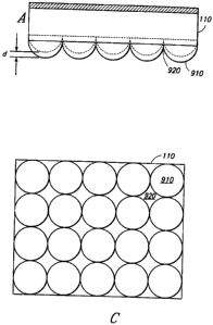
Figures 9A-9C illustrate side (9A-9B) and top (9C) views of a tissue
modulation
device 110 featuring recessed regions 920 and raised regions 910 of varying
heights.
"d" indicates the height difference between raised regions.
DETAILED DESCRIP'T10N
T"issue modulation refers to manipulating the tissue to which the method is
applied
so that measurements, such as spectroscopic measurements, can be made in botl:
blood replete and blood depleced states. One strategy for tissue inodulation
is the
application of pressure to an area of tissue, such as a finger tip. When
pressure is
appEed, the region of tissue is depleted of blood. When pressure is released
or
reduced, blood retums to the affected tissue. The difference bexween
measurements
taken in the blood replete and blood depleted states provides a measure
indicat:ive of
components in the blood while minunizing the effects of extraneous
spectroscopic
signals due to calluses, din, soap residue and or.her sources associ.ated with
the
surrounding tissuc. When tissue modulauon is employed during noninvasive
spectroscopy, for example, the analysis can include determining the difference
between the spectra collected in the blood replete and blood depleted states.
Definirions
All scientific and technical terms used in this application have me3nings
comnonly
used in the art unlcss otherwise speci$ed. As used in this application, the
following
words or phrases have the meanings specified.
As used herein, "tissue" rneans any portion of an organ or system of the body,
including, but not 3irnited to, skin, capillaiy beds, blood, rnuscle, breast
and brain.
A,NlENDED SHEET
CA 02315192 2000-06-16
Kl<. ~W:\:L-rri 1klrwLMt\ u! :_u- 8-J:1 : 21: 07 = 1:310641.8798-+ +=1:1 $J
239:14465:#10
1JlYJC,J=-1167 1 JO ~.11'11 CJ r=11'1L L.UUf"'~f~ Orp r 1[J P9~,,,JL7 --KJ J7
11 =
CJG
-4A-
As used herein, "Raman spectra associated with" a given component refers to
those
ernitted Runan spectra which one sld}led in rhe art would attribute to that
component.
~MENDED S~ =- .
A!lFAIDED SHEET
CA 02315192 2000-06-16
WO 99/37205 PCT/US99/01704
-5-
One can determine which Raman spectra are attributable to a given component by
irradiating that component in a relatively pure form, and coIlecting and
analyzing the
Raman spectra emitted by the component in the relative absence of other
components.
As used herein, "blood replete" refers to a state in which blood flow through
a tissue is
unobstructed by, for example, vasoconstriction induced by cooling or the
application of
pressure. The blood replete state can be enhanced by conditions which increase
vasodilation, such as warming.
As used herein, "blood depleted" refers to a state in which blood flow through
a tissue is
substantially restricted and blood volume is minimized. A blood depleted state
can be
achieved by, for example, cooling and/or applying pressure to the tissue.
As used herein, "opaque" refers to the optical property of an object such that
light is
substantially prevented from passing through the object. In preferred
embodiments of
the tissue modulation device, no light passes through the opaque regions.
As used herein, "optically transparent" refers to the optical property of an
object such
that light is permitted to pass through the object.
As used herein, "portion of tissue" refers to an area of tissue that light
penetrates, and
from which a signal is collected.
As used herein, "recessed region" refers to an area which is recessed relative
to the raised
area and may or may not be recessed relative to the immediately surrounding
surface.
Methods of the Invention
The invention provides a method of measurement of blood volume simultaneously
with
measurements of a signal or signals indicative of one or more blood analytes.
The blood
volume measurement permits normalization of blood analyte measurements to
allow
computation of concentration levels. Temperature and pressure can be used to
affect the
capillary content and, although these can be controlled to a large extent, it
is desirable to
use tissue modulation apparatus to aid in the normalization. The invention
provides a
method for normalization that is less vulnerable to error due to differences
between
individual anatomy and blood flow patterns.
The invention provides a method of noninvasive spectroscopic measurement of an
analyte in a subject. In one embodiment, the method comprises applying tissue
of the
CA 02315192 2000-06-16
WO 99/37205 PCT/US99/01704
-6-
subject to a tissue modulation device comprising a recessed region adjacent to
a raised
region so that the raised region depresses a first portion of tissue relative
to a second
portion of tissue in apposition to the recessed region. The method further
comprises
irradiating the tissue in a blood-replete state with electromagnetic radiation
having an
excitation wavelength and collecting the spectra emitted by the tissue in the
blood-replete
state. The method further comprises irradiating the tissue in a blood-depleted
state with
electromagnetic radiation having an excitation wavelength and collecting the
spectra
emitted by the tissue in the blood-depleted state. The method additionally
comprises
analyzing the collected spectra to determine a concentration of analyte
present in the
lo tissue, wherein the analyzing comprises determining the difference between
the spectra
collected in the blood-replete and blood-depleted states. Examples of spectra
that can be
collected include, but are not limited to, Raman, nuclear magnetic resonance
(NMR),
electron spin resonance (ESR), UV visible absorption, infrared absorption,
fluorescence
and phosphorescence spectra.
In one embodiment, the tissue is applied to the device with sufficient
pressure to achieve
the blood-depleted state in the first portion of the tissue that is in contact
with the raised
region. The pressure with which the tissue is applied can be such that the
blood-replete
state is simultaneously achieved in the second portion of the tissue that is
in contact with
the recessed region of the device. In another embodiment, the blood-replete
state and
the blood-depleted state are achieved at different points in time in the first
portion of the
tissue by varying the amount of pressure with which the tissue is applied to
the raised
region of the device. In another embodiment, the blood-replete state and the
blood-
depleted state are achieved in the first portion of the tissue by alternately
applying the
raised region and the recessed region to the first portion of the tissue.
Various modifications of the device can be made to accommodate different
embodiments of the method. For example, the recessed region can be recessed
relative
to an adjacent surface of the device. This modification can facilitate
achieving a blood-
replete state in tissue applied to the recessed region. In another example,
the recessed
region comprises a channel passing through the device so that the tissue can
be irradiated
through the channel. The provision of a channel in the device allows for an
unimpeded
light path between a light source used to irradiate the tissue and the
irradiated tissue as
well as between the tissue and a light collection and/or detection system used
in
conjunction with the method.
CA 02315192 2000-06-16
WO 99/37205 PCT/US99/01704
-7-
In preferred embodiments, the tissue has an ample supply of blood circulating
in
capillary beds, such as the fingertip. Other tissues can be used, such as ear
lobe, muscle,
skin, breast or brain. The subject is preferably a vertebrate, such as a
mammal, bird,
reptile or fish. Examples of mammals include, but are not limited to, human,
bovine,
porcine, ovine, murine, equine, canine, and feline. In a most preferred
embodiment, the
subject is human.
Tissue Modulation Device
The invention disclosed herein provides a device that can be used for
modulating blood
flow in a tissue. The device is suitable for use in conjunction with methods
for
1o measuring an analyte in the tissue. The device can be used noninvasively.
The device
comprises an upper surface and a lower surface. The upper surface comprises
one or
more recessed regions adjacent to one or more raised regions. The recessed
region can
be confluent with the upper surface of the device, or recessed relative to the
upper
surface. The raised region projects from the upper surface so that application
of a
portion of tissue to the raised region of the apparatus depresses that tissue
relative to a
second, adjacent portion of tissue.
In one embod'unent, the raised region projects about 50 m to about 2 mm from
the
upper surface of the device. Preferably, the raised region projects about 100
to about
300 m from the upper surface. The device can have a single raised region or
multiple
raised regions, including raised regions of differing heights. Likewise, the
device can
have a plurality of recessed regions, optionally varying in the extent to
which they are
recessed relative to the upper surface of the device. The regions can be
immediately
adjacent to one another, or spaced apart. Preferably, the recessed and/or
raised regions
are about 20 tn to about 2 mm apart, and more preferably, about 750 m apart.
In preferred embodiments, the device is less than about 8 nun in diameter.
More
preferably, the diameter of the device is about 4 to about 5 mm. The thickness
between
the upper surface and the lower surface of at least a portion of the device is
preferably
less than about 3 mm.
At least one recessed region and/or at least one raised region is optically
transparent. The
optically transparent region of the device is curved at the lower surface to
substantially
reduce backscattered light in a light path traveling through the optically
transparent
region to a light collection system. The device can be optically coupled with
a source of
electromagnetic radiation and/or with a light detector. In one embodiment, the
device
CA 02315192 2000-06-16
WO 99/37205 PCT/US99/01704
-8-
indudes a light collection system, which can include one or more lenses. In a
preferred
embodiment, a lens or other light collection system is integrated into one or
more raised
regions of the device. In another embodiment, the device is part of an
apparatus or
system that additionally indudes means for irradiating the tissue with a light
source
and/or means for collecting and detecting light emitted by the irradiated
tissue. One or
more beamsplitters and additional lenses, filters and collimators can be
introduced into
the light path to modify the light entering and or exiting the tissue.
As illustrated in Figure 1, a detector 140 can be used in conjunction with the
tissue
modulation device 110. Multiple detectors can be combined for use with a
single tissue
modulation device. In one embodiment, a quadrant detector 140 is used, with
four
sensitive light detectors 1601ocated on a single small substrate such that it
is possible to
image light onto each detector individually. Light from a laser 130 is
directed to a region
of tissue 100 where it penetrates the surface such as the skin. In this
embodiment, the
remitted light can have a characteristic spectral width and a wavelength other
than the
incident light wavelength. When this remitted light impinges on a detector
160, an
electrical current is produced in proportion to the power delivered by the
light.
Each of the four opto-mechanical elements 150 that are optically aligned with
the
quadrant detector 140 can be employed simultaneously, while each is
simultaneously
subjected to a chosen amount of tissue modulation. The pattern of tissue
modulation
that is utilized can define the set of connections made between each of the
four detectors
160 in the quadrant detector 140. These connections can be designed so that
the amount
of signal arriving to the detector from a blood depleted zone is subtracted
from the
amount of signal which simultaneously emanates from a blood replete zone.
Preferably, the signals are subtracted while in the analog domain, prior to
signal
digitization or amplification. This affords improved signal to noise and
dynamic range
compared to that obtainable by amplifying and digitizing the signals emanating
from the
blood depleted or blood replete tissue zones prior to signal subtraction. One
advantage
to subtracting the signals prior to digitization is that each detector is on
the same
substrate and therefore biased by the same power supply such that the noise
associated
with environmental fluctuations and the power supply are the same for each
detector.
The noise is then removed by simple analog subtraction. Because they can be
integrated
on the same "chip", the detectors and the amplification/subtraction circuitry
can be
designed and fabricated to share components such as load resistors in
amplifiers, so that
much of the noise present in the electrical currents produced by these
different detectors
CA 02315192 2000-06-16
WO 99/37205 PCT/US99/01704
-9-
is correlated. The noise can then be filtered out directly, and amplification
of the noise
prior to subtraction is avoided. Digitizing and then subtracting noise would
lead to an
increase in noise in the difference between the signal from a blood replete
zone and the
signal from a blood depleted zone.
The above quadrant detector embodiment combines in a single element the
simultaneous
production of spatially distinct regions of tissue modulation with a means to
account for
fluctuations in the power output of the light source employed. In this
embodiment, a
single light source can produce four distinct regions which simultaneously
experience the
same amount of fluctuation in the incident light.
1o Figure 2 is a representation of a quadrant detector 140 coupled to a tissue
modulator 110
and a light source 130. In the diagram, the filled circles and the open cirdes
indicate
blood replete and blood depleted regions which are interrogated by an array of
parallel
rays emanating from the light source 130. The signals emanating from the blood
replete
regions, represented by A and D, are imaged onto the corresponding quadrants
of the
detector 140 in a similar fashion as are the signals from the blood depleted
regions,
represented by B and C. The quadrant detector 140 is wired so that the
following
processing occurs:
Total quadrant detector output - (A+D) - (B+C)
_(total signal from blood replete regions) - (total signal from blood depleted
regions)
- (signal from blood).
The light from the light source 130 hits a beamsplitter 120 such that it is
entirely reflected
toward the backside of the modulator 110, which is anti-reflection coated. The
beamsplitter 120 is shaped so that the residual back-reflection is divergent.
This
minimizes the amount of the source light which gets directed back through the
beamsplitter 120, through a spectrograph/polarizer/notch filter and then to
the quadrant
detector 140.
The light which traverses the back surface of the modulator is focused by the
shape of
the front of the modulator 110, into the blood replete and depleted regions as
shown in
Figure 2. This light, which traverses the front surface of the modulator,
scatters from the
tissues in the interaction zone (represented by the intersection of lines in
Figure 2) and
some of the scattered Iight has a trajectory which causes it to re-enter the
front surface of
CA 02315192 2000-06-16
F2CV. ~ C\ =1_'I'A N1LE:NCHEN 01 :20 - 8- J5 = ? 1: l.)$ = l Lt 1 UE;4 l 8778-
+49 80 2:39E344t;6 : # 1 1
1 J l UU-/1 U f 7G tJP7 1 LZ Y71 VL I.IJUI' (;,r, Oti~J f1 1 I1uL,; ~U -
- .. 11 = C,Iõ~
-10-
the modulazor 110. Those rays are re-collimated and sent back toward the
beamsplicter 120. Traversing the bearnsplitter 120, these rays go through a
spectrograph/polarizer/notch hlter and then to the quadrant detector 140.
In the embodiment illustrated in Figure 2, the set of parallel rays
illuminates an area
spanning the various regions. The blood replete and depleted regions are
created by
the mechanical contact between the tissue modulator 110 and the finger tip 100
or
other portion of the body used in the measurement. The shape of the modulator
110
is designed so that there are four ba111ense.s which are incorporated into a
single
monolith. The centers of the balls creating r.he blood depleted zones
('mdicated by B
and C) are translated outward from the center of the m.odulator 110 so that
they
protrude far enough (at least about 200 microns) to push blood out of the
points
where contact is made with the fingertip 100. At this same position the other
two
balls (represented by A and D) do not make adequate contact to push blood out
of
their adjacent tissue.
The approach described above achieves a rejection of background light from the
primary light source, a tissue modulated spectroscopic signal, and an
automatic
analog processing of the signal to minimize noise and increase signal.
Static Tissue Modulation
One strategy for modulating blood flow in a region of livi,ng tissue 'snvolves
application of rnechanical pressure or other physical stress that does not
flucruate
with time. This strategy is referred to herein as static tissue modulation.
During
static tissue modulation, the blood content of the interrogated region is kept
as
constant as possible while measurements are made. One can then take thsee
measurements: one measurement that is indicative of blood volume, one
measurement related to the analyte of interest, and one measurement taken at a
non-
interacting wavelength to assess the quality of the optical connection to the
tissue of
interest. The quotient of the first two measurements is normalized using the
third
measurement and is proportional to analyte concentration. The proportionality
constant can be determined individual.}y for each user.
In one embodiment designed for static tissue modulation, an optical component
is
combined with the surface that is used for ur.plementing tissue modulation. In
one
embodiment, illustrated in Figure 4A-4B, a lens is integrated into a raised
region 150
that protrudes from the upper surface of the tissue modulation device 110. In
che
AMENDED SHEET
CA 02315192 2000-06-16
Kl 1.! W:\ t:l 1 11l t:\l.t1t::\ l) 1 :'_ll - i3 - JJ '? 1: Uti L i 1 Ufi } 1
k3! Uf3 +49 8U 2309}4C;Vc:#~
1..~1CJlJYll71 JO IJn 1 JJ I'IIYL, L.UUI'L11 U'YO r 1G r9õI~d GCJ ~=
a1.CJJ
-11-
e.carnple illustrated in Figure 4A-4B, a single plano-convex lens is used.
Different
lenses can be incorporated into the design in accordance with che desired
optical and
mechanical properties. The examples described herein are based on refractive
optics.
Those skiIled in the art wiU appreciate that diffractive optics can be
incorporated into
the device as well.
Pressure is typically applied in tissue modula,cion, requiring a surface that
makes
contact with the skin. T13is surface can be chosen in ways which utilize the
surface
for advantageous refraction properties and/or spatial encoding of the skin
response
to spatially encoded pressure. The use of this surface as the prirnary optical
collection
surface allows the most efficient light collection because it m+*+i~es the
number of
optical surfaces as well as the distance between the exposed tissue surface
and the
first surface of the iight collection system.
A device having multiple optically transparent regions pernzits encoding
information
from spatially distinct regions of tissue. Spatial encoding can provide
contrast
between one spatial location and another, each receiving different amounts of
pressure (tissue modulation) and providing a difference signal indicative of
the blood
volume per unic area of exposed tissue. Figure 1 gives one example of a system
utilizing the first surface as an optical surface. Figures 3A-3C suggest a few
types of
patterns wiuch could be useful from a spatial encoding sense. For example,
quadrant
detectors exist in which four detectors are oriented on an identical but
miniature
square grid which mimics the orientation of the mini-lenses functioning as
tissue
modulation sites. In a quadrant detector, the factors contributing to
intrinsic detector
noise tend to be equal for all the different spatial locations because of
their close
spatial proximity. Subtractive measurement approaches utili:-6ng the detectors
cancels
out detector noise.
Figures 5-7 illustrate various embodiments of the tissue rnodulation device
110 that
can be used to alter the light path. Figures 5 and 6 illustrate a device 110
integrated
with a polarizing beamsplitcer 120 and additional focusing elements 160. These
variations can be adapted for use with simultaneous imati ing and combinations
of
wavelengths. Figures 7A-7F show variadons on a cylind:.r lens 710-750 for use
with
the device 110. In addition, one can incorporate multiple cylinder lenses of
varying
aAdths to achieve Hadamard encoding and sophisticated signal processing. Use
of
confocal techniques allow depth of field rejection of skin surface effects and
enhancement of irraditaxion and collection efficiencies of light passing to
and from
AMENDED SHEET
CA 02315192 2000-06-16
a~, . . ..F . a... ..... .,. .~u . .. . ~u - u - .J.~ a = ...~ . l ai a vrr 1
U 1 ..'U~ r=YJ UJ 1JJ:1-t't!):~ . B 1 c~
~1JICJCJ'-IyV I JO = UIt I LJ 1'l1YL ~.UVI" CõIt O'ylJ f',yJ 11VV GtJ .7~
, 11 . CJJ
-11A-
capillary beds. By varying the height of the raised regions through which
light is
direcced, one can focus light and take measuremeW.s from skin using one height
and
from blood using a second height.
AMENDED SHEET
CA 02315192 2000-06-16
WO 99/37205 PCT/US99/01704
-12-
Dynamic Tissue Modulation
In some embodiments, the tissue modulation device is designed so that
information can
be obtained from a given region of tissue at different points in time. This
strategy is
referred to herein as dynamic tissue modulation. In dynamic tissue modulation,
a given
amount of stress and/or pressure is applied to the tissue and then released or
reduced.
Measurements are made during the time when the equilibrium distribution of
blood in
the interrogated tissue is reestablished by normal circulation. The components
of a
concentration measurement, analyte-related signal and blood volume-related
signal, are
obtained by processing the measurements to correlate the change in signals
with the
change in blood volume.
One advantage of the dynamic tissue modulation strategy is the amplification
of blood-
related signals achieved by distinguishing signals that change with blood flow
from non-
blood-related sigaals that remain constant as blood flow changes. In addition,
temporal
or dynamic modulation can be combined with spatial encoding to considerably
improve
both precision and accuracy of analyte measurements.
The invention provides a device for dynamic tissue modulation. The device
comprises
means for causing a region of tissue to become blood-depleted, means for
releasing the
cause of blood-depletion, and means for spectroscopic interrogation of the
region of
tissue before, during and after depletion of blood in the tissue region. Some
embodiments further comprise a means for iinposing an optically transparent
plate into a
position where it can exert sufficient pressure against a skin surface to
remove blood
from the adjacent capillary bed. Such a plate can comprise both raised and
recessed
regions to effect spatially selective tissue modulation.
One strategy for causing and subsequently releasing blood-depletion involves
use of a
continuous sequence of plates that form a circuit or conveyor belt
configuration that
translates around one or more sprockets. The plate can be rotated into
position and the
finger or other tissue placed on it so as.to achieve a blood-depleted region
of tissue. The
belt is then quic.kly translocated sideways, in 0.2 seconds or less for
example, by rotating
a sprocket. This translocation permits blood to flow back into the previously
blood-
depleted region. Throughout this process, interrogating light can impinge onto
the
modulated tissue and spectroscopic measurements can be taken. The amount of
pressure applied can be at least about 1 to about 100 g/cm2, and preferably
not more
than about 1 kg/cm2.
CA 02315192 2000-06-16
..r. ..1.~.1.L~
r.l..'~ . . L , = .1l.VJ
r 1JICJL'=11J Jl7 1.71~ 1 L..i 1'Y'IL I.UUfCf[ L=~ I Vtlt LU :JU-r r=t:J (7:9
~.,:JJ'l'1U:) . li i}
U-10 ~' 1'1 I~I:JU LCJ 7:J 11 .!Jy
-13-
To permit spectroscopic measurement before, during and after tissue
modulation,
adjacent plates in the conveyor belt can be selected to be opaque or
transparent, or to
have a gap in the structure. Opaque plates are useful to obtain measurements
immediately after pressure is removed, corresponding to the blood-depleted
condition. Measurements taken later would be associated with the blood replete
condition. With a transparent plate, it is possible to access the tissue of
interest
before and after the temporal modulation so as to obtain premodulation, steady
state
blood volume and analyte measurements averaged over a longer period of time.
These measurements produce numbers that can be used to calibrate the
temporally
varying values that are observed during ihe modulation process. Exclusion of a
plate,
or provision of a gap between or in the center of a plate, allows
spectroscopic
interrogation without light interacting with plates. This latter strategy
reduces
cont= +narion of spectroscopic measuremenr,s by unwanted back reflection from
a
plate.
Thus, in one embodiment, the device comprises a series of alternating recessed
and
raised regions coupled so as to form a continuous loop, and at least one
rotatable
sprocket engaged with the loop such that rotation of the sprocket effects
rotation of
the loop. The recessed regions can be flat, or have a depression in the
surface. In
one embodiment, the raised region comprises a substantially cylindrical
roller. In
some embodiments, the recessed region comprises a length having a first end
and a
second ?nd. The recessed region further comprises a substantially rectangular
cross-
section and is adjoined at an end by a poraon having a substantially circular
cross-
section.
One embodiment is depicted in Figures 8A-8C. A series of rollers 810 and slats
820
are connected by lirilcs 850 between their axles 860 or fraines 860 (rollers
have axles
and slats have frarnes). These rollrss 810 and slats 820 consritute a conveyer
belt type
of arrangement 800. The belt 800 is in turn mounted onto two sprockets 840.
The
sprockeu 840 are turned by a small motor. The device is oriented such that the
patient puts his fir,.ger (or other body part appropriar.ely posicioned
relarive to the
device) onto plates which fix the finger's position and orientation and
temperature
with respect to the motion of the rollers 810.
The rollers 810 can be nominaDy opaque and cylindrical with a round cross-
section
such that they extend outward from their radius sufficiently far that they
push on the
skin. The slats can be transparent, and as they rotate around to trade
positions with
AMENDED SHEET
CA 02315192 2000-06-16
== ' ' . . = =_= == . = y. =~==~iJ1UU~1lJ 1 JU UPt I CJ V , V . - I. A 1) i
.x,- r=rO
U:J _JiJJ41UJ . H 17
= l11YL LIJUrGf( CJYIJ !-iJ ~IUU GC! 7:7
õ 11=U~1
-13A-
the rollers, they do not push on the rissuc nearly as much as the rollers. The
slats can
be of a shape that they function as a cylinder lens in that they have a
conventional
plano convex or
AMENDED S1-IEET
CA 02315192 2000-06-16
WO 99/37205 PCT/US99/01704
-14-
biconvex cross-section. The motion of the rollers is such that they move the
blood in
and out of the capillaries as they push against the tissue relative to the
slats. The motion
of the slats is such that they allow efficient exposure of the tissue to light
and they allow
efficient collection of the light which scatters outward from the tissue. They
also allow
the light exposure and collection to occur in a precisely defmed temporal and
spatial
proximity to the region which was just squeezed by the preceding roller. The
combined
action of the slats and rollers is to repetitively squeeze and relax the
capillary bed while
synchronously probing the tissues with light so as to obtain the blood volume
and
spectral measurements.
As the rollers move across the finger, the blood is squeezed out into the
surrounding
regions. When the roller vacates a position, the slat which immediately
follows the roller
can allow a view of the blood re-entering the previously squeezed region. The
slat can be
a piece of optically transparent or specially chosen optical filter material
that allows light
to enter the skin immediately above it and also allows scattered light from
within the
exposed region to be collected and used for the blood volume and analyte
measurement.
The slat can also have a shape that is advantageous with regard to the
required optical
measurements. In one embodiment, the slats are shaped so as to function as
cylinder
lenses.
In another embodiment, the slats and rollers are shaped so that the pressure
on the tissue
is not uniform along the entire long axis of the roller. The capillary bed
therefore is not
uniformly evacuated. In a complementary fashion, the shape of the slat which
follows the
roller is shaped to expose and collect light into and from both the pressed
and
nonpressed regions. The collected light is imaged onto a monolithic spatially
selective
light detector such as a quadrant photodiode or grouping of discrete avalanche
photodiodes, so that the non-pressed regions can be automatic.ally subtracted
from the
pressed regions in the analog domain. This allows a direct background
subtraction to be
executed while simultaneously obtaining temporal information on the
intracapillary blood
flow.
The expected signal from a single detector observing scattered blue light
would appear as
a decreasing function with time, once the slat was in the position formerly
occupied by
the preceding roller. A decreasing amount of light will reach the detector as
time
increases. The apparatus has mechanical stops which allow precise and rapid
(50- 100
msec) exchange of the slats with the rollers. The temporal qualities of this
signal are
directly correlated with the temporal qualities of the desired blood analyte
signals. Thus,
CA 02315192 2000-06-16
WO 99/37205 PCT/US99/01704
-15-
phase sensitive or gated detection can be used with the amount of analyte
signal
modulation being directly traceable to the blood volume modulation. This will
also
effectively decrease the dynamic range of the signal, allowing an increase in
the gain of
the detection system (such as, but not limited to, an avalanche photodiode).
In another embodiment, the analyzing employs a fixed combination of opaque
rollers
and transparent slats. In this embodiment, the apparatus is essentially the
same as
described above except that the transparent and opaque regions are
mechanically fixed in
place. No conveyor belt is employed. The person presses his thumb or finger or
other
tissue region onto the combination, and then pulls it back or pushes it
forward while
1o maintaining the pressure of the tissue against the mechanically fixed
tissue modulation
device. In this embodiment, the transparent regions allow probing of the
regions that
have just come from the opaque rollers. The timing of this modulation is
determined by
how quickly the patient pulls or pushes his finger across the apparatus and
the amplitude
of the modulation is determined by how hard the person is pressing the finger
down
onto the apparatus.
In another embodiment, transparent rollers and transparent slats are employed.
Signal
normalization for this embodiment can employ additional correction.
Those skilled in the art will appreciate various modifications that can be
made to the
specific embodiments described and that are within the scope of the invention.
CA 02315192 2000-06-16