Note: Descriptions are shown in the official language in which they were submitted.
CA 02315812 2000-06-21
WO 99/32877 PCT/US98/27259
DETECTOR HAVING A TRANSMISSION GRATING BEAM SPLITTER
FOR MULTI-WAVELENGTH SAMPLE ANALYSIS
TECHNICAL FIELD
This invention relates to a detector system for
performing sample analysis, such as DNA sequencing, DNA
fingerprinting, absorption/emission spectroscopy, and the
like. More particularly, it pertains to a detector system
which employs a transmission grating beam splitter for
separating incoming light, either fluoresced or otherwise
emitted from one or more samples, into multiple order
diffraction bands and wavelengths.
BACKGROUND
Prior art techniques for detecting fluorescence from a
capillary used in DNA sequencing are well known.
Narrowband approaches typically call for filtering the
fluoresced light into discrete bands, through the use of
discrete filter elements or filter wheels, followed by further
processing and comparison of the resulting output. Such
approaches are rather limited in the quality and volume of
data that can be used for nucleotide identification.
Multi-wavelength approaches, such as that described in
Karger, A. et al., Multiwavelength Fluorescense Detection For
DNA Sequencing Using Capillary Electrophoresis, Nucleic Acids
Research, v. 19, no. 18, pp. 4955-4962, use a spectrometer to
separate the light into multiple bands for subsequent
analysis. However, spectrometers and the associated equipment
used, as shown in Fig. 1 of this reference, are both expensive
and bulky. The spectrometer used in this figure typically
comprises an entrance slit to spatially limit the incoming
light; a collimator lens having a focal point coincident with
the position of the entrance slit so as to convert the light
emerging from the slit into parallel rays; a reflection
1
CA 02315812 2000-06-21
WO 99/32877 PGT/US98/27259
diffraction grating to diffract the parallel rays from the
collimator lens to produce spectra; and an imaging lens to
focus the diffracted parallel rays onto a CCD imaging plane.
Thus, the arrangement of Fig. 1 in this reference is
expensive, bulky and has low light throughput.
SUMMARY OF THE INVENTION
The present invention provides a detector system which
provides the high data volume of the spectrometer, while not
incurring its cost, bulkiness and low light throughput. This
is realized by means of a device in accordance with the
present invention which employs a Transmission Grating Beam
Splitter ("TGBS") positioned between a capillary and an array
of detector pixels associated with a detector camera. The
TGBS spatially splits fluoresced light coming from a
fluorophore which has migrated through a capillary into at
least a Oth-order and a 1st-order region, at least the latter
of which is spatially spread out as a function of wavelength
over a multiplicity of pixels within the array. This allows
for a compact and inexpensive system with high light
throughput.
The detected intensities of a plurality of pixels
corresponding to the first order, and the detected intensities
of pixels corresponding to the 0th order, may then be used to
perform the necessary detection.
BRIEF DESCRIPTION OF THE DRAWINGS
The present invention can better be understood through
the attached figures in which
Fig. 1 shows a transmission grating beam splitter, such
as that used in the present invention;
Figs. 2a & 2b show two embodiments for a detector in
accordance with the present invention.
Fig. 3a shows detector array output with a detector not
having a transmission grating beam splitter;
2
CA 02315812 2000-06-21
WO 99/32877 PCT/US98/27259
Fig. 3b shows detector array output with a detector
having a transmission grating beam splitter;
Figs. 4a and 4b show the response of a detector of the
present invention to monochromatic light with a target
capillary present;
Fig. 5 shows the response of a detector of the present
invention with a capillary containing two dyes;
Figs. 6a and 6b shows a transmission grating beam
splitter separating incoming light comprising four wavelength
bands;
Fig. 7 presents a pixel sampling scheme for identifying
nucleotides with a detector of the present invention;
Fig. 8 presents synthetic data from the pixel sampling
scheme of Fig. 7; and
Fig. 9 presents experimental data which shows 0th and 1st
order components using a 16 capillary trial run.
DETAILED DESCRIPTION OF THE PREFERRED EMBODIMENT
Fig. 1 shows a typical transmission grating beam splitter
(TGBS) 100 of the sort used in the present invention. The
TGBS of Fig. 1 has a first side 102 formed with an incident
surface 104 on which incoming light 106 impinges, a
substantially transparent body 108 through which the incoming
light passes, and a second side 110 from which the split light
emerges. As shown in Fig. 1, the second side 110 is provided
with a plurality of grooves 112 having a width d, each groove
being provided with an angled exit surface 114 forming a wall
of that corresponding groove.
The incident light 10 passes through the first surface
104, the body 108, and emerges as a split beam 116 from one of
the angled exit surfaces 114. As seen in Fig. 1, the split
beam comprises a 0th order beam 116a, a +1 order beam 116b and
a -1 order beam 116c.
As is known to those skilled in the art, a TGBS is
typically formed from quartz, or other suitable, substantially
3
CA 02315812 2000-06-21
WO 99/32877 PCT/US98/27259
transparent material selected for its index of refraction.
The behavior of a TGBS is described in technical note TN 35-51
entitled "Transmission Gratings", published by the Richardson
Grating Laboratory Division of the Milton Roy Company of
Rochester, NY.
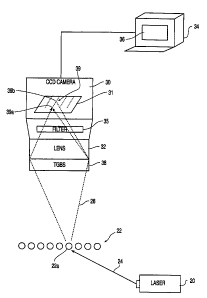
Fig. 2a shows the environment and preferred arrangement
of the present invention. A laser 20 is used to illuminate a
row of capillary tubes 22 which extend parallel to one
another, out of the plane of Fig. 2a. The capillary tubes
contain a gel in which fluorophore-tagged DNA molecules are
migrating during capillary electrophoresis. The capillaries
22, available from PolyMicro Technologies, are approximately
75 cm long and have an inner diameter of 75 ~.m and an outer
diameter of between 150-250 ~cm.
In the preferred embodiment, the laser 20 is an 100 mW
air cooled Argon ion laser, available from Spectra Physics.
The laser outputs monochromatic light 24 at 488 nm.
Alternatively, an Argon ion laser emitting a plurality of
discrete wavelengths between, for instance, 460-514 nm may be
used. In either case, the laser light 24 is focused onto the
row of capillary tubes 22. As shown in Fig. 2a, the laser
light is directed at an acute angle relative to the plane
formed by the row of capillary tubes 22, rather than being
directed normal thereto. Preferably, this angle is on the
order of 10-30° relative to the plane, and thus 60-80°
relative to the normal. For a row of 96 capillaries, each
having an outer diameter of about 200 ~.m, the laser light 24
would have to illuminate a total width of approximately 2 cm
to cover all the capillaries.
When the capillaries 22 are illuminated by the laser 20,
the fluorophore-tagged DNA molecules fluoresce and produce an
incoming light 26, represented by broken lines, directed
towards the CCD camera 30. In the preferred embodiment, the
camera 30 is a SpectraVideo #STOOlE, available from
PixelVision of Beaverton, OH. The camera 30 has a rectangular
4
CA 02315812 2000-06-21
WO 99/32877 PCT/US98/27259
detector array 31 comprising 165 rows and 1100 columns of 16-
bit pixels. From the detector array 31 within the camera 30,
the detected intensities are sent to a processing unit, such
as a personal computer 34, or like, having a display 38 and
associated memory storage (not shown). Before reaching the
detector array 31, the incoming light 26 passes through
additional lens and spectral filtering elements. In the
preferred embodiment, the light first passes through a 28 mm,
f 1.4 Nikon lens 32. Later on, the light also passes through
a filter 35 before impinging on the detector array 31. The
purpose of the filter 35 is to allow fluorescent light of
interest to pass therethrough, while attenuating light in
wavelengths not of fluorescent interest, such as the
wavelengths emitted by the laser 20. Examples of filters
which may be used include a Raman notch filter, available from
Kaiser Optical Systems of Ann Arbor, Michigan, and a longpass
filter having a cut-off of 515 nm, available from Spinder &
Hoyer Inc., of Milford, Massachusetts. In general, one may
use filters which pass wavelengths at which fluorescence of
interest is expected, and block wavelengths at which no
fluorescence of interest is expected. For instance, one may
wish to block the wavelength of the laser light 24 from the
laser 20.
However, before reaching the detector array 31 within the
camera 30, the incoming light 26 first passes through a
transmission grating beam splitter 38 ("TGBS"). In the
preferred embodiment, the TGBS is a Model #P46,068, available
from Edmund Scientific of Barrington, NJ. This particular
TGBS measures approximately 1" x 1", although other sizes and
shapes can be used. As shown in Fig. 2a, the TGBS, the lens
and the filter are all attached to the camera 30, thus
obviating the need for freestanding optical elements.
Fig. 2b depicts an alternate embodiment in which the TGBS
is positioned between the lens 32 and the filter 35. In such
case, the TGBS may be integrated into the camera 30, or be
5
CA 02315812 2000-06-21
WO 99/32877 PCTNS98/Z7259
fixed in some manner to the lens 32. In this alternate
embodiment, the spacing between the capillaries 22 and the
camera lens is about 3 cm, the spacing between the camera lens
and the TGBS about 0.5 cm and the spacing between the TGBS and
the detector array 31 is about 4 cm. As is known to those
skilled in the art, these spacings depend on the focal length
of the lens, and the thicknesses of the optical components.
It is noted that a TGBS normally requires substantially
parallel, or collimated, incoming light. Thus, in the
embodiment of Fig. 2b, the lens 32 has a long focal length so
that the light impinging on the TGBS 38 is substantially
parallel.
When using the detector system of Fig. 2b, the laser beam
from laser 20 is set to a width (in the dimension coming out
of Fig. 2b) of about 100-500 um, and a length sufficient to
extend along the breadth of the row of capillaries 22. This
causes the samples in each of the capillaries to fluoresce,
effectively causing a linear series of fluorescence spots.
The camera lens focuses this linear series onto a detector
array of the camera.
The fluorescence light 26 from the samples within the row
of capillaries 22 is collected by the camera lens from a wide
solid angle, and this light is focused onto the CCD. In the
embodiments of both Figs. 2a and 2b, the light is dispersed
within the TGBS, and emerges with spectral components of
different orders, with the first order having the greatest
intensity and dominating over the other orders. This provides
a light collection efficiency which is greater than that of a
prior art fluorometer having an entrance slit and collimator
lenses. The separated, fluoresced light from a given
capillary 22a is detected by pixels of a particular column 39
of the array 31, with the 0th order component being detected
by a first pixel 39a and the 1st order component being
detected by at least one of a plurality of second pixels 39b,
spaced apart from the first pixel.
6
CA 02315812 2000-06-21
WO 99/32877 PCT/US98/Z7259
Fig. 3a illustrates the effect of using a detector system
of the present invention, but with the TGBS omitted. In the
absence of the TGBS, each capillary creates a single
fluorescence spot 50 on the detector array 31, and all the
capillaries together form a row 52 of discrete florescence
spots. Nothing else appears on the detector array 31.
Fig. 3b illustrates the effect of using a detector system
of the present invention with the TGBS included. When the
TGBS is inserted into the system, the florescence from the
capillaries 22 is split into a plurality of bands, each band
representing a particular order. Thus, Fig. 3b depicts the
formation of a 0th order band 52, a 1st order band 54 and a
2nd order band 56. When imaged onto a detector array, each of
these bands occupies a plurality of rows of pixels in the
vertical direction, with different capillaries being imaged
onto different columns, at least one column of pixels for each
capillary. The 0th order band collects the fluorescence from
all wavelengths and the members in this band are tightly
focused, extending over only one or two rows of pixels for
each column corresponding to a capillary. By contrast, the
members in the 1st and 2nd order bands are dispersed, and
extend over several rows pixels, along the columns)
corresponding to each capillary.
To facilitate subsequent processing of the light
collected by the detector array, it is preferable that the 0th
order bands from the capillaries are imaged onto the same
rows) of pixels, and that their corresponding 1st order bands
are imaged onto substantially the same column(s). This
alignment obviates the need to later correct for any skew
among the received pixels in the sensed image, during
subsequent processing. To ensure this, however, one typically
may need to rotate the camera, and thus the detector array
therein, relative to the row of capillaries.
It should be noted here that TGBSes can be selected to
favor one or more orders over others. In other words, the
7
CA 02315812 2000-06-21
WO 99/32877 PC"T/US98/Z7259
transmittance in the favored orders can significantly exceed
the transmittance in the disfavored orders. Thus, one may
produce a TGBS which passes primarily 0th and 1st order
components, while -1st order and other orders are considerably
attenuated. The P46,068 model TGBS used in the preferred
embodiment is such a 0th and 1st order-favoring device.
In the preferred embodiment, the transmission grating
beam splitter has 70 grooves/mm. The angular difference
between the 0th and 1st order is about 2°, and the dispersion
angle within the 1st order for wavelengths between 500 nm and
700 nm is only about 0.8°. Thus, the 0th and 1st orders for a
single capillary can easily be separated from one another on a
detector array having a pixel width of about 25 ~.m, by
judiciously spacing the TGBS from the plane of the array.
Fig. 4a shows the relative separation between, and spread
of, the 0th, lst and 2nd orders scattered by a single
capillary with the resultant light impinging on a
lens/TGBS/detector array arrangement not having the filter 35.
The laser light is focused to a small spot occupying an area
of 2x2 pixels on the detector array. Thus, this image
represents the detector's system response. The intensity
distribution of 0th, 1st and 2nd orders is about 1, 7 and 0.6,
respectively.
Fig. 4b shows an expanded view of the peaks corresponding
to 0th, 1st and 2nd order in Fig. 4a, after normalizing each
to an intensity of 1.0, and co-locating them. This figure
shows that the spread for each of the peaks in response to
monochromatic light is substantially same for each order. In
particular, the widths of each peak at half normalized maximum
intensity are substantially similar, given the 1-pixel
detector resolution. Thus, the image dispersion of the
present detector is negligible for 0th, 1st and 2nd orders
using monochromatic light.
Fig. 5 shows the output from illuminating a capillary
carrying two dyes, fluorescein (1~",aX = 530 nm) and nile blue
8
CA 02315812 2000-06-21
WO 99/32877 PCT/US98/27259
(I~X = 625 nm), using the detector of the present system.
Both the 0th order and the 1st order peaks appear, and
distinct 1st order peaks appear for each of the two dyes. The
0th order confines all wavelengths of fluorescence within one
or two pixels in the detector array. In contrast, the 1st
order disperses the fluorescence from the two dyes across
multiple pixels.
Figs. 6a and 6b show the effect of a detector in
accordance with the present invention on incoming light 26
from tagged DNA samples of a single capillary. For
simplicity, only the TGBS 38 and one pixel column 31a of the
detector array 31, comprising a plurality of pixels 31b, is
shown in Fig. 6a. It is understood, however, that the lens 32
can be interposed between the TGBS and the pixel column 31a,
or, as described above, the incoming light 26 may have already
passed through the camera lens 32 at this point.
The incoming light 26 is separated into a 0th order
component 40 and a 1st order component 41. As shown in Fig.
6a, the 0th and 1st order components are spatially separated
from each other, as they impinge on the pixel column 31a.
This separation will subsequently allow one to use the
intensities of both the 0th order and the 1st order
transmitted incoming light components when performing
subsequent analyses for identifying particular fluorophores,
and hence, the corresponding nucleotides.
As is known to those skilled in the art of DNA sequencing
using capillary electrophoresis, each of the four DNA
nucleotides are typically tagged with one of four fluorophores
which fluoresce in overlapping wavelengths. Thus, in Fig. 6a,
the detected 1st order light 41 comprises four sub-bands,
designated 41a, 41b, 41c and 41d, each corresponding to a
region along the column of pixels 31a, in which a particular
one of the four fluorophores dominates.
Fig. 6b shows the relative intensity of fluorescence of
the four fluorophores as a function of relative pixel number.
9
CA 02315812 2000-06-21
WO 99/32877 PGTNS98/27259
Here, increasing pixel number corresponding to increasing
wavelength. In Fig. 6b, curves 42a, 42b, 42c and 42d
correspond to the fluorescence emission spectra of the four
fluorophores, each of which is shown to be dominant in a
corresponding one of the four pixel regions 41a, 41b, 41c and
41d of Fig. 6a.
As stated above, in Fig. 6a, the pixel column 31a
corresponds to the detector output for a single capillary.
And far that one capillary, data is available for a number of
contiguous pixels, including a small number of pixels which
have 0th order information, and a larger number of pixels
which have 1st order information. This offers some
flexibility in performing subsequent analysis to determine
exactly which fluorophore is present at any given time.
Fig. 7 shows how the pixels of the pixel column 31a may
be sampled to come up with a detection scheme which exploits
the detector of the present invention. In the preferred
embodiment, six pixels are monitored. Pixel 70 corresponds to
the 0th order component, and pixels 70a, 70b, 70c, 70d and 70e
correspond to various portions of the first order component.
In this example, fluorophores, whose spectral curves are
given by 72a, 72b, 72c and 72d, each tag a specific
nucleotide, Guanine ("G"), Adenosine ("A"), Thymine ("T") and
Cytosine ("C"), respectively.
Pixel 70a is positioned slightly to the left of the peak
for nucleotide G. Thus, it receives much energy contribution
from G, and virtually none from the A, T and C. Thus, energy
in pixel 70a indicates the presence of G.
Pixel 70b is positioned roughly at the intensity cross-
over point between fluorophores which correspond to
nucleotides G and A. Thus, signal energy from pixel 70b gets
substantially equal contribution from these two nucleotides,
and very little from T and C. Thus, energy in pixel 70b
indicates the presence of either G or A.
CA 02315812 2000-06-21
WO 99132877 PCT/US98/Z7259
Pixel 70c is positioned near the intensity cross-over
point for nucleotides A and T. Pixels 70c receives somewhat
less contribution from G, and virtually no contribution from
C. Thus, energy in pixel 70c is indicative of A or T, and, to
a lesser extent, of G.
Pixel 70d is positioned near the intensity cross-over
point for nucleotides T and C. This pixel receives nearly
equal contribution from these two nucleotides, somewhat less
from A, and considerably less from G. Thus, energy in pixel
70d is indicative of T or C, of A to a slightly lesser extent,
and of G to an even lesser extent.
Pixel 70e is positioned to the right of the intensity
peak for nucleotide C. At this point, there is relatively
little contribution from T, even less from A, and still less
from G. Thus, strong energy presence in pixel 70e is
indicative of C.
In practice, these six pixels, 70 and 70a-70e are
monitored continuously as electrophoresis takes place in at
least one capillary tube. Migration along the capillary tube
typically brings one nucleotide every 2-4 seconds, and the
detector array is sampled every one-half or so seconds. Thus,
a time series of the signal energy of each of these pixels can
be used to derive the sequence of nucleotides passing through
a window region of the capillary tube.
Fig. 8 shows a synthetic time series in which
hypothetical pixel values for pixels 70a-70e are presented for
the given set of nucleotides, each nucleotide causing one or
more peaks among the 1st order pixels for each frame number,
each frame number corresponding to the position at which a
nucleotide is expected. Pixel 70, which corresponds to the
0th order time series, shows a peak each time a nucleotide is
present. In contrast, the time series for the other pixels
70a-70e, which are normalized amongst themselves for each time
frame, exhibit regions devoid of peaks, and show varying
11
CA 02315812 2000-06-21
WO 99/328'17 PCT/US98/Z7259
signal intensities, depending on the nucleotide present during
that time frame.
DNA sequencing can be performed with the time series
shown in Fig. 8 by simultaneously examining the intensities of
all six pixels, during each time frame. For any given time
frame, the presence of a signal in pixel 70 (0th order),
indicates the presence of a nucleotide. Similarly, for each
time frame, a strong signal in pixel 70a indicates G; a strong
signal in both pixels 70b and 70c indicates A, a strong signal
in both pixels 70c and 70d indicates T, and a strong signal in
pixel 70e indicates C.
The time series data of Fig. 8 may be used in slightly
different ways for DNA sequencing. For instance, for each
time frame, the time series data for pixels 70a-70e may first
be normalized by the 0th order value for that time frame, and
the thus globally normalized values may be directly compared
in lieu of searching for co-occurrences of signal peaks in
multiple pixels, for each time frame.
Also, as stated above, TGBSes may be obtained which
weight the 0th and 1st orders in different ways. Thus, with
an appropriately weighted 0th and 1st order components, it may
even be possible to convert the detected signal intensities of
the selected pixels into logarithms through the use of a look-
up table, and then subtract these from one another, in
preparation for nucleotide identification. This approach may
be especially useful if special purpose hardware were employed
to implement the detection schemes.
It should be noted that the methodology of the present
invention is independent of the particular set of fluorophores
being used. Given a new set of fluorophores, one can derive
their spatio-spectral characteristics using known techniques
with a predetermined excitation wavelength, and then designate
appropriate pixels for a detection scheme. It should also be
noted that the present detection scheme provides one with more
measured values (six) than unknowns (four nucleotides), and
12
CA 02315812 2000-06-21
WO 99/32877 PCT/US98/Z7259
contrasts with known ratio-based methods which require
solution of underdetermined systems. Indeed, the present
detection scheme is not even limited to four fluorophores,
since a first number of such fluorophores may be used to
detect a second number of nucleotides, given appropriate
sampling of the first order pixel components.
Fig. 9 is a sample display output from an actual trial
using 16 capillaries containing DNA samples tagged with one of
four dyes. These figures show the 0th and 1st order
components from the trial, for a specific instant during
electrophoresis. As can be seen in these figures, the 0th
order band comprises compact members which do not extend much
along the vertical axis of the display. On the other hand,
the members of the 1st order band extend in this direction,
testifying to the spreading of 1st order data along this
dimension. However, it is important to note that the 0th
order and the 1st order do not overlap.
Thus, a detector in accordance with the present invention
simultaneously acquires the 0th and 1st order of the
transmitted florescence pattern with suitable relative
intensity for detection by a CCD, or other, detector. The 0th
order light contains the whole wavelength of undispersed
fluorescence from a target fluorophore going through a
detection window of one or more capillaries. The 0th order
light for all capillaries is collected by only one detector
channel. Typically, this channel comprises at least one row
of pixels in a CCD array, the number of pixels in each row
corresponding to at least the number of capillaries.
Meanwhile, the 1st order light is dispersed over a number of
detector channels (typically a number of rows of pixels). The
different detector channels (i.e., CCD rows in the preferred
embodiment) represent different wavelength portion of the
fluoresced light and are collected individually. The
availability of both 0th (whole incoming fluoresced light) and
13
CA 02315812 2000-06-21
WO 99/32877 PCT/US98/27259
1st order data (partial incoming fluoresced light) allow for
flexibility in subsequent data analysis
In addition, a detector having a TGBS has three
advantages over current used spectrograph-based techniques.
First, the detector of the present invention has higher
incoming light collection efficiency than a spectrograph, as
the light throughput in a TGBS-based detector is determined
only by the camera lens. This contrasts with a spectrograph,
which employs a reflective grating system, which is typically
not configured to retain the 0th order light energy. Second,
a detector using a TGBS allows one to dispense with the
bulkiness and high cost associated with a spectrograph,
thereby resulting in a less expensive and miniaturized system,
having greater flexibility and reliability. And third, a
TGBS-based detector system has negligible image distortion
when compared to the output from a spectrograph with its slits
and collimating optics.
While the above invention has been described with
reference to certain preferred embodiments, it should be kept
in mind that the scope of the present invention is not limited
to these. For instance, a compound lens may be used, and the
filter may be placed between the lens and the TGBS, or even
before both of them. Also, the samples may be illuminated by
the laser after emerging from the capillary tubes following
migration, rather than while they are still within the
capillary tubes. Thus, one skilled in the art may find
variations of these preferred embodiments which, nevertheless,
fall within the spirit of the present invention, whose scope
is defined by the claims set forth below.
14