Note: Descriptions are shown in the official language in which they were submitted.
CA 02318180 2000-07-14
WO 99/37204 PCT/US99/01723
-1-
FLUORESCENCE IMAGING ENDOSCOPE
RELATED APPLICATIONS
This application claims the benefit of U.S.
Provisional Application Serial No.60/072,455 filed on
January 26, 1998, the entire contents of which is
incorporated herein by reference.
Background
The following relates to the development of a laser-
induced fluorescence imaging endoscope for mapping
cancerous or precancerous tissues in hollow organs. In
initial clinical studies, on colon polyps, Ultraviolet (UV)
light was used at 370 nm to excite visible fluorescence
(400--700 nm}, the spectral signatures of which enabled
differentiating between normal and abnormal tissues.
Previously endoscopic imaging has been achieved using an
optics module mounted in one of the biopsy ports of a two-
port standard (white light) colonoscope. The optics module
employs a quartz optical fiber and associated optics to
deliver the UV light to the tissue, and a coherent quartz
fiber-optic bundle to transmit the resulting fluorescence
image to the proximal side of the endoscope, where a filter
removes the large background of reflected UV light and the
fluorescence image is then captured by a high-gain CID
detector array.
CA 02318180 2000-07-14
WO 99/37204 PCT/US99/01723
-2-
Endoscopically-collected autofluorescence images of
colonic mucosa can be used as a screening tool for
detecting pre-cursors to colorectal cancer (CRC).
Fluorescence has been used to distinguish between normal
mucosa and adenomas. In particular, spectra measured with
single point contact probes with the use of several
different excitation wavelengths.
Fluorescence spectra have been obtained through
optical fiber probes with several excitation wavelengths.
An in vitro study performed a search over a wide range of
excitation wavelengths, and concluded that 370 nm is
optimal for distinguishing between normal mucosa and
adenoma. Both in vitro and in vivo studies using
adenomatous polyps as a model for dysplasia have shown that
with this wavelength dysplasia has less peak intensity at
460 nm and may have increased fluorescence at 680 nm
compared with normal colonic mucosa. Furthermore, the
morphologic basis for these spectral differences have been
studied by fluorescence microscopy. The decreased
fluorescence intensity in polyps was attributed to its
raised architecture, increased vasculature, and reduced
collagen in the lamina propria. The red enhancements arise
from increased fluorescence of the crypt cells, which may
be caused by higher levels of porphyrin.
Summar~r of the Invention
The present invention relates to imaging endoscopes
and in particular to a flourescence imaging colonoscope
using a dual channel electronic endoscope that employs a
CA 02318180 2000-07-14
WO 9913'I204 PCT/US99/01723
-3-
charge coupled device (CCD) chip or other solid state
imaging device mounted on its distal tip to collect the
white light image. Of particular significance for the
present invention is that this chip can also collect the
fluorescence image, displaying it on the endoscope's video
monitor with much larger.signal size than that obtained
using the optics module and intensified CID camera. This
configuration was used to collect fluorescence images of
colonic dysplasia. Video images of two small FAP polyps,
have been taken with the standard white light image and the
unprocessed fluorescence image.
The CCD detector, which lacks gain intensification,
detects the weak fluorescence signals, which are six orders
of magnitude smaller in intensity than the diffusely
reflected white light image. In addition, it is surprising
that reflected 370 nm excitation light did not completely
flood the CCD, obscuring the fluorescence signal. This
results from the fact that the CCD spectral response falls
off to zero quickly at wavelengths below 400 nm. Thus, the
CCD effectively serves as its own long pass filter. Other
imaging devices can be used with a filter to reduce by at
least one half the detected intensity in the ultraviolet
region relative to the detected intensity in the visible
region.
In this particular embodiment, the CCD has a
resolution of 270 x 328 pixels and an objective lens of 2.5
mm in diameter. The images are collected in 33 ms in RGB
format. The advantages of this particular embodiment
include that the in vitro fluorescence images exhibit a
CA 02318180 2000-07-14
WO 99/37204 PCT/US99/01723
-4-
signal-to-noise ratio (SNR) of about 34 at clinical working
distances of 20 mm (distance between tip of endoscope and
tissue surface), which is superior to that obtained using
the UV Module/CID detector, which has a SNR of about 18 at
the same distance. The use of the CCD eliminates the need
for the optics module and greatly simplifies system design.
In addition, it also avoids problems associated with the
tendency of the UV module to rotate in the biopsy channel.
By using the same detector and optics for white light and
fluorescence images, perfect registration of these two
images can be obtained. Parallax between the white light
image of the CCD and the fluorescence image of the optics
module was a significant problem. The CCD in this
particular embodiment contains 88,560 pixels compared to
10,000 fibers for the UV module, resulting in higher total
image resolution. The objective lens on the Pentax
colonoscope has better imaging properties than the W
module. The characteristic width for the line spread
function of the lens of this embodiment is 200 mm compared
to 400 mm for the UV Module. The overall rigidity of the
spectral endoscope is not increased significantly with a
single UV illumination fiber.
The diagnostic methods employed can be based on the
overall fluorescence intensity difference between normal
mucosa and dysplasia. Thus, in certain applications it is
preferable to collect the fluorescence emission over the
full band between 400-700 nm. However, accurate
measurements can use a point contact device such that
diagnostic information can be obtained by sampling the
CA 02318180 2000-07-14
WO 99/37204 PCT/US99l01723
-5-
fluorescence at a plurality of specific wavelengths such as
460, 600 and 680 nm, for example. For many applications
the preferred range for.fluorescence excitation is between
350nm and 420 nm.Endoscopic imaging studies with the
electronic CCD endoscope can include the use of color
CCD's, which have the ability provide such information.
Brief Description of the Drawings
Figure 1 is a schematic view of an endoscopic system.
Figure 2 is a schematic view of a solid state imaging
device such as a CCD on the distal end of an endoscope.
Figure 3 is a schematic diagram of an endoscopic
system in accordance with the invention.
Figure 4 shows the relative sizes of the illumination
area and fluorescence area.
Figure 5 is a schematic diagram of an endoscopic
system.
Figure 6 is a graphical illustration of the average
fluorescence intensity and the measured and predicted
signal to noice(SNR)ratio.
Figures 7A and 7B are graphical illustrations of
variation fluorescence intensity between an average of 14
frames and a single frame for normal colonic mucosa and
ademoma, respectively.
Figure 8 is an illustration of the sensitivity of the
system as a function of detection threshold values.
Figures 9 and 10 show flourescence intensity profiles
of tissue with adenoma, and including the moving average
and percent ration values.
CA 02318180 2000-07-14
WO 99/37204 PCT/US99/01723
-6-
Figure 11 is the fluorescence intensity graph showing
adenoma, normal and intensity ratio values as a function of
pressure exerted on the site with the probe.
Figure 12 is an endoscope system showing the
difference in collection geometry between the endoscope and
a contact probe.
Figure 13 is a preferred embodiment of an endoscope
system in accordance with the invention.
Figure 14A is a preferred embodiment ofa fluorescence
imaging system in accordance with the invention.
Z5 Figure 14B illustrates graphically the dependence of
radiated power on the input power ofa light source emitting
in the ultraviolet region of the spectrum.
Figure 14C illustrates a timing diagram for a process
acquiring fluorescence and reference images.
Figure 15 is a preferred embodiment of a fluorescence
imaging system in accordance with the invention.
The equations describing the number of signal photons,
N9, collected by a given pixel in an endoscope as a
function of the separation distance d and the radial
distance p on the tissue surface, and the corresponding SNR
are as follows:
NOP)- ~7s~~8'ZTfTTofprisr('~~m~~~.~tan9;,Po('~~~~~
Z 3.5
jIC H(~-COSBm)IVfCIz 1-1-~
CA 02318180 2000-07-14
WO 99/37204 PCT/US99/01723
SNR = NS
(2)
~Ns+C~_)
The geometry and certain symbols are defined in Figure 1.
Note also, the emission wavelength Aem, pixel array size
gx g, fiber optic transmission efficiency T;, the bandwidth
of the filtered emission wavelength Dh, the fraction of the
transmitted energy in this wavelength region Tf, the
collective efficiency To of the system optics including the
long pass filter, lens and eyepiece, incident light energy
. Po(~,~~~t,h is Planck~s constant, c is the speed of light, fp
is the packing fraction of the fiber cores et is the
quantum efficiency of the tissue, and Nf is the total
number of resolution elements. The signal to noise ratio
(SNR) is a function of electronic noise 6~ and gain G.
Colorectal cancer constitutes a major national health
care problem. The incidence and mortality for carcinomas
of the colon and rectum are second only to those of lung in
the United States. This suggests that the current
screening methods are inadequate for controlling the spread
of colon cancer, and that little advancement in detection
has occurred in a long time. The five year survival rate
for all patients diagnosed is between 35-49~. Colorectal
cancer is relatively unresponsive to radiation and
chemotherapy, hence surgical resection with wide margins is
CA 02318180 2000-07-14
WO 99/37204 PCT/US99/01723
_g_
the only reliable method of preventing its growth. These
tumors spread by direct extension into adjacent structures
and by metastasis through the lymphatics and blood vessels.
The most common sites of metastatic spread in order are
regional lymph nodes, liver, lungs, and bones.
The pathophysiology of this disease begins in the
epithelial layer of colonic mucosa as dysplastic changes in
the crypts cells. This tissue can be accessed by
colonoscope, and if the pre-malignant lesions are detected
at an early stage, they can be removed for biopsy. Most
carcinomas of the colon and rectum are believed to arise
from visible precursor lesions called adenomatous polyps.
These benign masses evolve from a monoclonal expansion of
epithelial cells which develop irregularities in the size
and shape of the nuclei and cytoplasm, a condition known as
dysplasia. These lesions can be detected on colonoscopy by
their raised architecture. The medically accepted adenoma-
carcinoma sequence suggests that colorectal carcinoma
arises from adenomatous tissue that undergoes malignant
transformation, which is believed to occur through a multi-
step process in which genetic alterations accumulate. The
presence of a precursor stage in the development of CRC
provides a window of opportunity for early detection and
removal of these lesions to prevent future progression into
carcinoma.
The prevalent screening method of colonoscopy relies
on the observation of large structural changes in the
colonic mucosa in order to locate adenomatous tissue for
biopsy. However, this procedure is relatively insensitive
CA 02318180 2000-07-14
WO 99/3'7204 PCT/US99/01723
_g_
to adenomatous tissue which is flat. Patients diagnosed
with ulcerative colitis (UC), for example, have a high risk
of developing carcinoma from non-polypoid regions of
tissue. Moreover recent studies have concluded that some
forms of adenocarcinoma arise from small superficial
adenomas. Because of this risk, frequent screening by
colonoscopy must be performed with multiple biopsies
throughout the colon. However, the likelihood of sampling
error and missed diagnoses in these patients renders this
form of surveillance highly unsatisfactory. Also, the
examination of a tissue biopsy is time consuming and
costly. Moreover, considerable intra- and inter-observer
variation occurs in the identification of dysplasia. A
patient who is diagnosed as positive for dysplasia often
must return to the clinic for further screening and
possibly for surgical resection of the colon. Thus, the
current state of endoscopic surveillance with histologic
interpretation is an imperfect science and is in need of
improved methodologies with greater sensitivity and
specificity and less intra- and inter-observer variation.
The method of fluorescence endoscopic imaging offers
features which can overcome the present screening
limitations with white light endoscopy. This method is
sensitive to the biochemical constituents and
microarchitecture below the tissue surface. Furthermore,
combined with endoscopes, fluorescence images can scan wide
areas, and can resolve tissue surfaces on the sub-
millimeter scale. If sufficient information is present on
the fluorescence, computers can be used to determine the
CA 02318180 2000-07-14
WO 99/37204 PCT/US99/01723
-10-
presence and location of diseased regions in real-time.
Autofluorescence has demonstrated the ability to
distinguish between normal and neoplastic human tissue.
The first studies showed that single point fluorescence
spectra can be used to detect tumors in vitro from several
types of tissue,. Later, in vivo studies were performed
for detecting neoplasia in bladder, brain, colon, cervix,
esophagus, lung, oral mucosa, and skin. In addition,
fluorescence has been used to distinguish normal tissue
from diseased with the use of exogeneous agent such as
hematoporphyrin derivative (HpD).
The full length from the rectum to the cecum is
typically 1.5 m. Histologically, the mucosa is the layer
in contact with the lumen, and has a thickness of about 400
~Cm. The epithelium is the most superficial layer and
consists of absorptive columnar cells and intermittent
mucin-producing goblet cells, which function to reabsorb
water and to lubricate. These cells undergo continuous
turnover, and are replaced by rapidly dividing stem cells
at the base of the crypts, where the first signs of
dysplasia can be observed. The surrounding lamina propria
contains blood and lymphatic capillaries which supports the
secretory, absorptive and other highly active functions of
the mucosa. It consists of loose connective tissue, in
particular collagen, along with numerous inflammatory cells
which protect the intestinal wall from invasion by
microbes.
The muscularis mucosa is composed of several layers of
smooth muscle fibers which contract to expel secretions
CA 02318180 2000-07-14
WO 99/37204 PCT/US99/OI723
-11-
from the glandular crypts, prevents clogging, and enhances
absorption by maintaining contact between epithelium and
luminal contents. The submucosa contains the larger blood
vessels, lymphatics, and nerves, and are surrounded by
dense collagenous connective tissue which keeps the mucosa
attached to the muscularis propria. The muscularis propria
contains an inner circular and outer longitudinal muscle
layer, which are involved in the involuntary peristaltic
contractions of the colon for propagating the flow of fecal
matter. The outer serosal layer consists of connective
tissue which contain the major blood vessels and nerves.
Adenomatous polyps are raised protrusions of mucosa
which contain immature, poorly differentiated epithelial
cells with irregularity in size and shape of the nuclei.
These lesions are benign but they have the potential to
transform into colorectal carcinoma. The different
morphological types include tubular, villous, and
tubulovillous adenomas. Although all forms are raised,
each type can either contain a stalk, which is called
pedunculated, or can be hemispheric, which is known as
sessile. The malignant potential of polyps are greatest
with the villous form and least with the tubular. Also,
the probability of carcinoma developing increases with the
size of the polyp. There is about a 1% chance of finding
invasive tumor in a polyp less than 1 cm in diameter, 10%
for polyps between 1 and 2 cm, and 45% for polyps larger
than 2 cm. The sub-cellular changes associated with these
polyps are frequently histologically identical to the
dysplasia found in ulcerative colitis.
CA 02318180 2000-07-14
WO 99/37204 PCT/US99/01723
-12-
Results of molecular biology studies suggest that the
steps involved in the malignant transformation of adenoma
into carcinoma involves the mutational activation of an
oncogene coupled with the sequential loss of several tumor
suppressor genes. Also, it was found that several genes
must incur mutations before malignant tumors arise.
Several specific genetic alterations have been identified
during the process of tumorigenesis. Activational
mutations have been found in the ras oncogene of 50% of
colorectal carcinoma. Furthermore, allelic deletions were
identified in portions of chromosomes 5, 17, and 18, which
may involve loss of tumor-suppressor genes.
Patients with the presence of over 100 neoplastic
polyps in their colon are diagnosed with the condition
called familial adenomatous polyposis. These people have a
genetic predisposition for developing numerous polyps in
their colon by adulthood. Most patients have between 500
and 2500 polyps, and on average, there are about 100
polyps. FAP is a rare disease, and accounts for only about
1% of the incidence of CRC in the Western world. Foci of
dysplasia usually become malignant, and FAP patients must
have their colons removed at a young age. The probability
for the onset of colon cancer for someone with this
condition is 10% at 10 years of age, 50% at 20 years, and
100% at age 30. Histologically, most of the polyps are
tubular adenomas with a high probability of malignant
transformation, and the dysplasia associated with FAP
polyps is identical to that found in sporadic polyps. An
autosomal dominant genetic defect is responsible for the
CA 02318180 2000-07-14
WO 99/37204 PCT/US99/01723
-13-
development of this disease.
A second form of CRC that is associated with familial
predisposition is hereditary nonpolyposis colorectal cancer
(HNPCC). HNPCC is defined as patients with at least three
relatives in two generations having CRC, and with at least
one relative being diagnosed at less than 50 years old.
This form is much more common than FAP, and accounts for up
to 13°s of the incidences of CRC in the Western world.
HNPCC patients do not have numerous adenomatous polyps, and
it is very difficult to distinguish it from sporadic cases.
Genetic linkage has been found between this disease and
anonymous microsatellite markers on chromosome 2.
In ulcerative colitis, the mucosa undergoes
cytological changes resulting in the formation of dysplasia
without the presence of polyp formation. These changes are
believed to be associated with repeated episodes of chronic
inflammation and repair of the colonic epithelium, and
flat, ulcerated tumors with poorly defined margins are
common. Patients who have had UC for over 8 years are
recommended to have periodic colonoscopy with random
biopsies taken. This screening process is not effective
because less than 0.1 percent of the total mucosal surface
area is sampled. However, it is important to note that
only 1% of new incidences of CRC arise from UC cases.
UC is an inflammatory disorder of the colorectal
mucosa of unknown cause. Patients with UC are at increased
risk for developing dysplasia or cancer. Recognition of
this increased risk has resulted in colonoscopic
surveillance strategies starting at 7-10 years after the
CA 02318180 2000-07-14
WO 99/37204 PCT/US99/01723
-14-
initial presentation of symptoms. Colonic surveillance
strategies include direct macroscopic visualization of
colonic mucosa and access to mucosal biopsies for
microscopic assessment of dysplasia. Although the
pathological classification of dysplasia was standardized
in 1983, differences and inconsistencies remain regarding
the interpretation of dysplasia.
Dysplasia is typically focal. Despite the practice of
taking 12-20 mucosal biopsies during surveillance
colonoscopy, less than l~ of the colonic surface is
sampled, so the likelihood of missing small foci of
dysplasia is high. Thus, cancers can develop in patients
without any previous or concurrent dysplasia. Although
performing prophylactic colectomy on all patients after the
first decade of disease would be the most definitive
solution to the cancer problem in UC, patients with minimal
or mild symptoms of the disease are understandably
reluctant to take this radical approach. Colonoscopic
surveillance with histologic interpretation remains an
imperfect science in need of improved methodologies with
greater sensitivity and specificity.
Furthermore, studies have suggested that flat
dysplasia may be the origin of sporadic colon cancer which
does not arise via the adenoma-carcinoma sequence. The
morphological characteristics of adenomas that proliferate
superficially in flat nonpolypoid mucosa have been observed
endoscopically as small plaquelike lesions with vague
redness or discoloration. In a comprehensive study, 33
such lesions were described as slightly elevated with a
CA 02318180 2000-07-14
WO 99/37204 PCT/US99/01723
-15-
reddish surface and a central depression. Foci of cancer
or severe atypia were found in 25% of lesions of diameter
up to 4 mm, 40% of lesions measuring between 5 and 8 mm,
and 80% of lesions with diameter between 9 and ZO mm.
There are several methods in practice for the early
screening for CRC, but each is limited in its
effectiveness. The goal of screening is to detect
localized superficial masses in asymptomatic individuals.
A sigmoidoscopy involves the clinician viewing the
patient s rectum and sigmoid colon with either a rigid or
flexible imaging device. This form of screening is based
on the finding that 60% of CRC occur within the distal 25
cm of the colon. This length is reachable with a rigid
sigmoidoscope, and a flexible one can reach up to 60 cm.
However, recent statistics have shown that an increasing
number of tumors are found beyond the reach of this device.
An advantage of this procedure is that it can be performed
without the patient undergoing anesthesia or taking a prep.
The most extensive method of screening for this disease is
a colonoscopy, where the patient is first prepped and
sedated. A colonoscope is inserted throughout the full
length of the colon, and the mucosal surface is viewed by
the physician under white light for polyps and other
abnormal masses. This procedure is adequate for
identifying raised lesions, but flat region of dysplasia
will go undetected.
The fluorescence of tissue occurs through a process in
which the electrons of a biological molecule enters an
elevated energy state upon absorbing laser light at a given
CA 02318180 2000-07-14
WO 99/37204 PCT/US99/01723
-16-
excitation wavelength Ae~. The excited state is unstable,
and the electrons will return to the ground state. Most of
this energy is lost as heat through molecular collisions,
but a small fraction of excited electrons undergo an
internal conversion and spontaneously radiates light at
longer emission wavelengths l~em. The fraction of molecules
which release energy by fluorescence is called the quantum
efficiency of the tissue, denoted as e~. The fluorescence
intensity depends on the product of the initial population
of the excited state and the tissue quantum efficiency.
The spectral lineshape is determined by the
fluorescence emission and absorption by biochemical
molecules which are unique to the composition of tissue.
The electronic levels of the singlet state are split into
vibrational and rotational states, which in large molecules
consists of small intervals and may overlap due to
molecular interactions. The electrons may decay to any of
the vibrational-rotational levels of the ground state,
thus, the fluorescence spectra of biomolecules are
typically broad. This lack of structure in the spectra
limits the amount of information that can be obtained from
fluorescence. The tissue components which produce
fluorescence are known as fluorophores, and endogenous
chromophores include aromatic amino acids, NADH, FAD, and
porphyrins. The local environment may have a large effect
on the fluorescence emission, which may become quenched or
shifted in wavelength. Further details regarding the use
of outafluorescence for imaging tissue can be found in U.S.
Patent Nos. 4,193,142, 5,421,337, 5,452,723, 5,280,788 and
CA 02318180 2000-07-14
WO 99/37204 PCT/US99/01723
-17-
5,345,941, the entire contents of these patents being
incorporated herein by reference.
A first step taken in evaluating the use of
fluorescence in colon was to determine the existence of
optimal wavelengths to differentiate between normal colon
mucosa and adenomatous polyps in vitro with single point
measurements on a sub-millimeter scale. For example, the
fluorescence emissions of 4 normal colon and 11 adenomatous
polyps were recorded with a spectrofluorimeter. The
excitation wavelengths used ranged between 250 to 500 nm in
10 nm steps, and the results were tabulated in an array
called an excitation-emission matrix (EEM). A ratio was
taken of the average EEM from the normal colon to that of
the adenomatous polyps, and excitation at 330, 370, and 430
nm were found to produce fluorescence spectra which
contained the greatest amount of diagnostic information.
Based on the results of these in vitro studies,
clinical trials were conducted to evaluate the ability of
fluorescence to distinguish among normal, adenomatous, and
hyperplastic colon tissue with 370 nm. In this study, a
pulsed nitrogen-pumped dye laser delivered 370 nm
excitation through an optical fiber probe with one
excitation and six collection fibers. This t~robe was
inserted through the biopsy channel of a colonoscope, and
placed in contact with the colonic mucosa during
colonoscopy. The probe consisted of six individual 200 ~.m
collection fibers arranged in a bundle with one fiber for
excitation. With this device, fluorescence emission was
detected from an area of tissue about 1 mm2. The
CA 02318180 2000-07-14
WO 99/37204 PCT/US99/01723
-18-
fluorescence spectra were detected by a spectrograph
coupled to an OMA. The spectra showed a difference at 460
nm where the normal mucosa produced about 6 times greater
fluorescence intensity than adenoma. This difference is
almost twice that found from the in vitro studies. Above
650 nm, the average of the adenomas were slightly greater
than that of normal.
From 20 patients, the fluorescence intensities at 460
and 680 nm were located on a scatter plot, and a straight
line was drawn to minimize the number of misclassifications
when compared to histology. The decision line correctly
classified 31 of 31 adenomas, 3 of 4 hyperplastic polyps,
and 31 of 32 normal colonic tissue specimens. The
sensitivity, specificity and positive predictive value of
the technique for diagnosing adenomas were 100%, 970, and
94o respectively. Because only a small number of
hyperplastic polyps were examined, it was unclear whether
adenoma could be reliably distinguished from hyperplasia
using fluorescence. The observed differences in the
fluorescence may arise from architectural differences
between polyps and the normal mucosa rather than from
dysplastic changes.
The next step was to use the data from this study to
provide prospective methods of evaluating the performance
of fluorescence. The data were randomly divided into two
equal sets, and the first was used to devise an algorithm
to distinguish the tissue type based on the fluorescence
intensity at 460 nm and at the ratio between intensities at
680 to that at 600 nm. A biopsy of tissue from each point
CA 02318180 2000-07-14
WO 99/37204 PCT/US99/01723
_19_
was classified histologically as adenomatous, hyperplastic,
or normal. From the prospective decision criteria, the
sensitivity, specificity and positive predictive value of
the algorithm for diagnosing adenomas were 90%, 95%, and
90% respectively.
Further attempts have been made to use fluorescence to
distinguish between normal mucosa, adenomatous polyps and
hyperplastic polyps in vivo with 337 nm excitation.
Fluorescence spectra were measured from 86 normal colonic
sites, 35 hyperplastic polyps, and 49 adenomatous polyps
with a single optical fiber. The fluorescence emission
displayed peaks at 390 and 460 nm, which was attributed to
the collagen in the submucosa. Also, this peak decreased
in intensity for normal mucosa, hyperplastic polyps, and
adenomas, respectively. The peak intensity of the normal
mucosa was found to be slightly less than twice that for
adenomas. Using a MVLR analysis, the sensitivity,
specificity, positive predictive value, and negative
predictive value of fluorescence to distinguish between
adenomatous and hyperplastic polyps were 86%, 77%, 86% and
77%, respectively. This study concluded that the
differences in fluorescence were due to polyp morphology
rather than to the fluorophores present in the polyps.
Other excitation wavelengths have been used to study
fluorescence in colon. A continuous wave He-Cd laser was
used to deliver 325 nm excitation to measure fluorescence
spectra from 35 normal mucosa and 35 adenomatous polyps in
vitro from a single optical fiber by an OMA. The peak
intensity from normal mucosa occurred at 375 nm and that
CA 02318180 2000-07-14
WO 99/37204 PCT/US99/01723
-20-
for adenoma appeared at 450 nm. A multi-variate linear
regression (MVLR) analysis established a set of scores for
each data point to determine a diagnostic criterion.
Fluorescence spectra from an additional 34 normal, 16
adenomatous, and 16 hyperplastic sites were taken and
analyzed prospectively using the established decision
criteria. The sensitivity, specificity and positive
predictive value of this study to distinguish between
normal and adenomatous tissue were found to be 100%, 100%,
and 94%, respectively. In addition, 15 of 16 hyperplastic
polyps were classified as normal, which is the correct
diagnosis because hyperplastic polyps are formed from a
thickening of the epithelial layer.
The fluorescence of colon was studied with 410 nm
excitation as well. The emission from 450-800 nm.was
collected with a spectrofluorimeter from 83 biopsy
specimens removed during colonoscopy from 45 patients. The
intensity of the emission band from 460-530 nm declined
from normal to carcinoma to adenomatous mucosa. The peak
intensity at 460 nm was about 2.5 times higher for normal
mucosa than for adenoma. A stepwise discriminant analysis
was performed on the spectra using nine variables. The
results compared to histology showed that the process
distinguished between normal mucosa and adenoma with a
sensitivity and specificity of 88.2% and 95.2%,
respectively. The fluorescence emission resulted from the
superposition of three bands centered at about 470, 485,
and 404 nm.
Thus, there has been extensive research performed
CA 02318180 2000-07-14
WO 99/37204 PCT/US99/01723
-21-
within the last 5 to 10 years to evaluate the use of tissue
autofluorescence to distinguish between adenomatous and
normal mucosa. In vitro studies have concluded that 330,
370, and 430 nm are optimal for excitation. Preliminary in
vivo results indicate that single point fluorescence
detection has a sensitivity, specificity, and positive
predictive value as high as 90~, 95~, and 90% respectively
for such discrimination. Also, the data suggest that the
intrinsic fluorophores can include collagen, NADH, and
porphyrin. Hemoglobin is an absorbing chromophore. In
order to make this technique suitable for a clinical
setting, wide area fluorescence detection and processing
must be performed in real time and adapted to conventional
white light endoscopy. These requirements demand the
development of a spectral imaging instrument.
Fluorescence microphotographs of unstained frozen
sections were studied to account for the morphological
structures in normal colonic mucosa and adenomatous polyps
which emit fluorescence. The 351 and 364 nm lines from an
argon-ion laser were used for fluorescence excitation, and
the emission was collected by a series of barrier filters
with cut-off wavelengths of 420, 475, 515, 570, and 610 nm.
The fluorescence intensity was graded semi-quantitatively
from 1+ to 4+ by a single observer. In normal mucosa,
fluorescence in the spectral band from 420 to 700 from
collagen in the lamina propria was graded at 3+ and that in
the submucosa at 4+ in the same emission bandwidth. in the
epithelium, there was faint fluorescence seen from
absorptive cells and none from goblet cells. The H&E
CA 02318180 2000-07-14
WO 99/37204 PCT/US99/01723
-22-
stained section identifies the tissue composition of normal
mucosa. The fluorescence image of the serial unstained
section indicated the fluorescent structures. Several
differences were observed on the fluorescence image of the
serial unstained section indicating the fluorescent
structures.
Several differences were observed on the fluorescence
micrographs of the adenomatous mucosa. First, fewer
collagen fibers were present in the lamina propria,
resulting in less fluorescence intensity from the
epithelium. Also, the level of fluorescence seen in the
cytoplasm of crypt cells was recorded at 2+, compared to
+/- seen in normal crypts. Finally, a larger number of
fluorescent granules were present in adenoma. The image of
the H&E stained section include crypt cells from an
adenomatous polyp. The fluorescence from the serial
unstained section shows an observable level of
fluorescence, and the number of eosinophils in the lamina
propria is significantly larger than that in normal mucosa.
The submucosa of the adenomatous polyp was graded at 4+,
which is the same as that of normal.
A procedure has been developed to describe the
clinically observed fluorescence in terms of its
microscopic origins. This process combined the intrinsic
fluorescence of each microstructure with its density as a
function of tissue depth and the optical turbidity of the
incident and return path. The concentrations of each
fluorophore from clinical fluorescence spectra can then be
extracted. From this procedure, the factors for observing
CA 02318180 2000-07-14
WO 99/37204 PCT/US99/01723
-23-
greater fluorescence intensity from normal mucosa compared
to that from adenomas include: (1) The submucosal
fluorescence is about 10 times brighter than that of the
overlying mucosa. (2) The mucosa attenuates both the
incoming excitation light and the returning fluorescence;
if the mucosa is sufficiently thick, the underlying
submucosa cannot contribute, but if it is thin, as in
normal mucosa, attenuation is smaller, resulting in
brighter tissue fluorescence. (3) In addition, the
fluorescence intensity of adenomas is less than that of
normal colonic mucosa, perhaps because the dysplastic
crypts tend to displace the collagen in the lamina propria,
which is the dominant fluorophore. (4) Adenomas exhibit
greater attenuation of both the 370 nm excitation light and
the return fluorescence, due to increased hemoglobin-rich
microvasculature.
A multi-spectral imaging system has been developed
which collects fluorescence at four different emission
wavelengths simultaneously. In this device, the output of
a fiber optic endoscope is passed through 4 spatially
separated interference filters. The 4 images are arranged
onto quadrants of an intensified CCD array by adjustable
segments of a multi-mirror system. The CCD or other
imaging device 40 as seen in Figure 2 can have 30,000 pixel
elements or more. The four wavelengths were selected to
optimize the contrast in the fluorescence spectra between
normal and diseased tissue. Fluorescence from human
cadaveric aorta was excited with 337 nm, and emissions from
400, 420, 450, and 480 nm were ratioed to produce a
CA 02318180 2000-07-14
WO 99/37204 PCT/US99/01723
-24-
dimensionless contrast function. This function indicated a
value for atherosclerotic plaque that was four times
greater than that for normal artery, and the results were
displayed using a false-color overlay. This instrument was
also able to distinguish between rat tumor and surrounding
muscle from fluorescence spectra. Further details
regarding this system are described in Wang, T.D. et. al.,
"Real-time In Vivo Endoscopic Imaging of Fluorescence from
Human Colonic Adenomas', Proceedings of SPIE 1998, 3259,
the entire contents of which is incorporated herein by
reference.
The resolution of this design is limited by the fibers
in the imaging bundle. The use of 4 fluorescence emission
wavelengths provides for greater contrast between normal
and diseased tissue and for flexibility in the development
of the disease detection process. However, by separating
the fluorescence emission in parallel, the signal is
reduced by a factor of 4, thus lowering the SNR. Also, the
4 spectral images must be aligned onto the detector at
different angles, which poses a challenge for image
registration. Furthermore, image processing algorithms
using multiple images increase the computation time, and it
is not clear that the fluorescence contains independent
information at 4 bands. Finally, the fluorescence images
are detected at the proximal end of the endoscope, which
poses difficulty in clinical use for registering the white
light image and in navigating the instrument.
A simpler version of the mufti-spectral imaging system
has been developed which collects only 2 emission bands.
CA 02318180 2000-07-14
WO 99/37204 PCT/US99I01723
-25-
This design splits the fluorescence emission with a beam
splitter onto two intensified CCD cameras. A helium
cadmium laser delivers excitation light at 442 nm via the
illumination bundle of a fiberoptic bronchoscope. The
fluorescence emission was filtered in 2 bands, one between
480 and 520 nm and the other at wavelengths greater than
630 nm. The two spectral images were aligned, and the
intensities were ratioed point by point for discriminating
normal from diseased tissue, and a color image was formed.
This method eliminates the effects of distance and angle of
the illuminating light, as well as tissue reflective
properties. A color camera is attached separately for
observing the white light image. This system was tested
clinically on 53 patients and 41 volunteers, and the
results were compared with conventional white light
bronchoscopy at 328 sites. The sensitivity on fluorescence
was 730, which was significantly greater than that of 48%
found on white light in detecting dysplasia and carcinoma
in situ. The two methods were found to have the same
specificity of 94%.
In the clinical system, the white light and
fluorescence images were collected with a dual-channel
electronic colonoscope (Pentax EC-3800TL). This model
contains two biopsy channels with diameters of 3.8 and 2.8
mm, respectively. The outer diameter of the endoscope is
12.8 mm, and the working length is 1.7 m. The field of
view of the multi-element objective lens has a divergence
half-angle of 60° with a depth of focus ranging between 5
and 100 mm. The white light illumination is produced by a
CA 02318180 2000-07-14
WO 99/37204 PCT/US99/01723
-26-
300 W short-arc xenon lamp. By using the same detector for
both white light and fluorescence imaging, perfect
registration can be obtained. This feature is ideal for
producing a diagnostic overlay.
An illumination probe consisted of a 200 /cm core
diameter optical fiber with NA = 0.40 coupled to a 3 mm
diameter BK7 glass microlens (F#~=-1). The illumination
probe was inserted into one of the instrument channels, and
the tip was placed flush with the distal end of the
colonoscope. A mode mixer clamped the excitation fiber at
the proximal end to maximize the divergence angle of the
light. The probe was attached at the proximal end of the
colonoscope by a leur lock to prevent movement. A power of
300 mV~1 was delivered to the tissue. The spectral response
of the CCD detector (TI TC210) cuts off at about 400 nm,
and is negligible at the excitation wavelengths AeX = 351
and 364 nm3, thus eliminating the need for a long pass
filter to block specular reflection from the excitation
light. The two instrument channels allow for the optical
fiber illumination probe and the biopsy forceps to be used
at the same time. Figure 1 shows a schematic view of the
endoscope 10 with an imaging bundle 20, biopsy view of the
endoscope 10 with an imaging bundle 20, biopsy channel 12,
lens 18, and illumination ports 14. The distal end of the
device is positioned at a distance d from the tissue. One
problem associated with such a system is the shadows
generated by the illumination system. An important feature
of the invention described below is a process to compensate
for shadows on the tissue 16 surface.
CA 02318180 2000-07-14
WO 99!37204 PCTNS99/01723
-27-
A footswitch was activated by the user to block the
excitation light when the white light was used for
illumination, and vice versa, using a pair of computer
controlled shutters (Uniblitz, VS14). The integration time
for acquiring each fluorescence image is 33 ms. As shown
in Figure 3, the clinical fluorescence imaging system 40
consists a video processor 48, computer 44, monitor 46,
mavigraph 50, and VCR 52, laser 42 and colonoscope 54.
An electronic colonoscope 54 detects photons at the
distal end with a CCD detector. An important aspect of the
present invention is that the spectral response of the
Texas Instrument TC-210 CCD detector dropped sufficiently
fast below 400 nm that no diffuse reflection from the UV
excitation was observed. In fact, virtually no specular
reflection, which is several orders of magnitude higher in
intensity than diffuse reflectance and fluorescence, was
observed either. Another aspect which made this system
possible was that the detector has sufficient sensitivity
to detect fluorescence from colonic mucosa without the use
of an intensifier. Because the detector is located at the
distal end, the optical transmission efficiency is
determined only by the multi-element objective lens
positioned between the detector and the tissue. Another
significant feature of this embodiment of the invention is
that the same chip detects both the white light and
fluorescence image, thus perfect registration occurs on the
pseudo-color overlay. Furthermore, no modifications are
necessary to the colonoscope which can impede the
clinician's ability to perform the pracedure.
CA 02318180 2000-07-14
WO 99/37204 PCT/US99/01723
-28-
One limitation of this system is the bandwidth
selectivity and spectral resolution of the chip. The TC
210 is a monochrome detector and collects fluorescence over
the full visible spectrum. It is difficult to employ
bandpass filters in front of the CCD because the light is
collected at angles as high as 60°. However, RGB detectors
exist which contain pixels which are sensitive to red,
green, and blue light, and can produce fluorescence images
in 3 frames. However the passbands are determined by the
integrated circuit manufacturer of the imaging circuit.
Note that a gating mechanism can also be used, which is
desirable for using pulsed lasers as the excitation source.
Other excitation sources can include CW lasers and broad or
narrow band light sources.
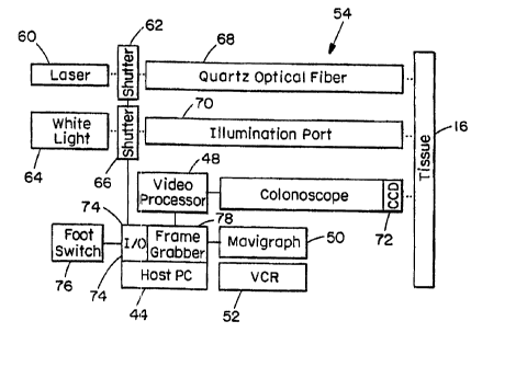
The block diagram of an electronic imaging system
operated by switch 76 is, shown in Figure 5. An argon-ion
laser 60 delivers UV light through a shutter 62 into a
quartz optical fiber coupled to a microlens located in ane
instrument channel of the colonoscope, while the white
light 64 is delivered through shutter 66 the illumination
fibers of port 70. The pair of shutters 62, 55 are
computer-controlled by a digital input/output (I/O) card
74. Both the fluorescence and white light images are
detected by the CCD 72 at the distal end. A frame grabber
78 digitizes the fluorescence and white light images
sequentially. A host microcomputer executes the image
processing algorithm and displays the pseudo-color overlay.
A mavigraph is used to convert the white light image with
overlay into a format which can be recorded by the VCR.
CA 02318180 2000-07-14
WO 99/37204 PCT/US99/01723
-29-
The plot in Figure 6 shows the fluorescence intensity
from the average of 14 frames collected with the electronic
imaging system. A row of pixels is shown from normal
colonic mucosa. Also plotted are the measured and the
predicted SNR. The SNR is approximately 30 at the center
and it falls to about 10 near the periphery. Thus, the
full field of view satisfies the minimum SNR requirement of
4 for the instrument-noise limited detection for
distinguishing between normal colonic mucosa and adenomas.
A frame-to-frame variation from average in the
fluorescence image intensities can be seen in Figures 7A
and 7B, which show the differences between the values
across a row of pixels in a single frame compared to the
average of 14 frames. The plot in Figure 7A is that for
the normal specimen shown in Figure 6, and the plot in
Figure 7B is from a sample of mucosa which contains an
adenoma in the center. The variation about the average is
small compared to the difference in fluorescence intensity
between normal and adenomatous tissue. Thus, the
occurrence of false positives resulting from pixel-to-pixel
variation is small.
A streaking artifact appeared in the fluorescence
images taken with the electronic imaging system. This
artifact arose because the UV excitation light was not
blocked while the CCD rows were being read out
electronically, which is performed under normal white light
illumination by a rotating wheel with spatially separated
filter. This artifact can be removed in the processing
software of the image data.
CA 02318180 2000-07-14
WO 99/37204 PCT/US99/OI723
-30-
A study was performed to determine the level of UV
light which can be safely delivered onto the colonic
mucosa. White light and fluorescence images were collected
sequentially. Fluorescence images from 30 patients with 14
colonic adenomas and 6 hyperplastic polyps were collected.
Finally, the fluorescence images were collected in parallel
with single point EEM spectra. From these studies, the
effectiveness of the realtime implementation of
fluorescence image collection, processing, and display with
movement in the colon were assessed. In addition, sources
of artifact present on the colonic mucosa such as mucous,
stool, and prep were evaluated. Also, the anatomy of the
colon makes it desirable to collect images at large
incident angles, and the effectiveness of the moving
average algorithm with these limitations were determined.
Finally, the intensities from fluorescence images were
compared to that from the single point optical fiber
probes.
The excitation source used was a Coherent Innova 328.
This laser is rated for 1 W in the UV, and requires 60 A at
208 V of electrical power and 3 gal/min of water. The
excitation light is coupled into an optical fiber device
including lengths of 12.5 and 16.5 m of fiber were required
to deliver the excitation light to the distal end of the
colonoscope.
First, the excitation fiber must be incorporated in
the colonoscope. Next, a method is used to rapidly switch
between white light and laser illumination. Finally, a
method of quickly and accurately registering the
CA 02318180 2000-07-14
WO 99/37204 PCT/US99/01723
-31-
fluorescence results with the white light images must be
implemented.
The colonoscopy procedures included prep of the
patient with 3 oz of Fleet phospha soda mixed with 4 oz of
water. There was no measurable fluorescence from the prep
mixture using an optical fiber contact probe on colonic
mucosa in vitro with 370 nm excitation.
Using the electronic endoscope, white light
reflectance and fluorescence images were collected
sequentially in vivo during routine colonoscopy. The white
light image can include a vascular pattern of arteries in
red, and an outline of a vein in blue. Patches of specular
reflection can be seen on the lower half of the images.
The fluorescence of normal mucosa appears uniform with an
arterial pattern interspersed as reduced fluorescence
intensity. This effect is attributed to the absorption of
fluorescence emission by hemoglobin. The vein does not
appear on the fluorescence image, and there is virtually no
specular reflection from the excitation light. The
illumination field on fluorescence is slightly smaller than
that on white light, as depicted in Figure 4.
An example illustrates the process of image
collection, processing and evaluation of adenomatous
polyps. A white light endoscopic image taken of a sporadic
polyp located in the rectum shows a polyp with visible
architectural features about 5 mm in diameter is located in
the lower half of the image near the middle. In the raw
fluorescence image the adenoma appears as a region of
reduced intensity surrounding a brighter central region.
CA 02318180 2000-07-14
WO 99/37204 PCT/US99/01723
-32-
This image was ratioed with its own moving average
image, and multiplied by 100 to produce the percent ratio
image. Thresholds on the processed fluorescence images
taken at 60%, 75~, and 90~ were used to determine the
contour lines which define regions of mucosa with various
likelihoods of containing dysplasia. The contours were
then filled in pseudocolor to highlight areas of tissue to
be targeted for biopsy. The pseudocolors red, green and
blue designate regions on the white light image which have
high, medium and low probability, respectively. The polyp
was found to be adenomatous on histology.
Overlay regions indicating disease included one
located at the site of the adenoma, and the other two
corresponded to shadows cast by mucosal folds. The shadows
appeared as regions of reduced intensity on the
fluorescence image. These effects were minimized by
directing the endoscope normal to the mucosal surface.
Moreover, the overlay regions which resulted from shadows
changed in size and shape as the angle of the endoscope to
the tissue surface varied, while those generated from the
adenoma remained fixed in size.
White light and fluorescence images were collected
from a total of 30 patients undergoing routine colonoscopy,
which included images from 14 adenomas and 5 hyperplastic
polyps. A biopsy was taken of each adenoma and one
adjacent normal site. The fluorescence images were
processed by the moving average algorithm, and the
sensitivity of detection was determined as a function of
threshold values ranging from 55a to 900. The results of
CA 02318180 2000-07-14
WO 99/37204 PCT/US99/01723
-33-
sensitivity are plotted in Figure 8.
Autofluorescence images of colonic mucosa can be
collected endoscopically in vivo and can be used to
identify and localize dysplasia in the form of adenomatous
polyps. The SNR of the fluorescence images was typically
above 30. The adenomas were correctly identified by the
fluorescence algorithm with high sensitivity. As shown in
Figure 8, the sensitivity of in vivo detection when the
images are collected at normal incidence is comparable to
that from the in vitro studies. At a threshold of 75%, the
sensitivity for detection of colonic adenomas was 86~,
compared to that of 92~ for the in vitro experiments. In
order to determine the specificity, the true negatives and
false positives must be identified. However, true
negatives (false positives) correspond to regions of normal
mucosa which were found to be normal (diseased) on
fluorescence. These results were not obtained because
additional biopsies incur additional risk of perforation.
Furthermore, the fluorescence from hyperplastic polyps,
which are not dysplastic, did not result in regions of
disease from the moving average algorithm.
In comparison of image size, the in vivo images
encompassed regions of mucosa as large as 10 x 10 cm2,
whereas the specimens of colonic mucosa were only 2 x 2 cm2
in the in vitro study. In such large fields of view, the
colon contains many mucosal folds, and these layers of
tissue blocked the excitation light from reaching the
posteriorly-located normal mucosa, thus creating shadows.
These folds were not present in the in vitro studies.
CA 02318180 2000-07-14
WO 99/37204 PCT/US99/01723
-34-
Diagnostic errors on the processed fluorescence image
resulted primarily from these shadows. The fluorescence
method used is based upon the difference in intensity
between normal and dysplastic mucosa. However, shadows
appear as regions of reduced intensity without dysplasia
being present. This artifact can be explained by the
fluorescence excitation geometry. The fluorescence
excitation is provided by one fiber located in the biopsy
port for convenience. The center of this instrument
channel is 8.3 mm away from the center of the CCD detector.
The white light image, on the other hand, is illuminated by
two fibers whose centers are located only 3.8 mm from the
detector. Thus, the shadows on the white light image are
much less pronounced that those on fluorescence.
The fluorescence technique used a single fluorescence
emission band for detection of adenomas. This method
worked well in vitro when the colonoscope is placed at
normal incidence to the lesion, and no mucosal folds were
present. However, during the clinical use of the
fluorescence prototype, the view of the endoscope was often
limited to the side of the adenoma. Because the colon is
a tube-shaped structure, some adenomas were anatomically
located at sites where it was virtually impossible to
orient the colonoscope at normal incidence to the lesion.
As a result, one side of the lesion may not be surrounded
by normal colonic mucosa. Another situation was that the
normal mucosa is far away to produce fluorescence
intensities sufficiently higher than that of the adenoma.
The fluorescence intensities were measured from the
CA 02318180 2000-07-14
WO 99/37204 PC'T/US99/01723
-35-
raw images. The normalized intensity values and the
intensity ratios were taken at three sites within the
adenoma (denoted by left, center, and right in Table 3).
The plot in Figure 9 contains fluorescence intensity
profiles through the adenoma, representing the raw
fluorescence and percent ratio values, respectively. The
adenoma was approximately 8 mm in diameter. On
fluorescence, the lesion is located between the 11 mm and
the 19 mm markings on the abscissa, which are labeled by
the vertical lines near the x-axis in Figure 9. Most of
the adenomas exhibited a single fluorescence intensity
minimum at the center of the lesion; the average ratio
between normal and diseased pixels was 1.8 ~ C.5 at the
center, and 2.0 ~ 0.6 and 2.0 ~ 0.7 at the left and right
midpoints, respectively. The average intensity ratio at
these sites was 2.0 ~ 0.6. The results of this procedure
show that the differences between normal colonic mucosa and
adenomas for in vivo fluorescence images are very similar
to that in vi tro.
Similarly, the fluorescence intensities were measured
from the raw images for hyperplastic polyps. The
normalized intensity values and the intensity ratios were
taken at three sites within the polyp (denoted by left,
center, and right in Table 3). The plot in Figure 10 shows
the fluorescence intensity profiles through the
hyperplastic polyp, representing the raw fluorescence and
percent ratio values, respectively. The hyperplastic polyp
was approximately 5 mm in diameter. On fluorescence, the
lesion is located between the 17 mm and the 22 mm markings
CA 02318180 2000-07-14
WO 99/37204 PCT/US99/01723
-36-
on the abscissa, which are labeled by the vertical lines
near the x-axis in Figure Z0. The hyperplastic polyps
exhibited an approximately uniform fluorescence intensity
across the lesion which was continuous with the normal
colonic mucosa. The average ratio between normal and
diseased pixels was 1.1 f 0.1 at the center, and 1.2 ~ 0.1
and 1.1 ~ 0.2 at the left and right midpoints,
respectively. The average intensity ratio at these sites
was 1.1 ~ 0.2. Because this average ratio value is not
significantly different from that of normal mucosa, it is
not surprising that no region of disease could be
identified by this intensity method.
In the in vivo images, the vascular pattern was
clearly displayed on both the white light and fluorescence
images. The vessels were not apparent on the in vitro
images, perhaps because the blood supply of the living
colon was no longer intact. The hemoglobin in the blood is
a well-known absorber of light, and produces linear
patterns of weak fluorescence intensity. Thus, the
intensities were measured from the raw fluorescence images
of blood vessels. As shown in Table 3, the intensity ratio
from the blood vessels is 1.3 ~ 0.1. This value is
significantly less than the average from adenomas, thus
blood vessels will not present as a source of artifact on
the overlay. Furthermore, image processing methods can be
used to remove the blood vessels based on their shape. In
Table 3, the intensity ratios for adenomas, hyperplastic
polyps, and blood vessels are summarized for comparison.
Endoscopic images and single point spectra can both
CA 02318180 2000-07-14
WO 99/37204 PCT/US99/01723
-37-
provide valuable information about tissue biochemistry.
Each method has its own advantages and disadvantages. The
endoscope collects images, and provides spatial information
with sub-millimeter resolution. The fluorescence intensity
between normal mucosa and adenomas can be compared from the
same image field within a fraction of a mm from each other.
Also, fluorescence images are collected remotely, thus the
pressure on the tissue is uniform throughout the image
field. However, it is more difficult to acquire spectral
information with fluorescence. Because of the larger areas
involved, the fluorescence energy may become to weak at
each pixel to maintain sufficient SNR, unless very large
excitation power is used.
On the other hand, single point optical fiber contact
probes collect fluorescence from an area of approximately 1
mm in diameter only. With an intensified optical
multi-channel analyzer (OMA), spectra over a wide bandwidth
can be measured with good spectral resolution and high SNR.
However, the probe must be placed at several sites.on the
mucosa to sample differences between normal and adenoma.
Typically, the normal mucosa sampled is several cm away
from the adenoma, and comparisons of the absolute intensity
can be affected by biological variability over distance.
The degree of contact of the probe on the polyp can
vary during the in viva measurements because the colonic
musculature is constantly contracting and expanding. As a
result, movement is created which makes probe placement
difficult. The adenoma is round and slippery, and the
movement of the colonic~wall renders complete contact with
CA 02318180 2000-07-14
WO 99/37204 PCTNS99/01723
-38-
the surface of the polyp very difficult. Furthermore, the
distal end of the optical fiber probe is not flat, but
there is a 17° bevel. Thus, the orientation of the beveled
side will affect the degree of contact as well.
Results of the colonoscopy procedure showed that it
was very difficult to place the probe onto the polyp for
the 0.5 seconds required to collect a full EEM. Light
escaping at various colors representing the excitation
sources was observed on the normal mucosa surrounding the
adenoma. This observation suggests that the delivery of
excitation energy to the polyp and collection of
fluorescence emission was not complete. Probe contact was
hindered by the physiological movement of the mucosa, and
by the fact that a flat probe was being placed on a
slippery, hemispherical surface. Contact is not a problem
for spectra collected on normal mucosa because this surface
is flat.
Moreover, the ratios between the intensities of normal
mucosa and adenomas can be affected by difference in the
pressure exerted on each site. An in vitro experiment was
conducted on a resected specimen of colonic mucosa which
contained an adenoma. The fluorescence intensity in the
spectral range between 400 and 700 nm was measured as a
function of pressure exerted by the probe which was passed
through the biopsy channel of a colonoscope. The pressure
was measured with a balance. As shown in Figure 11, the
fluorescence intensity increases with pressure, and the
intensity ratio does not change if equal pressure is
exerted on both the normal and adenoma sites. However,
CA 02318180 2000-07-14
WO 99/37204 PCT/US99/01723
-39-
this is usually not the case during the clinical
acquisition of spectra. The normal mucosa is relatively
flat, and measurements can be made with virtually complete
probe contact with a few grams of pressure. On the other
hand, the pressure on the polyp cannot be made the same as
that on the normal site because the probe will slip off.
The pressure on the normal site was estimated to be about 5
grams, while that on the adenoma was estimated to be close
to zero. Thus, the difference in pressure exerted on the
normal mucosa and the adenoma may result in the intensity
ratio increasing from 2 to 3, as shown in Figure 11.
Furthermore, on the recorded images of the colonoscopy
procedures, the normal mucosa showed an indentation at the
site where the probe was placed during the collection of
spectra. This observation confirmed the estimate that
several grams pressure was exerted on normal mucosa during
data collection. On the other hand, the probe was seen to
slide off the polyp when any significant pressure was
exerted, which resulted from the moistness of the surface.
Thus, the pressure exerted on adenomas was significantly
25' less.
Another procedure was conducted in vitro to compare
the fluorescence intensity ratio between normal mucosa and
adenoma as measured on imaging and single point. White
light and fluorescence images of a resected specimen of
colonic mucosa containing two adenomas were obtained. The
intensities were measured from 7 normal sites immediately
adjacent to the adenomas on both imaging and single point.
The results included the intensities that were normalized
CA 02318180 2000-07-14
WO 99/37204 PCT/US99/01723
-40-
so that the average value is 100 for each system. This
step allows for direct comparisons to be made at each
point, and reveals that the intensities are within about
10~ of each other. Furthermore, the normalized intensity
values range from 68 to 155 on imaging and from 63 to 136
on single point. Thus, the intensities measured on normal
mucosa depend on the site sampled with both methods, and
can vary by over a factor of 2.
In Table 4, the normalized intensities and the
intensity ratios are determined for the two adenomas on
imaging and single point. These values are determined at
the center and the left and right midpoints of the
adenomas. For the left adenoma, the average intensity
ratio was 1.43 on imaging and 1.54 on single point. For
the right adenoma, the average intensity ratio was 1.52 on
imaging and 1.72 on single point. These results indicate
there is little difference in the intensity ratios between
imaging and single point in vitro.
The fluorescence intensity ratio was calculated from
Monte-Carlo simulations to determine the fluorescence
intensity ratio, given the different excitation and
collection geometries of the imaging system and single
point. In Figure 12, a diagram of the collection geometry
for the endoscope 100 and the single point probe 102 is
shown. The endoscope contains a 2.5 mm diameter objective
lens 104, and is located in air at a distance 20 from the
surface of the tissue. This geometry corresponds to a
collection angle of 40°. The probe contains a quartz
shield 106 which is in contact with the tissue 16. The
CA 02318180 2000-07-14
WO 99/37204 PCT/US99/01723
-41-
optical fibers are located at a distance of 2 mm from the
tissue surface by this shield 106, and collect light at a
NA = 0.22, which corresponds to a collection angle of
12.7°. The optical parameters of colonic mucosa for the
excitation and emission wavelengths are shown in Table 2.
The excitation used in the simulation is an
infinitely-thin beam with a divergence angle of O°. The
fluorescence intensity at a point on the tissue from a
uniform thick excitation beam can be determined from the
fluorescence collected from a superposition of
infinitely-thin excitation beams which are incrementally
displaced in distance from the point to be measured.
However, this result is equivalent to integrating the
fluorescence intensity over the field of view. The LSF of
the tissue falls off quickly within several mm, thus the
simulation integrates over a 2 mm region within the
collection angle specified in Table 5. The results of the
simulation are shown in terms of the intensity ratio
between the light collected at the tissue surface with that
of the excitation. In Table 5, the intensity ratio between
normal colonic mucosa and adenomas is 3.0 and 2.9 for the
endoscope and the probe, respectively. The intensity ratio
is similar for the endoscope and the probe, a result which
is consistent with the in vitro.studies. The intensity
ratio for the endoscope is slightly higher than that of the
probe, which is consistent with the collection angle of the
endoscope being smaller. Light from the highly fluorescent
submucosa is more likely to reach the detector with a
smaller collection angle.
CA 02318180 2000-07-14
WO 99/37204 PCT/US99/01723
-42-
A model was developed to quantify the number of
photons collected-by the endoscopic imaging system over the
field of view at normal angle of incidence. This result is
valid for both white light reflectance and fluorescence
images, and can be applied to both the fiber optic imaging
bundles and electronic imaging systems. The spatial
distribution of the illumination and emission profiles of
in the center and to fall off towards the periphery of the
image. When combined with the detector noise statistics,
the SNR of the image can also be determined. This analysis
showed that distance and optical collection geometry
produces a profile in which the SNR at the periphery was
always lower than that in the center. This parameter is
needed for developing algorithms for identifying tissue
lesions. Also, the collected light intensity was found to
decrease with the square of the distance between the distal
end of the endoscope and the tissue. Furthermore, the
light collection by coherent imaging bundles is limited by
the numerical aperture of the optical fiber. This analytic
tool can be used to design the optical parameters of the
fluorescence imaging system and to identify the type of
light source required to excited the fluorescence.
The methods developed for endoscopic imaging model
were used to determine the excitation source, optics, and
detectors necessary for building two fluorescence imaging
systems. The first design consisted of a fiber optic
colonoscope which detected the fluorescence image at the
proximal end with an intensified CID camera. A 400 nm long
pass filter was used to block the reflected excitation
CA 02318180 2000-07-14
WO 99/37204 PCT/US99/01723
-43-
light, an a quartz optical fiber located external to the
colonoscope was used for image excitation. The second
design was a modification of the first to accommodate the
requirements for clinical use. This system used an
electronic colonoscope with dual instrument channels, and
detected fluorescence images at the distal end. The cutoff
in spectral sensitivity of the CCD detector below 400 nm
was used to avoid the reflected excitation light. An
illumination probe with a high NA quartz optical fiber was
coupled to a microlens and inserted into one instrument
channel for image excitation. In both systems, the
excitation source was an argon-ion laser which delivered
about 300 mW at J~eY = 351 and 364 nm, and microcomputer
with a frame grabber was used to acquire, process, and
display the diagnostic images.
Autofluorescence images from human colonic adenomas
were collected with the fiber optic system with high SNR in
vitro. For wide area surveillance of the colon wall,
regions of mucosa as large as 100 mm2 must be illuminated.
Furthermore, the endoscopic images are collected remotely,
and the intensity collected falls with distance d squared.
Previously, fluorescence spectra were collected from
contact probes which illuminated an area of about 1 mm2.
The results of this study showed that excitation sources,
optics, and detectors used in this design could collect
autofluorescence images with sufficient SNR to distinguish
between normal colonic mucosa and adenomas. In the fiber
optic system, an SNR of over 30 was attained, which
exceeded the minimum SNR requirement of 7.
CA 02318180 2000-07-14
WO 99/37204 PCT/US99/01723
_4a_
Fluorescence images were then collected from samples
of resected colonic mucosa in vitro to evaluate the
potential use of this technique for wide area surveillance
of dysplasia Colectomy specimens from three patients with
familial adenomatous polyposis containing polypoid and
non-polypoid adenomas were studied. Each raw image was
corrected for differences in distance and instrument light
collection efficiency by normalizing to a spatially
averaged image. Intensity thresholding was then used to
identify diseased regions of mucosa. The sensitivity and
specificity for detecting a region of dysplasia depended on
the threshold value selected. With the threshold set to
75% of the average normal intensity, a sensitivity of 90%
and a specificity of 92% were achieved. The average
fluorescence intensity from normal mucosa was found to be
greater than that from the adenomas by a factor of 2.2 ~
0.6. These results demonstrate the potential of this
technique to direct biopsy site selection.
The results from the in vitro studies provided
motivation for conducting an in vivo study. The electronic
system was used to collect autofluorescence images from
colonic adenomas in vivo. In the this system, an SNR of
over 30 was attained as well, which exceeded the minimum
SNR requirement of 4. Fluorescence images were collected
from 14 adenomas and 6 hyperplastic polyps from 30 patients
undergoing routine colonoscopy. The fluorescence images
were collected in a 33 ms frames, and were processed by
dividing the raw fluorescence image with a moving average
image. The processed images displayed regions of mucosa
CA 02318180 2000-07-14
WO 99/37204 PCT/US99/01723
-45-
with a probability of containing dysplasia in the form of
adenomas, as verified on histology. With the threshold set
to 75~ of the average normal intensity, a sensitivity of
86% was achieved for detecting adenomas and a specificity
of 100% was attained for hyperplastics. On average, the
ratio between the fluorescence intensity of normal mucosa
to that from adenomas was 2.0 ~ 0.6 and to that from
hyperplastic polyps was 1.1 ~ 0.2. The diseased regions on
fluorescence best corresponded to the adenoma on white
light when the colonoscope was at normal incidence. At
higher angles there were greater effects from shadows.
These results showed that dysplasia can be identified on
fluorescence images in vivo.
In the single point optical fiber contact probe
studies the average intensity ratio between the
fluorescence at 460 nm from normal colonic mucosa and
adenomas was found to be about 3, while that in endoscopic
imaging this ratio was measured to be 2.0 ~ 0.6. Direct
comparison of fluorescence imaging and single point
measurements in vitro revealed that there was little
difference between the intensity ratio measured on imaging
compared to that measured from single point. There are two
possibilities that can account for the difference in
intensity ratio between the two methods. First, the ratio
of 3 measured by the single point method was performed in
vivo. A lower ratio may have resulted in vitro because of
the loss of blood flow, which is known to absorb light.
Alternatively, the difference in the ratios may result
from contact and pressure artifacts. Videotapes of the
CA 02318180 2000-07-14
WO 99/37204 PCTNS99/01723
-46-
colonoscopy procedure showed that it was very difficult to
place the probe onto the polyp for the 0.5 seconds required
to collect a full EEM. Light at various color representing
the excitation sources was observed, which indicated that
the delivery of excitation energy to the polyp and
collection of fluorescence emission was not complete.
Probe contact was hindered by the physiological movement of
the mucosa, and by the fact that a flat probe was being
placed on a slippery, hemispherical surface. Contact is
not a problem for spectra collected on normal mucosa
because this surface is flat. Furthermore, increased
pressure was found to elevate the fluorescence intensity
collected. Higher pressures were exerted on the normal
mucosa compared to that on the polyp. The probe was seen
to slide off the polyp when any significant pressure was
exerted. Both differences in contact and pressure in vivo
resulted in a higher ratio between normal mucosa and
adenoma. On the other hand, the fluorescence images are
collected remotely, and the pressure and contact parameters
are identical for normal mucosa and adenoma.
Finally, the results of the clinical studies
identified future directions to improve the sensitivity and
clinical usefulness of fluorescence endoscopic imaging.
The shadow artifact can be reduced by illuminating the
tissue through the two white light ports. This
modification can be accomplished by replacing the glass
fibers with quartz, thus allowing for both white and
excitation light to be transmitted. Furthermore, the
shadow artifact, angle of incidence, and detection yield
CA 02318180 2000-07-14
WO 99/37204 PCT/US99/01723
-47-
can all be improved by collecting multi-spectral images
consisting of two or more fluorescence images. Lastly, the
concurrent collection of EEM spectra can be used to
identify new excitation wavelengths which result in higher
intensity contrast ratios.
Dysplastic tissue exhibits an increase in red
fluorescence which can be detected to improve the
sensitivity of disease detection. Thus another embodiment
includes the collection of multiple emission wavelengths.
One method of collecting multiple fluorescence emission
wavelengths is to use an electronic endoscope (e. g.
Olympus, Model CF I OOTL) with a CCD detector which is
sensitive to the red, green, and blue (RGB) regions of the
visible spectrum. Fluorescence images from each RGB frame
can be captured and processed, providing more detailed
information for use in a diagnostic procedure.
Furthermore, the use of spectral lineshape information from
images at different wavelengths reduces all geometric
distortions. The TI TC244 has a quantum efficiency of 30%
at 640 nm and 15% at 480 nm [TI Mannual, 1994].
Extrapolating from the 370 nm imaging data and the EEM
data, a SNR of lO:I in the red and 50:1 in the blue is
anticipated.
Performing the detection on the distal end of the
electronic colonoscope has many practical advantages.
First, the same detector can be used for both white light
and fluorescence imaging. A single detector not only
results in perfect registration of the two images, but
avoids the need to interchange of cameras, which can be
CA 02318180 2000-07-14
WO 99/37204 PCT/US99/017Z3
-48-
cumbersome. Second, fewer optical elements results in a
transmission efficiency of fluorescence photons which is
significantly higher than that of a fiber optic imaging
bundle. Third, the packing geometry of CCD pixels allow
for minimal loss of surface area of detection, unlike fiber
optic imaging bundles which have a hexagonal packing array.
While there are advantages to detecting the
fluorescence image with a distally located CCD, a fiber
optic imaging bundle with proximal detection has advantages
as well. The spectral bands of the distal CCD is limited
to the RGB response of the distal detector, while the
fluorescence collected by a fiber optic imaging bundle
could be filtered into an unlimited number of spectral
images. Also, detection of the fluorescence image at the
proximal can allow for detection with a gated intensifier.
This device enables use of pulsed lasers.
The EEM study provides valuable guidance about new
imaging strategies. The results indicate that excitation
near 410nm is useful. The contrast between normal and
adenoma tissues provided by the blue fluorescence is
greatly enhanced compared to that obtained with our current
excitation wavelength (10:1 versus 2:1). In addition, the
red fluorescence is quite pronounced for adenoma.
Extrapolation of the conclusions of the morphological model
developed using ?~eY = 365 nm to this new excitation
wavelength suggests that the blue fluorescence contains
information about both crowding of the crypts and mucosal
thickness, and that the red fluorescence contains
information about crypt cell dysplasia. Hence, collecting
CA 02318180 2000-07-14
WO 99/37204 PCT/US99/01723
_49_
images at red and blue emission wavelengths should provide
both high contrast diagnostic images and significant new
histological information. In addition, the ratio image can
be used to normalize out shadow effects. The next phase of
the imaging studies will use 410 nm excitation. A krypton
ion laser (Coherent Innova Model 302) will provide 500 mW
of power at the two lines 407 and 413 nm. This level of
power is adequate to achieve large fluorescence signals in
both red and blue bands. This laser will be installed at
the BWH Laser Laboratory along with the existing 365 nm
argon ion laser.
In addition, multiple excitation wavelengths can be
employed. One approach would be to use excitation from the
407 and 413 mn lines of a krypton ion laser to excite the
red fluorescence and to retain the 365 and 351 run lines
from argon ion laser to excite the blue fluorescence. Two
hardware configurations include (1) a fiber endoscope with
a switchable filter wheel between the scope and camera, and
(2) a dual-chip endoscope. Such a system has been
developed, for example, by American Hospital, Inc., for
stereo viewing during endoscopy. One can modify one of the
windows on the chip with a spectral cut-off mechanism. The
timing of the red-sensitive imaging channel can be
synchronized with the excitation light.
The diffuse reflectance image at 407-413 nm can be
explored to obtain information about the tissue hemoglobin
content. This image can be obtained by installing a filter
with the appropriate bandwidth on the rotating wheel in
front of the white light source. The approach is to ratio
CA 02318180 2000-07-14
WO 99/37204 PC'T/US99/017Z3
-50-
this reflectance image with the fluorescence images in the
red and blue frames. In order to develop the required
algorithms, and to decide how to optimize the spectral
information collected, an extensive contact probe study
with 410 nm excitation can be performed.
The shadow artifact obtained using the broadband
intensity algorithm with 365 nm excitation can be greatly
reduced by use of an improved excitation geometry.
Currently, excitation light is delivered through a single
quartz fiber located in the biopsy channel located 8.3 mm
from the CCD detector. The use of a single illumination
beam located a large distance from the CCD chip tends to
enhance shadows. In contrast, in the conventional white
light images produced by this colonoscope, shadows are
minimized by use of two closely spaced white light
illumination beams symmetrically positioned on opposite
sides of the CCD chip. By replacing the illumination
fibers with quartz fibers, the UV light can be delivered
through the two white light illumination ports, which are
located only 3.8 mm from the CCD detector. Implementing
this requires modifying the video processor to enable
alternate coupling of white light and laser excitation into
the illumination fibers.
Other spectral endoscope improvements can include: (I)
regulating the excitation light intensity on the tissue
surface via feedback control. This provides constant
illumination, regardless of viewing distance, and is also
important for patient safety; (ii) minimizing the streaking
effect of the fluorescence excitation on the white light
CA 02318180 2000-07-14
WO 99/37204 PCT/US99/01723
-51-
endoscopic imaging display by timing the fluorescence
excitation to occur during the "blank" periods of the
filter wheel used in the endoscope white-light source.
Feedback control of the excitation light can be
accomplished by measurement of the average intensity on the
fluorescence image. The intensity of this average value
will be used to modulate the open period on the shutter or
filter wheel. The streaking effect can be completely
removed by implementing the identical filter wheel for
blocking the excitation light that is used for producing
the RGB illumination on the white light mode.
As described above, a large argon-ion laser was used
as a near-LTV excitation source for the imaging studies.
Although adequate for these studies, this light source is
expensive and bulky and operates at only a few discrete
wavelengths. Such a laser system with its special
electrical and water cooling requirements cannot be easily
moved, preventing use at multiple sites. Alternative
excitation sources can be considered which include a pulsed
laser and a white-light source with filters, both of which
are compact and transportable.
For applications in which near-W excitation is
appropriate, a pulsed ND:YAG laser is used because it can
provide third harmonic radiation at 355 mm with sufficient
average power for spectral imaging. In both the in vitro
and in vivo studies, good SNR was obtained with 300 mW of
laser power, which corresponds to 10 mJ of energy per
fi-ame. Therefore, a frequency tripled ND:YAG laser with a
5 - 10 ns pulse duration operating at 30 Hz with an average
CA 02318180 2000-07-14
WO 99/37204 PCT/US99/01723
-52-
power of 300 mW at 355 nm will be adequate. Using a CCD
camera gated at about 10 ns, this short excitation. pulse
enables simultaneous acquisition of white light and
fluorescence images. Within this short temporal gate the
white light background is negligible, obviating the need to
chop the white light illumination. There are no special
power or water requirements for lasers of this type and a
fluorescence endoscope system with such a laser will be
easily transportable.
A mercury lamp can also be used as an excitation
source. Such a source is compact and lightweight and can
provide a~bright, narrowband illumination at a number of
excitation wavelengths. Employing this light source
simplifies system design and reduce cost, enabling less
expensive units to be produced for use at multiple sites.
The key issue is whether enough light in the desired
wavelength range can be coupled into the illumination
fiber(s). A commercial white light source with a 150 W
xenon lamp is capable of delivering as much as 80 mW of
white light at the distal end. Utilizing a 50 run
excitation bandwidth, about 20 mW of light can be used to
induce tissue fluorescence.
At selected wavelengths, mercury lamps have 5 to 10
times higher output powers than that of xenon. This
indicates that with a 500 w mercury lamp having a
relatively small filament, at least 300 mW of useful
excitation light should be available at the distal end of
the illumination fibery should be sufficier_t for collection
of good quality fluorescence images from colonic tissue.
CA 02318180 2000-07-14
WO 99/37204 PCT/US99/01723
-53-
In addition, to further enhance ShTR, either the total area
of illumination can be reduced or imaging elements can be
binned together. A lamp and power supply can be selected
for this application with the proper brightness, stability
and minimum electrical interference.
Currently, the image processing scheme is based on
ratioing the raw image to a spatially-averaged image, and
applying a threshold criterion for classifying a region of
tissue as normal or diseased. The averaging window and
detection threshold values are pre-flexed, regardless of
the polyp size, viewing angle and distance. These
predetermined values limit the range of polyp sizes which
can be accurately measured. Improved image processing and
thresholding methods will employ variable window sizes for
spatial averaging and variable thresholds. Information
from the raw digitized image about the diameter of the
largest lesion in the image will be used to determine these
parameters. This change in the window size as a fimction
of the lesion in the image field will maximize the
intensity ratio and optimize the performance of the
fluorescence method.
Image analysis methods for extracting information from
multivariate images can also be explored. A multivariate
image is a collection of congruent images of the same ob
ect measured with different variables, such as reflected
wavelengths, or fluorescence or Rainan band intensities.
Many methods are available for analyzing multivariate
images, and they can be adapted to image analysis. In
general, three steps will be followed, image processing,
CA 02318180 2000-07-14
WO 99/37204 PCTNS99/01723
-54-
object segmentation, and contrast measurement. The images
will first be processed based on the selected operation,
such as moving-window average, intensity difference or
ratio. The processed image will then be segmented based on
both frequency and intensity information. This can be done
either through thresholding, quick/slow descent, or region
growth. These methods can be coupled to the concomitant
identification and display of a lesions) based on a
probabilistic scheme.
When techniques for collecting multiple spectral
images are developed and a database of such images are
built, more advanced image analysis methods, such as
principal component analysis and regression analysis can be
used. Principal component analysis does not assume a known
(a priori) distribution, but instead employs a set of
calibrated data to extract information about structures
exhibiting pre-malignant changes. The regression technique
is based on the principle of building up a mathematical
relationship between two groups of variables, i.e., between
a number of independent variables and one or more dependent
.25 variables. As an example, a logistic regression to
correlate spectral intensities in the images with
histopatwogy of dysplastic lesions.
The development of the fluorescence imaging endoscope
has demonstrated the potential to perform wide-area
surveillance colonoscopy using fluorescence. The
fluorescence image can be analyzed in real time and can
provide the endoscopist with an instant interpretation of
the probability of dysplasia determined using a
CA 02318180 2000-07-14
WO 99/37204 PCT/US99/01723
-55-
previously-validated algorithm. In addition, the ability
to guide biopsy can be used with the present invention. In
patients with FAP, fluorescence imaging can be used to
direct mucosal biopsies to areas that are endoscopically
normal-appearing (non-polypoid) but, based on their
spectral characteristics, can have an increased likelihood
of being dysplastic. Histopathological assessment of
mucosal biopsies will be correlated with spectral data to
validate for detection of "flat" dysplasia.
The following method can be followed for determining
the capability of the fluorescence imaging system for
directing biopsy. The entire surface of the colon wall,
both at colonoscopy and using resected samples at
colectomy, is systematically imaged, and isolated areas
which are diagnosed as dysplastic selected for directed
biopsy. Random areas diagnosed as benign can also be
sampled, and the spectral diagnosis confirmed by
histological analysis. Again, the effects of complications
such as inflammation can be investigated. Once an imaging
algorithm has been validated, it can be adapted to the
detection of dysplasia in patients with UC. As in the case
of the contact probe studies, diagnostic algorithms for UC
must be capable of evaluating patients with various degrees
of background inflammation. The same patient groups
studied with contact probe EEMs will be studied with
fluorescence imaging. An important potential benefit of
wide area fluorescence surveillance is that one or more of
the otherwise random biopsies obtained during conventional
surveillance colonoscopy may be directed by the results of
CA 02318180 2000-07-14
WO 99/37204 PCT/US99/01723
-56-
fluorescence imaging. Those biopsies can be separated
from the remainder of the random biopsies to assess whether
fluorescence imaging can increase the yield of dysplasia
detection over random sampling.
The development of the rapid EEM and spectral imaging
systems represent two very important advances in
instrumentation. The two systems are complementary. The
imaging system views wide areas of mucosa in real time, and
the EEM system provides complete spectral characterization
of a given site of colonic mucosa. The two instruments can
be used simultaneously, where appropriate. The EEM probe
is placed through the second channel of a two channel
colonoscope. Thus, each system can be used to verify the
other. Also incorporated herein is the publication
attached hereto and entitled "Real-Time in vivo endoscope
imaging of fluorescence from human colonid adenomas".
CA 02318180 2000-07-14
WO 99/37204 PCT/US99/01723
-57-
The following signal size expected
table compares for
the
white light the
imaging, fluorescence
imaging observed
with
endoscope CCD,and fluorescenceimaging module
using
the
optics
with the intensified The parameters listedbelow
CID camera.
are taken fromeither the manufacturer's r from
specification
o
experimental
measurements.
Imaging DeviceDefinition Pentax(whiteht) Pentax ;fluorescence) j~V
lig
l
d
o
u ex wavelength - 356 356
e
(nm)
7l"0 (nm) em wavelength - 460 460
0~. (nm) em bandwidth 400-700400-700 400-700
Pa (mWJ power 1 300 300
~t (s) integration time0.0110.011 0.033
d (mm) distance 20 20 20
Diameter (mm) area illum 70 70 28
9,~ (degrees) max angle 60 60 35
Nf (pixels/fibers)number 8856088560 10000
rL lens (mm) radius 1.25 1.25 0.3
tissue efficiency1 S.OOE-05 S.OOE-OS
i~ packing fraction1 1 0.6
Tf % traps filter 1 1 0.8
Tj % traps imaging 1 1 0.9
Tp % traps optics 1 1 0.9
'0, photocathode 0.2 0.2 0.1
eff.
g group factor 1 1 1
N, sigaat photons 1.7x1052500 338
o, electronic noise55 55 50
G gain 1 1 10,000
SNR signaUnoise 407 34 18
TABLE 1
SUBSTITUTE SHEET (R~~I_E 2~}
CA 02318180 2000-07-14
WO 99/37204 PCT/US99/01723
-58-
TABLE 2
normal adenoma
(nm) ~,~~ 370 ~",-- 460 ~~x=
370 y",---
460
u, 0.9 0.45 2.1 1.I
15.0 9.5 8.5 5.9
g 0.9 0.9 0.9 0.9
mucosal thickness450 450 1000 1000
(um)
quantum eff(mucosa)0.1 0.1 0.8 0.8
quantum eff(submucosa) 0.1 0.8 0.8
0.1
T 3
BL
A
E
Adenoma Hy~~erplastic Blood Vessels
Normalized Intensitv
Normal 1 00.00127.5 1 OO.Of 18.1 100.00 16.1
Left 54.Of 16.3 87.9116.3
Center 59.9f 16.7 95.9f 16.7 75 .315.1
Right 54.4 17.7 98.1 f 17.7
Intensity Ratio
(normalllesion)
Average 2.0f0.6 1.1 X0.2 1.30.1
Left 2.0f0.6 1.20.1
Center 1.80.5 1.10.1
Right 2.0f0.7 1.110.2
TAB . . 4
IntensityRatio Intensity Ratio
Left Adenoma
Left 65 1.54 58 1.72
Center 74 1.35 74 1.35
Right 71 1.41 65 1.54
Avg 70 1.43 66 1.54
Right Adenoma
Left 61 1.64 57 I .75
Center 81 1.23 67 1.49
Right 59 1.69 52 1.92
Avg 67 I.52 59 1.72
TABLE 5
endoscope probe
normal adenoma normal adenoma
SUBSTITUTE SHEET (RULE 26)
nt 1.4 1.4 1.4 1.4
370 2.8E-02 2.8E-02 4.2E-03 6.2E-04
460 8.8E-05 2.9E-05 2.8E-03 9.7E-04
Rstio 3.0 2.9
CA 02318180 2000-07-14
WO 99/37204 PCT/US99/01723
-59-
Figure 13 presents a schematic outline cf the system
which has been demonstrated in clinical practice in a
format which will allow comparison with the improved
systems to be described below. The embodiment shown uses
an ultraviolet laser source 200, switched by a shutter 202
and focused with a lens 204 into a fused silica fiber probe
206 inserted into a biopsy channel of an endoscope 208 to
deliver it to a tissue site 210 so that it can illuminate
the tissue over an area 212. The UV illumination thus
comes from an aperture 214 which is different from the
endoscope~s own illumination ports 216. In the dual-
channel Pentax endoscopes used in the clinic this procedure
leaves one biopsy channel 218 free.
The endoscope camera 220 obtains its white light
illumination through its own fiberoptic illuminator 222
from a broadband Xenon arc lamp 224 and collection optics
226. A non-standard shutter 228 under computer 230 control
232 is attached to allow the white light illumination to be
turned off while fluorescence images are being taken. The
fluorescence image signal 234 is processed by the
endoscope~s video processor 236 to produce a standard video
signal 238 which is digitized by a framegrabber in computer
230. The processed image signal 240 with its information
on the state of the observed tissue is sent to monitor 242.
The entire diagnostic procedure is initiated by a foot
switch 244 attached to the computer by a cable 246.
Figure 14A shows an design for the fluorescence
imaging system which eliminates the tendency of the
previous system to identify shadows in the image as regions
CA 02318180 2000-07-14
WO 99/37204 PCT/US99/01723
-60-
of dysplasia. The improved design uses a 100W mercury (Hg)
arc lamp light source 302, dichroic mirrors 304 and 306,
wavelength filters 308 and 310 and rotating shutters 312
and 314 to provide precisely-timed, tissue-illumination
pulses in two separate wavelength bands. The first
wavelength band is centered on the near-ultraviolet (365
nm) mercury resonance line and is used to obtain the UV
autofluorescence image. The second wavelength band is at
the red end of the visible spectrum and is used to obtain a
simultaneous or near-simultaneous, reflectance image for
the purpose of identifying shadows and the extent of the W
illumination field.
A reflectance (non-fluorescing) image taken with an
endoscope camera system measures the brightness of the
tissue surface 316 in its field of view. To the extent that
the tissue surface is a Lambertian (non-specular) reflector
(generally the case) this image indicates the distance of
the tissue from a single illumination source (or a weighted
distance from multiple sources). If these illumination
sources are not in the direct line-of-sight from the camera
to the tissue source there will be shadows. A reflectance
image can thus be used to measure both the W illumination
318 at the tissue surface and the presence of shadows in
the fluorescence image as long as the UV illumination and
the visible illumination emanate from the same aperture 320
with the same angular divergence. Note that this condition
can be satisfied either by a two-color illumination fiber
322 passed through a biopsy channel of an endoscope 324 or
by the two-color illumination being passed through the
CA 02318180 2000-07-14
WO 99/37204 PCT/US99/01723
-61-
illumination bundle 326 of the endoscope. A shutter 328
switches off the normal white-light illumination of the
endoscope while the two diagnostic images are being
obtained. The closing of shutter 328 under computer
control 32~ occurs at the same time as the opening of
shutter 330 by control line 331. This action enables the
two-color light to reach the fiber 322 and thus the tissue
316.
The algorithm for using the visible reference image
along with the fluorescence image is as follows. The video
signals 332 from the CCD camera at the distal tip of the
endoscope 324 are converted by the video processor 334 to a
standard NTSC color video signal 336 and sent to a video
framegrabber in computer 338. The two images are first
corrected for the gamma factor applied to the video signal
by the video processor to insure that the digitized images
acquired by the framegrabber in the computer are linear
measurements of the tissue surface brightness. This is
accomplished in real time by the framegrabber input look-up
table. The two images are then normalized to their peaks,
which will generally be a region of non-dysplastic tissue
in the visual field. This normalizes the two illumination
fields. On a pixel-by-pixel basis the fluorescence image
value is then divided by the visible reference image value.
If the ratio falls below a predetermined threshold
(typically one-half to one-third) then that
pixel in the
image represents a region of reduced fluorescence which is
indicative of dysplasia. This pixel can then be set to a
false color state in an output video signal 340 sent to a
CA 02318180 2000-07-14
WO 99/37204 PCT/US99/01723
-62-
monitor 342 to indicate to the clinician the probability
of dysplasia. A prior threshold requirement on both images
insures that the ratio obtained is significant and
eliminates false color output in regions of shadow or low
illumination at the edges of the video field. This entire
operation occurs for every depression of the footswitch 344
which is connected to the computer through cable 346.
In the improved design both the UV-excitation light
pulse and the visible-reference light pulse are delivered
to the tissue through the same optical fiber 322 inserted
through a biopsy channel of the endoscope. The condition
that the two illumination sources have the same angular
distribution is assured by the design of the light
collection apparatus shown in Figure 14A. A single Hg arc
lamp 302 is used as the source of both wavelengths. A
dichroic mirror 304 reflects the UV portion of the spectrum
and transmits the visible portion. Filters in each path
further refine the bandwidth of the two beams. The UV
filter 308 must reject visible light to a high degree since
the efficiency of the 460 nm tissue fluorescence is only
about 0.1%. The filter 310 in the red path is less
critical but the chosen center wavelength should avoid
hemoglobin absorption bands to provide the best reference
image. Note that the design of the beam splitting optics
and beam combining optics have an even number of
reflections in both the UV and visible arms. This assures
that any angular deviations of the output beams due to
motion of the lamp track each other. Tt also makes the
directions of the output beams invarient under translations
CA 02318180 2000-07-14
WO 99/37204 PCT/US99/01723
-63-
and small rotations of the beam splitting and recombining
optics as a whole.
In the improved source design the current through the
Hg lamp 302 is boosted at appropriate moments to increase
the lamp output power for the UV exposure. This allows a
larger area of tissue to be scanned for dysplasia in a
single image. The data in Figure 14B show that the UV
output power from a 100 W Hg lamp is a linear function of
its input power to at least a factor of 3 over its nominal
rated power. Since the lamp discharge maintains a constant
voltage drop across the arc regardless of current, the lamp
output power is essentially proportional to current. At
least 50% power to the lamp must always be maintained,
however, to keep the mercury in the vapor phase. The lamp
power supply 348 in the improved fluorescence system
utilizes a DC current section to maintain the idle current
and a computer-controlled 350, pulsed current section which
can rapidly switch in multiple constant-current sources to
vary the output power of the lamp as required by the
imaging system. If the idling power is kept below the rated
power and the current pulses are kept to a sufficiently
small duty factor, then the pulsed UV output can be
sustained continuously.
The rotating shutter in the UV path 312 and in the red
path 314 of the light source are designed to provide pulsed
illumination light according to the timing diagram in
Figure 14C. This diagram shows how this fluorescence
imaging system is used with a monochrome camera, which uses
a xenon arc lamp 352 for illumination and a rotating blue-
CA 02318180 2000-07-14
WO 99/37204 PCT/US99/01723
-64-
green-red filter wheel 354 to synthesize a color image from
3 monochrome images. In this type of system a pulse of blue
light first illuminates the tissue for about 6 milliseconds
and the resulting tissue reflectance image is digitized for
the next 6 milliseconds. The illumination must be turned
off during the readout period because the monochrome camera
used continues to collect photo current in the pixels as
they are line shifted to the readout electronics.
Illumination during the readout causes a smearing artifact
in the image. The W illumination is switched in during the
normal blue exposure period and the red light illumination
is switched in during the red exposure period. The green
exposure period is not generally used but could be used to
obtain an additional reference image or an additional UV
fluorescence image. The shutters are timed to the video
acquisition system using an LM1881 Video Sync Separator
circuit to develop an even/odd frame synchronization pulse
356 from the standard composite video output signal. Phase-
locked-loops (PLL) 358 and 360 synchronize the phase of the
chopper wheels to this signal by varying a voltage to their
DC driving motors 362 and 364. This signal is also used to
synchronize the current pulser 348. In the schematic of
Figure 14A the chopper wheels are shown in collimated
portions of the beam. In practice, these chopper wheels are
placed at an internal focal point in the two arms of the
optics train (not illustrated) to provide for fast rise and
fall times for the light pulse.
Note that the dual-wavelength illumination method can
also be used with standard, color-CCD camera endoscopes. In
CA 02318180 2000-07-14
WO 99/37204 PCT/US99/01723
-65-
this case the W light illumination and red light reference
illumination are present simultaneously. The W-induced
fluorescence (primarily at 460 nm) is then detected by the
blue-responsive pixels in the CCD camera and the reference
reflectance image is detected by the red-responsive pixels.
Note that the visible blue light must still be removed from
the diagnostic illumination so as not to decrease the
contrast of the fluorescence image. The slight amount of
red tissue fluorescence seen in dysplastic tissue due to
the Uv excitation is much smaller than the level of direct
Z5 red illumination. The slight increase also acts to reduce
the fluorescence/reference ratio which properly increases
(slightly) the measured probability of dysplasia.
Figure 15 shows a preferred embodiment of the
fluorescence imaging system in which dual wavelength
illumination capability as well as white-light illumination
capability is built into the video endoscope system itself
400. This requires the illumination bundle 402 of the
endoscope 404 to be transmissive at UV wavelengths which is
not usually the case with current commercial systems. Such
a design fulfills the design requirement that the W-
excitation and visible-reference illumination emanate from
the same aperture or apertures 406. Such a system would
also be easier for an operator to use since the
illumination fiber would not have to be threaded down
through a biopsy channel 408 and those channels would be
free for their standard uses. The tissue surface 410 would
be illuminated over a larger area 412 with fewer shadows
since dual illumination ports 406 are standard. The video
CA 02318180 2000-07-14
WO 99/37204 PCTNS99/01723
-66-
signal 414 would be processed by the endoscope system 416,
formatted into a standard signal 416 and processed by the
computer 420. The output video signal with the false color
overlay 422 would be sent to a monitor 424 for the
clinician to see in real time. The diagnostic illumination
would be initiated by a footswitch 426 connected by a cable
428 to the computer 420 with the light pulses controlled by
signal lines 430 and 432 to the shutters in the light
source.
While this invention has been particularly shown and
described with references to preferred embodiments thereof,
it will be understood by those skilled in the art that
various changes in form and details may be made therein
without departing from the spirit and scope of the
invention as defined by the appended claims.