Note: Descriptions are shown in the official language in which they were submitted.
CA 02321204 2007-06-27
DESCRIPTION
DELIVERY SYSTEM AND METHOD FOR DEPLOYMENT AND ENDOVASCULAR
ASSEMBLY OF MULTI-STAGE STENT GRAFT
1. Field of the Invention
The present invention relates to the area of blood vessel graft systems in
general. More
particularly, this invention provides a catheter base deployment system for
multi-layered stent
grafts comprising multiple coaxial delivery mechanisms. By using the coaxial
delivery
mechanism system, the multiple layers of the stent graft can be assembled
endovascularly.
2. '* Description of Related Art
Aortic aneurysms are a common type of deteriorating disease caused by
weakening of
the wall of a blood vessel. The weakened wall, under the pressure of flowing
blood, balloons
outward. Such a deformity in the wall of a blood vessel not only affects its
ability to conduct
blood, but also is potentially fatal if a rupture occurs at the site of the
aneurysm.
Traditionally, the treatment for aneurysms entailed removing part or all of
the aneurysm
and implanting a replacement prosthetic section into the lumen. Alternatively,
a synthetic or
biomaterial graft is sutured' end-to-end completely replacing the excised
portion of the blood
vessel. However, surgical treatment or removal of the aneurysm involves
significant invasive
techniques, extended hospitalization and associated risk of complications.
Complications
include extensive blood loss, respiratory tract infections, wound infections,
and renal failure. In
addition, the mortality rates (8%) are significant for such surgeries.
A more contemporary method of treatment of aneurysms is to place a graft
within the
lumen of the weakened blood vessel via a catheter-based device. Conventional
tubular aortic
replacement sections, however, are generally larger in diameter than the
femoral artery, and
CA 02321204 2000-08-18
WO 99/43379 PCTIUS99/04431
2
therefore cannot be inserted through the lumen of the femoral artery. The
basic concept of a
transluminal placement of an endovascular prosthesis for decreasing risk
associated with the
surgical repair of aortic aneurysms was first experimentally investigated by
Balko (J. Surg Res
1986; 40:305-09). Since then, several investigators have studied the
feasibility of different
endovascular devices. For example Lazarus (U.S. Patent No. 5,669,936)
discloses a graft
system having a capsule catheter that is deployed after femoral arteriotomy.
To date, stent-
grafts used clinically for treatment of abdominal and thoracic aortic
aneurysms have required
large, 18-F to 30-F delivery systems. The large size of the delivery system
necessitated surgical
femoral arteriotomy, and sometimes retroperitoneal left iliac arteriotomy or
distal aorta
aortotomy, general anesthesia, and high levels of multidisciplinary
cooperation. Occasionally,
relatively healthy iliac vessels with large diameters are needed in patients
with highly sclerotic
tortuous iliac arteries; angioplasty with or without stenting was necessary
for performance of
endovascular grafting. None of the clinically used devices is suitable for
percutaneous
insertion; all require a femoral arteriotomy because of their size.
Recently, a catheter-based system for the delivery of grafts for repair of
aortic
aneurysms was disclosed by Taheri et al. (U.S. Patent No. 5,713,917 and U.S.
Patent
No. 5,591,195). The system includes a single stage graft comprised of two
nitinol springs. The
two nitinol springs are in physical communication with each other via a
nitinol connecting bar
and are embedded in graft material at each end and covered completely by
material so as to
prevent direct exposure to bodily fluids or tissues. The graft is deployed by
using an elongated
sheath introducer having an axially extending sheath passage for receiving the
graft and
maintaining it in a compressed condition. A flexible push rod around the
insertion catheter and
within the sheath passage is used to push the graft out of the sheath during
deployment.
In theory, one way to decrease the size of an endovascular device is to deploy
the stent
graft as separate parts. However, none of the delivery devices available are
suitable for delivery
of a multi-stage stent graft by a single percutaneous insertion. There is thus
an ongoing need
for graft delivery devices for treatment of aneurysms which require minimal
preparation and
hospitalization.
SUBSTITUTE SHEET (RULE 26)
CA 02321204 2000-08-18
WO 99/43379 PCT/US99/04431
3
SUMMARY OF THE INVENTION
In one aspect, the invention is an apparatus for delivering a stent graft
having first and
second stages and includes a first sheath and a second sheath. The first
sheath has a first
portion configured to enclose the fust stage. The first sheath has a second
portion smaller than
the first portion. The second sheath is configured to enclose the second
portion of the first
sheath. The second sheath is configured to enclose the second stent.
In other aspects, the apparatus may also include a pusher wire, having an end,
configured to fit within the first sheath. The apparatus may also include a
catheter configured
to enclose the second portion of the first sheath. The catheter may be
configured to fit within
the second sheath. The apparatus may also include a tip coupled to the end of
the pusher wire.
The tip may be configured to facilitate manipulation of the system within a
vessel. The
apparatus may also include a first blocking piece coupled to the pusher wire
in spaced relation
with the end of the pusher wire and a second blocking piece coupled to the
pusher wire between
the end of the pusher wire and the first blocking piece. The first sheath may
include one
contiguous piece. The apparatus may also include one or more fluid openings
defmed in the
second portion of the first sheath. The apparatus may also include a blocking
piece coupled to
the second portion of the first sheath in a location, the location being such
that the blocking
piece is positioned within the second sheath during operation of the
apparatus. The apparatus
may also include a microtubing configured to fit within the first sheath, the
microtubing having
an end. The apparatus may also include a guiding mechanism in operative
relation to the end of
the microtubing, the guiding mechanism being configured to facilitate
manipulation of the
system within a vessel. The guiding mechanism may include a guidewire
configured to fit
within the microtubing. The guiding mechanism may include a tip coupled to the
end of the
microtubing.
In another aspect, the invention is a stent graft delivery system including a
pusher wire
having an end, a tip, a first blocking piece, a first sheath, and a second
sheath. The tip is
coupled to the end of the pusher wire and configured to facilitate
manipulation of the system
within a vessel. The first blocking piece is coupled to the pusher wire in
spaced relation with
the end of the pusher wire. The first sheath is configured to enclose the
pusher wire. The first
SUBSTMUTE SHEET (RULE 26)
CA 02321204 2000-08-18
WO 99/43379 PCT/US99/04431
4
sheath has a first portion configured to enclose the first blocking piece. The
first sheath also
has a second portion smaller than the first portion. The second sheath is
configured to enclose
the second portion of the first sheath.
In other aspects, the system may also include a catheter configured to enclose
the
second portion of the first sheath. The catheter may be configured to fit
within the second
sheath. The system may also include a second blocking piece coupled to the
second portion of
the first sheath in a location, the location being such that the second
blocking piece is
positioned within the second sheath during operation of the apparatus. The
second blocking
piece may be coupled to the pusher wire between the tip and the first blocking
piece. The first
sheath may include one contiguous piece. The system may also include an inner
stage, an outer
stage, and a graft material. The inner stage may be configured to be
compressed so as to fit
within the first portion of the first sheath. The inner stage may have a
plurality of radially
compressible spring stents connected by connecting bars. The outer stage may
be configured to
be compressed so as to fit within the second sheath. The outer stage may have-
two radially
compressible spring stents connected by a connecting bar. The graft material
may enclose the
outer stage and may be coupled to the outer stage such that a portion of one
of the two radially
compressible spring stents of the outer stage may contact a vessel upon
delivery of the outer
stage into the vessel. The inner and outer stages may each be formed from a
single wire. The
graft material may be polyester. The system may also include a self-expanding
tube stent
configured to be constrained so as to fit within the first portion of the
first sheath. The system
may also include a self-expanding tube stent configured to be constrained so
as to fit within the
second sheath. The system may also include one or more fluid openings defined
in the second
portion of the first sheath.
In another aspect, the invention is a delivery system for inserting and
releasing a stent
graft having first and second stages into a vessel, and the system includes a
first means for
releasing the first stage into the vessel and a second means for releasing the
second stage into
the first stage. The second means is positioned so as to be inserted into the
vessel before the
first means is inserted into the vessel.
SUBSTITUTE SHEET (RULE 26)
CA 02321204 2000-08-18
WO 99/43379 PCT/US99/04431
In other aspects, the first means may include a first sheath. The second means
may
include a second sheath. The second means may include a second sheath having a
first portion
having a first caliber. The second sheath may also have a second portion
having a second
caliber, the second caliber being smaller than the first caliber. The second
sheath may be
5 formed from one contiguous piece. The first means may also include a
catheter for holding the
first stage in position during delivery thereof, the catheter being in
operative relation with the
first sheath. The system may also include one or more fluid openings defined
in the second
portion of the second sheath. The first stage may include two radially
compressible spring
stents connected by a connecting bar, and a graft material for enclosing the
first stage. The
graft material may be coupled to the first stage such that a portion of one of
the two radially
compressible spring stents of the first stage may contact a vessel upon
delivery of the first stage
into the vessel. The second stage may include a plurality of radially
compressible spring stents
connected by connecting bars. The first stage may be a self-expanding tube
stent. The second
stage may be a self-expanding tube stent.
In another aspect, the invention is a stent graft delivery system including a
pusher wire
having an end, a tip, a first blocking piece, a second blocking piece, a first
sheath, a second
sheath, and a catheter. The tip is coupled to the end of the pusher wire and
is configured to
facilitate manipulation of the system within a vessel. The first blocking
piece is coupled to the
pusher wire in spaced relation with the end of the pusher wire. The second
blocking piece is
coupled to the pusher wire between the end of pusher wire and the first
blocking piece. The
first sheath is configured to enclose the pusher wire. The first sheath has a
first portion
configured to enclose the first blocking piece. The first portion has a first
caliber. The first
sheath also has a second portion having a second caliber smaller than the
first caliber. The
second sheath is configured to enclose the second portion of the first sheath.
The catheter is
configured to enclose the second portion of the first sheath. The catheter is
also configured to
fit within the second sheath.
In another aspect, the invention is a method for endovascularly assembling a
stent graft
having an inner stage enclosed by a leading sheath and an outer stage enclosed
by a trailing
sheath. The inner stage and the outer stage is inserted into a vessel, the
stages being positioned
SUBSTITUTE SHEET (RULE 26)
CA 02321204 2000-08-18
WO 99/43379 PCT/US99/04431
6
such that the inner stage is inserted into the vessel before the outer stage
is inserted into the
vessel. The outer stage is positioned within the vessel. The outer stage is
released. The inner
stage is withdrawn so as to position it within the outer stage. The inner
stage is released into
the outer stage so as to endovascularly assemble the stent graft.
In other aspects, the step of releasing the outer stage may include pulling
back the
trailing sheath so as to release the outer stage. The step of releasing the
inner stage may include
pulling back the leading sheath so as to release the inner stage. The vessel
may be an aorta, an
iliac artery, an inferior vena cava, or a superior vena cava. The step of
inserting may include
inserting the inner stage and the outer stage into an aorta in a single
percutaneous insertion in a
femoral artery.
In another aspect, the invention is a method of endovascularly assembling a
stent graft
in an aorta. An inner and outer stage are provided. The stages are inserted
into the aorta in a
single percutaneous insertion through a femoral artery. The stages are
positioned within the
aorta, the inner stage being located cephalad of the outer stage. The outer
stage is released.
The inner stage is positioned within the outer stage. The inner stage is
released into the outer
stage so as to endovascularly assemble the stent graft.
In another aspect, the invention is a stent graft delivery system including a
microtubing
having an end, a guiding mechanism, a first blocking piece, a first sheath, a
second sheath, and
a catheter. The guiding mechanism is in operative relation to the end of the
microtubing and is
configured to facilitate manipulation of the system within a vessel. The first
blocking piece is
coupled to the microtubing in spaced relation with the end of the microtubing.
The first sheath
is configured to enclose the microtubing. The first sheath has a first portion
configured to
enclose the first blocking piece. The first portion has a first caliber. The
first sheath also has a
second portion having a second caliber smaller than the first caliber. The
second sheath is
configured to enclose the second portion of the first sheath. The catheter is
configured to
enclose the second portion of the first sheath. The catheter is configured to
fit within the
second sheath.
SUBSTIME SHEET (RULE 26)
CA 02321204 2000-08-18
WO 99/43379 PCT/US99/04431
7
In other aspects, the guiding mechanism may include a guidewire configured to
fit
within the microtubing. The guiding mechanism may include a tip coupled to the
end of the
microtubing. The microtubing may be made of nitinol. The first sheath may
include one
contiguous piece. The system may also include an inner stage, an outer stage,
and a graft
material. The inner stage may be configured to be compressed so as to fit
within the first
portion of the first sheath. The inner stage may have a plurality of radially
compressible spring
stents connected by connecting bars. The outer stage may be configured to be
compressed so as
to fit within the second sheath. The outer stage may have two radially
compressible spring
stents connected by a connecting bar. The graft material may enclose the outer
stage. The grafl
material may be coupled to the outer stage such that a portion of one of the
two radially
compressible spring stents of the outer stage may contact a vessel upon
delivery of the outer
stage into the vessel. The inner and outer stages may each be formed from a
single wire. The
graft material may be polyester. The system may also include a self-expanding
tube stent
configured to be constrained so as to fit within the first portion of the
first sheath. The system
may also include a self-expanding tube stent configured to be constrained so
as to fit within the
second sheath. The system may also include one or more fluid openings defined
in the second
portion of the first sheath. The system may also include a second blocking
piece coupled to the
microtubing between the first blocking piece and the end of the microtubing.
In another aspect, the invention is a stent graft delivery system including a
microtubing
having an end, a guiding mechanism, a first blocking piece, a first sheath, a
second sheath, and
a second blocking piece. The guiding mechanism is in operative relation to the
end of the
microtubing and is configured to facilitate manipulation of the system within
a vessel. The first
blocking piece is coupled to the microtubing in spaced relation with the end
of the microtubing.
The first sheath is configured to enclose the microtubing. The first sheath
has a first portion
configured to enclose the first blocking piece. The first portion has a first
caliber. The first
sheath also has a second portion having a second caliber smaller than the
first caliber. The
second sheath is configured to enclose the second portion of the first sheath.
The second
blocking piece is coupled to the second portion of the first sheath in a
location, the location
being such that the second blocking piece is positioned within the second
sheath during
operation of the system.
SUBSTITUTE SHEET (RULE 26)
CA 02321204 2000-08-18
WO 99/43379 PCf/US99/04431
8
In other aspects, the guiding mechanism may include a guidewire configured to
fit
within the microtubing. The guiding mechanism may include a tip coupled to the
end of the
microtubing. The microtubing may be made of nitinol. The first sheath may
include one
contiguous piece. The system may also include one or more fluid openings
defined in the
second portion of the first sheath. The system may also include a third
blocking piece coupled
to the microtubing between the fiust blocking piece and the end of the
microtubing.
In another aspect, the invention is a method for endovascularly assembling a
stent graft.
An inner stage, an outer stage, and a stent graft delivery system are
provided. The stages are
assembled within the delivery system. The delivery system is inserted into a
vessel, the stages
being positioned within the delivery system such that the inner stage is
inserted into the vessel
before the outer stage is inserted into the vessel. The delivery system is
positioned within the
vessel. The outer stage is released. The delivery system is positioned such
that the inner stage
is within the outer stage. The inner stage is released into the outer stage so
as to endovascularly
assemble the stent graft.
In other aspects, the stent graft may include a pusher wire having an end, a
tip, a
blocking piece, a first sheath, a second sheath, and a catheter. The tip may
be coupled to the
end of the pusher wire and configured to facilitate manipulation of the system
within a vessel.
The blocking piece may be coupled to the pusher wire in spaced relation with
the end of the
pusher wire. The first sheath may be configured to enclose the pusher wire.
The first sheath
may have a first portion configured to enclose the blocking piece. The first
sheath may also
have a second portion smaller than the first portion. The second sheath may be
configured to
enclose the second portion of the first sheath. The catheter may be configured
to enclose the
second portion of the first sheath. The catheter may be configured to fit
within the second
sheath. The step of assembling may include compressing the outer stage around
the second
portion of the first sheath; pulling the second sheath over the compressed
outer stage and the
fnst sheath; compressing the inner stage around the pusher wire; positioning
the first sheath
over the compressed inner stage; placing the pusher wire and the blocking
piece into the first
sheath; and placing the catheter over the second portion of the first sheath
and into the second
sheath. The step of releasing the outer stage may include the step of holding
the catheter in
SUBSTITUTE SHEET (RULE 26)
CA 02321204 2000-08-18
WO 99/43379 PCT/IJS99/04431
9
place while pulling back the second sheath. The step of releasing the inner
stage may include
the step of holding the pusher wire stationary while pulling back the first
sheath. The vessel
may be an aorta, an iliac artery, an inferior vena cava, or a superior vena
cava. The step of
inserting may include inserting the delivery system into an aorta through a
femoral artery. The
stent graft delivery system may include a microtubing having an end, a guiding
mechanism, a
blocking piece, a first sheath, a second sheath, and a catheter. The guiding
mechanism may be
in operative relation to the end of the microtubing and may be configured to
facilitate
manipulation of the system within a vessel. The blocking piece may be coupled
to the
microtubing in spaced relation with the end of the microtubing. The first
sheath may be
configured to enclose the microtubing. The first sheath may have a first
portion configured to
enclose the blocking piece. The first portion may have a first caliber. The
first sheath may also
have a second portion having a second caliber smaller than the first caliber.
The second sheath
may be configured to enclose the second portion of the first sheath. The
catheter may be
configured to enclose the second portion of the first sheath. The catheter may
also be
configured to fit within the second sheath. The step of assembling may include
positioning the
second sheath over the outer stage; positioning the second sheath over the
first sheath; placing
the inner stage over the pusher wire; placing the pusher wire and the blocking
piece into the
first sheath; and placing the catheter over the second portion of the first
sheath and into the
second sheath. The step of releasing the outer stage may include the step of
holding the
catheter in place while pulling back the second sheath. The step of releasing
the inner stage
may include the step of holding the microtubing stationary while pulling back
the first sheath.
BRIEF DESCRIPTION OF THE DRAWINGS
The following drawings form part of the present specification and are included
to
further demonstrate certain aspects of the present invention. The invention
may be better
understood by reference to one or more of these drawings in combination with
the detailed
description of specific embodiments presented herein.
FIG. !A is a perspective view of stage 1 of a two stage stent graft according
to one
embodiment of the present invention.
SUBSTITUTE SHEET (RULE 26)
CA 02321204 2000-08-18
WO 99/43379 PCT/US99/04431
FIG.1B is a perspective view of stage 2 of a two stage stent graft according
to one
embodiment of the present invention.
FIG.1C is a perspective view of the metal frame of stage I from FIG. 1A
according to
one embodiment of the present invention.
5 FIG. 1D is a magnified view of a portion of the metal frame in FIG. 1 C
showing the use
of a single nitinol wire for creating the metal frame according to one
embodiment of the present
invention.
FIG. 1E is a perspective view depicting the height of a spring stent according
to one
embodiment of the present invention.
10 FIG. 2A is an illustration of the longitudinal section of the coaxial
delivery system
according to one embodiment of the present invention.
FIG. 2B is a perspective view of the pusher wire according to one embodiment
of the
present invention.
FIG. 2C is a cross section view of the overlap between a leading and a
trailing sheath
according to one embodiment of the present invention.
FIG. 2D is an illustration of another embodiment of the coaxial delivery
system
according to one embodiment of the present invention.
FIG. 3 is a perspective view of a two stage stent graft after assembly
according to one
embodiment of the present invention.
FIG. 4 is a perspective view of a bifurcated frame of stage 1 of a two stage
stent graft
according to one embodiment of the present invention.
FIGS. 5A and 5B are perspective views depicting different configurations of
stage 2 of
a two stage stent graft according to one embodiment of the present invention.
SUBSTITUTE SHEET (RULE 26)
CA 02321204 2000-08-18
WO 99/43379 PCT/US99/04431
11
FIG. 6A is a perspective view of microtubing with adjustable plungers
according to one
embodiment of the present invention.
FIG. 6B is a perspective view of a partially assembled delivery system
according to one
embodiment of the present invention.
FIG. 7 is a perspective view of a graft material attached to a portion of a
spring stent in
a non-overlapping manner according to one embodiment of the present invention.
FIG. 8 is a front view of a graft material attached to a portion of a spring
stent in a non-
overlapping manner according to one embodiment of the present invention.
FIG. 9 is an end view of a graft material attached to a portion of a spring
stent in a non-
overlapping manner according to one embodiment of the present invention.
FIG. 10 is a perspective view of an adjustable plunger for use as a blocking
piece
according to one embodiment of the present invention.
FIG. 11 is a perspective view of microtubing according to one embodiment of
the
present invention.
FIG. 12 is a perspective view of a sheath formed from one contiguous piece and
having
two differently sized portions according to one embodiment of the present
invention.
FIG. 13 is a perspective view of a portion of a double coaxial delivery system
according
to one embodiment of the present invention.
FIG. 14 is a perspective view of a double coaxial delivery system loaded with
three
stages according to one embodiment of the present invention.
FIG. 15 is a perspective view of another double coaxial delivery system loaded
with
three stages according to one embodiment of the present invention.
FIG. 16 is a perspective view of a triple coaxial delivery system according to
one
embodiment of the present invention.
SUBSTITUTE SHEET (RULE 26)
CA 02321204 2000-08-18
WO 99/43379 PCT/US99/04431
12
FIG. 17 is a perspective view of a triple coaxial delivery system loaded with
three
stages according to one embodiment of the present invention.
FIG. 18 is a perspective view of a sheath having one or more fluid openings
defined
therein according to one embodiment of the present invention.
FIG. 19 is a perspective view of a bifurcated stage 1 of a two stage stent
graft according
to one embodiment of the present invention.
FIG. 20 depicts the loading of stages into and assembly of a double coaxial
delivery
system according to one embodiment of the present invention.
FIG. 21 is a perspective view of a double coaxial delivery system loaded with
two
stages according to one embodiment of the present invention.
FIG. 22 is a perspective view depicting the release of stage I into a vessel
using a
double coaxial delivery system according to one embodiment of the present
invention.
FIG. 23 is a perspective view depicting the release of stage 2 into stage 1
using a double
coaxial delivery system according to one embodiment of the present invention.
FIG. 24 is a perspective view of a stent graft delivered in an abdominal
aortic aneurysm
according to one embodiment of the present invention.
FIG. 25 is a front view of a double coaxial delivery system equipped with
various
control devices according to one embodiment of the present invention.
FIG. 26 is a front view of a portion of a double coaxial delivery system
equipped with a
removable sliding blocker according to one embodiment of the present
invention.
FIG. 27 is a cross-sectional view of the removable sliding blocker shown in
FIG. 26.
DESCRIPTION OF ILLUSTRATIVE EMBODIMENTS
Since endovascular grafting devices have to meet certain requirements of
strength and
durability, the possibility of reducing their size by decreasing the size of
their components is
SUBSTITUTE SHEET (RULE 26)
CA 02321204 2000-08-18
WO 99/43379 PCT/US99/04431
13
limited. However, by assembling the components of the graft endovascularly,
the size of the
delivery system can be reduced and the flexibility of the delivery system can
be increased.
Thus, the stent graft of the present invention is provided as a multi-stage
stent graft. Further, a
delivery system is provided for the insertion of the stent graft by a single
percutaneous insertion
and for the endovascular assembly of the stent graft.
As used herein, the term "pusher wire" means an elongated rod with a small
diameter
that may be somewhat rigid and flexible, and which may be inserted into a
vessel and used to
help navigate the pathway of the vessel. In addition, as a part of one of the
coaxial
mechanisms, and by means of an attached blocking piece (defined below) it
stabilizes one of
the stages (defmed below) during its release.
As used herein, the term "microtubing" means a small, hollow tube that, like a
pusher
wire, may be somewhat rigid and flexible, and which may be inserted into a
vessel and used to
help navigate the pathway of the vessel.
As used herein, the term "guidewire" means an elongated rod designed to allow
the safe
introduction of the delivery system to the vasculature, and which may be
inserted into a
microtubing.
As used herein, the term "blocking piece" means a small device that may be
attached to
a pusher wire or a microtubing, and which may fit within a sheath (defmed
below) and may
contact a stage (defmed below) of a stent graft (defmed below) that is placed
within the sheath
so as to support, push, or pull the stage during insertion of the stage and
delivery of the stage
into a vessel or another stage.
As used herein, the term "sheath" means a hollow tube or cover that may be
placed
around objects such as pusher wires, microtubings, blocking pieces attached to
a pusher wire or
a microtubing, stages of a stent graft, catheters (defined below), or other
smaller sheaths, and
which may enclose the object and prevent the object from contacting the vessel
into which the
object is placed.
SUBSTITUTE SHEET (RULE 26)
CA 02321204 2000-08-18
WO 99/43379 PCT/US99/04431
14
As used herein, the term "catheter" means, like a sheath, a hollow tube or
cover that
may be placed around objects such as pusher wires, microtubings, blocking
pieces attached to a
pusher wire or a microtubing, stages of a stent graft, sheaths, or other
smaller catheters, and
which may enclose the object and prevent the object from contacting the vessel
into which the
object is placed.
As used herein, the term "tip" means a small piece of material that may be
angled and
may be somewhat flexible, and that may be placed on the end of a pusher wire
or a microtubing
that first enters a vessel, and may serve to help control the direction of the
pusher wire or the
microtubing within the vessel.
As used herein, the term "guiding mechanism" means any suitable structure that
may be
configured to facilitate manipulation within a vessel or enclosure. Guiding
mechanism may
include, but are not limited to, tips and guidewires.
As used herein, the term "stent graft" means a small, hollow, compressible
tubular
medical device that is designed to be placed within a vessel having a weakened
vessel wall so
as to repair the damaged section of the vessel by providing a new passageway
through which
blood or other matter may flow. Stent grafts may consist of multiple layers or
stages (defined
below) which may be endovascularly assembled to form the stent graft.
As used herein, the term "stage" means a layer of a stent graft which may have
an
elastically deformable frame capable of being compressed or constrained,
covered with a
sheath, inserted into a vessel and then released into the vessel or into
another stage so as to
substantially return to its uncompressed or unconstrained form.
As used herein, the term "graft material" means a cover or jacket that may be
placed
around and attached to a stage so as to create a passageway through which
blood or other
material may flow.
As used herein, the term "self-expanding tube stent" means a small, hollow,
elongated
medical device having an elastically deformable structure that may serve as a
stage.
SUBSTITUTE SHEET (RULE 26)
CA 02321204 2000-08-18
WO 99/43379 PCT/US99/04431
As used herein, the term "radially compressible spring stent" means a small
elastically
defonnable spring formed from a wire that is bent several times and which may
serve as a stage
or a portion of a stage.
As used herein, the term "endovascular" means within a vessel.
5 As used herein, a device that is inserted into a vessel in a "single
percutaneous
insertion" is placed within the vessel following one small insertion or
puncture of the vessel
without using surgical methods such as cut-down or arteriotomy.
Referring to the drawings, as illustrated in FIG. lA, stage 1 of the two stage
stent graft,
termed the anchoring stent or the outer stage, comprises a tubular graft
formed by a plurality of
10 radially compressible spring stents preferably in the form of serpentine
stents. The radially
compressible spring stents are physically connected to each other by one or
more longitudinal
bars. The use of radially compressible spring stents advantageously allows
stage 1 to be
configured to be compressed so as to fit within the sheaths which may be
utilized in delivering
stage 1(discussed below). The spring means and the connecting bars can be made
as separate
15 units or as a single unit made from a single wire. In one embodiment, stage
1 comprises two, 4-
6 bend serpentine stents 3 and 4 connected by connecting bar 5. As shown in
FIG. 1 A, 6 bends
are used to form 6 fingers 3a on serpentine stent 3, and 5 bends are used to
form 5 fmgers 4a on
serpentine stent 4. It is to be understood that as few as 3 bends, and as many
as 10 bends,
including 4, 5, 6, 7, 8, or 9 bends, may be used to form serpentine stents 3
and 4. Serpentine
stent 3 is located close to one end of the graft while serpentine stent 4 is
located close to the
other end. In one embodiment, the use of radially compressible spring stents
advantageously
allows stage 1(and stage 2, to be discussed below) to be configured to be
compressed so as to
fit within the sheaths which may be utilized in delivering the stages as below
discussed.
Advantageously, by using a single wire to form the anchoring stent, the costs
and time
associated with connecting the two serpentine stents together with connecting
bar 5, formed
from a separate wire, may be eliminated.
Further, it is preferable to have the anchoring serpentine stents and the
connecting bar
made of the nickel-titanium alloy, nitinol. Nitinol is a biologically inert
alloy which possesses
SUBSTITUTE SHEET (RULE 26)
CA 02321204 2000-08-18
WO 99/43379 P(.'T/US99/04431
16
special shape-memory properties. The alloy is made of approximately equal
amounts of nickel
and titanium. The shape-memory properties of nitinol allow the wire coil
springs which are
initially fabricated with a desired shape and configuration to be reshaped
into a temporary
compressed configuration, which is more suitable for transluminal placement.
The alloy
typically is stable at room and body temperature, but can be forced to lose
its malleability and
permanently revert to its initially fabricated configuration. The transition
temperature of the
alloy can be controlled by varying its composition and processing. For
example, annealing the
stage at 500 degrees Celsius for 5 to 15 minutes, preferably 12 to 15 minutes,
may impart the
alloy with superelastic properties. At this same temperature, heating the
alloy for 60 to 120
minutes, preferably 90 to 120 minutes, may impart the alloy with temperature-
dependent
thermal-shape memory, which may advantageously allow it to be malleable at
room
temperature.
The anchoring stent graft is enclosed by graft material 6 and the serpentine
stents may,
for example, be stitched thereto with multiple stitches 7. As illustrated in
FIG. 1 C-and FIG. 1 D,
the unit can be made from one nitinol wire forming both the anchoring
serpentine stents and the
connecting bar(s). As further illustrated in FIG. 1C and 1D, when a single
wire is used to form
stage 1, portions of the wire may be supported or reinforced by pieces 50.
Pieces 50 may be
hollow pieces through which the wire is threaded during the shaping of the
stent, bearing any
feasible shape such as a cylinder, oval, triangle or rectangle. In another
embodiment, pieces 50
may be flat pieces the ends of which are bent around the portions of the wire
being supported or
reinforced. Pieces 50 may be made from any suitable material such as nitinol
or stainless steel,
and may be attached to the relevant portions of the wires by any suitable
means such as
welding, crimping, and the like.
In one embodiment of the present invention, when a single wire is used to form
stage 1,
the wire may have different caliber segments. For example, the caliber of the
portion(s) of the
single wire forming connecting bar(s) 5 may be larger than the caliber of the
portions of the
single wire forming the serpentine stents 3 and 4. As a result, the rigidity
of the connecting
bar(s) 5 may be increased, thereby increasing the likelihood that stage 1 will
maintain its shape
as it is being released into a vessel as below described. This variation in
caliber may be
SUBSTITUTE SHEET (RULE 26)
CA 02321204 2000-08-18
WO 99/43379 PCT/US99/04431
17
achieved, for example, by purchasing the wire from the manufacturer thereof in
the desired
configuration, or by physically removing portions from the wire using any
suitable means such
as chemical etching, and the like. Similarly, the portions of the wire which
are supported or
reinforced by pieces 50 may be decreased in caliber so as to reduce the
profile of stage 1
without jeopardizing the mechanical integrity of stage 1.
As shown in FIG. 4, in another embodiment of the present invention, stage 1
may be
formed from a single wire and have a bifurcated shape such that the distal end
of stage 1
comprises two serpentine stents with a smaller unconstrained profile than the
serpentine stent at
the proximal end. These three serpentine stents may also be formed from
separate wires. As
shown in FIG. 4, the single wire used begins and ends near the distal end of
stage 1. It is to be
understood, however, that the wire may begin and end at different locations.
Stage 1, as shown
in FIG. 4, was formed by first extending the wire as connecting bar 5 and
forming the proximal
larger profile serpentine stent 3. Then the wire returns as the other
connecting bar 5 and forms
the right serpentine stent 60, and then forms the left serpentine stent 62.
The left serpentine
stent may be positioned above the right serpentine stent, as shown, so that
the two smaller
serpentine stents do not overlap when they are compressed and inserted into or
are enclosed by
a sheath as below described. As a result, stage 1 maintains a smaller
constrained profile within
a delivery sheath than if the two small serpentine stents did overlap. Pieces
50 may be used to
reinforce the integrity of the design as shown. Graft material may be attached
to this
embodiment of stage 1, by any suitable means as below described. For example,
FIG. 19
shows bifurcated stage 1 covered by graft material using stitches.
The graft material for the stent graft of the present invention is chosen so
that the graft is
capable of substantially deforming to conform to an interior surface of a
blood vessel. Suitable
synthetic materials include, but are not limited to, woven polyester,
polytetrafluoroethylene
(PTFE), microporous urethane, nylon, and lycra. A preferred fabric material is
thin polyester.
Graft material that is minimally porous, or even non-porous may be utilized.
For example, a
material such as PeCap polyester (commercially available from Tetko, Inc.,
Briarcliff Manor,
NY) having a pore size of 5 micron, a fabric thickness of 65 micron, and an
open area of 2
percent may be used. In one embodiment of the invention, a photopolymerization
technique is
SUBSTITUTE SHEET (RULE 26)
CA 02321204 2000-08-18
WO 99/43379 PCT/US99/04431
18
used to treat the polyester. While not intending to be bound by any theory, it
is believed that
photopolymerization makes the surface of the polyester conducive to bonding of
proteins which
is necessary to create a collagen rich surface. This would enable a thinner,
higher porosity
fabric to be utilized without bleed-through and also would promote healing. In
addition,
cryogenically preserved biological materials, for example, veins including
umbilical cord veins,
also can be used. Further, the selection of the material may depend upon the
site of
implantation. For example, polyester (Dacron) may be more suitable for aortic
wall which
experiences a higher pressure change than for example, iliac artery, where
PTFE is the
preferred material.
The position of the graft material on the anchoring stent may be affected by
the location
of the damage to the vessel. For example, due to the short proximal infrarenal
neck of the
abdominal aortic aneurysm 250 shown in FIG. 24, the aneurysm may be stent
grafted such that
the fingers 3a of the anchoring stent are positioned in the renal flow. As
renal flow should not
be impeded, the graft material may be attached to the anchoring stent using
any suitable means
described below such that fmgers 3a are left substantially uncovered.
As shown in FIG. 1 B, the second part, stage 2 of the two-stage stent graft
termed the
scaffolding stent or the inner stage is also made of a plurality of radially
compressible spring
stents 9 connected by connecting bars 8. As with stage 1, the use of radially
compressible
spring stents advantageously allows stage 2 to be configured to be compressed
so as to fit
within the sheaths which may be utilized in delivering stage 2 (discussed
below). In one
embodiment, stage 2 fits longitudinally between the serpentine stents 3 and 4
of stage 1. In
another embodiment, to achieve a reliable seal between the vessel wall and the
edges of the
graft material enclosing the anchoring stent, stage 2 may be delivered or
released into stage 1
such that the scaffolding stent overlaps either or both serpentine stents of
the anchoring stent.
By doing so, the expansile or radial force of the scaffolding stent coupled
with the expansile
force of the serpentine stents of the anchoring stent may help to avoid any
leak from the newly
formed lumen of the stent graft into the aneurysmal sac. Although 3 spring
stents are shown in
FIG. 1 B, it is to be understood that in another embodiment of the present
invention, as few as
SUBSTITUTE SHEET (RULE 26)
CA 02321204 2000-08-18
WO 99/43379 PCT/US99/04431
19
one spring stents or as many as 12, or any number therebetween, may be used to
make up the
scaffolding stent.
Stage 2 may be made as a whole of one coherent element using only one wire or
may be
made of separate elements. If made from a single wire, the advantages
discussed above may be
achieved. In a preferred embodiment, the spring stents and the connecting
bar(s) are made of
nitinol. The unit may be bare or may be enclosed in coverings made of thin
polyester. While
not intending to be bound by any particular theory, it is believed that
covering the stent graft
with a thin polyester covering will decrease the permeability of the stent
graft for abdominal
aortic aneurysm treatment. Further, when a covering is used, the covering may
add to the
rigidity of the scaffolding stent, thereby decreasing the likelihood that the
separate spring stents
(if more than one is used) will cram into each other as the scaffolding stent
is being delivered as
below described. In one embodiment illustrated in FIG. 1 B, the unit is bare
and made of 4-6
bends of nitinol wire serpentine stents. In another embodiment, the number of
bends used in
forming the spring stent or stents of the scaffolding stent may be similar to
the number of bends
used in forming the spring stents of the anchoring stent above discussed.
Further, it is to be
understood that the number of bends may be decreased in a given spring stent
while retaining
the radial or expansile force of the spring stent by utilizing a larger
caliber of wire.
Additionally, the expansile force of a given spring stent may be increased by
decreasing the
height 260 of the bend, shown in FIG. 1 E. The stent bodies are connected to
each other with
nitinol Bar 8. It is important to note that neither the anchoring stent 1, nor
the inner scaffolding
stent 2, are equipped with barbs.
As shown in FIGS. 5A and 5B, the scaffolding stent 2 may be formed from one
wire.
FIGS. 5A and 5B show two possible shapes for scaffolding stent 2. As shown in
these figures,
the connecting bars 8 may be bent slightly, and pieces 50 may be utilized to
support the
scaffolding stent at locations where portions of the wire are positioned side-
by-side. As shown,
the connecting bars are formed on one side of the scaffolding stent.
In one embodiment of the present invention, stages 1 or 2 may be formed from
self-
expanding tube stents, including both slotted tube designs such as the
MEMOTHERM stent
(commercially available from CR Bard), and woven wire mesh stents, such as the
stent shown
SUBSTITUTE SHEET (RULE 26)
CA 02321204 2000-08-18
WO 99/43379 PCT/US99/04431
in U.S. Patent No. 4,655,771 to Wallsten (1987) (commercially available from
Schneider/Boston Scientific), or the SYMPHONY stent (commercially available
from Boston
Scientific). Advantageously, the nature of self-expanding tube stents is such
that they are
configured to be constrained so as to fit within the sheaths (discussed
below), and then may be
5 released into a vessel, or into other stages. These stents may be covered,
for example, by
treating the stents with a solvent and then dipping them into a polyurethane
bath for an
appropriate period of time to form a polyurethane cover thereon, a procedure
well known in the
art. Graft materials that are able to follow the movement of the wires making
up the tube stents
when the tube stents are compressed and/or elongated and do not hinder the
movement of the
10 wires, such as stretchable ultra-thin polyester fabric, may also be used.
Such graft materials
may be attached to the stents using monofilament sutures as below described.
At least one of
the stages of a multi-stage stent graft formed using self-expanding tube
stents should be
covered.
Radiopaque markers may be placed along the stages in a manner well-knovm in
the art,
15 to enable the operator to better view the stages using fluoroscopy.
In one embodiment of the invention, the stent graft comprises three stages.
The total
thickness of the graft material will depend upon the requirements. In one
embodiment, the total
thickness of the material is 0.18 mm with each layer of material being 0.06 mm
thick. The fust
stage has serpentine stents at the top and bottom of the graft connected by a
connecting bar.
20 The second stage has multiple serpentine stents connected by a connecting
bar. In one
embodiment, the longitudinal dimensions of the second stage are such that when
deployed
within the first stent, the second stent fits into the space between the upper
and the lower
serpentine stents of the first stage. In another embodiment, the longitudinal
dimensions of the
second stage may be such that when deployed within the first stage, the
serpentine stents of the
second stage overlap those of the first stage, thereby decreasing the
potential for leakage of
blood into the aneurysmal sac formed between the exterior of the covering
material of stage 1
and the stretched, weakened wall of the aneurysm.
In one embodiment, the distance between each of the five serpentine stents in
the
assembled stage one and stage two unit is approximately 5 mm. The third stage
of the stent
SUBSTITUTE SHEET (RULE 26)
CA 02321204 2007-11-06
21
graft is similar to the first stage with serpentine stents at the top and
bottom. The third stage
may be placed within the first and second stage so as to overlap both the
first and second stages.
The second stage which forms the backbone of the assembled graft, may be bare
or covered
with fabric. The fabric covering the various stages may be made of stretchable
or non-
stretchable materials. Examples of suitable covering materials and methods of
sewing the
stents within the graft have been disclosed in U.S. Patent No. 5,713,917 to
Taheri et al. In a
preferred embodiment, the fabric covering the first stent in made of a
stretchable material
enabling the upper and lower serpentine stents to conform to the diameter of
the wall of the
vessel and prevent any leaks around the edges of the graft (see U.S. Patent
No. 5,713,917 to
Taheri et al.). The second and the third stages are preferably made of non-
stretchable material to
provide strength around the area of the aneurysm. When assembled, the first
stage forms the
outermost layer, the second stage forms the middle layer and the third stage
forms the innermost
layer and is exposed to the lumen of the vessel.
As shown in FIGS. 7-9, in one embodiment of the present invention, the graft
material
used to cover the various stages may be attached to the appropriate portions
of the wire using a
non-overlap method. As shown in FIGS. 7-9, using the non-overlap method, the
graft material
6 does not overlap the wire to which it is attached, and thus the profile of
the stage will not be
increased as would be the case if the material overlapped the wire. Thus, when
graft material 6
is coupled to stage 1 using the non-overlap method, a portion of one of the
two radially
compressible spring stents of stage 1 may contact a vessel upon delivery of
the outer stage into
the vessel. As shown in FIG. 9, the cover or graft material 6 may be folded
one or more times
such that the thickness of the folded material is approximately as thick as or
less thick than,the
thickness of the wire. FIG. 8 shows the folded portion of graft material 6 on
the inside of the
stent. As shown in FIG. 7, for example, the material may then be attached to
the wire with
stitches of a single monofilament suture 100 such as a 5-0, 6-0, or 7-0
polypropylene suture.
Such sutures may be PROLENE sutures, commercially available from Ethicon.
After every 5
to 20 stitches of the continuous suture, one or more knots may be made. As a
result of the
knots, a mechanical failure of the suture should not result in clinically
significant consequences.
CA 02321204 2007-11-06
22
In another embodiment, the first stage is formed by a hollow foamed tube.
While this
stage may or may not have metal stents, it is preferable to have some
longitudinal support so as
to avoid problems of jamming of the layer in the delivery device or its
deformation upon
deployment. The longitudinal support may be a nitinol wire along the length of
the foamed
tube. Further, it is preferable to have one serpentine stent at the top so as
to enable easy release
during deployment. The second stage comprises two serpentine stents; one at
the top and one at
the bottom, and a connecting bar. The third stage is a scaffolding stent and
comprises multiple
serpentine bars that will fit in between the upper and the lower serpentine
stents of the second
stage. The fourth stage is similar to the second stage.
In one modification of the present invention, an adhesive is coated in between
the layers
or stages of the stent graft. Suitable adhesives include fibrin glue and
isobutyl 2 cyanoacrylate.
In another embodiment of the present invention, n-butyl 2 cyanoacrylate
(commercially
available as Histoacryl-Blau from B. Braun, Melsungen, Germany) which is not
considered
carcinogenic, also may be utilized. In one illustration of the embodiment, in
a two-stage or a
three-stage stent, fibrin glue is coated on the external surface of the
scaffolding stent. The
adhesive may be released in vivo as described in U.S. Patent No. 5,713,917. In
a four stage stent
comprising an outer foam layer, fibrin glue can also be applied to the top and
bottom portions on
the external surface of the foam layer so as to form a tight seal with the
wall of the blood vessel.
While not intending to be bound by any particular theory, it is believed that
the multiple layers
provide means for the ingrowth of cells from the blood vessel wall into the
graft. The fibrin
coating facilitates the attachment and growth of the cells thus strengthening
the graft.
The multi-stage stent graft may be deployed using devices well known in the
art. For
example, U.S. Patent Nos. 5,713,917 and 5,591,195 to Taheri et Ql. disclose
methods to deploy
graft to blood vessel. Thus, multiple layers of the graft can be loaded in
succession within the
introducer sheath with a plunger dividing the various stages within the
sheath. Thus, the first
stage could be delivered, and then the delivery system would have to be
advanced to deliver the
next successive stage. The multiple stage stent grafts also may be delivered
using the coaxial
delivery system of the
CA 02321204 2000-08-18
WO 99/43379 PCT/US99/04431
23
present invention. Since the stent graft is in the form of multiple stages,
the size of the delivery
system can be reduced so that it can be inserted percutaneously without the
need for femoral
arteriotomy. By using the coaxial delivery system of the present invention the
multiple stages
of the stent graft are assembled easily inside the blood vessel, and the
entire delivery can be
carried out quickly and continuously.
The various stages of the multi-stage stent graft also can be delivered
separately. If
delivered separately, it in preferable that the all the stages be placed
without delay, otherwise
thrombosis may occur between the graft material and aortic wall as well as
intraluminally
between the pleats of the partially expanded graft material. A clot formation
may jeopardize
patency of aortic side branches which is critically important for treatment of
thoracic aortic
aneurysms, decrease the lumen of the graft itself, and be a source of distal
embolization.
Recatheterization of the lumen of the graft material may be time consuming and
may even
cause the migration of the previously deployed part.
In the case in which multiple stages are loaded in succession within the
introducer
sheath, or in the case in which stages are delivered separately, special care
would be required to
avoid entanglement of the stages during deployment. With either method, after
deploying or
delivering the first.stage, the delivery sheath must be repositioned by
advancing it through the
lumen of the previously released stage. This manipulation could result in loss
of access to the
lumen of the previously released stage, entanglement of the delivery sheath
within the graft
material of the delivered stage, or dislodgment of the delivered stage. As a
result, vascular
damage such as intimal laceration, penetration or perforation of the vessel,
and, in the case of
aneurysm, eventually rupture of the aneurysm might occur. Further, regarding a
stage delivered
to the cranial area, cranial dislodgement of a delivered covered stage can
occlude the orifice(s)
of the renal artery(ies) threatening serious consequences.
Advantageously, using the double coaxial delivery system according to the
present
invention, the delivery system remains in the lumen of the graft material
during the entire
procedure. When delivering a two-stage stent graft, there is no need to
advance the delivery
system through the lumen of the previously deployed or delivered stage.
Positioning of the
second stage as well as removal of the delivery system requires withdrawal of
the delivery
SUBSTITUTE SHEET (RULE 26)
CA 02321204 2000-08-18
WO 99/43379 PCT/US99/04431
24
system. Therefore, the entire delivery can be carried out continuously and
quickly, eliminating,
for example, the risk of renal artery occlusion from cranial dislodgement of a
previously
released stage.
Furthermore, when delivering a three-stage stent grafft using the double
coaxial delivery
system of the present invention, the delivery system must only be advanced
once after the first
stage is delivered or released. That is, in one embodiment of the present
invention, the first
stage is positioned within the vessel and then released such that the first
stage engages the
vessel. Then the delivery system is advanced once to position the second stage
within the first
stage. After being so positioned, the second stage is released into the first
stage such that the
second stage engages the first stage. Next, the delivery system is withdrawn
to position the
third stage within the second stage. After being so positioned, the third
stage is released into
the second stage such that the third stage engages the second stage. In
another embodiment of
the present invention, the first stage is released within the vessel as just
described. Then, the
delivery system is withdrawn to position the second stage within the first
stage. After being so
positioned, the second stage is released into the second stage such that the
second stage engages
the first stage. Next, the delivery system is advanced once to position the
third stage within the
second stage. After being so positioned, the third stage is released into the
second stage such
that the third stage engages the first stage.
In contrast, when delivering a three-stage stent graft using either of the
methods above,
the delivery system would have to be advanced twice after the first stage is
released; once to
position the second stage within the first stage, and once more to position
the third stage within
the second stage.
Thus, advantageously, the multiple stage stent grafts of the present invention
may be
delivered using the coaxial delivery systems of the present invention. Since
the stent graft is in
the form of multiple stages, the size of the delivery system can be reduced so
that it can be
inserted percutaneously without the need for femoral arteriotomy. By using the
coaxial
delivery system of the present invention the multiple stages of the stent
graft are assembled
easily inside the blood vessel, and the entire delivery can be carried out
quickly and
continuously.
SUBSTITUTE SHEET (RULE 26)
CA 02321204 2000-08-18
WO 99/43379 PCT/US99/04431
It is to be understood that the delivery systems of the present invention, may
be inserted
by way of a single percutaneous insertion. The access vessel (most frequently
the right femoral
artery or vein) is punctured through the skin with an appropriate needle.
Through the lumen of
the needle, a guidewire is inserted into the body and the needle is removed.
Over the
5 guidewire, an introducer sheath may be advanced through which a delivery
system utilizing a
pusher wire may be advanced to the treatment site, or, in the case of a
delivery system utilizing
a microtubing, the microtubing and remainder of the delivery system may be
advanced to the
treatment site directly over the guidewire. Advantageously, procedures carried
out using a
single percutaneous insertion are minimally invasive and can usually be
performed on an
10 outpatient basis.
It is also to be understood that the delivery systems of the present invention
may be
surgically inserted into the vessel. For example, endovascular repair of a
lesion whose size
requires a 14-F delivery system may be suitably carried out with the delivery
systems of the
present invention via a femoral arteriotomy. As such, all the benefits of the
present delivery
15 systems sueh as flexibility and the like will be realized.
The size of the delivery system needed for placement of a self-expanding stent-
graft
made of serpentine (or Z-) stents is determined by several factors. One is the
required amount
of radial force exerted by the stents. If the radial force of the stent is
increased by increasing
the size of the stent wire and/or the number of bends (Fallone et al., 1986),
a larger delivery
20 system may be required because the compressed diameter of the stent would
also be increased.
Another factor influencing the required size of the delivery system in the
diameter of the
recipient vessel. To increase the unconstrained diameter of a serpentine (or Z-
) stent, more
terini.nal bends may be added which in turn increases the compressed diameter
of the stent. The
thickness of the covering material itself has a substantial impact on the
compressed diameter of
25 the graft and therefore the size of the delivery sheath. The amount of
friction between the graft
and the delivery sheath can affect the required size of the delivery system.
Friction is
influenced by the graft material, the radial force of the stems, and the
length of the stent
framework. An increase in the graft material's coefficient of friction, the
radial force of the
stents, and/or the length of the stent framework results in greater friction
between the device
SUBSTIT'U'TE SHEET (RULE 26)
CA 02321204 2000-08-18
WO 99/43379 PCT/US99/04431
26
and the delivery sheath which may necessitate use of a larger delivery system.
Yet another
factor influencing the size of the delivery system is the manner in which the
delivery system is
inserted into the vessel. Delivery systems that may be inserted into a vessel
using surgery may
be larger than those that may be inserted into a vessel in a single
percutaneous insertion.
Illustrated in FIG. 2A is a double coaxial delivery system according to the
present
invention for delivery or implantation of a two-stage stent graft as
illustrated in FIGS. IA and
1 B. Also described is a method of deployment and endovascular assembly of the
two-stage
graft. However, as those skilled in the art will appreciate, the invention
encompasses multiple
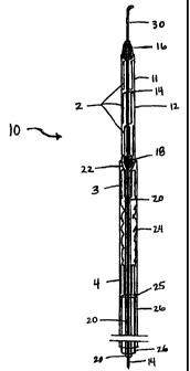
coaxial delivery systems. Referring to FIG. 2A, double coaxial system 10 of
the present
invention comprises two independent coaxial delivery sheaths 11 and 24. Sheath
24 is
constructed for the deployment or release of stage 1 of the two-stage stent
graft in FIG. IA,
while sheath 11 is for the delivery of the scaffolding stent (stage 2) of FIG.
1B, within the
lumen of the anchoring stent (stage 1).
As shown in FIG. 2A, leading portion 12 of sheath 11 is preferably made of a
thin
walled sheath. Sheaths 11 and 24 (and 40, to be discussed below) may be
constructed of any
suitable material such as TEFLON (TEFLON sheaths being commercially available
from
Cook), NYLON, or the like. In an embodiment useful for a single percutaneous
insertion, the
outer diameter of leading portion 12 may be between 7-F and 14-F, and is
preferably 8-F to 12-
F when the delivery system enters through vessels such as femoral arteries,
and even more
preferably 10-F to 12-F in such cases, and is preferably 8-F to 14-F when the
delivery system
enters through vessels such as femoral veins, and even more preferably 10-F to
12-F in such
cases, and is preferably 8-F to 10-F when the delivery system enters through
vessels such as the
carotid artery, and even more preferably 9-F to 10-F in such cases. In an
embodiment useful
for surgical insertion, the outer diameter of leading portion 12 may be
between 8-F and 24-F,
and is preferably 12-F to 16-F when the delivery system enters through vessels
such as femoral
arteries, and even more preferably 12-F to 14-F in such cases, and is
preferably 12-F to 24-F
when the delivery system enters through vessels such as femoral veins, and
even more
preferably 12-F to 16-F in such cases, and is preferably 8-F to 12-F when the
delivery system
enters through vessels such as the carotid artery, and even more preferably 8-
F to 10-F in such
SUBSTITUTE SHEET (RULE 26)
CA 02321204 2000-08-18
WO 99/43379 PCTJUS99/04431
27
cases. It is to be understood that the sizes above may differ according to the
manner of
insertion and the size of the vessel into which the delivery system is placed.
The distal end of the leading portion 12 of leading sheath 11 tapers into
small caliber
catheter 20. In an embodiment useful for a single percutaneous insertion, the
outer diameter of
catheter 20 may be 2.5-F to 5-F, and is preferably 2.5-F to 4-F, and even more
preferably 2.5-F
to 3.5-F. In an embodiment useful for surgery, the outer diameter of catheter
20 may be 3-F to
7-F, and is preferably 3-F to 5-F, and is even more preferably 3-F to 4-F. It
is to be understood
that the sizes above may differ according to the method of insertion and the
size of the vessel
into which the delivery system is placed. Leading portion 12 may be connected
to portion 20
with a tapered connecting piece 22. Connecting piece 22 may be made from
materials similar
to those from which sheaths 11 and 24 (and 40) may be made. The joint of the
distal end of the
leading portion 12 and the tapered connecting piece 22 should be strong enough
to be able to
withstand significant forces during delivery. In one embodiment of the present
invention,
leading portion 12 and small caliber catheter 20 of sheath 11 may be made from
one contiguous
piece, as shown in FIG. 12., thereby eliminating the joint between the two
pieces.
As further shown in FIG. 2A, the scaffolding stent 2 surrounds a pusher wire
14 and is
held within the leading portion 12 of the delivery system between two blocking
pieces 16 and
18, located in spaced relation to one another, one distal and one proximal to
the scaffolding
scent. In one embodiment of the present invention, blocking piece 16 may serve
to secure the
scaffolding stent in position. The front portion of blocking piece 16, the
portion that first enters
the vessel, may also be tapered so as to provide the front portion of the
delivery system with a
smooth profile, thereby facilitating the intravascular travel of the delivery
system. However, it
is to be understood that the use of blocking piece 16 in the embodiments of
the delivery system
disclosed herein is optional. Thus, in one embodiment, blocking piece 16 is
not coupled to
pusher wire 14 (or microtubing 31, to be discussed below). In one embodiment
of the present
invention, blocking piece 18 serves to prevent pusher wire 14, to which it may
be attached as
described below, from being pulled back through sheath 11. Blocking piece 18
serves this
function by contacting tapered portion 22. The portions of the blocking pieces
that may contact
SUBSTITtPI"E SHEET (RULE 26)
CA 02321204 2000-08-18
WO 99/43379 PCT/US99/04431
28
the stents may have circular indentions for keeping the compressed bends of
the stents together
within the respective delivery sheaths.
Pusher wire 14 may be made from any suitable material such as stainless steel,
nitinol,
or the like. In an embodiment useful for a single percutaneous insertion,
pusher wire 14 may
have a diameter ranging from 0.020 inches to 0.060 inches, and is preferably
0.020 inches to
0.045 inches, and even more preferably 0.020 inches to 0.038 inches. In an
embodiment useful
for surgery, pusher wire 14 may have a diameter ranging from 0.020 inches to
0.080 inches, and
is preferably 0.020 inches to 0.060 inches, and even more preferably 0.020
inches to 0.040
inches. It is to be understood that the sizes above may differ according to
the manner of
insertion and the size of the vessel into which the delivery system is placed.
Blocking pieces 16 and 18 (and those discussed below) may be fornzed from any
suitable material such as stainless steel, nitinol, plastic, or any suitable
material. In one
embodiment of the present invention, blocking pieces 16 and 18 may be coupled
to pusher wire
14 by welding, soldering, friction fit, taping, gluing, or any suitable means.
In another
embodiment of the present invention, an adjustable plunger may be used for
blocking piece 18,
as shown in FIG. 10. Such an adjustable plunger may use a tightening screw
mechanism to
achieve its adjustable nature along pusher wire 14 (or microtubing 31, to be
discussed below).
As shown in FIG. 2A and FIG. 2B, the front part of the pusher wire 14 is
equipped with
a short flexible angled tip 30 to facilitate manipulation within the
vasculature. In one
embodiment, tip 30 may be an angled piece of a guidewire formed from a
stainless steel coil
wrapped around a stainless steel core wire, and may be flexible. Tip 30 may be
attached to the
end of pusher wire 14 that first enters the vessel using any suitable means
such as soldering,
welding, gluing, taping, or the like. In another embodiment, pusher wire 14
may be tapered,
and tip 30 may be a coil attached to the tapered portion of the wire, using
any suitable means,
such as those just described. In one such embodiment, tip 30 may be made of a
highly
radiopaque metal such as tungsten, platinum, or the like, or it may be made of
a material such
as rubber, or the like. In one embodiment, the end of tip 30 may be rounded so
as to allow for
smooth passage through the vasculature.
SUBSTITUTE SHEET (RULE 26)
CA 02321204 2000-08-18
WO 99/43379 PCT/US99/04431
29
In an embodiment useful for a single percutaneous insertion, tip 30 may be 2
to 10 cm
in length, and preferably 3 to 8 cm in length for use in vessels such as
femoral arteries, and
even more preferably 3 to 5 cm in length in such cases, and preferably 2 to 10
cm in length for
use in vessels such as femoral veins, and even more preferably 3 to 8 cm in
length in such
cases, and is preferably 2 to 8 cm in length for use in vessels such as
carotid arteries, and even
more preferably 3 to 5 cm in length in such cases. In an embodiment useful
surgical insertion,
flexible tip 30 may be 2 to 8 cm in length, and preferably 2 to 6 cm in length
for use in vessels
such as femoral arteries, and even more preferably 2 to 4 cm in length in such
cases, and is
preferably 2 to 8 cm in length for use in vessels such as femoral veins, and
even more
preferably 2 to 4 cm in length in such cases, and is preferably 2 to 8 cm in
length for use in
vessels such as carotid arteries, and even more preferably 2 to 4 cm in length
in such cases. It is
to be understood that the sizes above may differ according to the manner of
insertion and the
size of the vessel into which the delivery system is placed.
In an embodiment useful for percutaneous insertion, tip 30 may have an outer
diameter
of 0.025 -inches to 0.038 inches, including 0.026, 0.027, 0.028, 0.029, 0.030,
0.031, 0.032,
0.033, 0.034, 0.035, 0.036, or 0.037 inches. In an embodiment useful for
surgical insertion, tip
30 may be shorter and have a larger diameter of 0.025 to 0.080 inches, and is
preferably 0.025
to 0.060 inches in diameter when a femoral artery approach is taken. It is to
be understood that
the sizes above may differ according to the manner of insertion and the size
of the vessel into
which the delivery system is placed. In an embodiment in which tip 30 is made
of soft plastic,
the portion of tip 30 that first enters the vessel may be tapered so as to
reduce its diameter along
the tapered portion.
In another embodiment of the present invention shown in FIG. 11, microtubing
31 may
be utilized instead of pusher wire 14. Advantageously, the microtubing may be
rigid and
flexible, thus providing stability as well as maneuverability to the system.
Further, a guidewire
(to be discussed below) may be utilized with microtubing 31. Blocking pieces
16 and 18 may
be attached to microtubing 31 in the same manner in which they may be attached
to pusher wire
14. As stated above, blocking piece 16 is optional. Microtubing 31 may be
formed of any
suitable material, such as nitinol.
SUBS'TIME SHEET (RULE 26)
CA 02321204 2000-08-18
WO 99/43379 PCT/US99/04431
In an embodiment useful for a single percutaneous insertion, microtubing 31
may have
an outer diameter of 0.025 inches to 0.060 inches, and preferably 0.025 to
0.050 inches for use
in vessels such as femoral arteries, and even more preferably 0.025 to 0.040
inches in such
cases, and preferably 0.025 to 0.060 inches for use in vessels such as femoral
veins, and even
5 more preferably 0.025 to 0.040 inches in such cases, and preferably 0.025 to
0.050 inches for
use in vessels such as carotid arteries, and even more preferably 0.025 to
0.040 inches in such
cases. In such an embodiment, the inner diameter of microtubing 31 may be
0.018 inches to
0.054 inches, and preferably 0.018 to 0.038 inches for use in vessels such as
femoral arteries,
and even more preferably 0.018 to 0.032 in such cases, and is preferably 0.018
to 0.054 inches
10 for use in vessels such as femoral veins, and even more preferably 0.018 to
0.03 8 inches in such
cases, and is preferably 0.018 to 0.045 inches for use in vessels such as
carotid arteries, and
even more preferably 0.018 to 0.035 inches in such cases.
In an embodiment useful for surgical insertion, microtubing 31 may have an
outer
diameter of 0.025 inches to 0.080 inches, and preferably 0.025 to 0.060 inches
for-use for use in
15 vessels such as femoral arteries, and even more preferably 0.025 to 0.040
inches in such cases,
and preferably 0.025 to 0.080 inches for use in vessels such as femoral veins,
and even more
preferably 0.025 to 0.040 inches in such cases, and preferably 0.025 to 0.060
inches for use in
vessels such as carotid arteries, and even more preferably 0.025 to 0.040
inches in such cases.
In an embodiment useful for surgical insertion, microtubing 31 may have an
inner diameter of
20 0.018 inches to 0.075 inches, and preferably 0.018 to 0.054 inches for use
in vessels such as
femoral arteries, and even more preferably 0.018 to 0.035 inches in such
cases, and preferably
0.0 18 to 0.075 inches for use in vessels such as femoral veins, and even more
preferably 0.018
to 0.035 inches in such cases, and preferably 0.018 to 0.054 inches for use in
vessels such as
carotid arteries, and even more preferably 0.018 to 0.035 inches in such
cases. It is to be
25 understood that the sizes above may differ according to the manner of
insertion and the size of
the vessel into which the delivery system is placed, as well as according to
the size of catheter
20 of sheath 11, into which the microtubing is placed.
In another embodiment of the present invention shown in FIG. 13, guidewire 33
may be
used with microtubing 31, such that the microtubing encloses the guidewire.
Thus, the
SUBSTITUTE SHEET (RULE 26)
CA 02321204 2000-08-18
WO 99/43379 PCT/US99/04431
31
microtubing may be advanced and pulled back while enclosing the guidewire. In
one
embodiment, a guiding mechanism may be positioned in operative relation to the
end of the
microtubing. In one embodiment, a tip, such as tip 30, may be coupled to the
end of the
microtubing in order to serve as a guiding mechanism. In another embodiment,
guidewire 33
may serve as a guiding mechanism.
In an embodiment in which guidewire 33 serves as a guiding mechanism,
guidewire 33
should be appropriately sized to fit within the lumen of microtubing 31. For
example, a
guidewire with a diameter of 0.018 inches fits within a microtubing with an
inner diameter of
0.020 inches. Thus, a difference of about 0.002 inches between the diameter of
guidewire 33
and the inner diameter of microtubing 31 may be utilized to size the
guidewire. In an
embodiment useful for a percutaneous insertion, guidewire 33 may have a
diameter of 0.018
inches to 0.052 inches, and preferably a diameter of 0.018 to 0.038 inches,
and even more
preferably a diameter of 0.018 to 0.032 inches. In an embodiment useful for
surgical insertion,
guidewire 33 may have a diameter of 0.018 inches to 0.052 inches, and
preferably a diameter of
0.018 to 0.045 inches, and even more preferably a diameter of 0.018 to 0.035
inches. It is to be
understood that the sizes above may differ according to the manner of
insertion and the size of
the vessel into which the delivery system is placed, as well as the size of
the microtubing
utilized.
Guidewire 33 may be formed of any suitable material, such as nitinol. It is to
be
understood that microtubing 31 may be utilized without guidewire 33. It is to
be understood
that in embodiments in which microtubing 31 is utilized without guidewire 33,
another suitable
guiding mechaaism may be used. For instance, as discussed above, tip 14 may
serve as a
guiding mechanism and may be coupled to microtubing 31 for facilitating
manipulation of the
microtubing within the vessel.
In one embod'nnent of the present invention, using guidewire 33, the operator
of the
delivery system may advantageously have more freedom to negotiate tortuous
vessels during
delivery than when a guidewire is not utilized. A guidewire made from nitinol,
for example,
may also provide the operator with excellent torque-control thereby improving
the
maneuverability of the delivery system. Guidewire 33 or pusher wire 14 may be
controlled by
SUBSTITUTE SHEET (RULE 26)
CA 02321204 2000-08-18
WO 99/43379 PCT/US99/04431
32
the operator with a wire control device 400, shown in FIG. 25 (pusher wire 14
is shown), such
as the CRICKETT device (commercially available from Microvena), the FLOSWITCH
HP
device (commercially available from Boston Scientific), the PIN VISE device
(commercially
available from Cook), or the like.
As shown in FIG. 2A, catheter 20 and tapered connecting piece 22 fit inside
the trailing
portion of the delivery system 24, which may be a thin-walled sheath. As shown
in FIG. 2A,
the anchoring stent encloses catheter 20 and is enclosed by trailing sheath
24. In one
embodiment, sheath 24 may be as large as, or slightly larger than sheath 11.
In one
embodiment, sheath 24 may also be slightly smaller than sheath 11.
In an embodiment useful for a single percutaneous insertion, the outer
diameter of
sheath 24 may be between 8-F and 14-F, and is preferably 9-F to 12-F for use
in vessels such as
femoral arteries, and even more preferably 10-F to 12-F in such cases, and is
preferably 8-F to
14-F for use in vessels such as femoral veins, and even more preferably 10-F
to 14-F in such
cases, and is preferably 8-F to 12-F for use in vessels such as the carotid
artery, and even more
preferably 9-F to 10-F in such cases. In an embodiment useful for surgical
insertion, the outer
diameter of leading portion 12 may be between 8-F and 24-F, and is preferably
12-F to 16-F for
use in vessels such as femoral arteries, and even more preferably 12-F to 14-F
in such cases,
and is preferably 12-F to 24-F for use in vessels such as femoral veins, and
even more
preferably 12-F to 16-F in such cases, and is preferably 8-F to 14-F for use
in vessels such as
the carotid artery, and even more preferably 10-F to 12-F in such cases. It is
to be understood
that the sizes above may differ according to the manner of insertion and the
size of the vessel
into which the delivery system is placed.
As shown in FIG. 2C, end 29 of trailing sheath 24 may overlap leading portion
12 of
sheath 11 by 5 to 10 mm. In such an embodiment, end 29 may be slightly tapered
and/or its
wall thickness may be reduced such that the profile of the transition from
sheath 11 to sheath 24
is smooth. The overlap also may serve to prevent the two sheaths from sliding
apart during
insertion and positioning of the delivery system. Further, the overlap may
serve to reduce the
amount of blood to which stage 1 (which may be enclosed by sheath 24 as
discussed below) is
exposed prior to the delivery of stage 1.
SUBSTtTUTE SHEET (RULE 26)
CA 02321204 2000-08-18
WO 99/43379 PCT/US99/04431
33
As shown in FIG. 2A, the anchoring stent 1 is held in place proximally by the
widened
leading portion of the delivery system and distally by a pusher catheter 26.
Thus, in one
embodiment, the proximal end of the anchoring stent may contact sheath 11, and
the distal end
of the anchoring stent may contact catheter 26. Pusher catheter 26 fits within
the lumen of
trailing sheath 24, and encloses catheter 20 of leading sheath 11. In one
embodiment, end 25 of
catheter 26 serves to release the anchoring stent from the delivery system
during implantation
by contacting the distal end of the anchoring stent as trailing sheath 24 is
pulled back; thus,
catheter 26 is in operative relation with sheath 24. Further, as the delivery
system is being
positioned within the vessel, catheter 26 may also serve to control the
position of the anchoring
stent within sheath 24 in a similar fashion. Catheter 26 may be formed from
similar materials
as those from which sheaths 11 and 24 may be formed.
The size of catheter 26 depends on many factors, including the sizes of
catheter 20 and
sheath 24. The outer diameter of catheter 26 may be about 2-F smaller than the
size of catheter
24. In an embodiment useful for a single percutaneous insertion, the outer
diameter of sheath
24 may-be between 6-F and 12-F, and is preferably 7-F to 10-F for use in
vessels such as
femoral arteries, and even more preferably 8-F to 10-F in such cases, and is
preferably 6-F to
12-F for use in vessels such as femoral veins, and even more preferably 8-F to
12-F in such
cases, and is preferably 6-F to 10-F for use in vessels such as the carotid
artery, and even more
preferably 7-F to 8-F in such cases. In an embodiment useful for surgical
insertion, the outer
diameter of leading portion 12 may be between 6-F and 22-F, and is preferably
10-F to 14-F for
use in vessels such as femoral arteries, and even more preferably 10-F to 12-F
in such cases,
and is preferably 10-F to 22-F when the delivery system is used in vessels
such as femoral
veins, and even more preferably 10-F to 14-F in such cases, and is preferably
6-F to 12-F for
use in vessels such as the carotid artery, and even more preferably 8-F to 10-
F in such cases. It
is to be understood that the sizes above may differ according to the factors
listed above, and
according to the manner of insertion and the size of the vessel into which the
delivery system
may be placed.
As shown in FIG. 2D, in one embodiment of the present invention, blocking
piece 23,
similar to blocking piece 18, may be attached to portion 20 in the manner
described above.
SUgSTiTUTE SHEET (RULE 26)
CA 02321204 2000-08-18
WO 99/43379 PCT/US99/04431
34
Blocking piece 23 may serve the functions of catheter 26, and may therefore be
used instead of
catheter 26. Thus, the location of blocking piece 23 is such that blocking
piece 23 is enclosed
by or positioned within the sheath 24 during operation of the delivery system.
Turning to the control of the delivery system, in one embodiment of the
present
invention, the delivery system may be controlled using the following well-
known control
devices, shown in FIG. 25. The position of microtubing 31 (not shown) with
respect to
guidewire 33 may be controlled using a standard attachment well known in the
art, such as the
FLOSWITCH HP device (commercially available from Boston Scientific).
The position of sheath 11 (which is configured to fit over and enclose pusher
wire 14 as
shown in FIG. 2A, or microtubing 31 as shown in FIG. 6B) with respect to
either pusher wire
14 or microtubing 31 may be controlled as follows. A female hub 402, such as a
Luer female
hub (commercially available from Cook), may be attached to the end of catheter
20 of sheath
11. A male connector, such as a Luer stopcock (not shown) (also commercially
available from
Cook), through which the wire or microtubing is threaded, may engage the
female hub, thereby
securing the position of sheath 11 with respect to the wire or the
microtubing. Further, the male
connector may be provided with a side arm for hemostasis. Saline may also be
injected through
the side arm and into sheath 11 in order to prevent thrombi from accumulating
within the lumen
of sheath 11. Contrast material may also be injected through the side arm and
into sheath 11 in
order to better identify the position of the delivery system under
fluoroscopy. In this regard, as
shown in FIG. 16, in one embodiment of the present invention, sheath 11 (and
sheath 40 to be
discussed below) may be provided with one or more fluid openings 51 defined in
at least
catheter 20, through which contrast material may flow in order to be
distributed to the areas
surrounding the openings. This may be advantageous in the situation in which,
for example,
sheath 24 has been withdrawn so as to deliver stage 1, and contrast material
may no longer be
delivered to the area which was enclosed by sheath 24. Two such male
connectors that may be
provided with side arms 403 are the CHECK FLO adapter 404 (shown in FIG. 25 as
connected
to catheter 26) (commercially available from Cook), and the TOUHY-BORST
adapter (also
commercially available from Cook).
SUBSTITUTE SHEET (RULE 26)
CA 02321204 2000-08-18
WO 99/43379 PCT/US99/04431
The position of catheter 26 with respect to portion 20 of sheath 11 may be
controlled
with the same devices for controlling sheath 11. In one embodiment of the
present invention,
the position of catheter 26 may be fixed relative to the position of catheter
20. The position of
sheath 24 with respect to catheter 26 may also be controlled using these same
devices. In one
5 embodiment, instead of attaching a female hub to sheath 24, sheath 24 may be
equipped with an
adapter 406, such as a CHECK FLO adapter, without a separate female hub, as is
well-known
in the art. In another embodiment, a slide-proof connection may be achieved
between sheath 24
and catheter 26 by providing removable sliding blocker 406, shown in FIGS. 26
and 27. This
blocker may be appropriately labeled (such as "Safety Lock") and may be
removed just prior to
10 delivery of stage 1 (to be discussed below).
In one embodiment of the present invention, at the end of the delivery system
nearest
the operator, the distances between the control devices are selected so as to
allow the stages to
be delivered without the control devices running into each other. For example,
distance 408
may be chosen such that sheath 24 may be pulled back far enough to allow stage
1 to be
15 released without the control devices attached to sheath 24 and catheter 26
interfering with each
other.
As shown in FIG. 2A, stages 1 and 2 are loaded within a delivery system,
according to
one embodiment of the present invention. To assemble the delivery system as
shown in FIG.
2A, small caliber catheter 20 may be positioned in the lumen of stage 1 of the
graft. Stage 1
20 may then be compressed around catheter 20 such that the two form a unit. In
one embodiment
of the present invention in which blocking piece 23 is utilized, stage 1 may
be positioned
between leading portion 12 and blocking piece 23, and then stage 1 may be
compressed around
catheter 20 to form a unit.
Sheath 24 may then be positioned or pulled over the unit so as to enclose the
unit and
25 hold it in place. It is to be understood that in another embodiment, stage
1 also may be
compressed and placed within sheath 24 before catheter 20 is placed within
stage 1, whether or
not blocking piece 23 is utilized.
SUBSTITUTE SHEET (RULE 26)
CA 02321204 2000-08-18
WO 99/43379 PCTIUS99/04431
36
Using a front-loading technique, pusher wire 14 may be placed into the leading
portion
12 of sheath 11, such that the blocking pieces are still outside leading
portion 12. It is to be
understood that microtubing 31 (not shown) may be used instead of pusher wire
14. The inner
or scaffolding stent 2 may be placed over pusher wire 14 so as to enclose
pusher wire 14, and
stage 2 may be positioned between the blocking pieces 16 and 18. It is to be
understood that
stage 2 may be placed over the wire before the wire is placed within sheath
11. The inner or
scaffolding stent may then be compressed around the wire, and leading portion
12 of sheath 11
may be positioned over the compressed scaffolding stent so as to enclose it
and hold it in place.
It is to be understood that stage 2 may also be compressed and positioned
within leading
portion 12, and pusher wire 14 may then be placed within stage 2. Pusher wire
14 may be
positioned within sheath 11 such that blocking piece 18 rests against the
tapered portion
connecting leading portion 12 and catheter 20.
Catheter 26 may then be placed coaxially over small caliber catheter 20 into
the lumen
of the outer thin-walled sheath 24 and may be used as a pusher/holding
catheter for stage 1 of
the graft. It is to be understood that catheter 26 may be placed over small
caliber catheter 20
before or after stage 2 is enclosed by sheath 11.
It also is to be understood that stage 1 may be positioned within sheath 24
before or
after stage 2 is positioned within sheath 11.
It is to be understood that any suitable stents may be loaded into the double
coaxial
delivery system of the present invention. For example, in one embodiment of
the present
invention, an anchoring stent that is similar to the one shown in FIG. 1 A,
but to which a graft
material is coupled using the non-overlap method described above, may be
utilized. By
coupling the cover material to the anchoring stent, at least a portion of one
of the two radially
compressible spring stents may contact a vessel upon delivery of the outer
stage into the vessel.
Like the anchoring stent shown in FIG. 1A, such an anchoring stent may be
compressed around
portion 20 of sheath 11 and sheath 24 may be placed around it so as to enclose
it. In another
embodiment of the present invention, the anchoring stent shown in FIG. 4 to
which a graft
material is coupled using the non-overlap method may also be utilized in the
same fashion. In
SUBSTITUTE SHEET (RULE 26)
CA 02321204 2000-08-18
WO 99/43379 PCT/US99/04431
37
yet another embodiment of the present invention, as discussed above, self-
expanding tube stents
may be utilized for either stage 1 or stage 2.
In one embodiment of the present invention shown in FIGS. 5A and 5B, during
deployment or delivery of a two stage stent (to be discussed in greater detail
below), if only the
distal most portion of the scaffolding stent (denoted by arrows 250) is
supported by blocking
piece 18, the separate serpentine stents might be crammed into each other
within the delivery
sheath because the serpentine stents are supported by only one connecting bar
between each
spring stent. To avoid that result, as shown in FIGS. 6A and 6B, adjustable
plungers 17, which
may have circular indentations as discussed above, may be attached to
microtubing 31 (or
pusher wire 14 not shown) in a manner similar to that described above with
respect to the
adjustable plungers that may be used for blocking piece 18. Plungers 17 may be
positioned in
spaced relation to each other so as to evenly transmit the longitudinal
delivery force applied to
scaffolding stent 2 during delivery thereof to each serpentine stent or body
of stage 2. In
another embodiment of the present invention, the longitudinal delivery force
may be evenly
transmitted to each serpentine stent of the scaffolding stent by attaching
firm connections (not
shown) between the bodies of the scaffolding stent such as reinforcement wires
(not made from
the same single wire as the scaffolding stent) at appropriate locations (such
as on the opposite
side of the scaffolding stent from the connecting bars 8). These reinforcement
wires may be
attached to the bodies by any suitable means such as welding, by crimping
metal clips on the
wires, and the like.
In one embodiment of the present invention, once the delivery system has been
loaded
with the stages and assembled, the devices for controlling the delivery system
may be placed in
the positions above described.
For insertion and deployment of a multi-stage stent graft, the loaded delivery
system
may be inserted into a blood vessel in a single percutaneous insertion or
through a surgical
insertion. Referring to FIG. 2A, stage 2, the inner stage enclosed by sheath
24, is positioned in
front of stage 1, the outer stage enclosed by sheath 11, such that stage 2 is
inserted into the
vessel before stage 1 is inserted into the vessel, just as sheath 11 is
inserted into the vessel
before sheath 24. The position of the stages within the device may be
monitored using
SUBSTlTUTE SHEET (RULE 26)
CA 02321204 2000-08-18
WO 99/43379 PCT/US99/04431
38
fluoroscopy, and further by injecting contrast material into the various
sheaths and the
microtubing as above described. Stage 1, enclosed by sheath 24, is positioned
within the
vessel. In an embodiment in which guidewire 33 is used, this positioning may
be accomplished
by inserting the guidewire into the vessel first, and then sliding the rest of
the delivery system
along the guidewire to the appropriate position. In an embodiment in which
pusher wire 14 is
used, or microtubing 33 is used without the guidewire, this positioning may
occur by guiding
the system to the appropriate position. After guiding the device to the
desired position, the
successive layers or stages of the graft are released with the outermost layer
being released first.
To accomplish the release, the trailing sheath 24 (the portion of the delivery
system surrounding
or enclosing the outer stage) is pulled back while holding in place the
pusher/catheter 26. The
portion of the delivery system immediately in front of the trailing portion is
then pulled back
into the outer stage. Then the inner stage is released into the outer stage by
pulling back sheath
11, which surrounds the inner stage, so as to endovascularly assemble the
stent graft. As will
be appreciated by those skilled in the art, by using successive stages and
coaxial delivery
systems, multi-stage stent grafts containing the desired number of layers or
stages can be
delivered and assembled endovascularly.
As stated above, prior to inserting the delivery system into a vessel an
introducer sheath
may be utilized. In one embodiment, this introducer sheath may be equipped
with a check flow
adapter and side arm fitting. In one embodiment in which a single percutaneous
insertion is
made in the femoral artery, the introducer sheath may be placed into the
artery after a
percutaneous puncture is made. The femoral introducer sheath may then remain
in the femoral
artery as the delivery system is inserted into it and positioned. The size of
the femora[ sheath
will depend on the size of sheaths 11 and 24. If a femoral introducer sheath
is used for access
via a femoral artery, the outer diameter of the introducer sheath may be 12-F,
or smaller. The
use of a femoral introducer sheath is optional, as with all catheter-related
interventions, and, if
used, may be withdrawn after the delivery system has been inserted into the
vessel to avoid
interfering with the operation of sheath 24. Introducer sheaths may also be
used when
insertions are made in vessels such as the carotid artery, or in the femoral
vein. For the latter,
an outer diameter of 14-F is acceptable. Introducer sheaths are not needed for
surgical
insertions.
SUBSTtTUTE SHEET (RULE 26)
CA 02321204 2000-08-18
WO 99/43379 PCT/US99/04431
39
To further illustrate this embodiment, the delivery and assembly of a two
stage stent
system of FIG. lA and FIG. 1B by the delivery system of FIG. 2 is described.
The delivery
system is positioned in the aorta via the optional femoral sheath so that the
mid-portion
containing the covered graft stage 1 is infrarenal and the leading portion 12
of the delivery
system containing the inner scaffolding stent 2 is located more cephalad in
the aorta. Thus, the
inner stage is located cephalad of the outer stage, or closer to the head of
the patient than is the
outer stage. Once stage 1 is in the appropriate position, the pusher/holding
catheter 26 is held
stationary and the outer sheath 24 is pulled back releasing stage 1 into the
vessel, such that
stage 1 engages the vessel. The outer sheath 24 containing the pusher/holding
catheter 26 is
then withdrawn into the femoral sheath so that it does not interfere with the
last phase of graft
deployment. If blocking piece 23 is utilized instead of holding catheter 26 as
described above,
blocking piece 23 is held stationary by so holding pusher wire 14 or sheath 11
to which the
blocking piece may be attached. Stage 1 may then be delivered by pulling back
sheath 24 as
just described.
In-the second step of the delivery, sheath 11 is withdrawn until the inner
stage 2 is
positioned within the lumen of the polyester graft tube 6 between the two
serpentine stents 3
and 4. In another embodiment, the inner stage may be chosen so as to overlap
the serpentine
stents of the outer stage. Once the inner scaffolding stent 2 is in place, the
pusher wire 14, or
microtubing 31 depending on which is being used, is held stationary and the
inner scaffolding
stent 2 is released from within the front portion 12 and into stage 1 by
pulling the small caliber
catheter 20 back. The endovascularly assembled aortic graft is illustrated in
FIG. 3, the
scaffolding stent 2 is positioned within graft material 6 of the anchoring
stent 1.
In another embodiment of the present invention, a three-stage stent graft may
be
delivered utilizing the coaxial delivery system just described. In a first
version in which a
three-stage stent graft may be so delivered shown in FIG. 14, the stages may
be loaded into the
delivery system as the delivery system is assembled in a very similar manner
to that above
described. Stage 1 may be loaded as above described. Then, the third stage,
denoted as 300,
may be loaded next, followed by the loading of stage 2. Both the third stage
and stage 2 may
be loaded in a manner similar to that described above for loading stage 2. The
difference being
SUBSTiTUTE SHEET (RULE 26)
CA 02321204 2000-08-18
WO 99/43379 PCTIUS99/04431
that blocking piece 34 may be placed on microtubing 31 (or pusher wire 14, not
shown)
between blocking pieces 16 and 18 in order to separate stage 2 from the third
stage. Blocking
piece 34 may have a circular indention on one or both surfaces for the reasons
above described.
During delivery, blocking piece 34, may serve to "push" stage 2 as the
delivery system is
5 advanced and it may also serve to "pull" the third stage when the delivery
system is withdrawn.
Further, when stage 2 is delivered or released into stage 1, blocking piece 34
may serve to
support the distal end of stage 2 as sheath 11 is pulled back. Thus, as shown
in FIG. 14, the
three stages are positioned such that stage 2, the intermediate stage, is
inserted into the vessel
first; the third stage, the inner stage, is inserted into the vessel second;
and stage 1, the outer
10 stage, is inserted into the vessel last.
In the first version, after the stages have been loaded into the delivery
system and the
delivery system has been assembled, the three-stage stent graft may be
inserted into a vessel
and endovascularly assembled as follows. The stages may be inserted into the
vessel in the
order above described, in a single percutaneous insertion. Stage 1 may be
positioned and
15 released into the vessel, thereby engaging the vessel, in the manner above
described. Stage 2
may then be positioned within stage 1. Stage 2 may then be released into stage
1, thereby
engaging stage 1, in the manner above described. Leading portion 12 should
remain positioned
around and enclosing stage 3 subsequent to the release of stage 2.
Next, sheath 11 and microtubing 31 (or pusher wire 14, not shown) may be
advanced
20 proximally over guidewire 33 (a cephalad movement--towards the head--when,
for example, an
abdominal aortic aneurysm is being stent grafted) while holding the guidewire
stationary until
the third stage (denoted as 300) is positioned within the lumen of stage 2.
The third stage may
then be released into stage 2, thereby engaging stage 2, by holding
microtubing 31 stationary
and withdrawing or pulling back the small caliber catheter 20 of sheath 11. It
is to be
25 understood that if the third stage is longer than stage 2, it may overlap
stage 2 and also engage a
portion of stage 1.
In a second version in which a three-stage stent graft may be delivered
utilizing the
coaxial delivery system as shown in FIG. 15, stage 1 may be loaded within
trailing portion 24
as above described. Stage 2 may be loaded so as to be positioned distally
behind stage 1. Thus,
SUBSTITUTE SHEET (RULE 26)
CA 02321204 2000-08-18
WO 99/43379 PCT/US99/04431
41
stage 2 may be positioned between an intermediate blocking piece 35 and
catheter 26. Instead
of catheter 26, a blocking piece may be attached to catheter 20 (or pusher
wire 14, not shown)
as above described. Blocking piece 35, like blocking piece 34, may have
circular indentions on
either or both surfaces and serves the same function with respect to stages 1
and 2 as
intermediate blocking piece 34 serves with respect to stage 2 and the third
stage as above
described. As shown in FIG. 15, the third stage (denoted as 300) may be loaded
so as to be
positioned within leading portion 12 of sheath 11. Thus, as shown in FIG. 15,
the three stages
are positioned such when inserted into a vessel, the third stage, the inner
stage, is inserted into
the vessel first; stage 1, the outer stage, is inserted into the vessel next;
and stage 2, the
intermediate stent, is the last of the three stages to be inserted into the
vessel.
As shown in FIG. 15, blocking piece 35 is positioned between catheter 26 (or
the
blocking piece (not shown) that may be used instead of catheter 26) and the
proximal end of
sheath 24.
In the second version, after the stages have been loaded into the delivery
system as
described above, the three-stage stent graft may be inserted into a vessel and
endovascularly
assembled as follows. The stages may be inserted into the vessel in the order
above described,
in a single percutaneous insertion. Stage 1 may be positioned and released
into the vessel,
thereby engaging the vessel, in the manner above described. Sheath 24 should
remain
positioned around and enclosing stage 2 subsequent to the release of stage 1.
Then, the delivery
system may be advanced proximally (a cephalad movement in the case of an
abdominal aortic
aneurysm) over the guidewire, which may be held stationary, until stage 2 is
positioned within
the lumen of stage 1. Once stage 2 is properly positioned, stage 2 may be
released into stage 1,
thereby engaging stage 1, in the manner in which stage 1 was released. Then,
sheath 11 and
microtubing 31 (or pusher wire 14, not shown) may be withdrawn distally over
guidewire 33,
which may be held stationary, until the third stage (denoted 300) is
positioned within the lumen
of stage 2. Once stage 3 is properly positioned, stage 3 may be released into
stage 2, thereby
engaging stage 2, in the manner in which stage 2 and the third stage were
released in version 1.
As with version 1, it is to be understood that if the third stage is longer
than stage 2, it may
overlap stage 2 and also engage a portion of stage 1.
SUBSTITUTE SHEET (RULE 26)
CA 02321204 2000-08-18
WO 99/43379 PCTIUS99/04431
42
In yet another embodiment of the present invention, a three-stage stent graft
may be
delivered and endovascularly assembled utilizing the triple coaxial delivery
system shown in
FIGS. 16 and 17. This delivery system operates in a similar fashion to the
delivery system
described above, and is equipped with a third sheath 40 which is similar in
form and function to
sheath 11. As shown in FIG. 16, sheath 40 has a first portion 41 having a
caliber of about 10-F
to 12-F, including 10.5-F, 11-F, and 11.5-F, and a second portion 43 having a
caliber of about
2-F to 3-F, including 2.5-F. Portions 41 and 43 are connected by tapered
portion 42. In one
embodiment of the triple coaxial delivery system, sheath 11 may overlap sheath
40 as above
described. As with sheath 11, sheath 40 may be formed of one contiguous
sheath. In another
embodiment, sheath 40 may be formed of two separate sheaths 41 and 43, and
connecting piece
42 that may be separate from both sheaths or formed contiguously with either.
Although not
shown, it is to be understood that connecting piece 42 (as well as connecting
piece 22) may be
formed so as not to be tapered. The size of catheter 20 of sheath 11 may be
increased slightly
to ensure that portion 43 can fit within and be enclosed by it. Additionally,
the inner diameter
of blocking piece 18 may be adjusted so as to allow it to fit around portion
43 instead of
microtubing 31. Also shown in FIG. 16 is blocking piece 36, which is similar
in form and
function to blocking piece 18. Blocking piece 36 may be attached to the
microtubing (which
may enclose guidewire 33, not shown) or pusher wire 14 (not shown) as
described above with
respect to blocking piece 18.
FIG. 17 shows the triple coaxial delivery system assembled and loaded with the
three
stages of the stent graft. Stages 1 and 2 may be loaded in the manner
described above regarding
a two-stage stent, and the third stage (denoted as 300) may be loaded like
stage 2. Thus, as
shown in FIG. 17, the three stages are positioned within the triple coaxial
delivery system such
that the third stage, the inner stage, may be inserted into a vessel first;
stage 2, the intermediate
stage, may be inserted into the vessel after the third stage; and stage 1, the
outer stage, is
inserted into the vessel after stage 2.
Control of the triple coaxial delivery system may be achieved using the
devices above
described.
SUBSTITUTE SHEET (RULE 26)
CA 02321204 2000-08-18
WO 99/43379 PCT/US99/04431
43
After the stages have been loaded into the triple coaxial delivery system as
just
described, the three-stage stent graft may be inserted into a vessel and
endovascularly
assembled as follows. The stages may be inserted into the vessel in the order
above described,
in a single percutaneous insertion. Stage 1 may be positioned and released
into the vessel,
thereby engaging the vessel, in the manner above described. Stage 2 also may
be positioned
and released into stage 1, thereby engaging stage 1, as above described with
regard to delivery
of a two-stage stent graft. The only difference being that instead of holding
microtubing 31
stationary while withdrawing sheath 11, portion 43 of sheath 40 may be held
stationary while
sheath 11 is withdrawn. Then, the third stage may be positioned within the
lumen of stage 2
and then released into stage 2, thereby engaging stage 2, in a manner similar
to the release of
stage 2, that is, by holding the microtubing stationary and withdrawing sheath
40. It is to be
understood that if the third stage is longer than stage 2, it may overlap
stage 2 and also engage a
portion of stage 1.
For any of the above embodiments involving the delivery of three stages, any
suitable
stent may-be used for the third stage, as was the case with stages 1 and 2.
The delivery systems of the present invention described herein may be useful
in the
treatment of abdominal aortic aneurysms. As discussed, in such cases, a multi-
stage stent graft
may be loaded into a delivery system which may then be inserted into a femoral
artery, thus
taking a femoral approach. It is to be understood that the delivery systems of
the present
invention described herein may also be useful for endovascularly assembling
multi-stage stent
grafts in the following vessels, using the following approaches: in the
treatment of thoracic
aortic aneurysms ("TAA") using a femoral, or carotid approach; in the
treatment of damaged
iliac arteries using a carotid, subclavian, or brachial approach; and in the
treatment of venous
stenoses on larger veins such as the inferior vena cava ("IVC") using a
femoral vein approach,
or the superior vena cava ("SVC") using a femoral vein approach. It is also to
be understood
that the delivery systems of the present invention described herein may be
useful in stent-
grafting any tubular structure capable of receiving a covered or multi-layer
stent graft, such as a
biliary system stenosed by a tumorous lesion using a percutaneous insertion on
the biliary tree
or endoscopic approach through the mouth; a ureter stenosed or obstructed by a
tumorous
SUBSTITUTE SHEET (RULE 26)
CA 02321204 2000-08-18
WO 99/43379 PCT/US99/04431
44
lesion using a percutaneous antegrade nephrostomy; or a tracheo-bronchial
system such as a
main bronchi stenosed by a tumorous lesion using a tracheal tubing as an
approach. It is also to
be understood that the delivery systems of the present invention described
herein may be useful
in creating transjugular intrahepatic portosystemic shunts ("TIPS") to
decrease portal
hypertension using a percutaneous intemal jugular vein approach, or in
repairing colonic
strictures caused by malignant tumors using an endoscopic approach, and the
like.
The sizes of sheaths 11 (both portions, 12 and 20) and 24, and the size of
catheter 26
would be similar to the sizes listed above for treating a TAA from a carotid
or subclavian
approach using surgery or percutaneous entry. For treating the iliac arteries
via a carotid,
subclavian, or brachial approach via surgical or percutaneous entry, such
sizes would be
proportionately reduced from those listed above such that entry diameter would
preferably be
10-F or smaller. For treating the IVC from a femoral vein using either
insertion, such sizes
would be similar to those listed above for the aortic system. For treating a
biliary system via
percutaneous entry, up to a 12-F outer diameter system may be used. For
treating a main
bronchi via a tracheal tubing, an even larger system than that described above
may be used. For
treating a TIPS via a right inten-al jugular vein, up to a 14-F outer diameter
system may be
used. For a colon via endoscopy, a system with an outer diameter of 10-F may
be used.
The following example is included to demonstrate specific embodiments of the
invention. It should be appreciated by those of skill in the art that the
techniques disclosed in
the example which follows represent techniques discovered by the inventors to
function well in
the practice of the invention, and thus can be considered to constitute
specific embodiments for
its practice. However, those of skill in the art should, in light of the
present disclosure,
appreciate that many changes can be made in the specific embodiments which are
disclosed and
still obtain a like or similar result without departing from the spirit and
scope of the invention.
SUBSTtTUTE SHEET (RULE 26)
CA 02321204 2000-08-18
WO 99/43379 PCT/US99/04431
Example 1
MATERIALS AND METHODS
Graft Construction
An aortic stent graft was made that consisted of two separate stages. As shown
in FIG.
5 20, the first stage, the anchoring stent, consisted of two five-bend
anchoring Z-stents 200 made
of 0.012-inch stainless steel wire each with an unconstrained diameter of 14-
16 nun. The stents
were connected by two stainless steel wire (0.010-inch) struts (not
shown)creating a unit 78-
82 mm long. Eyes were formed at the inner bends of each stent by placing a
bead of solder just
above the bend. A tube 204 of polyester fabric 10 mm in diameter was formed by
heat-sealing
10 the edges. The fabric (PeCap(D polyester; Tetko Inc., Briarcliff Manor, NY)
had a thickness of
61 m and pore size of 95 m. The tube was attached to the eyes of the
anchoring stents with
polypropylene suture (Prolene-5; Ethicon Inc., Sommerville, NJ) so that one-
third of each Z-
stent was covered by the fabric.
The second stage of the stent graft was a triple-body inner scaffolding stent
composed
15 of three six-bend Z-stents 208 constructed from 0.010-inch stainless steel
wire. Each stent was
14 mm in length and 13-14 mm in unconstrained diameter. The stents were
connected to each
other with two stainless steel wire (0.010-inch) struts. Neither the anchoring
stent nor the
scaffolding stent contained barbs.
Delivery System
20 As shown in FIG. 20, a delivery system composed of two independent coaxial
delivery
mechanisms was constructed for the two-stage deployment of the graft. One
delivery
mechanism consisted of a 0.014-inch stainless steel pusher wire 210. The front
part of the wire
was equipped with a 5-cm long piece of 0.028-inch flexible stainless steel
guide wire 212 that
had an angled tip and a tapered piece of a dilator 214 to facilitate
manipulation within the
25 vasculature. The section of the pusher wire that was located just behind
the inner scaffolding
stent was widened, forming a blocking piece 216, so that the wire would be
suitable for pushing
or stabilizing the stent. The remainder of the delivery mechanism was
constructed of a 10-F
OD thin-walled Teflon sheath 218 connected to a 4-F Teflon catheter 220 by
using a tapered
Teflon connecting piece 222. The connecting piece and the 4-F Teflon catheter
were flared
SUBSTITUTE SHEET (RULE 26)
CA 02321204 2000-08-18
WO 99/43379 PCT/US99/04431
46
together and then the pieces were joined with a Silastic medical adhesive (Dow
Coming,
Midland, MN).
The other delivery mechanism consisted of a 9-F Teflon catheter 224 placed
coaxially
over the 4-F Teflon catheter, and a 12-F OD Teflon sheath 226 pulled over
catheter 224. The
9-F Teflon catheter was used as a pusher/holding catheter for the first stage
of the graft.
Assembly of the Delivery System
First, using a front-loading technique, the 0.014-inch stainless steel pusher
wire 210 was
placed into the 10-F OD Teflon sheath 218 attached to the 4-F catheter 220, as
indicated by
arrow 230 in FIG. 20. The inner scaffolding stent was placed over the wire and
positioned
between the tapered dilator 214 and the blocking piece 216. The scaffolding
stent was then
compressed around the pusher wire and placed into the 10-F OD Teflon sheath
218. The
proximal end of the tapered segment of dilator 214 fit into the distal end of
the 10-F OD sheath
218, as shown in FIG. 21. The catheter 220 was positioned in the lumen of the
outer covered
portion of the graft which was then compressed around the catheter. The 12-F
OD Teflon
sheath 226 was pulled over the covered portion of the graft to hold it in
place, as indicated by
arrow 232. The 9-F Teflon catheter 224 stabilized the covered portion of the
graft within the
lumen of the 12-F OD sheath. FIG. 21 shows the parts of the grafts in relation
to each other
after assembling the delivery system.
Animal Evaluation
All experimentation involving animals was approved by the Institutional Animal
Care
and Use Committee of our institution. Animals were maintained in facilities
approved by the
Association for Assessment and Accreditation of Laboratory Animal Care (AAALAC
International) and in accordance with current U.S. Department of Agriculture,
Department of
Health and Human Services, and National Institutes of Health regulations and
standards.
Four adult mongrel dogs (22.5-31.2 kg) were used to test deployment of the
graft using
the double coaxial delivery system. Each dog was anesthetized by intravenous
injection of
pentothal (18.25 mg/kg), and then placed on inhalation anesthesia consisting
of halothane
(1.5%), nitrous oxide (0.3 L/min), and oxygen (0.8 L/min). The right carotid
and right femoral
SUBSTITUTE SHEET (RULE 26)
CA 02321204 2000-08-18
WO 99/43379 PCT/US99/04431
47
arteries were surgically isolated and a 6-F and a 12-F introducer sheath was
inserted into the
respective vessels. Sodium heparin (100 units/kg) was given intravenously.
Biplane abdominal
aortography was performed through a 6-F catheter placed via the carotid
approach.
As shown in FIG. 22, the delivery system was introduced through the right
femoral
artery and positioned in the aorta so that the portion containing the first
stage was infrarenal and
the leading portion of the delivery system containing the inner scaffolding
stent was located
more cephalad in the aorta. The front Z-stent of the anchoring stent was used
to guide graft
placement within the aorta. Once the covered portion of the graft was in the
appropriate
position, the 9-F pusher/holding catheter (not shown) was held stationary and
the 12-F OD
Teflon sheath 226 was pulled back releasing the covered unit. The 12-F OD
sheath containing
the pusher/holding catheter was then removed.
In the second step of the delivery shown in FIG. 23, the 10-F OD sheath /4-F
catheter
unit was withdrawn until the inner scaffolding stent was positioned within the
lumen of the
polyester tube between the two anchoring stents. Once the inner scaffolding
stent was in place,
the pusher wire was held stationary and the stent was released from the 10-F
OD Teflon sheath
218 by pulling the 4-F catheter 220 back.
Once the graft was deployed, the delivery system was completely removed from
the
animal. Aortography was then repeated.
RESULTS
Graft delivery was uneventful and completely successful in all cases. The
insertion and
advancement of the delivery system was easy and smooth. The parts of the
delivery system and
the graft were adequately radiopaque so every moment of graft delivery and
deployment could
be easily monitored under fluoroscopy.
Using the front anchoring stent of the covered portion of the graft for
guidance of the
first step of deployment, accurate placement of the first stage was achieved
in all cases. The
front anchoring stent was successfully positioned infrarenally, just below the
orifice of the more
caudal renal artery.
SUBSTITUTE SHEET (RULE 26)
CA 02321204 2007-06-27
48
Graft deployment was prompt and continuous, and less than two minutes elapsed
between placement of the two separate graft parts. Both coaxial delivery
mechanisms worked
dependably and smoothly. No dislodgment of the first stage or entanglement of
the delivery
system in the graft material occurred during positioning and deployment of the
second stage of
the graft.
It is to be understood that the present invention is by no means limited to
the specif c
embodiments which have been illustrated and described herein and that various
modifications
thereof may indeed be made. All of the methods and apparatus disclosed and
claimed herein
can be made and executed without undue experimentation in light of the present
disclosure.
While the apparatus and methods of this invention have been, described in
terms of illustrative
embodiments, it will be apparent to those of skill in the art that variations
may be applied to the
apparatus and in the steps or in the sequence of steps of the method described
herein without
departing from the concept, spirit and scope of the invention. More
specifically, it will be
apparent _that certain agents which are both chemically and physiologically
related may be
substituted for the agents described herein while the same or similar results
would be achieved.
All such similar substitutes and modifications apparent to those skilled in
the art are deemed to
be within the spirit, scope and concept of the invention as defined by the
appended claims.
25