Note: Descriptions are shown in the official language in which they were submitted.
CA 02321443 2000-09-29
DEVICE FOR PERMANENTLY MARKING A SELECTED TISSUE
LOCATION WITHIN A PATIENT'S BODY
BACKGROUND OF THE INVENTION
1. Field of the Invention
The present invention relates to medical
devices for localizing selected tissue sites in a
body and, more particularly, to an improved device
adapted to individually percutaneously deliver a
selected number of permanent markers to desired
tissue locations within a patient's body.
2. Description of the Prior Art
More and more, since the advent of breast
screening program by mammography and extensive
regulation of quality control, very small lesions are
found before they become clinically palpable.
Since the eighties, there are technologies
rendering these non-palpable lesions accessible for
percutaneous biopsy, such as fine-needle aspiration
biopsy, core biopsy or vacuum assisted biopsy. It is
necessary to sample a portion of the lesion to obtain
a diagnosis to prevent surgical excision in lesions
that are benign and plan the best treatment in
patients when cancerous cells are present. A minimum
amount of tissue must be obtained to be able to have
a representative sample of the lesion. In some cases,
the lesions being biopsied are so small that after
percutaneous biopsies, the lesions are either
difficult to locate or completely removed.
Also, more and more, preoperative
chemotherapy is administered after a diagnosis of
breast cancer has been established by these
percutaneous biopsies. After chemotherapy, the
lesion completely disappears and there is a need to
mark the precise location (the superior margin, the
inferior margin, the lateral and medial margin) of
the cancer so that the area can be properly excised.
- 1 -
CA 02321443 2000-09-29
Accordingly, various devices have been
developed to enable relatively precise identification
of a biopsy site for subsequent surgical procedures.
For instance, United States Patent No. 5,853,366
issued on December 29, 1998 to Dowlatshahi discloses
a tissue marking device comprising a hollow
hypodermic needle having a distal piercing end
adapted to be inserted into a patient's body to
deliver a marker to a lesion site, using conventional
imaging techniques. The marker is pushed out of the
needle by a plunger mounted for free sliding motion
with respect to the needle.
Although the tissue marking device
disclosed in the above mentioned patent is effective,
it has been found that there is a need for a tissue
marking device adapted to safely and individually
deliver a desired number of markers to selected
tissue locations without having to reload said
device.
SUMMARY OF THE INVENTION
It is therefore an aim of the present
invention to provide a tissue marking device adapted
to individually deliver a desired number of markers
without having to be reloaded.
It is also an aim of the present invention
to provide a tissue marking device which is
relatively simple and economical to manufacture.
It is a further aim of the present
invention to provide a tissue marking device which
can be conveniently used to deliver a desired number
of markers to selected tissue locations.
Therefore, in accordance with the present
invention, there is provided a kit for marking a
selected tissue location within a patient's body,
comprising an elongated guide member having a distal
end adapted to be inserted into a patient's body to a
selected tissue location, a proximal end extending
- 2 -
CA 02321443 2000-09-29
outwardly from the patient's body once said distal
end has been introduced therein, a passageway
extending from said proximal end to said distal end,
at least first and second markers adapted to be
successively loaded into said passageway, and an
actuator adapted to be inserted through said proximal
end, into said passageway, to a first position in
which further insertion of said actuator is
temporally prevented so that a predetermined length
of said actuator extends into said passageway to
cause only said first marker to be discharged through
said distal end, and from said first position to a
second position in which a sufficient length of said
actuator now extends into said passageway to cause
said second marker to be discharged from said distal
end, thereby ensuring individual discharge of said
first and second markers.
In accordance with a further general aspect
of the present invention, there is provided a kit for
marking a selected tissue location within a patient's
body, comprising an elongated guide member defining a
passageway extending longitudinally therethrough
between a distal end and a proximal end, said distal
end being adapted to be inserted into a patient's
body to a selected tissue location, at least two
markers adapted to be successively loaded into said
passageway, an actuator adapted to be inserted into
said passageway, through said proximal end, for
successively pushing said at least two markers out of
said passageway through said distal end, and an
indexing mechanism for releasably blocking said
actuator at successive predetermined levels of
insertion into said passageway, wherein each said
predetermined level of insertion is associated with
the individual discharge of one of said at least two
markers.
- 3 -
CA 02321443 2000-09-29
BRIEF DESCRIPTION OF THE DRAWINGS
Having thus generally described the nature
of the invention, reference will now be made to the
accompanying drawings, showing by way of illustration
a preferred embodiment thereof, and in which:
Fig. 1 is a schematic, partly exploded,
side view of a tissue marking device in accordance
with a first embodiment of the present invention;
Fig. 2 is a schematic perspective view of a
stopper forming part of the device of Fig, l;
Fig. 3 is a schematic, partly exploded,
side view of a tissue marking device in accordance
with a second embodiment of the present invention;
and
Fig. 4 is enlarged side view of a proximal
end portion of an actuator forming part of the device
of Fig. 3.
DESCRIPTION OF THE PREFERRED EMBODIMENTS
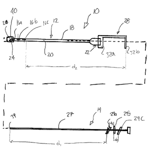
Now referring to the drawings, a device 10
for permanently marking a selected tissue location
within a patient's body will be described.
As shown in Fig. 1, the device 10 generally
includes an elongated guide member 12 and an actuator
14 movable within the elongated guide member 12 in a
predetermined sequence for individually releasing a
certain number of markers 16a, 16b and 16c (three in
the illustrated embodiment) that are pre-loaded into
the elongated guide member 12.
The elongated guide member 12 can be
provided in the form of a hollow needle or cannula 18
defining a passageway 20 extending longitudinally
therethrough between a funnel-shaped proximal end 22
and a sharp distal end 24 defining an opening for
percutaneously delivering the markers 16a, 16b and
16c to selected tissue locations within a patient's
body, using conventional imaging systems. The distal
- 4 -
CA 02321443 2000-09-29
nd 24 is preferably beveled or angled to form a
cutting edge 26 in order to facilitate the
introduction thereof into the tissues of a patient's
body, as is well known in the art. According to a
preferred embodiment, the needle used is an 18-gauge
spinal needle and is about 88 mm in length. It is
understood that the dimensions of the needle 18 can
vary depending upon the type and location of the site
to be marked.
The markers 16a, 16b and 16c are preferably
provided in the form of mechanically closed clips
made of a biocompatible radiopaque non ferromagnetic
material, such as titanium or the like, radioactive
material, and have identical cross-sections which
generally correspond to that of the passageway 20 to
prevent the markers 16a, 16b and 16c from moving
along the passageway 20 during manipulation of the
needle 18. The term radiopaque or non ferro magnetic
or radioactive is herein intended to include any
material that can be detected using conventional
radiographic, sonographic or magnetic techniques or
with a gamma probe . The markers 16a, 16b and 16c are
adapted to be pre-loaded into the needle 18 through
the proximal end 22 thereof down to the distal end
24. The markers 16a, 16b and 16c are configured to be
wholly received within a patient's body and can have
any appropriate anchoring means to prevent migration
thereof after they have been delivered to a selected
tissue location.
According to the illustrated embodiment,
the actuator 14 is provided in the form of a stylet
27 properly sized to be slidably inserted into the
passageway 20 through the proximal end 22 in order to
successively pushed the markers 16a, 16b and 16c out
of the distal end 24 of the needle 18.
As shown in Fig. 1, a stopper 28 is adapted
to be mounted to the proximal end 22 of the needle 18
_ 5 _
CA 02321443 2000-09-29
to cooperate with a predetermined number of spaced-
apart index pins 29a, 29b and 29c (the number of
index pins matching the number of markers)
distributed along the proximal end portion of the
stylet 27 and oriented in different radial directions
so as to releasably block the stylet 27 at selected
depths of insertion within the needle 18, as will be
described hereinbelow.
As shown in Fig. 2, the stopper 28 includes
a substantially C-shaped body 30 having a pair of
legs 32a and 32b spaced-apart by a web member 34. The
distal leg 32a defines an opening 36 for allowing the
stopper 28 to be tightly fitted over the proximal end
22 of the needle 18. The proximal leg 32b defines an
opening 38 which is aligned with the opening 36 to
form a passage for allowing the stylet 27 to be
introduced into the passageway 20. The opening 38 is
sized to successively receive the index pins 29a, 29b
and 29c when the same are respectively aligned
therewith.
The distance dl between a leading end 39 of
the stylet 27 and the first index pin 29a is selected
to correspond to the distance d2 between the trailing
end of the third marker 16c and the outer surface of
the leg 32b such that when the stylet 27 is
introduced into the needle 18 through the stopper 28
with the first index pin 29a abutting against the
outer surface of the leg 32b, the leading end 39 of
the stylet 27 will be located immediately upstream of
the third marker 16c.
To further insert the stylet 27 into the
needle 18, the physician manipulating the device or
needle has to rotate the stylet 27 about its
longitudinal axis so as to angularly place the first
index pin 29a in phase with the opening 38, thereby
allowing the stylet 27 to freely slide into the
needle 18 along a distance d3, that is until the
- 6 -
CA 02321443 2000-09-29
second index pin 29b, which is 90 degrees offset with
respect to the first index pin 29a, comes in contact
with the outer surface of the leg 32b. The length of
the stylet 27 that extends into the needle 18 when
the second index pin 29b abuts the leg 32b is such
that only the first marker 16a is pushed out of the
distal end 24 of the needle 18 by the stylet 27.
Indeed, the distance d3 between the front face of the
first index pin 29a and the second index pin 29b is
equal to the length of the markers 16a, 16b and 16c.
When it is desired to discharge the second marker
16b, the physician has to rotate the stylet 27 90
degrees upon itself to place the second index pin 29b
in phase with the opening 38, thereby allowing the
stylet 27 to be further introduced into the needle 18
until the stylet stroke is blocked by the third index
pin 29c, which is 90 degrees angularly offset
relative to the second index pin 29b. The distance d4
between respective front faces of the second and
third index pins 29b and 29c also corresponds to the
length of the markers 16a, 16b and 16c and, thus,
only the second marker 16b will be ejected from the
needle 18 during this second dispensing operation.
To eject the third marker 16c, the
physician has to rotate the stylet 27 90 degrees upon
itself so as to place the third index pin 29c in
phase with the opening 38, and then pushed the stylet
27 further into the needle 18 to a fully inserted
position thereof.
The above-described indexing mechanism
advantageously prevents two or more markers from
being inadvertently simultaneously expelled out of
the needle 18 during a marking operation.
Furthermore, the present invention advantageously
allows to individually deliver a plurality of markers
without having to re-load the device after each
single marker delivery.
_ 7 _
CA 02321443 2000-09-29
The needle 18, the markers 16a, 16b and 16c
and the stylet 27 are preferably manufactured in the
form of a kit wherein the markers 16a, 16b and 16c
are pre-loaded into the needle 18 and packaged in a
sterile fashion with a safety cap 40 fitted over the
distal end 24 of the needle 18 and with the stopper
28 and the first index pin 29a cooperating to prevent
the stylet 27 from releasing the markers 16a, 16b and
16c during shipping.
In use, the needle 18 is first inserted
into the patient's body with the markers 16a, 16b and
16c loaded in the needle 18 so that when the distal
end 24 thereof is located proximate to a selected
tissue location, the stylet 27 can be manipulated as
described hereinbefore to successively wholly
position a selected number of markers 16 within the
patient's body. A conventional visualization aid is
used to confirm the position of the distal end 24.
The markers 16a, 16b and 16c can remain in
the body even after the procedure is completed to
allow subsequent identification and observation of
the marked site(s). If desired, the markers 16a, 16b
and 16c may be removed using conventional surgical
techniques.
Figs. 3 and 4 illustrate another embodiment
of the present invention wherein the index pins 29a,
29b and 29c are replaced by three externally threaded
zones 129a, 129b and 129c that are spaced-apart along
a stylet 127 to successively threadably engage an
internally threaded zone 128 provided in a proximal
end 122 of a hollow needle 118 which is otherwise
similar to needle 18. The distance between the
beginning of successive threaded zones 129a, 129b and
129c is equal to the length of the pre-loaded markers
116a, 116b and 116c positioned at a distal end 124 of
the needle 118.
_ g
CA 02321443 2000-09-29
The stylet 127 is sized to be freely
slidable into the needle 118 until its first threaded
zone 129a encounters the internally threaded zone 128
of the needle 118, thereby preventing the stylet 127
from being further slidably inserted into the needle
118. In this position a leading end 139 of the stylet
127 is located immediately adjacent the trailing end
of the third marker 116c. To eject the first marker
116a, the physician must screw the stylet 127 so as
to cause the first externally threaded zone 129a
thereof to pass translativally beyond the internally
threaded zone 128 of the needle 118, thereby allowing
the stylet 127 to be subsequently further slidably
inserted into the needle 118 until its second
threaded zone 129b encounters the internally threaded
zone 128 of the needle 118. The length of the stylet
127 which extends into the needle 118 when the second
externally threaded zone 129b of the stylet 127
engages the internally threaded zone 128 of the
needle 118 is such as to only cause the ejection of
the first marker 116a.
The second marker 116b can be subsequently
ejected by first screwing the stylet 127 into the
needle 118 to cause the second externally threaded
zone 129b of the stylet 127 to pass beyond the
internally threaded zone 128 of the needle 118 and
then pushing the stylet 127 further into the needle
118 until the third externally threaded zone 129c of
the stylet 127 contacts the internally threaded zone
128 of the needle 118. The advancement of the stylet
127, i.e. the distance between the beginning of the
second and third threaded zones 129b and 129c,
corresponds to the length of the second marker 116b
and, thus, cause the ejection thereof.
Finally, the third marker 116c can be
discharged from the distal end 124 of the needle 118
by screwing the stylet 127 so as to cause the
_ g _
CA 02321443 2000-09-29
externally threaded zone 129c to enter into the
needle 118 past the internally threaded zone 128 and
subsequently pushing the stylet 127 further into the
needle 118 to a fully inserted position thereof.
Other indexing mechanisms adapted to
releasably block the stylet at different depths of
insertion could be used as well.
- 10 -