Note: Descriptions are shown in the official language in which they were submitted.
CA 02328535 2003-09-30
1
ROCHE DIAGNOSTICS CORPORATION 5431/00
BIOSENSOR APPARATUS
Field of the Invention
The present invention relates to a sensor, more particularly to a top dose
sensor.
Background of the Invention
Electrochemical biosensors are known. They have been used to determine the
concentration
of various analytes from biological samples, particularly from blood.
Electrochemical
biosensors are described in U.S. Patent Nos. 5,413,690; 5,762,770; and
5,798,031; as well as
in International Publication No. W099/30152,
Summary of the Invention
According to the present invention, a biosensor apparatus is provided that
comprises a base,
1~5 electrodes positioned on the base, a cover including ports, at least one
plate positioned
between the electrodes and the cover, and a reagent situated on at least one
plate. At least one
plate includes apertures and microstructures spaced-apart from the apertures.
In addition, according to the present invention a biosensor apparatus is
provided that
comprises a base, electrodes positioned on the base, a plate positioned on the
base, a cover,
2o and at least one reagent positioned between the plate and the cover. The
plate includes
apertures in communication with at least one of the electrodes and the cover
includes ports
disposed through the cover. The ports are in corivmunication with the plate
and offset from the
apertures.
Still further, the present invention provides a biosensor apparatus that
comprises a base,
25 electrodes positioned on the base, a first plate positioned on the base, a
second plate
positioned on the first plate, a cover positioned on the second plate, and a
reagent positioned
on at least one of the first and second plates. The first and second plates
each include
CA 02328535 2004-04-28
2
apertures in an offset relationship relative to one another and the cover
includes ports in
an offset relationship to the apertures of the second plate.
Additionally, in accordance with the present invention a biosensor apparatus
for
detecting an analyte in a fluid sample is provided. The biosensor apparatus
comprises a
base, electrodes positioned on the base, a cover spaced-apart from the
electrodes and
being formed to include ports sized to receive the fluid sample a reagent, and
means for
distributing the fluid sample from the ports in the cover to the electrodes.
The
distributing means is formed to spread the fluid sample radially outwardly
from the port
in the cover and to also permit the fluid to flow in a direction generally
perpendicular to
the cover toward the electrodes.
In accordance with one aspect of the invention there is a biosensor apparatus
comprising:
a base, electrodes positioned on the base, a cover including ports, at least
one plate
positioned between the electrodes and the cover, the at least one plate
including an upper
face facing the cover, a lower face, apertures, and microstructures extending
from the
upper face toward the cover spaced-apart from the apertures, and a reagent
situated on
the at least one plate.
In accordance with another aspect of the invention there is a biosensor
apparatus
comprising: a base, electrodes positioned on the base, a cover including
ports, at least
one plate positioned between the electrodes and the cover, the at least one
plate including
an upper face, a wall extending from the upper face and cooperating with the
upper face
to define an upper recess, apertures, and microstructures spaced-apart from
the apertures
and extending from the upper face into the upper recess, and the
microstructures being
cone-shaped, and a reagent situated on the at least one plate.
In accordance with yet another aspect of the invention there is a biosensor
apparatus
comprising: a base, electrodes positioned on the base, a plate positioned on
the base, the
plate being formed to include an upper face, a lower face facing the base,
first and
second ends, and apertures in fluid communication with at least one of the
electrodes,
wherein one of the first and second ends is connected to the base, a cover
being formed
to include ports disposed through the cover, the ports being in fluid
communication with
DOCSMTL: 1394415\1
CA 02328535 2004-04-28
2a
the plate and offset from the apertures, at least one reagent positioned
between the plate
and the cover, and wherein the plate is formed to include an upper recess
defined by the
upper face and a wall extending from the upper face.
In accordance with another embodiment of the invention there is a biosensor
apparatus
comprising: a base, electrodes positioned on the base, a plate positioned on
the base, the
plate being formed to include apertures in fluid communication with at least
one of the
electrodes, an upper face, a wall extending from the upper face and
cooperating with the
upper face to define an upper recess, a lower face, a wall extending from the
lower face
and cooperating with the lower face to define a lower recess, and
microstructures
extending from the upper face into the upper recess, wherein the apertures
extend
between the upper and lower faces, a cover being formed to include ports
disposed
through the cover, the ports being in fluid communication with the plate and
offset from
the apertures, and at least one reagent positioned between the plate and the
cover,
wherein the microstructures extend into the ports of the cover.
In accordance with yet another embodiment of the invention there is a
biosensor
apparatus comprising: a base, electrodes positioned on the base, a plate
positioned on the
base, the plate being formed to include apertures in fluid communication with
at least
one of the electrodes, a cover being formed to include ports disposed through
the cover,
the ports being in fluid communication with the plate and offset from the
apertures, at
least one reagent positioned between the plate and the cover, and a hinge
extending
between the cover and the plate.
In accordance with a further embodiment of the invention there is a biosensor
apparatus
comprising: a base, electrodes positioned on the base, a plate positioned on
the base, the
plate being formed to include apertures in fluid communication with at least
one of the
electrodes, a cover being formed to include ports disposed through the cover,
the ports
being in fluid communication with the plate and offset from the apertures, at
least one
reagent positioned between the plate and the cover, and a hinge extending
between the
base and the plate.
DOCSMTL: 1394415\1
CA 02328535 2004-04-28
2b
In accordance with yet a further embodiment of the invention there is a
biosensor
apparatus comprising: a base, electrodes positioned on the base, a first plate
positioned
on the base and including an inner side facing the base, an outer side, and
first and
second ends, a second plate positioned on the first plate, the second plate
including an
inner side facing the first plate, an outer side, and first and second ends,
the first and
second plates each being formed to include apertures in an offset relationship
relative to
one another and the first ends of the first and second plates being connected,
a cover
positioned on the second plate, the cover being formed to include ports in an
offset
relationship to the apertures of the second plate, and a reagent positioned on
at least one
of the first and second plates.
In accordance with a further aspect of the invention there is a biosensor
apparatus
comprising: a base, electrodes positioned on the base, a first plate
positioned on the base,
the first plate including microstructures extending into the apertures of the
second plate,
a second plate positioned on the first plate, the first and second plates each
being formed
to include apertures in an offset relationship relative to one another, a
cover positioned
on the second plate, the cover being formed to include ports in an offset
relationship to
the apertures of the second plate, and a reagent positioned on at least one of
the first and
second plates.
In accordance with yet a further aspect of the invention there is a biosensor
apparatus for
detecting an analyte in a fluid sample, the apparatus comprising: a base,
electrodes
positioned on the base, a cover spaced-apart from the electrodes and being
formed to
include ports sized to receive the fluid sample a reagent, and means for
distributing the
fluid sample and the reagent from the ports in the cover to the electrodes,
the distributing
means being formed to spread the fluid sample radially outwardly from the port
in the
cover and to also permit the fluid to flow in a direction generally
perpendicular to the
cover toward the electrodes, wherein the distributing means comprises an upper
face
facing the cover, a lower face, and microstructures extending from the upper
face toward
the cover.
DOCSMTL: 1394415\1
CA 02328535 2004-04-28
2c
Additional features of the invention will become apparent to those skilled in
the art upon
consideration of the following detailed description of the preferred
embodiment
exemplifying the best mode of carrying out the invention as presently
perceived.
BRIEF DESCRIPTION OF THE DRAWINGS
The detailed description particularly refers to the accompanying figures in
which:
FIG. 1 is a perspective view of an electrochemical sensor according to an
aspect of the
invention in a partially expended position;
FIG. 2 is a perspective view of the sensor of FIG. 1 in a folded position;
FIG. 3 is a view taken along lines 3--3 of FIG. 2;
FIG. 4 is an enlarged perspective view of a microstructure of the sensor of
FIG. 3;
FIG. 5 is a perspective view of an electrochemical sensor according to a
further aspect of
the invention in a partially expanded position;
FIG. 6 is a top plan view of an upper plate of the sensor of FIG. 5;
FIG. 7 is a cross-sectional view of the sensor of FIG. 5 in a folded position;
FIG. 8 is a perspective view of an electrochemical sensor according to a
further aspect of
the invention in a fully expanded position;
DOCSMTL: 1394415\1
CA 02328535 2000-12-13
3
Fig. 9 is a perspective view of the sensor of Fig. 8 is a partially etpanded
position; and
Fig. 10 is a perspective view of the sensor of Fig. 8 in a folded position.
Detailed Description of the Drawings
The present invention relates to a top dose sensor that provides a
manufacturer with the ability
to transport a liquid sample both horizontally and vertically in relatively
short times. The
sensor of the present invention, comprises a series of perforated plates that
are formed so that
adjacent plates are situated in a generally parallel overlapping relationship
to form a fluid
distribution gap between the adjacent plates. The apertures of the plates are
offset from one
another. Thus, the liquid flows through the series of plates, alternating in
vertical flow
1 o through the apertures and horizontal flow through the fluid distribution
gap between the
plates.
Various aspects of the invention are presented in Figs. 1-10, which are not
drawn to scale and
wherein like components in the several views are numbered alike. Referring now
specifically
to Figs. 1-3, a sensor 10 of the present invention includes a base 12, an
electrode set 14
positioned on base 12, a cover 16, a series of plates 18, and a series of
hinges 23 connecting
base 12, cover 16, and series of plates 18 together. Cover 16 and plates 18
cooperate to
distribute a liquid sample 24 horizontally as sample 24 travels toward
electrode set 14. As will
be discussed later in detail, base 12, cover 16, series of plates 18 and
hinges 23 are formed
from a single piece of molded mufti-resinous material.
2o Electrode set 14 and series of plates 18 are supported on base 12 of sensor
10. Base 12
includes a top surface 26 facing series of plates 18, a bottom surface 28, a
front end 30, a back
end 32, and side walls 34, 36. A cavity 38 is formed through top surface 26
and front end 30.
Cavity 38 is sized to receive an electrical insulator 40 therein. While base
12 is shown to be
generally rectangular in shape, it is contemplated that base 12 may be formed
in a variety of
shapes and sizes in accordance with this disclosure.
As shown in Figs. 1 and 3, insulator 40 is coupled to base 12 within cavity
38. Insulator 40
includes an upper side 42, a lower side 44 engaging base 12, a front end 46
positioned
adjacent to front end 30 of base 12, a back end 48, and sides 50, ~2. As shown
in Fig. 1,
electrode set 14 extends across upper side 42 of insulator 40 from front end
46 toward back
CA 02328535 2000-12-13
end 48. Insulator 40 is formed to prevent an electrical connection from
existing between the
electrodes of electrode set 14. Non-limiting examples of a suitable insulator
40 include glass,
ceramics, and polymers such as a polyester or polyimide. Specific examples of
a suitable
material include glass; the polyimide UPILEX from UBE INDUSTRIES, LTD., Japan,
which
is available pre-coated with gold. palladium or platinum from TECHNI-MET of
Connecticut,
USA; or ULTEM 1000 (polyetherimide) from GE, available coated with copper.
Preferably,
the insulator is constructed of glass and electrode set 14 is positioned in
the glass.
Additionally, insulator 40 is coupled to base 12 by an adhesive. It is
contemplated, however,
that insulator 40 can be coupled to base 12 using solvent-based adhesives,
ultrasonic welding,
or mechanical fasteners such as dovetails, pins, snaps, rivets, screws,
staples, or the like in
accordance with this disclosure.
As shown in Fig. 1, electrode set 14 includes two electrically conductive
tracks 54, 56 that are
laid down into upper side 42 of insulator 40. Track 54 may be a working
electrode and track
56 may be a counter electrode. Tracks 54, 56 are constructed from electrically
conductive
materials. Examples include aluminum, carbon (such as graphite). cobalt,
copper, gallium,
gold, indium, iridium, iron, lead, magnesium, mercury (as an amalgam), nickel,
niobium,
osmium, palladium, platinum, rhenium, rhodium, selenium, silicon (such as
highly doped
polycrystalline silicon), silver, tantalum, tin, titanium, tungsten, uranium,
vanadium, zinc,
zirconium, mixtures thereof, and alloys or metallic compounds of these
elements. Preferably,
2o the tracks include gold, platinum, palladium, iridium, or alloys of these
metals, since such
noble metals and their alloys are unreactive in biological systems. Most
preferably, track 54 is
a working electrode made of gold, and track 56 is a counter electrode that is
also made of gold
and is substantially the same size as the working electrode.
Tracks 54, 56 that serve as working and counter electrodes respectively have
contact pads 59
that are electrically connected to a sensing region 61. The values for the
dimensions illustrated
in Figs. 1-3 are for a single specific embodiment and these values may be
selected as need for
the specific use. For example, the length of electrode set 14 may be 1.5 to
250mm, the width
may be 0.4 to 40 mm, the gap between contact pads 59 may be 0.1 pm to 5 mm,
and the width
of each contact pad 59 may be 0.1 to 20 mm. The electrode pattern shown in
Fig. 1 is
3o symmetric; however this is not required, and irregular or asymmetric
patterns (or electrode
shapes) are possible. It is also contemplated that electrode set 14 can be
coupled to insulator
CA 02328535 2000-12-13
40 using a wide variety of techniques, such as adhesives, dovetail
connections. hook-and-loop
type fasteners, etc. in accordance with this disclosure. It is also
contemplated that electrodes
may be positioned on base 12 using commercially available techniques such as
screen
printing, sputtering, laser ablation, photolithography, etc. in accordance
with this disclosure.
5 Series of plates 18 extends between base 12 and cover 16 and distributes
sample 24 in a
horizontal direction as sample 24 travels from cover 16 toward electrode set
14. Series 18
includes a first plate 22 resting upon base 12, a second plate 20 resting upon
first plate 22, and
a third plate 25 adjacent to cover 16 and resting upon second plate 20. See
Fig. 2. It is
contemplated that series 18 may have as few as one or two plates or may have
greater than
o three plates. Series of plates 18 are coupled together so that plates 20,
22, 25 are positioned in
a generally parallel relationship relative to one another when in a folded
position, as shown in
Fig. 2.
Each plate 20, 22, 25 in series 18 includes an inner side 58 and an outer side
60. As shown in
Figs. 2 and 3, plates 20, 22, 25 are positioned with respect to base 12 so
that outer side 60 of
~5 each lower plate in series 18 supports inner side 58 of an adjacent upper
plate in series 18.
Referring now to Fig. 3, outer side 60 of each plate 20, 22, 25 includes an
upper recess 66 that
is defined by an upper face 70 and a wall 72 extending from upper face 70.
Likewise, inner
side 58 of plates 20, 22, 25 includes a lower recess 68 defined by a lower
face 74 and a wall
76 extending from lower face 74. Walls 72, 76 are in general alignment with
one another to
20 limit the amount of horizontal distribution of sample 24 on plates 20, 22,
25. Upper face 70 of
each plate 20, 22, 25 is preferably hydrophilic to aid in the distribution of
sample 24. While
upper and lower faces 70, 74 each have a generally circular shape (Fig. 1), it
is contemplated
that faces may be oblong, triangular, square, rectangular, trapezoidal, etc.
in shaped in
accordance with this disclosure.
25 When sensor 10 is in the folded position, as shown in Figs. 2 and 3, plates
20, 22, 25 are
stacked on top of one another. Lower recess 68 of cover 16 and upper recess 66
of plate 25
cooperate to define a first horizontal distribution gap 81. Lower recess of
plate 25 and upper
recess of plate 20 cooperate to define a second horizontal distribution gap
83. Likewise, lower
recess 68 of plate 20 and upper recess 66 of plate cooperate to define a third
horizontal
3o distribution gap 85 and lower recess 68 of plate 22 and insulator 40
cooperate to define a
fourth horizontal distribution gap 87. Distribution gaps 81, 83, 85. 87 are
generally
CA 02328535 2000-12-13
perpendicular to apertures 88 in plates in series 18. In addition plate 22
includes an air vent
102 that extends between lower recess 68 and an edge 62 of plate 22. It is
contemplated that
vent 102 may have a variety of sizes and paths and may extend through any one
or greater
than one of the plates 22, 20, 25 or from upper recess 66 in accordance with
this disclosure, so
long as air is vented from sensor 10 as sample 24 travels toward electrode set
14.
Sensor 10 of the present invention pulls sample 24 from cover 16 toward
electrode set 14.
This movement is accomplished both by gravity and by increasing capillary pull
as sample 24
moves from cover 16 toward electrode set 14. The capillary strength of series
of plates 18
increases from cover 16 to insulator 40 as the height of distribution gaps 81,
83, 85. 87
1o decreases. Horizontal distribution gaps 81, 83, 85, 87 range in height from
about 5gm to
1000~m, preferably about l Opm to 200pm, and most preferably about 25~m to
100g.m. For
example, first distribution gap 81 has a height of about 100~.m, second
distribution gap 83 has
a height of about 75~m, third distribution gap 85 has a height of about 50~m,
and fourth
distribution gap 87 has a height of about 25g.m. It is contemplated that the
height of
15 distribution gaps 81, 83, 85, 87 may be substantially equal, or may vary so
long as height of
gap 81, 83, 85, 87 is sufficient to pull sample 24 across the corresponding
plate 25. 20, 22 or
insulator 40 by capillary action.
As shown in Fig. 3, each plate 20, 22, 25 includes microstructures 86
extending from upper
face 70 into recess 66 and apertures 88 extending through upper and lower
faces 70, 74.
2o Referring now to Fig. 4, microstructures 86 are cone-shaped and are formed
to include an
interrupted face 87 suitable for providing an edge for sample 24 and make a
smooth transition
between plates 20, 22, 25. Interrupted face 87 of microstructures is defined
by four V-shaped
grooves 89 positioned in spaced-apart relation to one another. It is
contemplated that grooves
may vary in number and positioning about the surface of interrupted face 87
and that
25 microstructures may be formed with a smooth face in accordance with this
disclosure. In
addition, it is contemplated that microstructures may be formed to include
platforms that
protrude from interrupted face 87.
Microstructures 86 also guide movement of sample 24 in a generally horizontal
direction in
gaps 81,83, 85 as shown by arrows 90 in Fig. 3. Microstructures 86 are aligned
with apertures
30 88 in the vertically elevated plate in series 18. Microstructures 86 extend
through an opening
of aperture 88 in adjacent plate in series 18. It is contemplated that
microstructures 86 may
CA 02328535 2000-12-13
have a variety of heights and angles and may be formed as cylinders, bumps,
triangles,
pyramids, blocks, etc. in accordance with the present disclosure. It is also
contemplated that
apertures 88 may take on a variety of shapes and sizes through plates 20, 22,
and 25.
Moreover, plates 20, 22, 25 may include greater or fewer than the illustrated
microstructures
and apertures and plates 20, 22, 25 may be formed to include microstructures
and apertures in
a variety of patterns in accordance with this disclosure.
Each illustrative plate 20, 22, 25 includes opposite ends 82, 84 and edges 62,
64 that extend
across the length of each plate 20, 22, 25 between opposite ends 82. 84. As
shown in Fig. 1,
plates 20, 22, 25 in series 18 are coupled together at each opposite end 82,
84, which allows
to series 18 to be situated in an expanded position during manufacture. Hinges
23 extend
between base 12 and second end 84 of plate 22, between first ends 82 of plates
20, 22 and
second ends 84 of plates 20, 25 respectively, and between first end 82 of
plate 25 and cover
16. While hinges 23 are illustrated, it will be contemplated that straps,
cords, adhesives,
snaps, rods, pins, staples, and the like may be used to couple adjacent plates
20, 22, 25
t 5 together.
As shown in Fig. 3, cover 16 of sensor 10 directs the flow of sample 24 toward
series of
plates 18. Upper face 70 of cover 16 is formed to receive a user's finger
thereon to deposit
sample 24. In addition, cover 16 includes ports 92 extending through upper and
lower faces
70, 74. A tapered portion 94 and a generally cylindrical portion 96 define
each port 92. It is
2o contemplated, however, that ports 92 may take on a variety of shapes and
sizes through cover
16. Ports 92 are generally aligned with microstructures 86 of third plate 25
and are spaced
apart from apertures 88. It is contemplated that while Fig. 2 illustrates
cover 16 with ports in a
circular pattern, it is contemplated that cover may include greater or fewer
than the illustrated
ports, ports may be positioned in a variety of patterns through cover 16, and
ports may vary in
25 diameter in accordance with this disclosure.
Reagent 100 provides electrochemical probes for specific analytes. The choice
of specific
reagent 100 depends on the specific analyte or analytes to be measured, and
are well known to
those of ordinary skill in the art. An example of a reagent that may be used
in sensor 10 of the
present invention is a reagent for measuring glucose from a whole blood
sample. A non-
30 limiting example of a reagent for measurement of glucose in a human blood
sample contains
62.2 mg polyethylene oxide (mean molecular weight of 100-900 kilodaltons), 3.3
mg
CA 02328535 2004-04-28
8
NATROSOL~'250M, 41.5 mg AVICEL~RC-591 F. 89.4 mg monobasic potassium
phosphate,
157.9 mg dibasic potassium phosphate, 437.3 mg potassium ferricyanide, 46.0 mg
sodium
succinate, 148.0 mg trehalose, 2.6 mg TRITOI~X-100 surfactant. and 2,000 to
9,000 units of
enzyme activity per gram of reagent. The enzyme is prepared as an enzyme
solution from
12.5 mg coenzyme PQQ and 1.21 million units of the apoenzyme of quinoprotein
glucose
dehydrogenase. This reagent is further described in WO 99/30152.
When hematocrit is to be determined, the reagent includes oxidized and reduced
forms of a
reversible electroactive compound (potassium hexacyanoferrate (III)
("ferricyanide") and
l0 potassium hexacyanoferrate (II) ("ferrocyanide"); respectively), an
electrolyte (potassium
phosphate buffer), and a microcrystalline material (Avicel~RC-591 F - a blend
of 88%
microcrystalline cellulose and 12% sodium carboxymethyl-cellulose, available
from FMC
Corp.). Concentrations of the components within the reagent before drying are
as follows: 400
millimolar (mM) ferricyanide, SS mM ferrocyanide, 400 mM potassium phosphate,
and 2.0%
(weight: volume) Avicel. A further description of the reagent for a hematocrit
assay is found
in U.S. Patent No. 5,385,846.
Other non-limiting examples of enzymes and mediators that may be used in
measuring
particular analytes in sensor 10 of the present invention are listed below in
Table 1.
* NATROSOL, AVICEL and TRITON .are all trade-marks
CA 02328535 2000-12-13
TABLE 1
Analyte Enzymes Mediator Additional Mediator
(Oxidized Form)
Glucose Glucose Dehydrogenase Ferricyanide
and
r
(Quinoprotein)
Cholesterol Cholesterol Esterase and Ferricyanide2,6-Dimethyl-1,4-
Cholesterol Oxidase Benzoquinone
2,5-Dichloro-1,4-
Benzoquinone or
Phenazine Ethosulfate
HDL Cholesterol Esterase Ferricyanide 2,6-Dimethyl-1,4-
Cholesterol and Cholesterol Oxidase Benzoquinone
2,5-Dichloro-1,4-
Benzoquinone or
Phenazine Ethosulfate
Triglycerides Lipoprotein Lipase,Ferricyanide or Phenazine Methosulfate
Glycerol Kinase, and Phenazine
Glycerol-3-Phosphate Ethosulfate
Oxidase
Lactate Lactate Oxidase Ferricyanide 2,6-Dichloro-1,4-
Benzoquinone
Lactate Lactate Dehydrogenase Ferricyanide
and Diaphorase Phenazine
Ethosulfate, or
Phenazine
Methosulfate
Lactate Diaphorase Ferricyanide Phenazine Ethosulfate, or
Dehydrogenase Phenazine Methosulfate
D........,.+,. D.._......+.. n..:a,.",. ~......:.,......:a,.
Alcohol AlcoholOxidase Phenylenediamine
Bilirubin Bilirubin Oxidase 1-Methoxy-
Phenazine
Methosulfate
Uric Acid Uricase Ferricyanide
In some of the examples shown in Table 1, at least one additional enzyme is
used as a reaction
catalyst. Also, some of the examples shown in Table 1 may utilize an
additional mediator,
which facilitates electron transfer to the oxidized form of the mediator. The
additional
mediator may be provided to the reagent in lesser amount than the oxidized
form of the
mediator. While the above assays are described, it is contemplated that
current, charge,
impedance, conductance, potential, or other electrochemically indicated
property of sample 24
CA 02328535 2000-12-13
may be accurately correlated to the concentration of the analyte in sample 24
with sensor 10
in accordance with this disclosure.
Sensor 10 is manufactured by multi-resin injection molding. Such a molding
process is
commercially available from H. Weidmann AG, Neue Jonastrasse 60, CH-8640
Rapperswil,
5 Switzerland. Multi-resin injection molding requires that a suitable multi-
resinous material be
selected to impart desired characteristics to base 12, plates 20, 22. 25,
hinges 23, and cover
16. The minti-resinous material enables base, 12, plates 20, 22, 2~. hinges
23, and cover 16
each have an individualized stiffness. Although sensor 10 is preferably
manufactured using
minti-resin injection molding, it is contemplated that cover 16, series of
plates 18, and base 12
to may be formed separately and coupled together without exceeding the scope
of this
disclosure.
Sensor 10 is constructed from a thermoplastic polymeric material. for example
acrylonitrile
butadiene styrene (ABS), acetal, acrylic, polycarbonate (PC), polyester,
polyethylene,
fluoroplastic, polyimide, nylon, polyphenylene oxide, polypropylene (PP),
polystyrene,
polysulfone, polyvinyl chloride, poly (methacrylate), poly (methyl
methacrylate), or mixture
or copolymers thereof. More preferably, base 12, plates 18, and cover 16 are
formed from a
polycarbonate, such as those used in making compact discs and hinges 23 are
constructed of a
thermoplastic rubber (TPR). Specific examples of polycarbonates include
MAKROLONTM
2400 from Bayer AG of Leverkusen, Germany; and NOVAREXTU 7020 HF, from
Mitsubishi
2o Engineering-Plastics Corporation of Tokyo, Japan. Non-limiting examples of
TPR include a
polyolefin such as a polypropylene or polyethylene. Specifically, the TPR is
Cawiton
commercially available from Shell Chemical. The material injection molded to
form base 12,
series of plates 18, hinges 23, and cover 16, is either a thermoplastic
polymeric material, or
components that will react to form the material of the thermoplastic polymeric
material, such
as monomers or polymeric precursors.
The starting reagents are the reactants or components of reagent 100, and are
often
compounded together in liquid form before application to upper face 70 of each
plate 20, 22,
25 when sensor is in the expanded position. Referring now to Fig. 3, the
liquid is then
evaporated, leaving reagent 100 in solid form coating upper face 70 and
microstructures 86 in
3o upper recess 66. While a single reagent 100 may be coated on upper face 70
of each plate 20,
22, 25, it is contemplated that reagent 100 may be separated into different
components in
CA 02328535 2000-12-13
accordance with this disclosure. For example, a first enzyme may be situated
on first plate 25,
a second enzyme situated on second plate 20, and a mediator may be positioned
on third plate
22.
A chemical adhesive is applied to inner side 58 of plates 22, 20, 2~ and cover
16. Cover 16
and plates 20, 22, 25 are then folded upon one another until sensor 10 is in
the folded position
of Fig. 2. It is contemplated that sensor 10 can alternatively be bonded
together by diffusion
or anodic bonding, ultrasonic welding, laser welding, solvent-based adhesives,
or
mechanically held in the folded position with fasteners, dovetails. pins,
snaps, rivets, screws,
staples, or the like. When a mechanical connection is utilized, it is
beneficial to position a seal
1o such as a gasket between each of the plates 20, 22, 25 to block the flow of
sample and reagent
from sensor 10.
In use, liquid sample 24 is deposited in upper recess 66 of cover 16. Sample
24 flows into
ports 92, as shown by arrow of 98 in Fig. 3. While traveling through ports 92,
sample 24
engages reagent-coated microstructures 86, which guide the flow of sample 24
horizontally
into first distribution gap 81. Sample 24 dissolves reagent 100 as sample 24
flows across
microstructures 86 and along upper face 70 of plate 25 by capillary action, as
shown by arrow
90. Sample 24 is pulled by capillary action across plate 25 until sample 24
encounters
apertures 88 in plate 25. Sample 24 then flows vertically through aperture 88
and into
engagement with corresponding reagent-coated microstructure 86 of second plate
20.
2o Second distribution gap 83 creates a stronger capillary pull than first
distribution gap 81 to
pull sample 24 from apertures 88 in plate 25 across plate 20.
Microstructures 86 of plate 20 extend into apertures 88 of plate 2~ and guide
the flow of
sample 24 in a generally horizontal direction. See Fig. 3. As sample 24 is
pulled along plate
20, reagent 100 that coats microstructures 86 and surface 70 of plate 20 is
dissolved. Sample
24 continues its travel across plate 20 until sample 24 encounters apertures
88 in plate 20.
Sample then flows vertically through aperture 88 and into engagement with
corresponding
reagent-coated microstructure 86 of first plate 22.
Third distribution gap 85 creates a stronger capillary pull than second
distribution gap 83 to
pull sample 24 across plate 22. Microstructures 86 of plate 22 extend into
apertures 88 of
CA 02328535 2000-12-13
12
plate 22 and guide the flow of sample 24 in a generally horizontal direction.
See Fig. 3. As
sample is pulled along plate 22, reagent 100 that coats microstructures 86 and
surface 70 of
plate 22 is dissolved. Sample 24 continues its travel across plate 22 until
sample 24
encounters apertures 88 in plate 22. Again, fourth distribution gap 87 creates
a stronger
capillary pull than third distribution gap 85 and pulls sample 24 from
apertures 88 in plate 22
and across electrode set 14.
When sample 24 containing the analyte dissolves reagent 100 on plates 20, 22,
25, the analyte
is oxidized and the oxidized form of the mediator is reduced. The reaction
between the
analyte and reagent 100 is permitted to go to completion. (Completion is
defined as sufficient
to reaction involving analyte, enzyme, and mediator (oxidized form) to
correlate analyte
concentration to diffusion limited current generated by oxidation of the
reduced form of the
mediator at the surface of the working electrode.) After reaction is complete,
a power source
(e.g., a battery) applies a potential difference between electrodes. When the
potential
difference is applied, the amount of oxidized form of the mediator at the
counter electrode and
15 the potential difference must be sufficient to cause diffusion-limited
electrooxidation of the
reduced form of the mediator at the surface of the working electrode. A
current measuring
meter (not shown) measures the diffusion-limited current generated by the
oxidation of the
reduced form of the mediator at the surface of the working electrode. The
measured current
may be accurately correlated to the concentration of the analyte in sample 24
when the
2o following requirements are satisfied:
1. The rate of oxidation of the reduced form of the mediator is governed by
the rate of
diffusion of the reduced form of the mediator to the surface of the working
electrode.
2. The current produced is limited by the oxidation of reduced form of the
mediator at
the surface of the working electrode.
25 Sensor 10 of the present invention satisfies the above requirements by
employing reagent 100
that includes a readily reversible mediator and by supplying reagent with the
oxidized form of
the mediator in an amount sufficient to insure that the current produced
during diffusion
limited electro-oxidation is limited by the oxidation of the reduced form of
the mediator at the
surface of the working electrode. For current produced during electro-
oxidation to be limited
3o by the oxidation of the reduced form of the mediator at the surface of the
working electrode,
CA 02328535 2003-09-30
13
the amount of the oxidized form of the mediator at the surface of the counter
electrode must
always exceed the amount of the reduced form at the surface of the working
electrode.
Sensor 10 is used in conjunction with the following:
1. a power source in electrical connection with the working and counter
electrodes and
capable of supplying an electrical potential difference between the working
and counter
electrodes sufficient to cause diffusion limited electro-oxidation of the
reduced form of the
mediator at the surface of the working electrode; and
2. a meter in electrical connection with the working and counter electrodes
and capable
of measuring the diffusion limited current produced by oxidation of the
reduced form of the
1 o mediator with the above-stated electrical potential difference is applied.
The meter will normally be adapted to apply an algorithm to the current
measurement,
whereby an analyte concentration is provided and visually displayed.
Improvements in such
power source, meter, and biosensor system are the subject of commonly assigned
U.S. Pat.
No. 4,963,814, issued Oct. 16, 1990; U.S. Pat. No. 4,999,632, issued Mar. 12,
1991; U.S. Pat.
1s No. 4,999,582, issued Mar. 12, 1991; U.S. Pat. No. 5,243,516, issued Sep.
7, 1993; U.S. Pat.
No. 5,352,351, issued Oct. 4, 1994; U.S. Pat. No. 5,366,609, issued Nov. 22,
1994; White et
al., U.S. Pat. No. 5,405,511, issued Apr. 11, 1995; and White et al., U.S.
Pat. No. 5,438,271,
issued Aug. 1, 1995.
Sensor 10 of the present invention may be used to determine the concentration
of an analyte
2o in a fluid sample by performing the following steps:
a. placing a fluid sample on upper face 70 of cover 16;
b. allowing the sample to travel through series of plates 18, whereby the
sample contacts
reagent 100 and permits the reaction between the analyte and the oxidized form
of the
mediator to go to completion, as defined herein;
25 c. subsequently applying a direct potential difference petween the
electrodes sufficient to
cause diffusion limited electro-oxidation of the reduced form of the mediator
at the surface of
the working electrode;
CA 02328535 2000-12-13
14
d. thereafter measuring the resulting diffusion limited current: and
e. correlating the current measurement to the concentration of analyte in the
sample.
Many fluid samples may be analyzed. For example, human body fluids such as
whole blood,
blood serum, urine, and cerebrospinal fluid may be measured. Also foods,
fermentation
products and in environmental substances, which potentially contain
environmental
contaminants, may be measured.
Referring now to Fig. 5, a sensor 110 is provided in accordance with the
present invention
that provides a manufacturer with the ability to transport a liquid sample
both horizontally and
vertically in relatively short times. Sensor 110 also enables the user to
conduct multiple
to assays with a single sample by separating the sample into discrete chambers
for contact with
different reagents and separate electrode sets. For example, sensor 110 may be
used to
measure glucose and hematocrit concentrations and to measure blank current.
Base 12 of
sensor 110 receives an insulator 114 that supports three sets of electrodes
116, 118, 120 and a
reference electrode 122 that corresponds to electrode set 118. Each electrode
set 116, 118, 120
includes two electrically conductive tracks 54, 56 that correspond to a
working and counter
electrode respectively.
Series of plates 18, shown in Fig. 5, includes a first plate 150 extending
from base 12 and a
second plate 152 extending between first plate 150 and cover 16. It is
contemplated that the
series of plates of may have as few as one plate or may have greater than two
plates in
2o accordance with this disclosure. Plates 150, 152 are positioned so that
they are stacked in a
generally parallel relationship relative to one another when sensor 110 is in
a folded position
as shown in Fig. 7.
Referring now to Figs. 5 and 6, plates 150, 152 are formed similarly to plates
20, 22 except
that upper face 70 of plates 150, 152 includes partitions 132, 134 that
cooperate with wall 72
to separate recesses 66, 68 into three distinct regions 136, 140, 142.
Partitions 132, 134 extend
from face 70 and through recesses 66, 68 to a height sufficient to engage
lower face 70 of
vertically elevated plate in series 18. Thus, when sensor 110 is in the folded
position, (Fig. 7)
partitions 132, 134 of plate 1 SO engage plate 152 and partitions 132, 134 of
plate 152 engage
cover 16 to limit the amount of horizontal distribution of sample on plate
150, 152. While
CA 02328535 2000-12-13
regions 136, 140, 142 are illustrated in Fig. 6 in a specific pattern. this is
not required, and
symmetric, irregular or asymmetric patterns are possible in accordance with
this disclosure.
Moreover, it is contemplated that greater or fewer than three regions may be
formed on each
plate 150, 152.
5 Regions 136, 140, 142 cooperate with electrode sets 118, 116, and 120
respectively to enable
the user to conduct multiple assays. For example, a glucose assay is conducted
by partitioning
a portion of sample 24 into region 136 for contact with electrode set 118, and
reference
electrode 122. A hematocrit assay is conducted by partitioning a portion of
sample 24 into
region 140 for contact with electrode set 116. Additionally, blank current is
measured
to partitioning a portion of sample 24 into region 142 for contact with
electrode set 120. It is
contemplated that a variety of assays including those described in Table 1 can
be used with
sensor 110 of the present invention. Additionally, sensor 110 can be used to
measure
temperature of sample by partitioning a portion of sample 24 into a region for
contact with a
thermistor (not shown).
15 Sensor 110 is constructed in a similar manner to sensor 10 using a multi-
resin injection
molding. Sensor 110 is also constructed from a thermoplastic polymeric
material as discussed
above with reference to sensor 10. Preferably, base 12, plates 150. 152, and
cover 16 are
formed from a polycarbonate, hinges 23 are constructed of a thermoplastic
rubber, and
partitions are formed from a TPR. When glucose, hematocrit, and blank current
are to be
2o measures, a common mediator 160, such as ferricyanide, is applied in liquid
form to plate 152
in each region 136, 140, 142. Discrete enzymes are applied in liquid form to
plate 152 in
regions 136, 140 respectively. The liquid is then evaporated, leaving the
reagents in solid
form coating upper face 70 and microstructures 86. The choice of specific
reagents depends
on the specific analytes to be measured, and are well known to those of
ordinary skill in the
art.
In use, liquid sample 24 is deposited in upper recess 66 of cover 16. Sample
24 flows into
ports 92, as shown in Fig. 7. While traveling through ports 92, sample 24
engages reagent-
coated microstructures 86, which guide the flow of sample 24 horizontally into
first
distribution gap 81 in regions 136, 140, 142. Sample 24 dissolves mediator 160
as sample 24
3o flows across microstructures 86 and along upper face 70 of plate 152 by
capillary action, as
shown by arrow 90. Partitions 132, 134 limit the amount of horizontal flow of
sample 24
CA 02328535 2000-12-13
16
across plate 150. Sample 24 is pulled by capillary action across plate 152 in
region 136. 140,
142 until sample 24 encounters apertures 88 in plate 152. Sample 24 then flows
vertically
through aperture 88 and into engagement with reagent-coated microstructure 86
of plate 150
in a corresponding region 136, 140, 142.
Second distribution gap 83 creates a stronger capillary pull than first
distribution gap 81 to
pull sample 24 across plate 150. Microstructures 86 of plate 20 extend into
apertures 88 of
plate 152 and guide the flow of sample 24 in a generally horizontal direction.
See Fig. 3. As
sample 24 is pulled along plate 1 S0, enzymes 162, 164 that coat
microstructures 86 and
surface 70 of plate 150 in regions 136, 140 are dissolved. Sample 24 continues
its travel
to across plate 20 until sample 24 engages partition 142, 134 or encounters
apertures 88 in plate
150. When sample 24 encounters apertures 88, sample 24 flows vertically
through aperture 88
toward electrode set 116, 118, 120 that corresponds with region 136, 138, 142
from which
sample is flowing.
When sample 24 containing the analyte dissolves reagents on plates 152, 150
the analyte is
oxidized and the oxidized form of the mediator is reduced. For current
measurement, the
reaction between the analyte and reagent 100 is permitted to go to completion
and a power
source (e.g., a battery) applies a potential difference between electrodes of
sets 116, 118. A
current measuring meter (not shown) measures the diffusion-limited current
generated by the
oxidation of the reduced form of the mediator at the surface of the working
electrode. A
2o potential difference is also applied between electrodes of set 120 to
measure the diffusion-
limited current generated by the oxidation of the reduced form of the mediator
at the surface
of the working electrode in the absence of enzyme, e.g. the blank current. The
effects of blank
current of the system is therefore accounted for and the measured current of
the glucose and
hematocrit assays can be used to accurately correlated to the concentration of
the analyte in
sample as discussed above with reference to sensor 10.
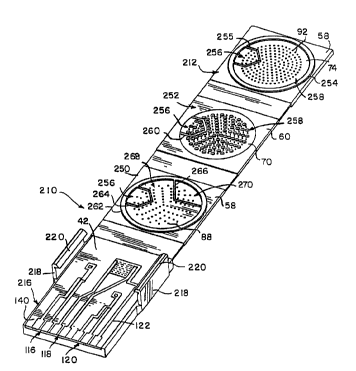
Referring now to Figs. 8-10, a sensor 210 is provided in accordance with the
present
invention that provides a manufacturer with the ability to transport a liquid
sample both
horizontally and vertically in relatively short times. Sensor 210 also enables
the user to
conduct multiple assays with a single sample by separating the sample into
discrete chambers
3o for contact with different reagents and separate electrode sets. For
example, sensor 210 may
be used to measure glucose, hematocrit, and a blank current.
CA 02328535 2000-12-13
17
Sensor 210 includes base 216 that supports insulator 140. Base 216 includes
side panels 218
extending from side walls 34, 36. Each panel 218 includes a tab 220 that is
formed to hold
cover 212 and series of plates 18 securely on base 216. Series of plates 18
shown in Figs. 8
and 9 includes a first plate 250 extending from base 12 and a second plate 252
extending
between first plate 250 and cover 16. It is contemplated that the series of
plates of may have
as few as one plate or may have greater than two plates in accordance with
this disclosure.
Plates 250, 252 are positioned so that they are stacked in a generally
parallel relationship
relative to one another when sensor 210 is in a folded position as shown in
Fig. 10.
Referring now to Fig. 8, cover 212 is similar to cover 16, except that cover
212 includes a seal
254 that extends about the periphery of lower face 74. Seal 254 also includes
an inner portion
255 that extends across face 74 to form two distinct regions 256. 2~8. When
sensor 210 is in
the folded position of Fig. 10, seal 254 engages outer side 60 of plate 252 to
form a sealing
engagement between cover 212 and plate 252. Seal 254 is preferably constructed
of the TPR
as previously discussed.
15 As shown in Fig. 8, plates 250, 252 are formed similarly to cover 16 and
plates 20, 22, except
that upper face 70 of plate 252 includes a partition 260 that lies in general
alignment with
inner portion 255 of seal 254. Partition 260 divides upper face into regions
256, 258. In
addition, plates 250, 252 include a seal 262 that extends about the periphery
of lower face 74.
Seal 262 also includes first and second inner portions 264, 266 that extends
across face 74 to
2o form three distinct regions 256, 268, 270. Seals 262 are preferably
constructed of the TPR as
previously discussed.
When sensor 210 is in the folded position of Fig. 10, seal 254 of cover 212
and seal 262 of
plate 252 engage outer sides 60 of plates 252, 250 respectively. Thus, a
sealing engagement is
formed between cover 212 and plate 252 and between plates 252, 250. Likewise,
seal 262 of
25 plate engages upper side 42 of insulator 140 to form a sealing relationship
between plate 250
and insulator 140. While regions 256, 258, 268, 270 are illustrated in Fig. 8
in a specific
pattern, this is not required, and symmetric, irregular or asymmetric patterns
are possible in
accordance with this disclosure. Moreover, it is contemplated that greater or
fewer than two
regions may be formed on cover 212 and greater or fewer than three regions may
be formed
3o on lower face 74 on plates 252, 2~0.
CA 02328535 2000-12-13
18
Regions 256, 268, 270 cooperate with electrode sets 118, 116, and 120
respectively to enable
the user to conduct multiple assays. For example, a glucose assay is conducted
by partitioning
a portion of sample 24 into region 268 for contact with electrode set 118, and
reference
electrode 122. A hematocrit assay is conducted by partitioning a portion of
sample 24 into
region 256 for contact with electrode set 116. Additionally, blank current is
measured by
partitioning a portion of sample 24 into region 270 for contact with electrode
set 120. It is
contemplated that a variety of assays including those described in Table 1 can
be used with
sensors 210 of the present invention.
Sensor 210 is constructed in a similar manner to sensor 10, using a mufti-
resin injection
o molding. Sensor 210 is also constructed from a thermoplastic polymeric
material as discussed
above with reference to sensor 10. Preferably, base 12, plates 150. 152, and
cover 16 are
formed from a polycarbonate, hinges 23, partitions 260, and seals 253, 262 are
formed of
TPR.
Sensor 110 is constructed in a similar manner to sensor 10 using a mufti-resin
injection
molding. Sensor 110 is also constructed from a thermoplastic polymeric
material as discussed
above with reference to sensor 10. Preferably, base 12, plates 150. 152, and
cover 16 are
formed from a polycarbonate, hinges 23 are constructed of a thermoplastic
rubber, and
partitions are formed from a TPR. When glucose, hematocrit, and blank current
are to be
measures, a common mediator, such as ferricyanide, is applied in liquid form
to plate 252 in
2o regions 256, 258. Discrete enzymes are applied in liquid form to plate 250
in regions 256,
288. The liquid is then evaporated, leaving the reagents in solid form coating
upper face 70
and microstructures 86 of plates 250, 252. The choice of specific reagents
depends on the
specific analytes to be measured, and are well known to those of ordinary
skill in the art.
In use, sensor 210 operates similarly to sensor 110, except that seals 254,
262 cooperate with
partitions 260, 132, 134 to guide the flow of sample liquid sample 24 into
regions 256, 268,
270. The glucose, hematocrit, and blank measurements are conducted as
discussed above with
reference to sensor 110.
Although the invention has been described in detail with reference to a
preferred embodiment,
variations and modifications exist within the scope and spirit of the
invention as described and
3o defined in the following claims.