Note: Descriptions are shown in the official language in which they were submitted.
CA 02330136 2000-10-23
WO 99/55239 PCT/US99/07399
, APPARATUS AND METHOD FOR VASCULAR EMBOLIZATION
2
3 CROSS-REFERENCE TO RELATED APPLICATIONS
4 Not Applicable
6 FEDERALLY-SPONSORED RESEARCH OR DEVELOPMENT
7 Not Applicable
8
9 BACKGROUND OF THE INVENTION
This invention relates generally to the field of vascular occlusion
õ devices and methods. More specifically, it relates to an apparatus and
12 method for occluding a blood vessel by embolizing a targeted site (such as
13 an aneurysm) in the blood vessel.
14 The embolization of blood vessels is desired in a number of clinical
situations. For example, vascular embolization has been used to control
16 vascular bleeding, to occlude the blood supply to tumors, and to occlude
17 vascular aneurysms, particularly intracranial aneurysms. In recent years,
18 vascular embolization for the treatment of aneurysms has received much
19 attention. Several different treatment modalities have been employed in
the prior art. U.S. Patent No. 4,819,637 - Dormandy, Jr. et al., for
21 example, describes a vascular embolization system that employs a
22 detachable balloon delivered to the aneurysm site by an intravascular
23 catheter. The balloon is carried into the aneurysm at the tip of the
24 catheter, and it is inflated inside the aneurysm with a solidifying fluid
(typically a polymerizable resin or gel) to occlude the aneurysm. The
26 balloon is then detached from the catheter by gentle traction on the
27 catheter. While the balloon-type embolization device can provide an
28 effective occlusion of many types of aneurysms, it is difficult to retrieve
or
29 move after the solidifying fluid sets, and it is difficult to visualize
unless it
is filled with a contrast material. Furthermore, there are risks of balloon
31 rupture during inflation and of premature detachment of the balloon from
CA 02330136 2000-10-23
WO 99/55239 PCT/US99/07399
2
1 the catheter.
2 Another approach is the direct injection of a liquid polymer
3 embolic agent into the vascular site to be occluded. One type of liquid
4 polymer used in the direct injection technique is a rapidly polymerizing
liquid, such as a cyanoacrylate resin, particularly isobutyl cyanoacrylate,
6 that is delivered to the target site as a liquid, and then is polymerized in
7 situ. Alternatively, a liquid polymer that is precipitated at the target
site
a from a carrier solution has been used. An example of this type of embolic
9 agent is a cellulose acetate polymer mixed with bismuth trioxide and
dissolved in dimethyl sulfoxide (DMSO). Another type is ethylene glycol
11 copolymer dissolved in DMSO. On contact with blood, the DMSO
12 diffuses out, and the polymer precipitates out and rapidly hardens into an
13 embolic mass that conforms to the shape of the aneurysm. Other
14 examples of materials used in this "direct injection" method are disclosed
in the following U.S. Patents: 4,551,132 - Pdsztor et al.; 4,795,741 -
16 Leshchiner et al.; 5,525,334 - Ito et al.; and 5,580,568 - Greff et al.
17 The direct injection of liquid polymer embolic agents has proven
18 difficult in practice. For example, migration of the polymeric material
19 from the aneurysm and into the adjacent blood vessel has presented a
problem. In addition, visualization of the embolization material requires
21 that a contrasting agent be mixed with it, and selecting embolization
22 materials and contrasting agents that are mutually compatible may result
23 in performance. compromises that are less than optimal. Furthermore,
24 precise control of the deployment of the polymeric embolization material
is difficult, leading to the risk of improper placement and/or premature
26 solidification of the material. Moreover, once the embolization material
27 is deployed and solidified, it is difficult to move or retrieve.
28 Another approach that has shown promise is the use of
29 thrombogenic microcoils. These microcoils may be made of a
CA 02330136 2000-10-23
WO 99/55239 PCT/US99/07399
3
1 biocompatible metal alloy (typically platinum and tungsten) or a suitable
2 polymer. If made of metal, the coil may be provided with Dacron fibers
3 to increase thrombogenicity. The coil is deployed through a
4 microcatheter to the vascular site. Examples of microcoils are disclosed
in the following U.S. patents: 4,994,069 - Ritchart et al.; 5,133,731 -
6 Butler et al.; 5,226,911 - Chee et al.; 5,312,415 - Palermo; 5,382,259 -
7 Phelps et al.; 5,382,260 - Dormandy, Jr. et al.; 5,476,472 - Dormandy, Jr.
a et al.; 5,578,074 - Mirigian; 5,582,619 - Ken; 5,624,461 - Mariant;
s 5,645,558 - Horton; 5,658,308 - Snyder; and 5,718,711 - Berenstein et al.
The microcoil approach has met with some success in treating
small aneurysms with narrow necks, but the coil must be tightly packed
12 into the aneurysm to avoid shifting that can lead to recanalization.
13 Microcoils have been less successful in the treatment of larger aneurysms,
14 especially those with relatively wide necks. A disadvantage of microcoils
is that they are not easily retrievable; if a coil migrates out of the
16 aneurysm, a second procedure to retrieve it and move it back into place is
17 necessary. Furthermore, complete packing of an aneurysm using
18 microcoils can be difficult to achieve in practice.
19 A specific type of microcoil that has achieved a measure of success
is the Guglielmi Detachable Coil ("GDC"). The GDC employs a
21 platinum wire coil fixed to a stainless steel guidewire by a solder
22 connection. After the coil is placed inside an aneurysm, an electrical
23 current is applied to the guidewire, which heats sufficiently to melt the
24 solder junction, thereby detaching the coil from the guidewire. The
application of the current also creates a positive electrical charge on the
26 coil, which attracts negatively-charged blood cells, platelets, and
27 fibrinogen, thereby increasing the thrombogenicity of the coil. Several
28 coils of different diameters and lengths can be packed into an aneurysm
29 until the aneurysm is completely filled. The coils thus create and hold a
CA 02330136 2000-10-23
WO 99/55239 PCT/US99/07399
4
1 thrombus within the aneurysm, inhibiting its displacement and its
2 fragmentation.
3 The advantages of the GDC procedure are the ability to withdraw
4 and relocate the coil if it migrates from its desired location, and the
enhanced ability to promote the formation of a stable thrombus within the
6 aneurysm. Nevertheless, as in conventional microcoil techniques, the
7 successful use of the GDC procedure has been substantially limited to
s small aneurysms with narrow necks.
9 There has thus been a long-felt, but as yet unsatisfied need for an
aneurysm treatment device and method that can substantially fill
11 aneurysms of a large range of sizes, configurations, and neck widths with
12 a thrombogenic medium with a minimal risk of inadvertent aneurysm
13 rupture or blood vessel wall damage. There has been a further need for
14 such a method and device that also allow for the precise locational
deployment of the medium, while also minimizing the potential for
16 migration away from the target location. In addition, a method and
17 device meeting these criteria should also be relatively easy to use in a
18 clinical setting. Such ease of use, for example, should preferably include
a
19 provision for good visualization of the device during and after
deployment in an aneurysm.
21
22 SUMMARY OF THE INVENTION
23 Broadly, one aspect of the present invention is an embolic device,
24 comprising a thrombogenic medium, that is deployed in a soft, compliant
state, and that is controllably transformed into a rigid or semi-rigid state
26 after deployment. In another aspect, the present invention is an apparatus
27 for deploying the aforesaid embolic device in the interior of an aneurysm.
28 Still another aspect of the present invention is a method for embolizing a
29 vascular site, particularly an aneurysm, using the aforesaid embolic
CA 02330136 2000-10-23
WO 99/55239 PCT/US99/07399
1 device.
2 In a first preferred embodiment, the embolic device comprises a
3 continuous, filamentous extrusion of polymeric "transition material" that
4 is inserted into an aneurysm while in a soft, self-adherent, compliant
state.
5 The insertion of one or more such embolic devices results in a mass of
6 material that substantially fills the aneurysm and that substantially
7 conforms to the interior shape of the aneurysm. Depending on the
8 particular polymeric material employed, any of several mechanisms is
9 then employed controllably to transform the transition material into a
rigid or semi-rigid state, in which the material forms a stable,
11 thrombogenic "plug" inside the aneurysm. For example, the material
12 may be injected at a temperature slightly above body temperature and
13 then cooled into its rigid or semi-rigid state by contact with the
patient's
14 blood, or by the injection of a cooler saline solution. Alternatively, the
polymeric material may be exposed to a hardening agent that reacts
16 physically or chemically with the material to effect the transition to the
17 rigid or semi-rigid state. As still another alternative, the polymeric
18 material may be mixed with a water soluble, biocompatible plasticizer
19 that dissolves out in the vascular blood to leave a rigid or semi-rigid
polymeric structure.
21 In another preferred embodiment, the embolic device comprises an
22 elongate, flexible microcoil, the interior of which contains the transition
23 material. The microcoil is deployed in the aneurysm with the transition
24 material in its soft, compliant state, and then the transition material is
rigidified by any suitable mechanism, as mentioned above, thereby
26 rigidifying the microcoil in situ.
27 In another preferred embodiment, the embolic device comprises an
28 elongate, flexible chain of articulated segments linked together so as to
29 form a limp segmented filament that is installed in the aneurysm. After
CA 02330136 2000-10-23
WO 99/55239 PCT/US99/07399
6
1 placement in the aneurysm, the segmented filament is rigidized by fusing
2 the segments through one of several mechanisms, depending on the
3 material of the segments. For example, if the segments are metal, the
4 segments can be fused together by electrolytic corrosion resulting from a
current being passed through the device. If the segments are made, at
6 least in part, of a polymeric "transition material", the transition of the
7 device to a rigid or semi-rigid state can be induced by one of the
8 mechanisms discussed above.
s In still another preferred embodiment, the embolic device is a
highly-compliant chain-like structure comprising a plurality of
11 interconnected hollow links or segments. Each of the segments has a
12 slotted, mushroom-shaped head portion and a socket portion that is
13 shaped and dimensioned to receive the head portion of an adjacent
14 segment. The hollow segments allow the embolic device to be inserted
into an aneurysm over a guide wire (not shown), if desired. Once the
16 device is inserted, a polymeric transition material is injected, while in
the
17 soft, compliant state, into the hollow interior of the device, and the
18 transformation into its rigid or semi-rigid state can be effected as
described
19 above. Alternatively, the segments can be made of a metal and then fused
together by electrolytic corrosion.
21 A preferred embodiment of the apparatus for deploying the embolic
22 device comprises a flexible, elongate, hollow deployment tube having an
23 axial passage and a cup-shaped holding element at its distal end. The
24 holding element, which is configured and dimensioned to hold the
proximal end of the embolic device by a frictional engagement, has a base
26 with an opening that communicates with the axial lumen. The
27 deployment tube (or at least its distal end) is preferably made of a
28 radiopaque material, such as a biocompatible metal alloy, thereby
29 facilitating visualization during the deployment of the embolic device,
CA 02330136 2000-10-23
WO 99/55239 PCT/US99/07399
7
1 without requiring the inclusion of a radiopaque substance in the embolic
2 device itself.
3 The preferred method of deploying the embolic device using this
4 apparatus is as follows: The deployment tube, with the embolic device
thus attached to it, is inserted into and pushed through a microcatheter
6 that has been advanced intravascularly to the aneurysm site by means
7 well known in the surgical arts. Passage of the flexible deployment tube
8 and the limp embolic device through the microcatheter is assisted and
s facilitated by a flow of fluid (e.g., saline solution) through the
microcatheter around the exterior of the deployment tube and the embolic
11 device. The deployment tube is pushed through the microcatheter until
12 the embolic device has been fully inserted into the aneurysm. Finally, a
13 fluid (e.g., saline solution) is injected through the axial lumen and into
the
14 holding element of the deployment tube. The pressure of the fluid pushes
the embolic device out of the holding element, thereby detaching the
16 embolic device from the deployment tube. The deployment tube is then
17 withdrawn from the microcatheter. If more than one embolic device is
18 necessary to fill the aneurysm, the above-described process can be
19 repeated until the aneurysm is filled.
The present invention offers a number of advantages over prior art
21 embolization methods and devices. For example, the embolic device of
22 the present invention is deployable within an aneurysm in a soft,
23 compliant state, thereby minimizing the risk of aneurysm rupture or
24 vascular damage. The location of the embolic device can be controlled
with some precision, and, until it is detached from the deployment tube,
26 its deployment can be reversed. Thus, the risks of migration out of the
27 aneurysm are minimized. Furthermore, the embolic device of the present
28 invention can be used in aneurysms having a wide variety of shapes and
29 sizes; it is not limited to small aneurysms or those with narrow necks.
CA 02330136 2000-10-23
02-06-2000 US 009907399
. . .. .. .. .. .. ..
.. .. .. . . .. . .. .
. . . : . . . . ... . . . .
. . . . . . .. . . .. .
. : . . . . .. . . .. .
. . .. .... .. .. . .. ..
8
These and other advantages of the present invention will be more fully
appreciated from the detailed description that follows.
BRIEF DESCRIPTION OF THE DRAWINGS
Figure 1 is an elevational view of a preferred embodiment of an
apparatus for deploying an embolic device in accordance with the present
invention;
Figure 2 is a cross-sectional view taken along line 2 - 2 of Figure 1,
lo showing the apparatus with an embolic device in accordance with a first
preferred embodiment of the present invention;
Figures 3 and 4 are idealized views of an emboiic device in
accordance with present invention in the process of being deployed in an
aneurysm by means of the apparatus of Figures 1 and 2;
Figure 5 is an elevational view of one embodiment of an embolic
device in accordance with a second preferred embodiment of the present
invention;
Figure 6 is a detailed view taken within the area of Figure 5
designated by the broken outline 6;
Figure 7 is a cross-sectional view of a portion of an embolic device
that is a modification of the embodiment of Figures 5 and 6;
Figure 8 is a cross-sectional view similar to that of Figure 7, showing
the device at a later stage of deployment;
Figure 9 is an elevational view of a portion of an embolic device that is
another modification of the embodiment of Figures 5 and 6;
Figure 10 is a cross-sectional view taken along line 10 - 10 of Figure
9;
Figure 11 is an end elevational view of an embolic device in
accordance with a third preferred embodiment of the present invention;
Figure 12 is a cross-sectional view taken along line 12-12 of Figure
11; and
AMENDED SHEET
CA 02330136 2000-10-23
WO 99/55239 PCT/US99/07399
9
1 Figures 13-16 are cross-sectional views, similar to that of Figure 10,
2 showing further modifications of the third preferred embodiment of the
3 embolic device of the present invention.
4
DETAILED DESCRIPTION OF THE INVENTION
6 Figures 1 and 2 illustrate a preferred embodiment of an apparatus
7 10 for deploying an embolic device 12 in accordance with the present
8 invention. The apparatus 10 comprises a microcatheter 14 having an
9 axial lumen 15, and a deployment tube 16 that is insertable through the
lumen 15 of the microcatheter 14. The microcatheter 14 is of
11 conventional design, and many suitable microcatheters for the apparatus
12 10 are commercially available. The proximal end of the microcatheter 14
13 is provided with a fitting 18 for coupling to a source (not shown) of a
fluid
14 (such as saline solution), the flow of which is used to facilitate the
passage
of the deployment tube 16 through the microcatheter 14, as will be
16 described below. The microcatheter 14, or at least its distal end, is
17 preferably made of a radiopaque material, such as a biocompatible metal.
18 Alternatively, it may be made of a suitable plastic, with a radiopaque
19 insert (not shown) proximate its distal end, as is well known in the art.
The deployment tube 16 is a long, thin, hollow, highly flexible
21 tube, having an axial passage 20 and an overall length that is somewhat
22 greater than that of the microcatheter 14. The deployment tube 16 has a
23 proximal end to which is attached an inlet fitting 22 that communicates
24 with the axial passage 20 and that is adapted for coupling to a liquid
source (not shown). The source contains a biocompatible liquid that can
26 be delivered to the inlet fitting 22 under pressure for purposes to be
27 described below. The distal end of the deployment tube 16 is provided
28 with a cup-like fitting 24 that serves as a holding element that is
29 configured for frictional engagement with the proximal end of the embolic
CA 02330136 2000-10-23
WO 99/55239 PCT/US99/07399
1 device 12. The interior of the holding element 24 communicates with the
2 axial passage 20 of the deployment tube 16 by means of an axial bore 26.
3 A substantial portion of the length of the deployment tube 16 extending
4 proximally from the holding element 24 is formed as a highly flexible and
5 compliant outer portion 28 formed from a continuous length of helically-
6 coiled metal wire. The outer portion 28 concentrically surrounds an inner
7 portion 30, formed from a highly-flexible polymeric material, the interior
8 of which defmes a distal portion of the axial passage 20 that is coupled to
9 the axial bore 26 of the holding element 24. The proximal ends of both
10 the outer portion 28 and the inner portion 30 are connected to the distal
11 end of an internal transition fitting 32, the proximal end of which is
12 connected to the distal end of a proximal tube section 34, which may be
13 made of a flexible polymeric material. An axial bore 36 traverses the
14 length of the transition fitting 32, providing fluid communication between
the distal portion of the axial passage 20 that is within the inner portion
16 30, and the proximal portion of the axial passage 20 that is defmed within
17 the proximal tube section 34. The aforementioned inlet fitting 22 is
18 connected to the proximal end of the proximal tube section 34.
19 As shown in Figures 1 and 2, the embolic device 12 comprises a
continuous, filamentous extrusion of polymeric "transition material".
21 This transition material is initially in a soft, self-adherent, compliant
22 state. While the material is in this state, the embolic device 12 is
inserted
23 into an aneurysm. The insertion results in a web-like mass of material
24 that substantially fills the aneurysm and that substantially conforms to
the
interior shape of the aneurysm. Depending on the particular polymeric
26 material employed, any of several mechanisms is then employed
27 controllably to transform the transition material into a rigid or semi-
rigid
28 state, in which the material forms a stable, thrombogenic "plug" inside the
29 aneurysm. For example, the embolic device 12 may be injected at a
CA 02330136 2000-10-23
WO 99/55239 PCT/US99/07399
11
I temperature slightly above body temperature and then cooled into its rigid
2 or semi-rigid state by contact with the patient's vascular blood, or by the
3 injection of a cooler saline solution. Alternatively, the polymeric material
4 may be exposed to a hardening agent that reacts chemically or physically
with the material to effect the transition to the rigid or semi-rigid state.
6 As still another alternative, the polymeric material may be mixed with a
7 water-soluble, biocompatible plasticizer (as described below) that
8 dissolves out in the vascular blood to leave a rigid or semi-rigid polymeric
9 structure.
Prior to deployment, and while the material of the embolic device
11 12 is in its initial soft, compliant state, the proximal end of the embolic
12 device 12 is pushed into the holding element 24 of the deployment tube
13 16, where it is frictionally retained in place. With the distal end of the
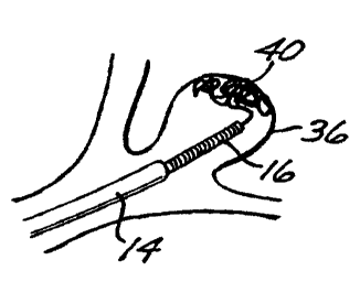
14 microcatheter 14 having previously been deployed adjacent the targeted
aneurysm (designated by the numera136 in Figures 3 and 4), the distal
16 end (not shown) of the embolic device 12 is then inserted into the fitting
17 18 at the proximal end of the microcatheter 14. As the embolic device 12
18 and the deployment tube 16 are pushed through the lumen 15 of the
19 microcatheter 14, a liquid, such as a saline solution, is caused to flow
through the microcatheter 14, as indicated by arrows designated by the
21 numera138 in Figure 2. The flow of the liquid assists in carrying the
22 embolic device 12 and the deployment tube 16 through the microcatheter
23 14 until the distal end of the deployment tube 16 is well within the
24 aneurysm 36 (Figure 3), at which point the embolic device 12 starts to
form a web-like, thrombogenic mass or plug 40 within the aneurysm. The
26 proximal end of the embolic device 12 is detached from the deployment
27 tube 16 by the pressure of a fluid (such as saline solution) injected
through
28 the axial passage 20 of the deployment tube and the axial bore 26 of the
29 holding element 24.
CA 02330136 2000-10-23
WO 99/55239 PCT/US99/07399
12
1 If the size of the aneurysm 36 requires more than one embolic
2 device 12 to fill it completely, the deployment tube 16 is withdrawn
3 through the microcatheter 14 and reloaded with another embolic device
a 12, and the above-described deployment process is repeated as often as is
needed to fill the aneurysm 36 completely (Figure 4). As shown in Figure
6 4, the fmal embolic device 12 is then detached from the deployment tube
7 16 in the manner described above, and the deployment tube 16 is
8 withdrawn from the microcatheter 14.
s The fluid used to carry the deployment tube 16 and the embolic
device 12 through the microcatheter 14, and the fluid used to detach the
11 embolic device 12 from the deployment tube (i.e., the "deployment
12 fluids"), are selected so that they do not effect the transition of the
embolic
13 device material from its soft state to its rigid or semi-rigid state. Thus,
for
14 example, if the transition material effects the transition by being cooled
from slightly above body temperature (e.g., from about 40 C) to
16 approximately normal body temperature (37 C), these deployment fluids
17 are injected at about the higher temperature, so that the transition does
18 not take place prematurely.
19 Once the web-like thrombogenic mass 40 completely fills the
aneurysm 36, as shown in Figure 4, the transition material of the embolic
21 device(s) 12 installed within the aneurysm 36 can be transformed to its
22 rigid or semi-rigid state by means of one of the aforementioned
23 mechanisms, depending on the nature of the material itself. For example,
24 a "transition fluid", such as saline at the required temperature, can be
injected through the microcatheter 14 to bathe the mass 40, thereby
26 effecting the desired transition.
27 Figures 5 and 6 illustrate an embolic device 50 in accordance with
28 a second preferred embodiment of the invention. The embolic device 50
29 comprises a hollow metal microcoi152, the interior of which is filled with
a core 54 of polymeric transition material. The embolic device 50 is
CA 02330136 2000-10-23
WO 99/55239 PCT/US99/07399
13
1 rigidified by the transformation of the material of the core 54 from its
soft,
2 compliant state to its rigid or semi-rigid state effecting a temperature
3 change, as described above. The deployment of the embolic device 50 is
4 performed by essentially the same method as that used for the deployment
s of the previously-described embodiment.
6 Modifications of the embolic device 50 are shown in Figures 7
7 through 10. In Figures 7 and 8, an embolic device 50' comprises a hollow
8 metal microcoi152', the distal end of which is closed by an end cap 56.
9 The device 50' lacks a core. Instead, when the microcoil 52' is inserted
into an aneurysm, but before it is detached from the deployment tube 16,
11 a flowable transition material is injected into the interior of the
microcoil
12 52' through the axial passage 20 of the deployment tube 16 and the axial
13 bore 26 of the holding element 24. The injection of the transition material
14 is illustrated in Figure 7 by the arrows designated by the numera158. The
flexing and bending of the installed microcoi152', as shown in Figure 8,
16 causes interstices between the coils to open up, allowing the transition
17 material to flow out of the microcoil, as indicated by the arrows
18 designated by the numera160. The transition material then can be
19 transformed into its rigid or semi-rigid state, thereby rigidifying the
microcoi152'. The exposed transition material that has flowed out of the
21 interstices between the coils provides further rigidity and enhances the
22 thrombogenicity of the device 50'.
23 The advantages of the embolic device 50' of Figures 7 and 8 can
24 also be realized in another modification shown in Figures 9 and 10. In
this latter modification, an embolic device 50" comprises a hollow metal
26 microcoi152" having an end cap 56" closing its distal end. The microcoil
27 52" has a plurality of apertures 62 along its length, only one of which is
28 shown in the drawings. The apertures 62 provide additional paths for the
29 outflow of the transition material, as shown by the arrows indicated by
the numera164 in Figure 10.
CA 02330136 2000-10-23
WO 99/55239 PCT/[7S99/07399
14
1 A third preferred embodiment of the embolic device is shown in
2 several variations in Figures 11-16. Referring first to Figures 11 and 12,
3 an embolic device 70 in accordance with this third embodiment is a
a chain-like structure comprising a plurality of interconnected metal links or
segments 72, each of which has a socket 74 at one end and a slotted ball
6 76 at the other end. Each socket 74 is dimensioned to receive the ball 76
7 of the adjacent segment 72, the slotted configuration of the balls 76
8 allowing them to be slightly compressed to fit into the sockets 74. The
9 balls 76 are loosely received in the sockets 74, and the segments 72 are
dimensioned so that there is a gap between each adjacent pair. Thus, the
11 entire chain-like structure of the device 70 can be flexibly deformed and
12 twisted much like a microcoil to form the web-like mass 40 when
13 deployed inside an aneurysm by means of the above-described method.
,a When it is desired to rigidify the device 70, an electric current is passed
through it, resulting in the fusing of the balls 76 in the sockets 74 by
16 electrolytic corrosion. The electric current can be applied through the
17 deployment tube 16, provided that the deployment tube 16 (including the
18 holding element 24) is made of a conductive metal with suitable
19 electrodes (not shown) that connect the embolic device 70 to a current
source (not shown).
21 A modification of the third embodiment is shown in Figure 13. An
22 embolic device 70' is a chain-like structure comprising a plurality of
23 interconnected metal links or segments 72', each including a socket 74' at
24 one end and a slotted ball 76' at the other end. The balls 76' are received
in the sockets 74' as described above. The modification comprises an
26 annular collar 78 around the socket 74' of each segment 72'. The collar 78
27 extends axially away from the ball 76' to abut against, or at least be
28 closely adjacent to, the next adjacent segment 72'. The collar 78 is formed
29 of a polymeric transition material that is initially in the soft, compliant
state when the device 70' is inserted into an aneurysm, and that is
CA 02330136 2000-10-23
02-06-2000 US 009907399
. . .. .. .. .. .. ..
.. .. . .. . . .. . .. .
. . . . . . . .... .. .
. . . . . . . .. . : .. .
. . . . . . .. . . .. .
, = . . .. .... .. .. . .. ..
1 transformed into its rigid or semi-rigid state, in the manner described
above,
2 when the aneurysm is filled. Since the collars 78, when rigidified, form
3 interlinking elements between adjacent segments 72, the transformation of
4 the material of the collars 78 rigidifies the entire device 70'. A similar
effect
5 can be achieved, at some cost savings, by the modified embolic device 70"
6 of Figure 14, in which only altemating segments 72' are provided with the
7 collar 78.
8 Figures 15 and 16 illustrate still another modification of the third
9 preferred embodiment. In this modification, an embolic device 70"' is a
1o highly-compliant chain-like structure comprising a plurality of
interconnected
11 links or segments 72", each of which is hollow. Each of the segments 72"
12 has a slotted, mushroom-shaped head portion 80, and a socket portion 82
13 that is shaped and dimensioned to receive the head portion 80 of an
adjacent
14 segment 72". The hollow segments 72" allow the embolic device 70"' to be
15 inserted into an aneurysm over a guide wire (not shown), if desired. Once
16 the device 70"' is inserted, a transition material 84 (Fig.16) is injected,
while
17 in a flowable state, into the hollow interior of the device 70"', and the
18 transformation of the device 70" from a soft compliant state into its rigid
or
19 semi-rigid state can be effected as described above. Alternatively, the
segments 72" can be made of a metal and then fused together by electrolytic
21 corrosion, as described above.
22 For the selection of transition materials which are used in accordance
23 with the present invention to fill the aneurysm in a relatively soft, semi-
rigid
24 state as described above, and which thereafter harden to fill the aneurysm
in
a sufficiently rigid state, the skilled artisan may refer to the self-
hardening
26 polymeric materials described in United States Patent No. 5,634,936.
27 Generally speaking, the materials described in this reference are polymers
28 that, due to the judicious addition of cross-linking agents and/or cross-
linking
29 catalysts, are in a soft , compliant state while being
AMENDED SHEET
CA 02330136 2000-10-23
02-061-20p0 US 009907399
. .. .. .. .. .. ..
.. .: . .. . . .. . .. .
. . . . . . . .... .. .
. . . . . . . .. . : .. .
. . . . . . .. . . .. .
. .. .... .. .. . .. ..
16
1 introduced through a catheter, and harden only after they have been
2 deposited in the aneurysm. Materials described in United States Patent No.
3 5,725,568 can also be selected for use in the present invention.
4 A presently preferred material for use in the present invention
constitutes a microcrystalline wax composition that is of the appropriate
6 compliant consistency a few degrees above body temperature, but becomes
7 sufficiently rigid when cooled to body temperature. As is known, waxes are,
8 generally speaking, fatty acids having more than 12 carbon atoms and a
9 straight alkyl chain. A microcrystalline wax material is readily formulated
within the state-of-the-art to have the appropriate transition temperature.
11 Another presently preferred material for use in the present invention is
12 cellulose-acetate polymer that is softened with ethyl lactate or
13 dimethylsulfoxide (DMSO) plasticizer. Still other presently preferred
14 materials are a class of polyurethane based copolymers that are available
under the TECOPHILIC trademark from Thermedics Corporation. Specific
16 commercial designations of these copolymers are HP-60D-60, SP-80A-150
17 and SP-93A-100. These polyurethane-based copolymers are softened with
1 s a plasticizer or mixture of plasticizers that are selected primarily from
DMSO,
19 ethanol, and ethyl lactate, with DMSO being most suitable for HP-60D-60,
2o and ethanol or ethyl lactate or mixtures thereof for SP-80A-1 50 and SP-93A-
21 100. The above-noted plasticizers are sufficiently water soluble that after
the
22 intimate mixture of polymeric material and plasticizer has been deposited
in
23 the aneurysm, percolation of blood gradually washes out the plasticizer
from
24 the polymeric material to render it rigid.
A composition that is well-suited for the transition material in the
26 hollow microcoil embolic devices 50' and 50" of Figures 7 through 10,
AMENDED SHEET
CA 02330136 2000-10-23
02-06-2000 US 009907399
. .. .. .. .. .. ..
. .: . .. . . .. . .. .
. . . . . . .... .. .
: . . . . . . .. : ..
. . . . . . .. : . .. :
. . .. .... .. .. . .. ..
17
~ and for the transition material 84 of the embolic device 70"' of Figures 15
and
2 16, is cyanoacrylate. The cyanoacrylate rigidifies by polymerization when
3 contacted by vascular blood which seeps into the embolic device 70"'
4 between the segments 72".
In addition to the foregoing, a number of biocompatible polymers and
6 copolymers, such as ethylene vinyl alcohol copolymers, polycarbonate
7 urethane copolymers, and hydrogels may be formulated with a sufficient
a amount of biocompatible plasticizer, such as DMSO, to render them semi-
9 rigid and suitable for application in the present invention through the
1o catheters described above. Thereafter, these materials harden sufficiently
in
11 the aneurysm due to the removal of the plasticizer by percolating blood.
12 While several preferred embodiments have been described above, as
13 well as a number of variations and modifications, it will be appreciated
that
14 other variations and modifications will suggest themselves to those skilled
in
the pertinent arts. Such variations and modifications are considered to be
16 within the scope of the invention, as set forth in the claims that follow.
AMENDED SHEET