Note: Descriptions are shown in the official language in which they were submitted.
CA 02336825 2001-O1-05
WO 00/01301 pCT/ZA99/00048
APPARATUS FOR EVALUATION OF SKIN IMPEDANCE VARIATIONS
Copyright Notice
The matter contained herein is subject to Copyright protection in Berne
Convention countries, which copyright is the property of the inventor.
Publication of
the patent specification, or any act by any patent office, does not constitute
a waiver of
to these rights.
Background to the Invention
This invention relates to an apparatus and method for automatic evaluation of
skin impedance variations in order to estimate the state of health of the
internal organs
of a human or animal.
Existing methods of utilising skin impedance values for organ diagnostics base
their results on non-ratiometric measurements of basic skin impedance and
produce
2u inconsistent and unreliable results which depend on numerous variables,
including the
emotional state of the patient, muscular tension, measurement time, the
contact area
and pressure of the measuring electrode and various physiological differences
between
individuals.
CA 02336825 2001-O1-05
WO 00/01301 p~/T,Agg~ppp4g
After many years of research the inventor now believes that the internal
organs
of the body of a human or animal have corresponding areas on the skin where '
information regarding the corresponding internal organs can be retrieved by
measuring
the electrical properties of said skin. The inventor further believes that
said
corresponding areas of the skin have other properties related to the science
of reflexive
pyhsiotherapy (including acupuncture), for example, the ability to heal and
/or relieve
pain caused by the corresponding organs.
The inventor believes yet further that these corresponding areas of the skin
may
to be mapped, which map is applicable to various individuals.
The inventor has found that the ear auricle may be particularly accurately
mapped and is most suitable for the method of the invention since in most
cultures the
skin of the ear is exposed and may be examined without any garments having to
be
1 s removed.
In this specification, unless the context clearly indicates to the contrary,
the
term "impedance", is to be understood to include resistance.
zt~ Summary of Invention
The impedance variation can be measured in two ways:
2
CA 02336825 2001-O1-05
wo oorot3oi rcrrc~~rooo4s
Method 1: AC evaluation. '~'
The difference between the AC impedance measured at a specific frequency
and at a specific skin area with a calibration electrode and a reference
electrode and the
impedance measured at a similar frequency and in the same area with a
measurement
electrode and a reference electrode, is used to determine the state of health
of the
internal organ corresponding to the examined skin area. The calibration
electrode and
reference electrode contact areas are relatively larger than the measurement
electrode
to skin contact area.
Method 2: DC evaluation
The term "break-through effect" refers to the sudden and significant drop in
1 i skin electrical resistance witnessed after a sufficient potential
difference is applied
between the electrodes.
The skin between the electrodes is exposed to a DC potential of a magnitude
selected to give the break-through effect. The DC resistance of the skin is
measured
2o between a measurement electrode polarised negatively with respect to a
reference
electrode, and the DC resistance of the same skin area is again measured but
with the
measurement electrode polarised positively with respect to the reference
electrode.
The ratio of these two values is used to determine the state of health of the
internal
organ corresponding with the examined skin area.
3
CA 02336825 2001-O1-05
wo ooroi3oi pcrz~~rooo4s
An apparatus broadly in accordance with the invention may include the ~'~
following functional blocks:
A measurement and/or calibration electrode, a reference electrode, a voltage
generator block, a measurement block, a control block, a user interface block,
a result
presentation block and, optionally, a data storage block.
The voltage generator block generates a potential difference between the
to measurement electrode and the reference electrode, or the calibration
electrode and the
reference electrode. The voltage generator block is connected to and
controlled by
the control block. The measurement block is connected to the measurement
electrode
and the reference electrode (Fig 1 ).
The measurement block determines the impedance between the measurement
electrode and the reference or calibration electrode. Alternatively the
voltage
generator block can be connected through the measurement block to the
measurement
electrode or the reference electrode (Fig 2). The ultimate purpose of the
measurement
block is to measure a parameter (such as voltage or current) that can be used
to
2o determine the impedance or resistance between the measurement electrode and
the
reference electrode. The measurement block is connected to the control block.
The control block is connected to the user interface block (if present), the
data
storage block (if present), the result presentation block, the voltage
generator block
a
CA 02336825 2001-O1-05
WO 00101301 Pt:T/ZA99/00048
and the measurement block. The control block sets the voltage generated by the
voltage generator block. The control block uses information received from the
~'-
measurement block to detect the break-through effect, and the resistance
asymmetry.
The control block can store and retrieve information in the data storage block
(if
present). The control block informs the user of the results of the
measurements
through the result presentation block. The result presentation block may be
generate a
visual or audio indication to inform the user of the result i.e. the state of
health of the
internal organ, obtained by the control block.
to Description of Operation
After a great deal of experimental work the inventor has found that an
apparatus broadly in accordance with the invention may be operated as
described
below to obtain reliable results.
is
Technique I: AC evaluation
The calibration electrode is placed in contact with the relevant skin area
corresponding to the internal organ of a subject the sate of health of which
is to be
2o determined. The reference electrode is placed in contact with any other
skin area,
usually the hand of the subject. The control block uses the voltage generator
block to
generate an AC signal of specific frequency and magnitude between the
calibration and
reference electrodes. The control block determines the impedance between the
electrodes via the measurement block. The control block stores the impedance
value
5
CA 02336825 2001-O1-05
WO 00/01301 pCT~ZA99/00048
in the data storage block (referred to as "calibration impedance"). The
control block
signals that the calibration impedance has been determined via the result
presentation%'- "
block. The calibration electrode is removed and the measurement electrode is
placed
on the skin area undergoing investigation. The control block uses the voltage
s generator block to generate an AC signal of similar frequency and magnitude
between
the calibration and reference electrodes.
The control block determines the ratio between the calibration impedance and
the impedance measured with the measuring electrode and converts this ratio to
an
to indication of the state of health of the internal organ. The control block
displays the
result on the result presentation block (e.g. on a disease intensity
percentage scale).
Conveniently the result is displayed in percentage format, calculated
according
to the following equation:
1~
% Disease = ( 1 - 1 ,nea~urement ~ Ireference ) X 100; Or
% Disease = ( 1 - lZreferencc ~ Rmeaxuremenl ) x 1 fl0.
Various percentage ranges corresponding to different states of health of the
2n organ. Typically 0 to 40% indicates a healthy state, 40 to 60% indicates
the upper
limits of the healthy state, 60 to 80% indicates a sub-acute state, and 80 to
100%
indicates an acute condition of the internal organ in question.
r,
CA 02336825 2001-O1-05
wo ooroi3oi rcrrr,~~rrooo4s
Technique 2: DC evaluation
The reference electrode is placed in contact with any skin area. The
measurement electrode is placed in contact with a specific skin spot
corresponding
with an internal organ the state of health of which is to be determined. The
control
block uses the voltage generator block to generate a DC potential difference
between
the electrodes. The control block determines the resistance between the
electrodes via
the measurement block. The control block adjusts the DC potential difference
and
checks the resistance until the resistance falls below a certain threshold or
suddenly
to starts decreasing rapidly (break-through effect). The control block checks
the
resistance until a stable value is reached. The control block stores this
resistance value
in the data storage block (referred to as "reference resistance")
The control block inverts the polarisation of the measurement and reference
electrodes with respect to each other and uses the voltage generator block to
apply a
DC potential across the electrodes. The control block determines the
resistance
between the electrodes via the measurement block (referred to as "measurement
resistance").
2o The control block determines the ratio between the "measurement resistance"
and the "reference resistance'' and calculates the intensity of disease from
this ratio.
The following equation is used for the calculation:
Disease = ( ~ - 1",eaqne,nrnt ~ Ircferencc ~ ~ 1 ~~; or
CA 02336825 2001-O1-05
wo ooroi3oi pcrizn99iooo4s
% Disease = ~ 1 - )~f~,ce ~ Rmeaaurement ) X 100
The control block displays the result on the result presentation block (e.g.
on a
disease intensity percentage scale).
Conveniently the result is displayed in percentage format, with various
percentage ranges corresponding to different state of health of the organ.
Typically 0
to 40% indicates a healthy state, 40 to 60% indicates the upper limits of the
healthy
state, 60 to 80% indicates a sub-acute state, and 80 to 100% indicates an
acute
condition of the internal organ in question.
For best break-through effect induction the reference resistance measurement
should be made with the measurement electrode polarised negatively with
respect to
the reference electrode, although with higher potential differences between
the
t5 electrodes it is believed that the break-through effect may also be
observed if the
polarity is inverted.
When using the DC technique, if the internal organ is not healthy a higher
resistance will be measured with the measurement electrode polarised
positively with
respect to the reference electrode than with the measurement electrode
polarised
negatively with respect to the reference electrode e.g. 300 kSZ as opposed to
30 kS2.
Similarly, when using the AC technique, the measurement obtained using the
measurement electrode will have an impedance reading which is higher than that
of the
measurement obtained using the calibration electrode.
X
CA 02336825 2001-O1-05
WO 00/01301 PCT/ZA99/00048
It is also possible to use an AC signal when using the DC technique. ''
Although both the AC and DC evaluation techniques are effective, for thin skin
regions of the body, such as the ear auricle, the DC evaluation technique is
preferred,
while for thicker skin areas of the body, such as the feet, the AC method is
preferred as
thicker skin areas require higher voltages for the breakthrough effect to
occur, which
could be painful for the subject.
1o The inventor believes that an apparatus such as that described above, uses
new
measurement technologies and ratiometric techniques and achieves consistent
and
repeatable diagnostic results which are independent of various physiological
differences
between individuals, the emotional state of a patient, muscular tension and
the
measurement time. The results depend on the intensity of the disease, the
effect of
t5 pressure is insignificant.
Eaampies
Example I:
2o In a first test in which a gastric ulcer was diagnosed, the following
results were
obtained:
Auricular projection areas (thin skin - DC measurement):
reference resistance = 10 kS2;
2i ~ Stomach projection area: measurement resistance = 200kS2;
9
CA 02336825 2001-O1-05
WO 00/OI301 pGT/ZA99/00048
i.e. 95% of disease activity
Healthy organ projection areas: measurement resistance = 10 - '' '
25kS2
i.e. 0-60% of disease activity
Pedal projection areas (thick skin - AC measurement):
Reference resistance = l S kS2 (at 250 Hz)
to ~ Stomach projection area: measurement resistance = 300 kS2
i.e. 95% of disease activity
Healthy organ projection areas: measurement resistance =
15-37.SkSZ
i.e. 0-60% of disease activity
Example 2:
Pyelonephritis (kidney infection)
Auricular projection areas (thin skin - DC measurement):
2o Reference resistance = 10 kS~
Kidney projection area: measurement resistance = 100 kS~
i.e. 90% of disease activity
Healthy organ projection areas: measurement resistance = ! 0 -
kS2
to
CA 02336825 2001-O1-05
WO 00/01301 pGT/ZA99/00048
i.e. 0-60% of disease activity
Pedal projection areas (thick skin - AC measurement):
Reference resistance = I 0 kSZ (at 250 Hz)
~ Kidney projection area: measurement resistance = 100 kSZ
i.e. 90% of disease activity
Healthy organ projection areas: measurement resistance = 10 -
25 kit
i.e. 0-60°,% of disease activity
Description of the Drawings
Figure 1 shows, in schematic block diagram representation, an apparatus for
evaluation of skin impedance variations and state of health of a corresponding
internal
t; organ, using the DC or AC technique of measurement, broadly in accordance
with the
invention;
Figure 2 shows, in schematic block diagram representation, an alternative
implementation of an apparatus for evaluation of skin impedance variations and
state of
health of a corresponding internal organ, using the DC or AC technique of
2o measurement, broadly in accordance with the invention;
Figure 3 shows, in schematic block diagram representation, an apparatus for
evaluation of skin impedance variations and state of health of a corresponding
internal
organ, using the DC or AC technique of measurement, in accordance with the
invention;
CA 02336825 2001-O1-05
wo ooroi3oi rc~rr~~ioooos
Figure 4 shows, in schematic circuit diagram representation, a power supply
unit according to Figure 3;
Figure 5 shows, in schematic circuit diagram representation, a control block
and a user interface block according to Figure 3;
Figure 6 shows, in schematic circuit diagram representation, a result
presentation block according to Figure 3;
Figure 7 shows, in schematic circuit diagram representation, a voltage
generator block and a measurement block according to Figure 3;
Figure 8 shows, in schematic circuit diagram representation, a data storage
to block according to Figure 3;
Figure 9 shows a simplified flow diagram of the software used in the
microcontroller in the control block in Figure 5;
Figure 10 shows, in graphic representation, the breakthrough effect obtained
using the apparatus of the invention in DC measurement mode;
Figure 11 shows, in graphic representation, the dependence of skin resistance
on applied voltage for a skin spot corresponding to a healthy organ (Figure 11
a) and a
skin spot corresponding to a diseased organ (Figure 116), obtained when
applying the
technique of the invention using the DC measurement technique;
Figure 12 shows, in graphic representation, the dependence of skin impedance
on applied frequency, when applying the technique of the invention using the
AC
measurement technique;
Figure 13 shows, in graphic representation, skin spots on a human ear auricle
corresponding to internal organs; and
Figure 14 shows, in graphic representation, skin zones on the sole of a human
12
CA 02336825 2001-O1-05
WO 00/01301 PGT/ZA99/00048
foot corresponding to particular internal organs.
;,
In the figures reference numeral 1 broadly indicates an apparatus for
determining the state of health of an internal organ of a subject by the
impedance
variation evaluation method, broadly in accordance with the invention
The apparatus 1 includes a voltage generator block 5 which generates a
potential difference between a measurement I 1.1 or calibration 11.2 electrode
and a
reference electrode 11.3. The voltage generator block 5 is connected to and
controlled
to by the control block 2. The measurement block 6 is connected to the
measurement
electrode 11.1 or calibration electrode 11.2 and the reference electrode 11.3.
In figure 2 reference numeral 1.2 broadly indicates an alternative apparatus
for
determining the state of health of an internal organ of a subject by the
impedance
variation evaluation method, broadly in accordance with the invention.
The voltage generator block 5 generates a potential difference between the
measurement 11.1 or calibration electrode 11.2 and the reference electrode
11.3. The
voltage generator block 5 is connected to and controlled by the control block
2. The
2o measurement block 6 is connected to the measurement electrode 1 I.1 or
calibration
electrode 11.2 and the reference electrode 11.3.
The measurement block 6 determines the impedance between the measurement
electrode 11.1 or calibration electrode 11.2 and the reference electrode 11.3.
13
CA 02336825 2001-O1-05
WO 00/01301 PCT/ZA99/00048
Alternatively the voltage generator block 5 can be connected through the
measurement
block 6 to the measurement electrode l 1.1 or the calibration electrode 11.2
or the' '
reference electrode 11.3. The ultimate purpose of the measurement block 6 is
to
measure a parameter (such as voltage or current) that is dependent on the
impedance
or resistance between the measurement electrode I 1.1 or calibration electrode
I 1.2 and
the reference electrode 11.3. The measurement block G is connected to the
control
block 2.
The control block 2 is connected to the user interface block 8 , the data
storage
to block 3, the result presentation block 7, the voltage generator block 5 and
the
measurement block 6. The control block 2 sets the voltage generated by the
voltage
generator block 5. The control block 2 uses information received from the
measurement block 6 to detect the breakthrough effect and the resistance
asymmetry.
The control block 2 can store and retrieve information in the data storage
block 3. The
is control block 2 informs the user of the results of the measurements through
the result
presentation block 7. The result presentation block 7 may generate a visual or
audio
indication to the user of the result i.e. the state of health of the internal
organ, obtained
by the control block 2.
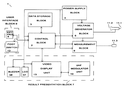
2o Referring to Figure 3, an electrodermal diagnostic unit I is provided which
is
primarily intended for use in diagnosing internal organ pathology in humans
through
electric stimulation and impedance measurement of remotely located skin spots.
In the electrodermal diagnostic unit 1 the control block 2 displays
instructions
1:l
CA 02336825 2001-O1-05
WO 00/01301 pGTIZA99/00048
to the operator through LCD display 37 exclusively or in combination with a
video
monitor connected to the video display unit 13 or a television set connected
to the~~
UHF modulator unit 10. Lists of organs that can be diagnosed are displayed on
the
monitor or television set. The operator selects internal organs to diagnose
via the
keypad 9.
The control unit 2 selects a method of diagnosis based on the location of the
skin spot/zone to be investigated. Two methods of diagnosis are available
specifically
alternating current measurement (AC measurement) or direct current measurement
to (DC measurement). AC measurement is better suited to areas with thicker
skin such as
the soles of the feet. DC measurement is better suited to areas with thin skin
such as
the ear.
The control unit 2 displays a picture of the region to be investigated (such
as
15 the foot or ear). A flashing zone or spot indicates where the measuring
electrode I l .l
must be placed by the operator . The control unit 2 controls the voltage
generator
block S through an optical link to execute the selected test.
For DC measurement: The voltage generator block 5 generates a small
2o constant potential dif~'erence between the measurement electrode 11.1 and
the
reference electrode I 1.3, with the measurement electrode polarized negatively
with
respect to the reference electrode. The current is continuously monitored by
the
control block 2 using the measurement block 6 and when the current rises above
a
preset threshold, it is assumed that both probes are in contact with the skin.
The
CA 02336825 2001-O1-05
WO 00/01301 PCT/LA99/00048
potential difference between the probes is slowly increased and the current
through the
probe measured continuously. The skin resistance is calculated by dividing the
value o~
the potential applied to the probes by the value of the measured current
running
through the probes. When a sudden significant drop in this resistance is
detected (see
i figure 9 for algorithm) the potential difference between the probes is
continuously
adjusted so as to maintain the measured current through the probes at a
predetermined
level. This continues until the rate of change in skin resistance fails below
a
predetermined level. The value of the potential difference at this time is
stored by the
control block 2 in the data storage block 3. The control block 2 uses the
voltage
to generator block to apply the same potential difference between the probes
with
opposite polarization ( the measurement electrode positively polarized with
respect to
the reference electrode). The current is continuously measured. The ratio
(measured
current)/(preset current) is believed to give an indication of the degree of
pathology in
a particular organ. If this ratio is close to zero, the relevant organ is
believed to be
1, diseased. If this ratio is greater than 0.6 then the relevant organ is
believed to be
healthy. The closer this ratio is to zero, the greater the degree of pathology
(e.g.
cancer) is believed to be present in the organ.
The control block 2 monitors the current via the measurement block 6 and
2o displays the test result on the LCD display 37 on a disease percentage
scale until the
operator presses the foot pedal 12. The control block stores the test result
in the data
storage block 3, and activates the buzzer 36 to indicate the completion of the
test.
For AC measurement: The operator places the reference electrode 1 I.3 and the
CA 02336825 2001-O1-05
WO 00/01301 PCT/ZA99/OOOd8
calibration electrode 11.2 on the skin of the subject when prompted via the
LCD
display 37. The voltage generator block 5 generates a small constant potential
~~
difference between the electrodes I 1.2,11.3. The current is continuously
monitored
and when the current rises above a preset threshold, it is assumed that both
probes are
in contact with the skin. An sinusoidal alternating voltage is now applied
between the
electrodes. The current is monitored continuously by the control block 2 using
the
measurement block 6 and the voltage adjusted anti( the current reaches a
preset level.
This process is referred to as calibration.
to The control unit 2 displays a picture of the region to be investigated
(typically
the foot ) on the video monitor or television screen using the result
presentation block .
The control unit 2 displays a message on the LCD display 37 informing the
operator
that the measuring electrode I I . I and the reference electrode 11.3 must now
be used.
A flashing zone or spot indicates where the measuring electrode 1 l . t must
be placed
l5 by the operator. The current is continuously measured. The ratio (measured
current)/(preset current) is believed to give an indication of the degree of
pathology in
a particular organ. If this ratio is close to zero, the relevant organ is
believed to be
diseased. If this ratio is greater than 0.6 then the relevant organ is
believed to be
healthy. The closer this ratio is to zero, the greater the degree of pathology
(e.g
2o cancer) is believed to be present in the organ. The control block 2
monitors the current
via the measurement block 6 and displays the test result on the LCD display 37
on a
disease percentage scale until the operator presses the foot pedal 12. The
control block
stores the test result in the data storage block 3, and activates the buzzer
36 to indicate
the completion of the test.
17
CA 02336825 2001-O1-05
wo ooro><3o><
The control unit 2 consists of a microcontroller 16 (typically an 8051 ). An
%~-
oscillator 28 provides a clock signal for the microcontroller 16. A standard
adress latch
( I 1 ) conf guration is used to create a 16 bit adress bus ( 11.1,16.3) which
connects to
32K random access memory 56 and 32K read only memory 57. A bidirectional data
bus 16.1 transports data to and from the microcontroiler 16. The
microcontroller 16
interfaces which a keypad 9 using keypad interface 17, (typically 74HC922)
The electrodes are galvanicalfy isolated 55 from the main circuit by a
1o transformer 43 in the voltage generator block 5. The measurement block 6 is
optically
isolated from the main circuit by optocouplers 51,52,53,54. A sinusoidal
volta;e
applied to the primary coil of transformer 43.1 is stepped up by the secondary
coil
43.2. Voltage doubling circuit 44 doubles and rectifies the sinusoidal output
of coil
43.2 so that a constant voltage appears over capacitor 38. When relay 40 is
off an
t 5 alternating voltage is fed through to the relay 42. When relay 40 is on,
the constant
voltage over capacitor 38 is fed through to the relay 42.. Relay 42 is used to
switch the
polarity of the signal from relay 40. This combination of relay 42 and 40 is
used to set
the voltage between the probes to an alternating voltage or a constant voltage
and
allows the polarity of the probes to be reversed.
The primary coil 43.1 of then transformer 43 is driven by an opamp 41 which is
used in inverter amplifier mode. A programmable sinewave generator 39
(typically
ML2036) provides the input signal to the opamp 41. Sinewave generator 39 is
controlled by the microcontroller 16 via lines 22.1, 22.3, 22.4. The frequency
is
is
CA 02336825 2001-O1-05
WO 00/01301 PCT/ZA99/00048
digitally programmed via this serial bus. The magnitude of the output
sinusoidal signal
(half peak to peak) is equal to the voltage on the output 24.1 of a digital to
analoguea'~
converter 24. The output voltage of the digital to analogue converter 24 is
set via bus
16.1 by the microcontroller 16.
The current is measured using the measurement block 6. When the circuit
between the electrodes is closed by an impedance such as the human body,
current is
conducted through the electrodes to ground via measuring resistor 46. The
voltage
appearing over this resistor with respect to ground is therefore proportional
to the
current through the probes. An opamp buffer 45 feeds the signal to the
precision
rectifier formed by the opamps referred to by numerals 47 and 48.These
e~ctract the
absolute value of the signal which is fed into a serial analogue to digital
converter
(ADC) 49. The ADC 49 communicates with the microcontroller 16 via a serial bus
consisting of lines 22.1, 22.2 and 22.3 through an optical link provided by
three
15 optocouplers 52,53,54. A zero crossing detector 50 detects the polarity of
the voltage
over the measuring resistor 46 and transmits this information as a binary one
or zero to
the microcontroller 16 through an optocoupler 51. When an alternating voltage
is
applied to the electrodes, current is measured at every voltage peak. The
microcontroller 16 waits for a zero to one transition to occur on the output
22.5 of
20 optocoupler 51. The microcontroller 16 waits for a time period equal to one
quarter of
the period of the output voltage frequency before requesting a conversion from
the
analogue to digital converter 49.
The microcontroller displays relevant information on a monitor through the
CA 02336825 2001-O1-05
WO 00/01301 PGT/ZA99/00048
video display unit 13. A dual port ram 30 contains a bit mapped version of the
screen.
The microcontroller 16 can read and write data to the dual port ram 30 through
a datae'
bus 16.1 using control lines 16.6 and 16.7 and the adress bus, referred to by
numerals
11.1 and 16.3. A field programmable gate array (FPGA) 25 uses counters and
shift
registers to sequentially read bytes of screen data and to write red green and
blue
(RGB) pixel information as well as vertical and horizontal retrace information
to an
RGB to PAL encoder 31. A universal sync generator 33 generates PAL video
standard
synchronization pulses. These pulses are locked to the main system clock using
a phase
locked loop 34. The pixel clock is derived from the main system clock 16.5
usin;~
to counters in the FPGA 25. The pixel clock is used to read and serialize data
at the
correct rate and in the correct manner from the dual port ram 30 so that the
bit streams
fed to the RGB to PAL encoder can be encoded into a PAL standard composite
sync
video signal that can be fed directly into the video input 31.1 of a standard
video
monitor.
l.i
The L7HF modulator unit 10 converts the composite sync video signal to an
ultra high frequency signal that can be directly fed into the aerial port 35.1
of a
television set. An integrated UHF modulator 35 modulates the composite sync
PAL
signal from the video display unit to a frequency determined by external
components.
2t1
Power is delivered to the circuit by the power supply block 4. A transformer
14.1 with primary coupling connected to mains and secondary coupling connected
to
rectifier bridge 14.2 converts the mains 220VAC to 7.2 VAC. The output of the
rectifier is fed into SV regulator 14.3. A monolithic voltage inverter 15.1
typically a
2t1
CA 02336825 2001-O1-05
wo ooio><3o>< pcrrra~rooo4s
MAX660 generates a -5V supply from the main SV supply.
:;~-
The power supplied to the voltage generator block 5 and the measurement
block 6 is galvanically isolated from the main power supply. DC to DC
converter 15.2
s (typically NMA0505) is used to supply +5V and -5V to the isolated patient
interface
c~rcmtry.
Figure 10 shows a graph of voltage over a period of time when the
breakthrough effect (x) is achieved. At the point of breakthrough (x) a sudden
and
to significant decrease in resistance occurs, and thus a sudden and
significant decrease in
voltage is also observed. The reference value is measured once the voltage
stabilises
after the brekthrough effect.
Figures lla and llb show how resistance is affected when an organ is
t5 diseased. In Figure Ila, the two curves represent resistance values at
different
voltages for a healthy organ, while figure 11 b shows two curves of resistance
values at
different voltages for an unhealthy organ. Lines 50 represent the reference
resistance
values, and lines 52 represent the measurement resistance values. Figure l lb
shows
two curves of resistance values at different voltages for an unhealthy organ.
When an
zo organ is healthy, the reference and measurement resistance values are
similar, but as
the state of disease of the organ increases, so the measurement value
increases and the
greater the difference between the reference and measurement values.
Similarly, Figure 12 shows how the measurement impedance changes when an
21
CA 02336825 2001-O1-05
wo ooro><3o1 rcr~~rooo4s
organ is diseased. Line 56 shows reference or resistance values at different
frequencies. Line 58 shows measurement resistance values for a healthy
organ,''- '
whereas line 60 shows measurement resistance values for an unhealthy organ.
The
more diseased an organ becomes, the higher the resistance of the measurement
values.
s and consequently the greater the difference between the measurement and
reference
values.
Referring to Figures 13 and 14, the measurement or calibration electrodes are
placed on one of the spots indicated in order to obtain a diagnosis of a
specific organ.
to The spot on which the electrode is placed depends on the specified organ,
and Tables 1
and 2 set out below show the organs to which the numbered spots refer.
Table 1
Number on Fi re 13 Portion of Bodv
1 Heart (R
_
2 Th ~roid Gland
3 Lun s (U r Lobe)
:l Lun s
Oeso ha s (Cardia)
G Stomach
7 Liver (Le8 Lobe) R)
8 Li~~er (Ri ht Lobe)
(L)
9 S teen (R)
Kidne ~s
11 Pancreas(R)
12 Gall Bladder (L)
13 Duodenum (L)
1-1 Transverse Colon
Left Colon (R)
1G Ri ht Colon (A endix)
(L)
17 Small Intestine
18 Ureter
I 9 Bladder
Prostate
21 Distal Colon
22 Mamma ~ Gland
23 Wa ~ & Adnexa Uteri
2:1 Utems
2~ Pons
______ ___ 2 f _ Thalamus
22
CA 02336825 2001-O1-05
WO 00/01301 Pfy">r/ZA99/00048
27 H ~ thalamus
28 H ~ o h ~sis
29 Cortex Frontal Lobe)
30 Midcortex
31 Cortex (Posterior)
32 Cerebellum
33 Medulla Oblon ata
3d Cen~ical S ine
3S Thoracic S inc
3G Lnmbo-Sacral S ine
3 7 Shoulder
3 8 Elbow
39 Wrist
40 Metaca us
a 1 Fin ers
i 2 Hi
~13 Knee
~-1 Ankle
:1 i Metatarsus
-lG Toes
(L) - left ear auricle
(R) - riglU ear auricle
Table 2
Number on Fi ure la Portion of Bodv
I Brain
2 Pituita
3 Th roid Gland
:1 Oeso ha Is
Lun s
G Heart
7 Li~~er
8 Cardiac S hincter
9 Stomach
S Teen
11 Gall Bladder
12 Adrenal Glands
la Pancreas
1.1 Duodenum
Colott
I G Kidne~~s
17 Small Intestine
18 Ureters
l9 Fallo tan Tubes
Ovaries
2 I Bladder
22 A eudix
23
CA 02336825 2001-O1-05
wo ooromoi rcrrr,~~roooes
The invention is not limited to the precise constructional details disclosed
in
this specification and it will be clear to those skilled in the art that the
above principles-~'~ '
may be applied produce other apparatus embodying these principles.
Specifically, the
apparatus as described uses impedance and resistance measurements to
calaculate the
s state of health of the organ in question, but it will be clear to those
skilled in the art
that other values which are either directly or indirectly proportional to
resistance or
impedance may be used as measurements and in the calculations.
2d