Note: Descriptions are shown in the official language in which they were submitted.
CA 02339157 2001-01-31
WO 00/09024 PCT/US99/16289
- 1 -
SYSTEMS AND METHODS
FOR PLACING MATERIALS INTO BONE
FIELD OF THE INVENTION
The invention generally relates to the
treatment of bone conditions in humans and other
animals.
BACKGROUND OF THE INVENTION
Injection devices similar to a household
caulking gun are used to inject bone cement into
bone. A typical bone cement injection device has a
pistol-shaped body, which supports a cartridge
containing bone cement. A trigger actuates a spring-
loaded ram, which forces a vo:Lume of bone cement in
a viscous condition through a suitable nozzle and
into the interior of a bone targeted for treatment.
According to the teachings of U.S. Patent Nos.
4,969,888 and 5,108,404, a cavity can be first
formed by compacting cancell.ous bone inside the
bone, into which the bone cement is injected.
Conventional cement injection devices provide no
opportunity to override the spring action and
quickly terminate the flow of cement, should the
cavity fill before the spring-actuated load cycle is
completed. Furthermore, once the spring-actuated
mechanism is triggered, conventional cement
injection devices do not permit the injection volume
or inject rate to be adjusted or controlled in real
time, in reaction to cancellous bone volume and
density conditions encountered inside bone.
In a clinical procedure called
ii.
CA 02339157 2001-01-31
WO 00/09024 PCT/US99/16289
- 2 -
vertebroplasty, bone cement is injected at high
pressure (typically, about '700 psi) into the
interior of a vertebral body, without the prior
formation of a cavity. Becau,se high pressure is
used, there is little opportunity to quickly and
accurately adjust cement flow in reaction to bone
volume and density conditions encountered. Momentum
generated by high pressure-induced cement flow
continues to propel oement in-to the targeted bone
site even after termination of' the high pressure.
As a result of the relatively high pressure
that conventional procedures rely upon, coupled with
the effective lack of a short response time, the
targeted bone interior can suddenly overfill.
Excess filling material can be forced outside the
bone interior, and into adjoining tissue regions,
where the presence of filling material is not
required or desired.
For these and other reasons, there is a
need for new systems and methods for placing
material into bones, with greater rate and volume
control, a f,aster response time, and without
requiring the use of high pre,ssure.
SUMMARY OF THE INVENTION
The invention provides instruments,
systems, and methods, which, in use, enable greater
control over the placement of materials into bone.
One aspect of the invention provides an
instrument for tamping material into bone through a
subcutaneous path. The instrument comprises a body
having a length and a terminus. The body includes
markings located along the length at increments from
the terminus. The markings allow the physician to
gauge the position of the instrument in the
subcutaneous path, as material is being tamped into
II
CA 02339157 2001-01-31
WO 00/09024 PCT/US99/16289
- 3 -
bone. In particular, the markers allow the
physician to tell at a glance the location of the
terminus, in terms of how far beyond or short of the
end of the subcutaneous path it is.
In one embodiment, the instrument is used
by deploying a cannula to establish a subcutaneous
path into bone.. A material is introduced into bone
through the cannula. The terminus of the instrument
is advanced through the cannula to urge material
residing in the cannula into bone.
Another aspect of the invention provides an
apparatus for introducing material into bone through
a subcutaneous cannula. The apparatus includes a
delivery device to convey the material at a low
delivery pressure. As used herein, a "low delivery
pressure" is equivalent to the pressure at which
liquid is expressed from 1 cc syringe by the
application of moderate force to the syringe piston,
which amounts to a pressure that is no greater than
about 360 psi.
According to this aspect of the invention,
the apparatus also includes a nozzle instrument
capable of advancement through the subcutaneous
cannula into bone. The nozzle: comprises a proximal
fitting to couple the nozzle instrument to the
delivery device. The nozzle further comprises a
nozzle terminus through which the material conveyed
by the delivery device enters bone at the delivery
pressure.
In one embodiment, the delivery device
comprises a syringe.
In one embodiment, =the apparatus further
includes a tamping instrument, which is capable of
advancement through the subcutaneous cannula. The
tamping instrument has a tamping terminus which,
CA 02339157 2001-01-31
WO 00/09024 PCT/US99/16289
- 4 -
during the advancement, urges material residing in
the subcutaneous cannula into bone.
In one embodiment, the tamping instrument
includes markings to visually gauge the advancement
of the tamping terminus through the subcutaneous
cannula.
In one embodiment, the apparatus is used by
deploying a cannula to establislh a subcutaneous path
into bone. The delivery device is actuated to
convey material at the delivery pressure through the
nozzle terminus into bone.
Another aspect of the invention provides a
tool for deployment into bone. The,tool comprises
a catheter tube having a distal region and an
expandable structure carried by the distal region
for compacting cancellous borie. The tool also
includes an introducer sleeve slidably carried by
the catheter tube for movement between a retracted
position spaced from the expandable structure and an
advanced position overlying the expandable
structure. The introducer sleeve includes a tubular
main body dimensioned to compress the expandable
structure when the introducer sleeve is in the
advanced position. A collar extends beyond the
distal region of the catheter tube when the
introducer sleeve is in the advanced position. The
collar is dimensioned larger than the tubular main
body to releasably engage an end of a cannula.
Thus, the introducer sleeve both sizes and aligns
the expandable structure for passage into the
cannula through the end of the cannula.
Another aspect of the invention provides
apparatus for introducing material into bone through
a subcutaneous cannula. The apparatus includes a
delivery device to convey the material at a low
CA 02339157 2001-01-31
WO 00/09024 PCT/US99/16289
- 5 -
delivery pressure, i.e., a pressure no greater than
about 360 psi. The apparatus also includes a nozzle
instrument capable of advancement through the
subcutaneous cannula into boine and comprising a
proximal fitting to couple the nozzle instrument to
the delivery device. The nozzle also includes a
nozzle bore, through which the material conveyed by
the delivery device enters bone at the delivery
pressure. The apparatus further includes a stylet
capable of advancement into the nozzle bore through
the proximal fitting to close the nozzle bore and,
with the nozzle instrument. Together, the nozzle
and the stylet form a tamping instrument capable of
advancement through the subcutaneous cannula to urge
residual material from the subcutaneous cannula.
Another aspect of the invention provides a
method for delivering materj.al into bone. The
method deploys a cannula through soft tissue to
establish a subcutaneous path into bone. The method
introduces a material into bone through the cannula.
The method advances a tamping :Lnstrument through the
cannula to urge material residing in the cannula
into bone.
In one embodiment, the method delivers
material at a low delivery pressure, i.e., a
pressure no greater than about 360 psi.
In one embodiment, the introducing step
uses a manual syringe.
The material can comprise medication or a
material that sets to a hardened condition e.g.,
bone cement, or autograft tissue, or allograft
tissue, or synthetic bone substitute, or
combinations thereof.
In one embodiment, the method further
includes the step of deploying a cavity forming
CA 02339157 2007-03-26
60895-1603
- 6 -
instrument through the cannula to compress cancellous bone
and form a cavity. In this embodiment, the introducing and
advancing steps convey material into the cavity.
According to one broad aspect of the present
invention, there is provided a system comprising an access
tool sized and configured to establish an access path
through soft tissue to bone having an interior volume
occupied, at least in part, by cancellous bone, a void
forming tool sized and configured to be introduced through
the access path to form a void in cancellous bone, a nozzle
sized and configured to pass through the access path and
including an interior bore defining a fixed interior volume
to receive and deliver a measured volume of filling material
into the void, and an auxiliary tool sized and configured to
be advanced through the interior bore and urge filling
material from the nozzle.
According to another broad aspect of the present
invention, there is provided a system comprising a cannula
sized and configured to establish an access path through
soft tissue to bone having an interior volume occupied, at
least in part, by cancellous bone, a void forming tool sized
and configured to be introduced through the cannula to form
a void in cancellous bone, a nozzle that can be manipulated
independent of the cannula and that is sized and configured
to pass through the cannula, the nozzle including an
interior bore to receive and deliver a measured volume of
filling material into the void, and an auxiliary tool that
can be manipulated independently of the nozzle and the
cannula and that is sized and configured to be advanced
through the interior bore and urge filling material from the
nozzle, the auxiliary tool, when fully advanced,
substantially fully occupying the entire interior bore of
the nozzle.
CA 02339157 2007-03-26
60895-1603
- 6a -
According to still another broad aspect of the
present invention, there is provided apparatus for
delivering material into bone comprising a cannula for
establishing a subcutaneous path into bone and including at
least one radiopaque marker, and a tamping instrument having
a tamping terminus, the tamping instrument being sized and
configured for manipulation independent of the cannula to
enable insertion of the tamping instrument into the cannula,
advancement of the tamping terminus in the cannula to urge
material residing in the cannula into bone, and withdrawal
of the tamping terminus from the cannula.
According to yet another broad aspect of the
present invention, there is provided apparatus for
delivering material into bone comprising a cannula for
establishing a subcutaneous path into bone, and a tamping
instrument having a tamping terminus and including at least
one marking to visually gauge the advancement of the
terminus relative to the distal end of the cannula, the
tamping instrument being sized and configured for
manipulation independent of the cannula to enable insertion
of the tamping instrument into the cannula, advancement of
the tamping terminus in the cannula to urge material
residing in the cannula into bone, and withdrawal of the
tamping terminus from the cannula.
According to a further broad aspect of the present
invention, there is provided apparatus for delivering
material into bone comprising a cannula for establishing a
subcutaneous path into bone; and a tamping instrument for
advancement through the cannula including at least one
marking to visually gauge the advancement of the terminus
relative to the distal end of the cannula, and comprising a
body portion and a handle portion, the body portion being
CA 02339157 2007-03-26
60895-1603
- 6b -
sized and configured to substantially fill the cannula when
the tamping instrument is fully inserted into the cannula.
According to yet a further broad aspect of the
present invention, there is provided apparatus for
delivering material into bone comprising a cannula for
establishing a subcutaneous path into bone and including at
least one radiopaque marker; and a tamping instrument for
advancement through the cannula comprising a body portion
and a handle portion, the body portion having a
substantially constant diameter along its length.
Features and advantages of the inventions are set
forth in the following Description and Drawings, as well as
in the appended Claims.
BRIEF DESCRIPTION OF THE DRAWINGS
Fig. 1 is a plane view of a kit housing a system
of functional instruments, which, in use, gain subcutaneous
access to the inside of a bone to compact cancellous bone
and form a cavity for therapeutic purposes;
Fig. 2 is an exploded perspective view of the kit
shown in Fig. 1;
Fig. 3 is a perspective view of the subcutaneous
access instrument group that forms a part of the system
shown in Fig. 1;
Fig. 4A is a perspective view of the cavity
forming instrument that forms a part of the system shown in
Fig. 1;
Fig. 4B is a section view of the catheter tube of
the cavity forming instrument, taken generally along line
42-4B in Fig. 1;
CA 02339157 2007-03-26
60895-1603
- 6c -
Fig. 4C is an end view of an alternative
embodiment of the cavity forming instrument shown in
Fig. 4A, having a prebent stylet;
Fig. 5 is a perspective view of the material
introducing instrument group that forms a part of the system
shown in Fig. 1;
Figs. 6 and 7 are, respectively, top and side
views of a human vertebral body;
Fig. 8 is a top view of a vertebral body during
insertion of a spinal needle instrument to begin a bone
access procedure;
i~.
CA 02339157 2001-01-31
WO 0009024 PCT/US99/16289
- 7 -
Figs. 9 to 11 are top views showing
subsequent steps, after insertion of the spinal
needle instrument shown in Fig. 8, of inserting a
guide pin instrument into the vertebral body;
Fig. 12 is a perspective view showing a
subsequent step, after insertion of the guide pin
instrument shown in Figs. 9 to 11, which deploys an
obturator instrument deployed over the guide pin
instrument with aid of a hand].e;.
Fig. 13 is a top, view of the vertebral
body, with the obturator instrument shown in Fig. 12
deployed;
Fig. 14 is a perspective view showing a
subsequent step, after insertion of the obturator
instrument shown in Fig. 12, which uses the handle
shown in Fig. 12 to aid in the deployment of a
cannula instrument over the obturator instrument;
Fig. 15 is a top view of the vertebral
body, with the cannula instrument shown in Fig. 14
deployed;
Fig. 16 is a perspective view showing a
subsequent step, after insertion of the cannula
instrument shown in Fig. 14, which removes the
obturator instrument from the cannula instrument, to
leave the cannula instrunient and guide pin
instrument in place;
Fig. 17 is a top view of the vertebral
body, after the obturator removal step shown in Fig.
16, leaving the cannula instrument and guide pin
instrument in place;
Fig. 18 is a perspective view showing a
subsequent step, after removal of the obturator
instrument shown in Fig. 16, which uses the handle
shown in Fig. 14 to aid in the deployment of a drill
bit instrument through the cannula instrument along
i ~.
CA 02339157 2001-01-31
WO 00/09024 PCT/US99/16289
- 8 -
the guide pin instrument;
Fig. 19 is a top view of the vertebral
body, as the drill bit instrumient shown in Fig. 18
is deployed with aid of the handle to open a passage
into the interior volume of the vertebral body;
Fig. 20 is a perspective view showing a
subsequent step, after removal of the drill bit
instrument and guide pin instrument shown in Fig.
18, of deploying the cavity forming instrument into
the vertebral body;
Fig. 21 is a top view of the vertebral
body, as the expandable structure carried by the
cavity forming instrument shown in Fig. 20 is
deployed into the interior volume of the vertebral
body;
Fig. 22 is a top view of the vertebral
body, as the expandable structure shown in a
collapsed condition in Fig. 21 is expanded to
compact cancellous bone and fcsrm a cavity;
Fig. 23 is a top view of the vertebral
body, after removal of the expandable structure,
showing the cavity formed by compacting cancellous
bone;
Fig. 24 is a perspective view of the
syringe of the material introducing instrument
group, shown in Fig. 5, being filled with a material
selected for introduction into the cavity shown in
Fig. 23;
Fig. 25 is a perspective view of the
syringe shown in Fig. 24 being joined to a nozzle,
which also forms a part of the material introducing
instrument group shown in Fig. 5;
Fig. 26 is a perspective view showing the
syringe and attached nozzle shown in Fig. 25 being
deployed through the cannula instrument in
CA 02339157 2001-01-31
WO 00/09024 PCT/US99/16289
- 9 -
preparation of introducing material into the cavity;
Figs. 27 and 28 are perspective and top
views, respectively, showing the syringe and
attached nozzle shown in Fig. 26 in use to inject
material into the cannula instrument for passage
into the cavity;
Fig. 29 is a top view of the vertebral body
after a measured volume of material has been
injected and the syringe and attached nozzle
withdrawn from the cannula instrument;
Fig. 30 is a top view showing the
deployment of a tamping instrument, which forms a
part of the material introducing instrument group
shown in Fig. 5, being dep:loyed in the cannula
instrument;
Fig. 31 is a top view showing advancement
of the tamping instrument in the cannula instrument
to displace and distribute material from the cannula
instrument into the cavity;
Fig. 32 is a top view of the vertebral body
after removal of the tamping instrument and cannula
instrument, showing the cavity, now filled with the
material;
Fig. 33 is a perspective view of a reduced
diameter cannula instrument and associated reduced
diameter material introducing instruments, which
embody features of the invention;
Fig. 34 is a perspective view of a cavity
forming instrument having an expandable cavity
forming structure, which, in use, is deployed using
the reduced diameter cannula instrument shown in
Fig. 33, the cavity forming instrument having a
sliding introducer sleeve shown in its rearward
position;
Fig. 35 is a perspecltive view of the cavity
iI
CA 02339157 2001-01-31
WO 00/09024 PCT/US99/16289
- 10 -
forming instrument shown in Fig. 34, with the
introducer sleeve moved forward to overlie and
compress the expandable cavity forming structure;
Fig. 36 is a perspective view of the cavity
forming structure shown in Fig. 35, with the
introducer sleeve (shown partially in. section)
coupled to the proximal end of the cannula
instrument, to guide the expandable structure
compressed within the sleeve into the reduced
diameter cannula instrument without damage; and
Fig. 37 is a perspective view of the cavity
forming structure shown in Fig. 36, after the
expandable structure has been guided by the
introducer sleeve into the canriula instrument and is
being advanced through the cannula instrument for
deployment in bone.
The invention may be embodied in several
forms without departing from its spirit or essential
characteristics. The scope of the invention is
defined in the appended claims, rather than in the
specific description preceding them. All embodi-
ments that fall within the meaning and range of
equivalency of the claims are therefore intended to
be embraced by the claims.
DETAILED DESCRIPTION OF THE PREFERRED EMBODIMENTS
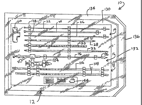
Figs. 1 and 2 show a system 10 of
functional instruments. In use, certain instruments
of the system 10 are deployed in a purposeful manner
to penetrate tissue and gain subcutaneous access to
the inside of a bone. Inside bone, other instruments
of the system 10 are deployed to form a cavity in
cancellous bone, into which a material is placed for
therapeutic purposes.
In the illustrated embodiment, the system
10 is arranged as a prepackage kit 12 in three
CA 02339157 2001-01-31
WO 00/09024 PCT/US99/16289
- 11 -
functional instrument groups 14, 16, and 18. The
first group 14 (which Fig. 3 shows outside the kit
12) comprises instruments whose purpose is to gain
subcutaneous access to a bone interior. The second
group 16 (which Fig. 4 shows outside the kit 12)
comprises an instrument whose function is to create
a cavity in cancellous bone. The third group 18
(which Fig. 5 shows outside the kit 12) comprises
instruments whose function is to introduce a
material into the cavity.
The kit 12 can take various forms. In the
illustrated embodiment, the kit 12 comprises a
sterile, wrapped assembly.
Further details of each functional
instrument group 14, 16, and 18 and the kit 12
follow.
I. The Subcutaneoias Access Instrument
Group
The number and type of instruments in the
group 14 can vary. Fig. 3 shows five representative
instruments, each having a different size and
function.
A. The Spinal Needle and Guide Pin
As Fig. 3 shows, one: instrument comprises
a conventional spinal needle assembly 20 and a guide
pin instrument 26.
In use, the spinal needle assembly 20
establishes the initial subcutaneous path leading to
the targeted treatment site. The guide pin
instrument 26 is deployed through this path,
followed by progressively larger instruments, as
will be described later.
The spinal needle assembly 20 comprises a
stylet 22, which is slidably deployed within a
stylus 24. The stylus 24 typically has, for example,
ic
CA 02339157 2001-01-31
WO 00/09024 PCT/US99/16289
_ 12 _
about an eleven gauge diameter. Other gauge
diameters can be used, according to the gauge.of the
guide pin instrument 26 used.
In use, the guide pin instrument 26 is
deployed through the subcutaneous path established
by the spinal needle assembly 20, by exchange with
the needle stylet 22. The guide pin instrument 26
serves to guide the establishment of the main
operative pathway to the targeted treatment site.
The remaining instruments 28, 30, and 32 in
the group 14 share some common features, although
they are intended, in use, to perform different
functions. These instruments 28, 30, and 32 are each
made of a rigid, surgical grade plastic or metal
material. These instruments :28, 30, and 32 each
comprises an elongated, cylindrical body having a
proximal end 34 and a distal end 36.
B. The Obturator Ins:trument
The instrument 28 functions as an
obturator. Its distal end 36 is tapered to present
a penetrating surface 38. In use, the surface 38 is
intended to penetrate soft tissue in response to
pushing or twisting forces applied by the physician
at the proximal end 34.
The proximal end :34 of the obturator
instrument 28 presents a flariged surface 40, which
tapers from a larger outer diameter to a smaller
outer diameter in the direction of the proximal end
34. The flanged surface 40 includes an array of
circumferentially spaced teeth 42.
An interior lumen 44 extends through the
obturator instrument 28 from the distal end 36 to
the proximal end 34. The interior lumen 44 is sized
to accommodate the guide pin instrument 26, as will
be described in greater detail later.
CA 02339157 2001-01-31
WO 00/09024 PCT/US99116289
- 13 -
C. The Cannula instruiaent
The instrument 30 functions as a cannula or
guide sheath. The cannula instrument 30 is somewhat
larger in diameter than and not as long as the
obturator instrument 28. The.cannula instrument 30
includes an interior lumen 46 that extends from its
distal end 36 to its proximal end 34. The interior
lumen 46 is sized to accept the obturator instrument
28. The size of the interior lumen 46 permits a
physician to slide and rotate the cannula instrument
30 relative to the obturator instrument 28, and vice
versa, as will be described in greater detail later.
The distal end 36 of the cannula instrument
30 presents an end surface 48. In use, the end
surface 48 of the cannula instrument 30 is intended
to penetrate soft tissue surrounding the obturator
instrument 28 in response to pushing or twisting
forces applied at the proximal end 34.
The proximal end 34 carries an enlarged
fitting 50. The fitting 50 tapers from a larger
diameter to a smaller diameter in the direction of
the proximal end 34. Like the tapered flange 40 on
the obturator instrument 28, the tapered fitting 50
has an array of circumferentially spaced teeth 52.
The tapered fitting 50 of the cannula instrument 30
possesses a larger maximum outer diameter than the
maximum outer diameter of the tapered flange 40 of
the obturator instrument 28.
The cannula instrument 30 includes measured
markings 118 along its length(see Fig. 3). The
measured markings 118 gauge the depth of insertion.
The markings 118 can be placed, for example, at one
centimeter intervals. As Fig. 3 shows, the markings
118 can be consecutively numbered, beginning at the
distal end 36, so that the physician can ascertain
i I!
CA 02339157 2001-01-31
WO 00/09024 PCT/US99/16289
.. 14 _
the insertion depth at a glance.
D. The Drill Bit Instrument
The instrument 32 functions as a drill bit.
The drill bit instrument 32 has generally the same
physical dimensions as the obturator instrument 28.
Like the obturator iristrument 28, the drill bit
instrument 32 is intended, in use, to fit for
sliding and rotational movement within the interior
lumen 46 of the cannula instrument 30.
The distal end 36 of the drill bit
instrument 32 includes machined cutting edges 54. In
use, the cutting edges 54 are intended to penetrate
hard tissue in response to rotation and longitudinal
load forces applied at the proximal end 34 of the
drill bit instrument 32.
The proximal end 34 presents a tapered
flange 56, which is substantially identical to the
flange 40 on the obturator instrument 28. Like the
obturator instrument 28, the tapered flange 56
changes from a larger diameter to a smaller diameter
in the direction of the proximal end 34. The
tapered flange 56 of the drill bit instrument 32
also includes an array of circumferentially spaced
teeth 58. The form and orientation of the teeth 58
on the drill bit instrument 32 correspond to the
form and orientation of the teeth 42 on the
obturator instrument 28.
E. The Handle
The group includes a handle 60. The handle
60 engages the functional instruments 28, 30, and 32
in a removable, slip fit fashion to aid a physician
in manipulating the instruments during use.
The handle 60 is made from a molded or cast
rigid plastic or metal material. The handle 60 is
shaped to be comfortably and securely grasped by a
CA 02339157 2001-01-31
WO 00/09024 PCT/US99/16289
- 15 -
normal human hand. The shape and size to accommodate
this function can, of course, vary. In the
illustrated embodiment, the handle 60 is elongated
along a main axis to fit comfortably across the palm
of the hand.
The handle 60 includles a center post 62,
which is integrally molded tci the handle 60 about
its geometric center. The center post 62 extends
downward to give the handle 60 a general T-shape.
The handle 60 includes two interior
cavities or sockets 64 and 66 in the center post 62.
The sockets guide the attachment between the handle
60 and the instruments 28, 30, and 32. The first and
second sockets 64 and 66 are. sized to present
unique attachment sites for different functional
instruments.
The first socket 64 includes an array of
circumferentially spaced grooves 68, which, in form
and orientation, match the teeth 42 and 58 at the
proximal ends 34 of the obturator instrument 28 and
the drill bit instrument 32. The first socket 64
accepts the tapered flange 40 or 56 of either the
obturator instrument 28 or the drill bit instrument
32. The teeth 42 and 58 of either tapered flange 40
or 56 mesh in a slip-fit with the grooves 68 of the
first socket 64. The running slip-fit allows
longitudinal force to be applied to either
instrument 28 or 32 through the handle 60. The
running slip-fit also prevents relative rotation
between either instrument 28 or 32 and the first
socket 64, thereby permitting torsional or twisting
forces to be applied to either instrument 28 or 32
by the handle 60, with an increased mechanical
advantage.
The second socket 66 is larger than the
CA 02339157 2007-03-26
60895-1603
- 16 -
first socket 64 and is sized to accept the larger
tapered fitting 50 of the cannula instrument 30. The
second socket 66 includes an array of
circumferentially spaced grooves 70, which, in form
and orientation, match the teeth 52 on the tapered
fitting 50. The teeth 52 of the tapered fitting 50
mesh in a slip-fit with the grooves 70 of the second
socket 66. The running slip-fit allows both
longitudinal and torsional forces to be applied to
the cannula instrument 30 through the handle 60,
with increased mechanical advantage.
As shown in phantom lines in Fig.3, a first
passage 72 extends through the top of the handle 60,
through the center post 62, and into the first
socket 64. The passage 72 is generally aligned with
the center of the first socket 64 and is sized to
pass the guide pin instrument 26 (see Fig. 12).
Likewise, as also shown in phantom lines in
Fig. 3) a second passage 74 extends through the top
of the handle 60, through the center post 62, and
into the second socket 66. The passage 74 is
generally aligned with the center of the second
socket 66 and is sized to pass the either obturator
instrument 28 or the drill bit instrument 32 (see
Fig. 14).
Further details of the handle 60 can be
found in copending U.S. Patent Serial No. 6,468,279,
filed January 27, 1998, and entitled "A Slip-Fit
Handle for Hand-Held Instruments that Access Interior
Body Regions."
Further details regarding the use of the
handle 60 and the associated instruments 26, 28, and
30 will be provided later.
II. The Cavity Forming Instrument
As Fig. 4A shows, the group 16 includes an
CA 02339157 2001-01-31
WO 00/09024 PCTlUS99/16289
- 17 -
instrument 76, which is deployed through the cannula
instrument 30 to a location inside bone (see Fig.
20). When so deployed, the instrument 76 serves to
form a cavity in cancellous bone.
The instrument 76 can be constructed in
various ways. In the illustrated embodiment, the
instrument 76 includes a flexible catheter tube 78
having a proximal end 80 and a distal end 82. The
proximal end 80 carries a handle grip 84 to
facilitate gripping and maneuvering the catheter
tube 78. The materials for the: catheter tube 78 are
selected to facilitate its advancement through the
cannula instrument 30. The catheter tube 78 can be
constructed, for example, using standard flexible,
medical grade plastic materials, like vinyl, nylon,
polyethylenes, ionomer, polyurethane, and
polyethylene tetraphthalate (PET). The catheter
tube 78 can also include more rigid materials to
impart greater stiffness and thereby aid in its
manipulation. More rigid materials that can be used
for this purpose include stainless steel, nickel-
titanium alloys (NitinolTM material), and other metal
alloys.
The distal end 82 of the instrument 76
carries an expandable structure 86. In the
illustrated embodiment, the expandable structure 86
is made from a polyurethane or an elastomer (e.g.,
silicone or nylon) material. 'rhe structure 86 has
been preformed to possess a. desired shape by
exposure to heat and pressure, e.g., through the use
of conventional thermoforming techniques.
As Fig. 4B shows, thie catheter body 78
includes an interior lumen 88, which communicates
with the interior of the structure 86. A fitting 90
on the proximal end 80 of the catheter tube 78 (see
CA 02339157 2001-01-31
WO 00/09024 PCT/US99/16289
- 18 -
Fig. 4B) communicates with thE: lumen 88. The fitting
90 couples the lumen 88 to a source 92 of fluid,
e.g., sterile saline (see Fig. 21), or a radiopaque
contrast medium.
The fluid is introduced from the source 92
into the structure 86 under positive pressure,
causing the structure 86 to expand. During expansion
inside bone, the material selected for the structure
86 preferably resists deformation, so that the
expanded shape inside bone essentially corresponds
to its expanded shape outside bone, i.e., when in an
open air environment. This allows the physician to
select in an open air environment a structure 86
having an expanded shape desired to meet the
targeted therapeutic result, with the confidence
that the expanded shape inside bone will be similar
in important respects. In addition to being able to
expand its volume while resisting deformation inside
bone, the material of the structure 86 preferable
withstands abrasion, tearing, and puncture when in
contact with cancellous bone.
The shape of the structure 86, when
expanded inside bone, is selected by the physician,
taking into account the morphology and geometry of
the site to be treated. The shape of the cancellous
bone to be compressed, and the local structures that
could be harmed if bone were moved inappropriately,
are generally understood by medical professionals
using textbooks of human skeletal anatomy along with
their knowledge of the site and its disease or
injury. The physician is also able to select the
expanded shape inside bone based upon prior analysis
of the morphology of the targeted bone using, for
example, plain film x-ray, fluroscopic x-ray, or MRI
or CT scanning. The expanded shape inside bone is
CA 02339157 2001-01-31
WO 00/09024 PCT/US99/16289
- 19 -
selected to optimize the formation of a cavity that,
e.g., when filled with a suitable material, provides
support across the region of the bone being treated.
As one general guideline, in cases where
the bone disease causing fracture (or the risk of
fracture) is the loss of cancellous bone mass (as in
osteoporosis), the selection of the expanded shape
of the structure 86 inside bone should take into
account that from 30% to 90% of the cancellous bone
volume should be compacted. Another general
guideline is the amount that the: targeted fractured
bone region has been displaced or depressed. The
expansion of the structure 86 within the cancellous
bone region inside a bone can elevate or push the
fractured cortical wall back to or near its anatomic
position occupied before fracture occurred.
In the illustrated embodiment (see Fig.
4A), the structure 86 possesses a preformed hour-
glass or peanut shape. This shape is selected in
contemplation of deploying the structure 86 in a
vertebral body, as will be described in greater
detail later.
To facilitate deployment of the structure
86 through the cannula instrument 30, the catheter
tube 78 includes a second interior lumen 94. The
lumen 94 extends from a second fitting 98 on the
proximal end 80 of the catheter tube 78, through the
body of the cannula tube 78, and through the
interior of the structure 86 to the tip end 172 of
the structure 86. The lumen 94 receives a generally
stiff stylet 96, which can be made from a molded
plastic or stainless steel mate:rial. The stylet 96
is inserted through the fitting 98 into the lumen
94, and includes a threaded coupling 100 to secure
the stylet 96 against movement. The presence of the
CA 02339157 2007-03-26
60895-1603
- 20 -
stylet 96 serves to keep the structure 86 in the
desired distally straightened condition during
passage through the cannula instrument 30 into the
targeted tissue region. Once the structure 86 is
free of the cannula instrument 30 andinside bone,
the stylet 96 can be withdrawn (shown by arrow 174
in Fig. 4A). This returns normal flexibility to the
catheter tube 78 and facilitates manipulation of the
structure 86 inside bone. With the stylet 96
withdrawn, the lumen 94 can also serve as a pathway
for introducing rinsing liquid or to aspirate debris
from the bone.
In the illustrated embodiment, the stylet
96 is biased toward a generally straight condition.
In an alternative embodiment (see Fig. 4C), a stylet
102 can have a preformed memory, to normally bend
its distal region. The memory is overcome to
straighten the stylet 102 when confined within the
cannula instrument 30. However, as the structure 86
and distal region of the preformed stylet 102
advance free of the cannula instrument 30, to pass
into the targeted region, the preformed memory bends
the distal region of the stylet 102 and thereby
shifts the main axis of the expandable structure 86.--
The prebent stylet 102, positioned within the
interior of the structure 86, aids in altering the
orientation of the structure 86, bringing it into
better anatomic alignment with the targeted region.
Other types of instruments that can form
cavities in cancellous bone and other interior body
regions are described in copending U.S. Patent
Serial No. 6,440,138, entitled "Structures and
Methods for Creating Cavities in Interior Body
Regions," filed April 6, 1998.
III. The Material Introducing Instrument
CA 02339157 2001-01-31
WO 00/09024 PCT/US99/16289
- 21 -
Group
The group 18 includes irtstruments 104, 106,
and 108 which serve to convey and compact a selected
material inside the cavity formed by the structure
86. The material in the cavity provides a desired
therapeutic result, e.g., rep:lacement of tissue
mass, or.renewed interior suppo:rt for the bone, or
the delivery of medication, or combinations thereof.
Accordingly, the material to perform this function
can be selected from among, e.g., a material that
sets to a hardened condition, including bone cement,
autograft tissue, allograft tissue, synthetic bone
substitute, as well as a medication, or combinations
thereof.
In the illustrated embodiment, the group 18
comprises material injection instruments 104 and 106
and a material tamping instrument 108, which deliver.
material at a low delivery pressure, i.e., a
pressure no greater than about :360 psi.
A. Low Pressure Material Injection
Instruments
In the illustrated embodiment, the material
is injected by use of a conventional syringe 104, to
which a specially designed injection nozzle 106 is
coupled. A manual actuated syringe with a push
plunger can be used. Alternatively, a LeVeen
Inflation Syringe with threaded plunger can be used,
which can be actuated manualJLy or by use of a
mechanical actuator.
In the illustrated embodiment, the syringe
104 is made from a clear plastic material. The
syringe 104 includes a chamber 110, which receives
the material to be injected. The material is
expressed from the chamber 100 by a manually
advanced syringe piston 112 (see also Fig. 25).
CA 02339157 2001-01-31
WO 00/09024 PCT/US99/16289
- 22 -
The injection nozzle 106 connects by a
threaded connector 114 to the endlof the syringe 104
9 (see also Fig. 25). In the illustrated embodiment,
the nozzle 106 is made from a qenerally flexible,
inert plastic material, such as such as polyethylene
or an other suitable polymer. Alternatively, the
nozzle 106 can be made from a generally rigid
plastic or metal material.
The injection nozzle 106 is sized to be
advanced through the cannula instrument 30 (see Fig.
26). The nozzle 106 includes measured markings 116
along its length. The markings 116 can be placed,
for example, at one centimeter intervals, to
correspond with the markings :L18 on the cannula
instrument 30, so that the relative position of the
nozzle 106 within the cannula instrument 30 can be
gauged. The markings 118 can, e.g., include a set
point 176. Alignment of the sc:t point 176 at the
proximal end 34 of the canniala instrument 30,
indicates that the distal end of the nozzle 106 is
located in an aligned relationship with the distal
end 36 of the cannula instrument 30. In this
arrangement, the markings 118 are consecutively
numbered with positive numbers proximally of the set
point 176 and with negative numbers distally of the
set point 176. The physician is thereby able to
tell at a glance the location of the distal end of
the nozzle 106, in terms of how far beyond or short
of the distal end 36 of the cannula instrument 30 it
is.
In use, the distal end of the nozzle 106 is
located beyond the distal end 36 of the cannula
instrument 30 within the cavity formed in the
targeted tissue region. As Fig. 5 shows, the distal
end of the nozzle 106, when made from a plastic
CA 02339157 2001-01-31
WO 00/09024 PCT/US99/16289
- 23 -
material, can carry at least one radiopaque marker
208, to enable remote visualization of the nozzle
position within the body. The syringe 104 ejects a
predetermined volume of material into the nozzle 106
in a low pressure stream into the cavity. As the
material fills the cavity, the nozzle (still
ejecting material) is retracted from the cavity and
into the cannula instrument 30 itself. Further
details of this function and resiult will be provided
later.
B. The Material Tamping Instrument
The group 18 also includes a material
tamping instrument 108. The tamping instrument 108
is made from generally rigid, inert plastic or metal
material. The tamping instrumeint 108 is also sized
to be advanced into the cannula. instrument 30 (see
Fig. 30). The free end 124 of the tamping instrument
108 is ribbed or contoured to facilitate gripping
the instrument 108 during use.
The tamping instrunient 108 includes
measured markings 122 along its length. The markings
116 can be placed, for example, at one centimeter
intervals, to correspond with the markings 118 on
the cannula instrument 30, sc> that the relative
position of the tamping instrument 108 within the
cannula instrument 30 can be gauged. Like the nozzle
106, the markings 122 on the tamping instrument 108
includes a set point 178, which indicates when the
distal end of the tamping instrument 108 aligns with
the distal end 36 of the cannula instrument 30. Also
like the nozzle 106, the markings 122 on the tamping
instrument 108 are consecutively numbered with
positive numbers proximally of -the set point 178 and
with negative numbers distally of the set point 178.
The physician is thereby able to tell at a glance
CA 02339157 2001-01-31
WO 00/09024 PCT/US99/16289
- 24 -
the location of the end of the tamping instrument
108, in terms of how far beyond or short of the
distal end 36 of the cannula instrument 30 it is. As
Fig. 5 also shows, the end of 'the tamping instrument
108, when made from a plasticimaterial, can carry at
least one radiopaque marker 210, so that its
position can be visualized from outside the body.
After withdrawal of the nozzle 106 from the
cannula instrument 30, residual material is left in
the cannula instrument 30. The purpose of the
tamping instrument 108 is to displace the residual
material out the distal end. 36 of the cannula
instrument 30 and into the cavity, to thereby fill
the cavity without exerting undue pressure within
the bone. The tamping instrument 108 thereby serves
to clear residual material from the cannula
instrument 30, to assure that the desired volume of
material is delivered into the cavity. The removal
of residual material from the cannula instrument 30.
by the tamping instrument 108 also prevents seepage
of material into surrounding tissue regions upon
removal of the cannula instru:ment 30. The tamping
instrument 108 also compacts the material uniformly
within the cavity, again without undue pressure.
Further details of these functions and results will
be discussed later.
IV. The Kit
As Figs. 1 and 2 show, in the illustrated
embodiment, the kit 12 includes an interior tray 126
made, e.g., from die cut cardboard, plastic sheet,
or thermo-formed plastic material. The tray 126
includes spaced apart tabs 1,28, which hold the
various instruments in a secure position during
sterilization and storage prioz- to use.
When packaged as a sterile assembly, the
CA 02339157 2001-01-31
WO 00/09024 PCT/US99/16289
- 25 -
kit 12 includes an inner wrap 130, which is
peripherally sealed by heat or the like, to enclose
the tray 126 from contact iwith the outside
environment. One end of the inner wrap includes a
conventional peal-away seal 132,, to provide quick
access to the tray 126 at the instant of use, which
preferably occurs in a sterile erivironment, such as
within an operating room.
When packaged as a sterile assembly, the
kit 12 also includes an outer wrap 134, which is
also peripherally sealed by heat or the like, to
enclosed the inner wrap 130. One end of the outer
wrap includes a conventional pea:1-away seal 136, to
provide access to the inner wrap 130 and its
contents. The outer wrap 134 can be removed -from
the inner wrap in anticipation of imminent use,
without compromising sterility of the contents of
the kit 12.
As Fig. 2 shows, each iinner and outer wrap
130 and 134 includes a peripherally sealed top sheet
138 and bottom sheet 140. In the illustrated
embodiment, the top sheet 138 is made of transparent
plastic film, like polyethylene or MYI,AR material,
to allow visual identification of the contents of
the kit 12. The bottom sheet 1.40 is made from a
material that is permeable to ETO sterilization gas,
e.g., TYVEK plastic material (available from
DuPont).
In the illustrated embodiment, the tray 126
presents the instruments groups 14, 16, and 18 in an
ordered, organized layout, which is arranged to aid
the physician in carrying out the intended
procedure. For example, the layout of the tray 126
can present the instruments groups 14, 16, and 18 in
top-to-bottom order, accordinq to sequence of
CA 02339157 2001-01-31
WO 00/09024 PCT/US99/16289
- 26 -
intended use. For example, in a typical bone access
procedure (as will be demonsti-ated in greater detail
later), the stylet 22 and stylus 24 of the spinal
needle assembly 20 are deployed first, followed by
the guide pin instrument 26, followed by the
obturator instrument 28, then the cannula instrument
30, then the drill bit instrument 32, then the
cavity forming instrument 76, then the syringe 104
and nozzle 106 instruments, and lastly the tamping
instrument 108. Accordingly, the tray 126 packages
these instruments and componeints in a top-to-bottom
order, with the spinal needle: assembly 20 topmost,
the guide pin instrument 26 next, the obturator
instrument 28 next, and so on, with the tamping
instrument 108 lowermost on the tray 126.
In this layout, the handle 60 is packaged
to the side of the access instrument group 14. The
tray 126 can include written labels (not shown)
identifying the components contained in the kit 12.
The kit 12 also preferably includes in the
tray 126 directions 144 for using the contents of
the kit 12 to carry out a dlesired procedure. An
exemplary procedure which thie directions 144 can
describe will be explained later.
When packaged as a sterile assembly, the
directions 144 can also include the statement "For
Single Patient Use Only" (or comparable language) to
affirmatively caution against reuse of the contents
of the kit 12 whose performance characteristics and
efficacy degrade after a single use. The spinal
needle assembly 20, the cavity forming instrument
76, and the material introducing instruments 104,
106, and 108 should, for these reasons, be used but
a single time and then discarded. The directions 144
also preferably affirmatively instruct against
CA 02339157 2001-01-31
WO 00/09024 PCT/US99/16289
- 27 -
resterilization of at least these contents of kit
12, and also instructs the physician to dispose of
at least these contents of the :kit 12 upon use in
accordance with applicable biological waste
procedures.
The presence of the instrument groups 14,
16, and 18 packaged in the ster9Lle kit 12 verifies
to the physician that the contents are sterile and
have not been subjected to prior use. The physician
is thereby assured that the instrument groups meet
established performance and sterility
specifications.
It should be appreciated that the various
instruments contained in the kit 12 can be packaged
into several, smaller functional kits. For example,
a first kit can package the access instrument group
14, a second kit can package the cavity forming
instrument group 16, and a third kit can package the
material introduction instrument group 18. Figs. 1
and 2 illustrate one of many different possible
embodiments.
V. Illustrative Use of the System
The following describes use of the
instrument groups 14, 16, and 18 packaged in the kit
12 in the context of treating bones. This is
because the instruments of the giroups 14, 16, and 18
can be advantageously used for this purpose. Still,
it should be appreciated that one or more of the
instrument groups, used alone or in association with
other instruments, can perform other diagnostic or
therapeutic functions in other interior regions of
the body.
In particular, the instrument groups 14,
16, and 18 will described with regard to the
treatment of human vertebra. It should be
CA 02339157 2001-01-31
WO 00/09024 PCT/US99/16289
- 28 -
appreciated, however, their use is not limited to
human vertebrae. The instrument groups 14, 16, and
18 can be used in association with hand-held
instruments in the treatment of diverse human or
animal bone types.
A. The Vertebral Body
As Figs. 6 and 7 show, a typical vertebra
146 includes a vertebral body 148, which extends on
the anterior (i.e., front or chest) side of the
vertebra 146. The vertebral body 148 has the shape
of an oval disk. The vertebral body 148 includes an
exterior formed from compact cortical bone 150. The
cortical bone 150 encloses an interior volume of
reticulated cancellous, or spongy, bone 152 (also
called medullary bone or trabecular bone).
The spinal cord 154 passes through the
spinal canal 156 of the vertebr=a 146. The vertebral
arch 158 surrounds the spinal canal 156. The
pedicles 160 of the vertebral arch 158 adjoin the
vertebral body 148. The spinous process 162 extends
from the posterior of the vertebral arch 158, as do
the left and right transverse processes 164.
B. Treatment of a Vertebral Body
During a typical procedure, a patient lies
on an operating table. The patient can lie face down
on the table, or on either side, or at an oblique
angle, depending upon the physician's preference.
The physiciari or surgical assistant removes
the outer and inner wraps 130 and 134 of the kit 12,
exposing the tray 126 for use. The physician
acquires the spinal needle assembly 20 from the tray
126. As Fig. 8 shows, the physician introduces the
spinal needle assembly 20 into soft tissue ST in the
patient's back. Under radiologic or CT monitoring,
the physician advances the spina.l needle assembly 20
CA 02339157 2001-01-31
WO 00/09024 PCT/US99/16289
- 29 -
through soft tissue down to and into the targeted
vertebra 146. The physician will typically
administer a local anesthetic, for example,
lidocaine, through assembly 20. In some cases, the
physician may prefer other forms of anesthesia.
The physician directs the spinal needle
assembly 20 to penetrate the cortical bone 150 and
the cancellous bone 152 of the targeted vertebral
body 148. Preferably the depth of penetration is
about 60% to 95% of the vertebral body 148.
Fig. 8 shows gaining access to cancellous
bone through the side of the vertebral body 148,
which is called postero-lateral access. However,
access may be indicated through a pedicle 160, which
is called transpedicular access. The type of access
is based upon the objectives of the treatment or for
other reasons, based upon the preference of the
physician.
As Fig. 9. shows, after positioning the
spinal needle assembly 20 in caLncellous bone 152,
the physician holds the stylus 24 and withdraws the
stylet 22. The physician acquires the guide pin
instrument 26 from the tray 126. As Fig. 10 shows,
while still holding the stylus 24, the physician
slides the guide pin instrumeint 26 through the
stylus 24 and into the cancellous bone 152. The
physician now removes the stylus 24 (see Fig. 11) ,
leaving the guide pin instrument 26 deployed within
the cancellous bone 152.
The physician next acquires the obturator
instrument 28 and the handle 60 from the tray 126.
The physician slides the obturator instrument 28
over the guide pin instrument 26, distal end first.
The physician slides the guide pin instrument 26
through the first passage 72 and the first socket 64
CA 02339157 2001-01-31
WO 00/09024 PCT/US99/16289
- 30 -
of the handle 60. As Fig. 12 shows, the physician
slides the handle 60 along thie guide pin instrument
26 toward the tapered flange 40 of the obturator
instrument 28, until achieving a running slip-fit
between the first socket 64 and the tapered flange
40, in the manner previously described. The
obturator instrument 28 is now ready for use.
As Fig. 12 shows, lthe physician makes a
small incision I in the patient's back. The
physician twists the handle 60 while applying
longitudinal force to the handle 60. In response,
the surface 38 of the obturator instrument 28
rotates and penetrates soft tissue ST through the
incision I. The physician may also gently tap the
handle 60, or otherwise apply appropriate additional
longitudinal force to the handle 60, to advance the
obturator instrument 28 through the soft tissue
along the guide pin instrument: 26 down to the entry
site (see Fig. 13). The physician can also tap the
handle 60 with an appropriate striking tool to
advance the surface 30 of the obturator instrument
28 into the side of the vertebral body 148 to secure
its position (as Fig. 13 shows).
The physician next slides the handle 60
along the guide pin instrument 26 away from the
obturator instrument 28 to disengage the tapered
flange 40 from the first socket 64. The physician
then proceeds to slide the handle 60 completely off
the guide pin instrument 26.
The physician acquires the . cannula
instrument 30 from the tray 126. As Fig. 14 shows,
the physician slides the cannu]La instrument 30 over
the guide pin instrument 26, distal end first, and,
further, over the obturator instrument 28, until
contact between the end surface 48 and soft tissue
CA 02339157 2001-01-31
WO 00/09024 PCT/US99/16289
- 31 -
tissue ST. The physician now slides the guide pin
instrument 26 and obturator instrument 26 through
the second passage 74 and secorid socket 66 of the
handle 60. The physician slides the handle 60 toward
the tapered fitting 50 of the cannula instrument 30
until a running slip-fit occurs between the second
socket 66 and the tapered fitting 50, as previously
described. The cannula instrument 30 is now ready
for use.
As Fig. 14 shows, the physician applies
appropriate twisting and longituidinal forces to the
handle 60, to rotate and advance the cannula
instrument 30 through soft tissue ST along the
obturator instrument 28. As Fig. 15 shows, when the
end surface 48 of the cannula instrument 30 contacts
cortical bone, the physician caii appropriately tap
the handle 60 with a striking tool to advance the
end surface into the side of the vertebral body 148
to secure its position.
As Fig. 16 shows, the physician now
withdraws the obturator instrument 28, sliding it
off the guide pin instrument 26. This leaves the
guide pin instrument 26 and the cannula instrument
in place, as Fig. 17 shows. The physician next
25 slides the handle 60 along the guide pin instrument
26 away from the cannula instrument 30 to disengage
the tapered fitting 50 from the second socket 66.
The physician then slides the handle 60 completely
off the guide pin instrument 26.
30 The physician now acquires the drill bit
instrument 32 from the tray 126. As Fig. 18 shows,
the physician slides the drill bit instrument 32
over the guide pin instrument 26,. distal end first,
through the cannula instrument 30 until contact
between the machined surface 54 and bone tissue
CA 02339157 2001-01-31
WO 00/09024 PCT/US99/16289
32 -
occurs. As Fig. 18 also shows, the physician next
leads the guide pin instrument 26 through the first
passage 72 and first socket 64 of the handle 60. The
physician slides the handle 60 along the guide pin
instrument 26 toward the tapered flange 56 of the
drill bit instrument 32, until a running slip-fit
occurs between the first socket, 64 and the tapered
flange 56, as previously described. The drill bit
instrument 32 is now ready for use.
As shown by Fig. 18, guided by X-ray (or
another external visualizing sy:gtem), the physician
applies appropriate twisting and longitudinal forces
to the handle 60, to rotate and advance the cutting
edge 54 of the drill bit instz.=ument 32 to open a
passage 166 (see Fig. 19) through the bone tissue
and completely into the cancellous bone 152. The
drilled passage 166 preferable extends no more than
95% across the vertebral body 148.
The physician now slides the handle 60
along the guide pin instrument 26 away from the
drill bit instrument 32 to disengage the tapered
flange 56 from the first socket 64. The physician,
further, slides the handle 60 completely off the
guide pin instrument 26.
The physician can now remove the drill bit
instrument 32 and the guide pin instrument 26,
leaving only the cannula instrument 30 in place. The
passage 166 made by the drill bit instrument 32
remains.. Subcutaneous access to the cancellous bone
152 has been accomplished.
The physician can now acquire the cavity
forming instrument from the tray 126. As Fig. 20
shows, the physician can advance the expandable
structure 86 through the cannula instrument 30 and
passage 166 into the interior volume of the
CA 02339157 2001-01-31
WO 00/09024 PCT/US99/16289
- 33 -
vertebral body 148, as Fig. 21 also shows. The
structure 86 is in its normally collapsed and not
expanded condition during deployment. The stylet 96
or 102 is inserted in the lumen. 94 of the catheter
tube 78 to provide added stiffness to the structure
86 while being passed through the cannula instrument
30.
As shown in phantom lines in Fig. 20, the
physician can, if desired, reconnect the handle 60
to the cannula instrument 30, to help stabilize the
cannula instrument 30 while deploying the structure
86. The second passage 74 of the handle accommodates
the catheter tube 78 and the structure 86, when
collapsed.
As Fig. 21 shows, the structure 86 is
oriented in the desired way in the passage 166. As
before explained, the bent stylet 102 can aid in
this task. Before, during, or after the orientation
process, the stylet 96 or 102 can be withdrawn (as
Fig. 21 shows), to open the lumen 94 for use to pass
a rinsing liquid or negative aspiration pressure.
Sterile liquid is conveyed under pressure
from the source 92 through the lumen 88 into the
structure 86. As Fig. 22 shows, the structure 86
expands inside bone. Expansion of the structure 86
compresses cancellous bone 152 in the vertebral body
148.
The compression forms an interior cavity
168 in the cancellous bone 152. As Fig. 23 shows,
subsequent collapse and removal of the structure 86
leaves the cavity 168 in a condition to receive a
filling material.
The compaction of cancellous bone 152 can
also exert interior force upon cortical bone., making
it possible to elevate or push broken and compressed
CA 02339157 2001-01-31
WO 00/09024 PCT/US99/1628.9
- 34 -
bone back to or near its original prefracture, or
other desired, condition.
Upon formation of the cavity 168, the
physician acquires the syringe 104 and injection
nozzle 106 from the kit 12. As Fig. 24 shows, the
physician fills the syringe chamber 110 with the
desired volume of filling material 170. As Fig. 25
shows, the physician attaches the nozzle 106 to the
filled syringe 104. As Fig. 26 shows, the physician
inserts the nozzle 106 a selected distance beyond
the distal end 36 of the cannula instrument 30 and
into the cavity, guided by the markings 116.
As shown in phantom 7Lines in Fig. 26, the
handle 60 can remain attached to the cannula
instrument 30 to provide stability, as the second
passage 74 of the handle accommodates the nozzle
106.
As Fig. 27 shows, the physician manually
advances the piston 112 to cause the material 170 to
flow through and out of the nos.zle 106 and into the
cavity. As material 170 fills the cavity, the
physician withdraws the nozzle from the cavity and
into the cannula instrument: 30. The cannula
instrument 30 channels the mate:rial 170 flow toward
the cavity 168. As Fig. 28 shows, the cement
material 170 flows in a stream into the cavity 168.
If the selected material 170 is bone
cement, the cement material 171) is placed into the
syringe chamber 110 shortly af-ter it is mixed from
two materials (e.g., in an external mixing device),
while it is in a low viscosity, relatively free
flowing liquid state, like a thin pancake batter. In
time (e.g., about two minutes after mixing), the
consistency of the cement material 170 will change
to a substantially putty-like character.
CA 02339157 2001-01-31
WO 00/09024 PCT/US99l16289
- 35 -
The physician operates the syringe 104 to
expel the cement material 170 from the chamber,
through the nozzle 106, first into the cavity and
then into the cannula instrument 30. Typically, at
the end of the syringe injection process, material
170 should extend from the cavity and occupy about
40% to 50% of the cannula instrument 30.
When a desired volume of cement is expelled
from the syringe 104, the physician withdraws the
nozzle 106 from the cannula instrument 30, as Fig.
29 shows. The physician may first rotate the
syringe 104 and nozzle 106, to break loose the
material 170 in the nozzle 106 from the ejected
bolus of material 170 occupying the cannula
instrument 30.
, The physician acquires the tamping
instrument 108 from the kit 12. As Fig. 30 shows,
the physician advances the tamping instrument 108
through the cannula instrument 30,. As phantom lines
in Fig. 30 show, the handle 60 can remain attached
to the cannula instrument 30 to provide stability,
as the second passage 74 of the handle accommodates
the tamping instrument 108.
The distal end of the tamping instrument
108 contacts the residual volume of cement material
170 in the cannula instrument 30. As Figs. 30 and 31
show, advancement of the tamping instrument 108
displaces progressively more of the residual
material 170 from the cannula instrument 30, forcing
it into the cavity 168. The flow of material 170
into the cavity 168, propelled by the advancement of
the tamping instrument 108 in the cannula instrument
30, serves to uniformly distribute and compact the
material 170 inside the cavity 168, without the
application of undue pressure.
CA 02339157 2001-01-31
WO 00/09024 PCTJUS99/16289
- 36 -
The use of the syringe: 104, nozzle 106, and
the tamping instrument 108 allows the physician to
exert precise control when filling the cavity with
material 170. The physician can immediately adjust
the volume and rate of delivery according to the
particular local physiological conditions
encountered. The application of low pressure (i.e.,
no greater than 360 psi), which is uniformly applied
by the syringe 104 and the tamping instrument 108,
allows the physician to respond to fill volume and
flow resistance conditions in a virtually
instantaneous fashion. The chance of overfilling and
leakage of material 170 outside the cavity is
significantly reduced.
When the physician is satisfied that the
material 170 has been amply distributed inside the
cavity 168, the physician withdraws the tamping
instrument 1.08 from the cannula instrument 30. The
physician preferably first twists the tamping
instrument 108 to cleanly break contact with the
material 170. The handle 60 can now be removed and
the cannula instrument 30 withdrawn, as Fig. 32
shows. The incision site is sutured closed. The bone
treatment procedure is concluded.
Eventually the material 170, if cement,
will harden a rigid state within the cavity 168. The
capability of the vertebral bc-dy 148 to withstand
loads is thereby improved.
The selected material 170 can be an
autograft or allograft bone graft tissue collected
in conventional ways. For example, the graft
material can be in paste form, as described by Dick,
"Use of the Acetabular Reamer to Harvest Autogenic
Bone Graft Material: A Simple r+iethod for Producing
Bone Paste," Archives of Orthopaedic and Traumatic
CA 02339157 2007-03-26
60895-1603
- 37 -
Surgery (1986), 105: 235-238, or in pellet form, as
described by Bhan et al, "Percutaneous Bone Grafting
for Nonunion and Delayed Union of Fractures of the
Tibial Shaft," International Orthopaedics (SICOT)
(1993) 17: 310-312. Alternatively, the bone graft
tissue can be obtained using a Bone Graft Harvester,
which is commercially available from SpineTech.
Using a funnel, the paste or pellet graft tissue
material is loaded into the cannula instrument 30.
The tamping instrument 108 is then advanced into the
cannula instrument 30 in the manner previously
described, to displace the paste or pellet graft
tissue material out of the cannula instrument 30 and
into the cavity.
The selected material 170 can also comprise
a granular bone material harvested from coral, e.g..
ProOsteonTM calcium carbonate granules, available
from Interpore. The granules are loaded into the
cannula instrument 30 using a funnel and advanced
into the cavity using the tamping instrument 108.
The selected material 170 can also comprise
demineralized bone matrix suspended in glycerol
(e.g., GraftonTM allograft material available from
Osteotech), or SRST'" calcium phosphate cement
available from Novian. These viscous materials,
like the bone cement previously described, can be
loaded into the syringe 104 and injected into the
cavity using the nozzle 106, which is inserted
through the cannula instrument 30 into the cavity.
The tamping instrument 108 is used to displace
residual material from the cannula instrument 30
into the cavity, as before described.
The selected material 170 can also be in
sheet form, e.g. CollagraftT"' material made from
CA 02339157 2001-01-31
WO 00/09024 PCT/US99/16289
- 38 -
calcium carbonate powder and collagen from bovine
bone. The sheet can be rolled into a tube and
loaded by hand into the cannula instrument 30. The
tamping instrument 108 is then advanced through the
cannula instrument, to push and compact the material
in the cavity.
VI. Alternative Embodiments
The use of low pressure delivery of
material 170 frees the system 10 from the need to
accommodate relatively large diameter, high pressure
delivery devices. The interior diameter of the
cannula instrument 30 can be downsized accordingly,
thereby minimizing the dimensions of the
subcutaneous pathway to gain access to the targeted
bone region.
Typically, when low pressure material
injection instruments are used, the largest tool
that the reduced-diameter car.inula instrument must
accommodate is the expandable cavity-forming
structure 82. The structure 82 presents a minimal
profile during deployment, as it can be collapsed
and, if desired, a lubricous coating may also be
applied to the exterior of the structure 82 to
facilitate its passage through. the reduced-diameter
cannula instrument.
A. Low Pressure Material Injection
Instruments
Fig. 33 exemplifies :Low pressure material
injection instruments 180 and 182 that function in
association with a cannula instrument 184 having a
reduced interior diameter, e.g. only about 3.4 mm or
less.
One instrument 180 comprises a reduced-
diameter nozzle. As Fig. 33 shows, the nozzle 180
is sized to pass through the reduced-diameter
CA 02339157 2001-01-31
WO 00/09024 PCT/US99/16289
- 39 -
cannula instrument 184, to thereby pass into bone in
the manner previously shown in Fig. 26. The reduced-
diameter nozzle 180 connects by a threaded connector
186 to the syringe 104. For material strength,
despite its reduced dimension, the nozzle 180 is
preferably formed from a rigid, metal material, e. g. ,
stainless steel.
As Fig. 33 shows, the reduced-diameter
nozzle 180 also includes measured markings 188 along
its length, as previously described. The markings
188 include a set point 190, as previously
described, which aligns with the proximal end of the
cannula instrument 184 when the distal ends of the
cannula instrument 184 and the nozzle 180 align.
The other reduced diameter instrument 182
comprises a stylet, which is sized to pass through
the interior bore of the nozz]Le 180. The stylet 182
includes a handle 192, which rests on the proximal
connector 186 of the nozzle 180 when the stylet 182
is fully inserted into the nozzle 180. When the
handle 192 is rested, the distal ends of the stylet
182 and nozzle 180 align. The presence of the stylet
182 inside the nozzle 180 closes the interior nozzle
bore.
In use, the nozzle 7.80 is coupled to the
syringe 104 and inserted through the cannula
instrument 184 into the material-receiving cavity
168 formed in cancellous bone, in the same manner
shown in Fig. 26. Material in the syringe 104 is
injected at low pressure throucjh the noz z le 180 into
the cavity 168. As before explained, as the cavity
168 progressively fills with material, the nozzle
180 is withdrawn back into the cannula instrument
184. Typically, when the injection of material is
completed, material extends from the cavity 168 and
CA 02339157 2001-01-31
WO 00/09024 PCT/US99/16289
- 40 -
occupies about 40% to 50% of the cannula instrument
184.
At this point, the nozzle 180 can be fully
withdrawn from the cannula instrument 184 and
unthreaded from the syringe 104. The stylet 182 can
be advanced into the nozzle 180, to bring the handle
192 at rest against the co:nnector 186, thereby
clearing residual material from the nozzle 180. The
nozzle 180 and stylet can then be inserted as a
nested unit into the cannula instrument 184. Nested
together, the nozzle 180 and stylet 182 form a
tamping instrument. Upon advancement through the
cannula instrument 184, the riested nozzle 180 and
stylet 182 displace residual material from the
cannula instrument 184 into the cavity 168, in
generally the same manner as previously shown in
Figs. 30 and 31, thereby uniformly compacting
material within the cavity 168 in a controlled
fashion and without undue pressure.
Alternatively, a single-piece tamping
instrument, separate from the nozzle 180, can be
provided,.downsized to fit =through the reduced-
diameter cannula instrument 184. In this embodiment,
the stylet 182 is not necessary, unless it is
desired to reclaim material from the nozzle.
B. cavity Forming Instrument
Fig. 34 shows a caviity forming instrument
194 intended to be deployed through the reduced-
diameter cannula instrument 184, shown in Fig. 33.
In many respects, the instrument 194 is like the
instrument 76, previously described and shown in
Fig. 4A, and common reference numerals will be
assigned to common structural elements. The
instrument 184 includes a flexible catheter tube 78
having a proximal end 80 and a distal end 82. The
i
CA 02339157 2001-01-31
WO 00/09024 PCT/US99/16289
- 41 -
proximal end 80 carries a handle grip 84, and the
distal end 82 carries an expandable structure 86,
which, when deployed in boneõ compacts cancellous
bone and forms the cavity 168õ
Unlike the previously-described instrument
76, the instrument 194 carries; an introducer sleeve
196. The introducer sleeve :196 slides along the
catheter tube 78 between the handle grip 84 and the
expandable structure 86. The introducer sleeve 196
includes a tubular main body 198 with a forward
collar 200 and a rear collar 202.
The introducer sleeve 196 normally occupies
an advanced position on the instrument 194, as shown
in Fig. 35. In this position, the main body 198
overlies and surrounds the expandable structure 86.
The main body 198 is sized to compress the structure
86 to an outside diameter that is slightly less than
the interior diameter of the reduced-diameter
cannula instrument 184.
As Fig. 35 shows, when the introducer
sleeve 196 occupies the advanced position, the
forward collar 200 extends beyond the distal end of
the compressed expandable structure 82. As Fig. 36
shows, in this position, the forward collar 200
presents itself for engagement with the proximal end
204 of the cannula instrument 184. The forward
collar 200 is sized to have an interior diameter
that makes friction-fit engagement about the
proximal end 204 of the cannula instrument 184.
As Fig. 36 shows, when it is time to deploy
the expandable structure 86 through the cannula
instrument 184, the physician engages the forward
collar 200 of the introducer sleeve 196 in a
friction fit about the proximal end 204 of the
cannula instrument 184. As Fig. 37 shows, advancing
CA 02339157 2001-01-31
WO 00/09024 PCT/US99/16289
- 42 -
the catheter tube 78 moves the compressed structure
86 through the main body 198 of the sleeve 196 and
into the bore of the cannula instrument 184. The
engagement of the forward collar 200 about the
proximal cannula end 204 aligns the axis of the
structure 86 with the axis of the cannula instrument
184, while compressing the structure 86 to a
diameter smaller than the interior of the cannula
instrument 184. Upon advancement of the catheter
tube 78, the introducer sleeve 196 guides the
structure 86 into the cannula instrument 194 without
tearing or other damage.
Once the expandable structure 86 is
advanced through the cannula instrument 184 and into
bone, the physician can slide the introducer sleeve
196 rearward away from the proximal cannula end 204,
to break the friction fit between the end 204 and
the forward sleeve. As Fig. 34 shows, the rear
collar 202 of the sleeve 196.is sized to make a snap
fit engagement about a stem 206, which surrounds the
catheter tube 78 near the handle 84. The snap fit
engagement stabilizes the posiition of the sleeve 196
during subsequent use and manipulation of the
cavity-forming instrument 194.
The features of the irivention are set forth
in the following claims.