Note: Descriptions are shown in the official language in which they were submitted.
CA 02345654 2001-03-28
WO 01/08596 PCT/USOO/40559
S P E C I F I C A T I O N
GUIDED FILTER WITH SUPPORT WIRE AND METHODS OF USE
Field of the Invention
The present invention relates generally to devices and methods for
providing temporary placement of a filter in a blood vessel. More
particularly, the
invention provides a guidewire system for entrapment of embolic material in an
artery or
vein during an endovascular procedure. The system also provides a support wire
for
directing and/or exchanging other "over the wire" devices, such as
angioplasty,
atherectomy, or stent deployment catheters, to a region of interest within the
vessel.
Background of the Invention
Treatment of thrombotic or atherosclerotic lesions in blood vessels using
an endovascular approach has recently proven to be an effective and reliable
alternative
to surgical intervention in selected patients. For example, directional
atherectomy and
percutaneous translumenal coronary angioplasty (PTCA) with or without stent
deployment are useful in treating patients with coronary occlusion.
Atherectomy
physically removes plaque by cutting, pulverizing, or shaving in
atherosclerotic arteries
using a catheter-drliverable endarterectomy device. Angioplasty enlarges the
lumenal
diameter of a stenotic vessel by exerting mechanical force on the vascular
walls. In
addition to using angioplasty, stenting, and/or atherectomy on the coronary
vasculature,
these endovascular techniques have also proven useful in treating other
vascular lesions
I
CA 02345654 2007-01-15
in, for example, carotid artery stenosis, peripheral arterial occlusive
disease (especially
the aorta, the iliac artery, and the femoral artery), renal artery stenosis
caused by
atherosclerosis or fibromuscular disease, superior vena cava syndrome, and
occlusive
iliac vein thrombosis resistant to thrombolysis.
It is well recognized that one of the complications associated with
endovascular techniques is the dislodgment of embolic materials generated
during
manipulation of the vessel, thereby causing occlusion of the narrower vessels
downstream and ischemia or infarct of the organ which the vessel supplies. In
1995,
Waksman et al. disclosed that distal embolization is common after directional
atherectomy in coronary arteries and saphenous vein grafts. See Waksman et
al.,
American Heart Journal 129 (3): 430-5 (1995). This study found that distal
embolization occurs in 28 %(31 out of 111) of the patients undergoing
atherectomy. In
January 1999, Jordan, Jr. et al. disclosed that treatment of carotid stenosis
using
percutaneous angioplasty with stenting is associated with more than eight
times the rate
of microemboli seen using carotid endarterectomy. See Jordan, Jr, et al.
Cardiovascular surgery 7 (1): 33-8 (1999). Microemboli, as detected by
transcranial
Doppler monitoring in this study, have been shown to be a potential cause of
stroke.
The embolic materials include calcium, intimal debris, atheromatous plaque,
thrombi,
and/or air.
There are a number of devices designed to provide blood filtering for
entrapment of vascular emboli. The vast majority of these devices are designed
for
permanent placement in veins to prevent pulmonary embolism. A temporary venous
filter device is disclosed in Bajaj, U. S. Patent No. 5,053,008.
2
CA 02345654 2007-01-15
The Bajaj device is an intracardiac catheter for temporary placement in the
pulmonary
trunk of a patient predisposed to pulmonary embolism due to, e.g., hip
surgery, major
trauma, major abdominal or pelvic surgery, or immobilization. The Bajaj device
includes an umbrella made from meshwork which traps venous emboli before they
reach the lungs. This device is designed for venous filtration and is not
suitable for
arterial use because of the hemodynamic differences between arteries and
veins.
There are very few intravascular devices designed for arterial use.
Arteries are much more flexible and elastic than veins and, in the arteries,
blood flow
is pulsatile with large pressure variations between systolic and diastolic
flow. These
pressure variations cause the artery walls to expand and contract. Blood flow
rates in
the arteries vary from about 1 to about 5 L/min. Ginsburg, U. S. Patent No.
4,873,978, discloses an arterial filtering system, which includes a catheter
with a
strainer device at its distal end. This device is inserted into the vessel
downstream from
the treatment site and, after treatment, the strainer is collapsed around the
entrapped
emboli and removed from the body. The Ginsburg device could not withstand flow
rates of 5 L/min. It is designed for only small arteries and therefore could
not capture
emboli destined for all parts of the body. Ing. Walter Hengst GmbH & Co,
German
Patent DE 34 17 738, also discloses another arterial filter having a folding
linkage
system which converts the filter from the collapsed to the expanded state.
Filters mounted to the distal end of guidewires have been proposed for
intravascular blood filtration. A majority of these devices includes a filter
which is
attached to a guidewire and is mechanically actuated via struts or a pre-
shaped basket
3
CA 02345654 2001-03-28
WO 01/08596 PCT/USOO/40559
which deploys in the vessel. These filters are typically mesh "parachutes"
which are
attached to the shaft of the wire at the distal end and to wire struts which
extend outward
in a radial direction at their proximal end. The radial struts open the
proximal end of the
filter to the wall of the vessel. Blood flowing through the vessel is forced
through the
mesh thereby capturing embolic material in the filter. These devices are self-
directing
and can be placed intravascularly. However, one major disadvantage associated
with the
current devices is that the steerability of the guidewire may be altered as
compared to the
conventional guidewires due to the size of the filter. The guidewire may bend,
kink,
and/or loop around in the vessel, making insertion of the filter through a
complex
vascular lesion difficult.
During endovascular procedures, it is not uncommon to exchange one
endovascular device for another over the guidewire. However, the guidewire
position is
often lost or compromised during the exchange of devices. For example, during
coronary
revascularization, it is often required to exchange of one guide catheter for
another guide
catheter possessing different qualities, e.g., a larger diameter guide to
deliver a
specialized angioplasty device, a smaller diameter guide to prevent deep
intubation
and/or pressure damping, a different guide shape, or a guide catheter
containing side
holes. It is known that there are few interventional maneuvers as challenging
as
attempting to maintain distal guidewire access while trying to exchange one
guiding
catheter for another-without compromising the guidewire position.
What is needed are simple and safe blood filtering and guidewire systems
which can be temporarily placed in the arteries and veins to prevent distal
embolization
during endovascular procedures, and can be used to introduce and/or exchange
various
4
CA 02345654 2001-03-28
WO 01/08596 PCT/US00/40559
instruments to a region of interest without compromising the position of the
filter or
guidewire. Existing devices are inadequate for this purpose.
Summarv of the Invention
The present invention provides devices and methods for introduction of
endovascular devices, e.g., guide catheters, atherectomy catheters,
angioplasty catheters,
intravascular ultrasound catheters, or stent-deployment catheters, and for
protecting a
patient from distal embolization during cardiovascular procedures. More
specifically, a
guided filter system with support wire is disclosed for capturing embolic
material
generated during the procedure and for directing or exchanging other devices
to a region
of interest in an artery or vein.
In one embodiment, the filter system comprises a guidewire and a support
wire having an expandable filter, e.g., a parachute, basket, or scroll,
mounted on a distal
region of the support wire. The support wire is adapted for percutaneous
insertion into an
artery or vein and is adapted to receive an endovascular instrument. The
distal region of
the support wire includes a wire guide, which slideably engages the guidewire.
In certain
embodiments, the wire guide comprises a ring having an aperture adapted to
receive the
guidewire.
In another embodiment, the filter comprises an expansion frame and a
mesh disposed over the frame. The filter can be placed in a collapsed
condition to
facilitate entry into a vessel and an enlarged condition to capture embolic
material in the
vessel. In certain embodiments, the frame comprises a plurality of struts
bonded to the
guidewire at a frst end, and the struts expand radially outward at a second
end. The
5
CA 02345654 2007-01-15
construction and use of expansion means and associated filter mesh have been
thoroughly discussed in earlier applications including Barbut et al., U.S.
Patent No.
5,650,126, issued July 22, 1997, Barbut et al., U.S. Patent No. 5,769,816,
issued June
23, 1998, and Barbut et al., U.S. Patent No. 5,662,671.
The methods of the present invention include deployment of a
percutaneous medical instrument during an endovascular procedure to remove
plaque
and/or thrombi from the coronary artery, aorta, common carotid artery,
external and
internal carotid arteries, brachiocephalic trunk, middle cerebral artery,
basilar artery,
subclavian artery, brachial artery, axillary artery, iliac artery, renal
artery, femoral
artery, popliteal artery, celiac artery, superior mesenteric artery, inferior
mesenteric
artery, anterior tibial artery, posterior tibial artery, and all other
arteries carrying
oxygenated blood. The methods also include prevention of distal embolization
during
an endovascular procedure to remove thrombi and/or foreign bodies in the
venous
circulation, including the superior vena cava, inferior vena cava, external
and internal
jugular veins, brachiocephalic vein, pulmonary artery, subclavian vein,
brachial vein,
axillary vein, iliac vein, renal vein, femoral vein, profunda femoris vein,
great
saphenous vein, portal vein, splenic vein, hepatic vein, and azygous vein.
In a first method of using the guided filter system, the distal end of the
guidewire is inserted percutaneously through an artery or vein and advanced
into or
6
CA 02345654 2001-03-28
WO 01/08596 PCT/US00/40559
beyond a region of interest, typically a stenotic lesion caused by buildup of
atherosclerotic plaque and/or thrombi. In a collapsed condition, the filter
and the distal
region of the support wire are advanced over the guidewire, having the wire
guide of the
support wire engaging the guidewire, i.e., like a monorail catheter engaging a
guidewire.
The filter is expanded downstream of the vascular occlusion, and the guidewire
is
withdrawn and removed from the body. The distal region of an endovascular
device,
such as an atherectomy, stent-deployment, or angioplasty catheter, is inserted
over the
support wire and advanced to the region of interest. After the stenotic lesion
is removed
or otherwise treated by the endovascular device and an adequate lumenal
diameter is
established, the filter is collapsed and removed, together with the captured
embolic
debris, from the vessel by withdrawing the support wire.
In another method, after the guidewire and the support wire with the
expanded filter are positioned in a vessel distal to the region of interest,
the endovascular
device is inserted over both the guidewire and the support wire to position
within the
region of interest. During certain cardiovascular procedures, especially
coronary
revascularization, exchange of endovascular instruments and catheters is
needed and is
difficult to accomplish because the initial guidewire positioning across the
region of
interest is often lost as the first device is withdrawn. Using the guided
filter system, the
guidewire and the support wire are both advanced distal to the region of
interest. If the
position of the guidewire is lost during the withdrawal of the first device,
the second
device that needs to be exchanged can be advanced over the support wire to be
positioned
within the region of interest.
It will be understood that there are several advantages in using the devices
7
CA 02345654 2001-03-28
WO 01/08596 PCT/USOO/40559
and methods disclosed herein for capturing and removing embolic debris during
endovascular procedures. For example, the guided filter system (1) is
particularly well
suited for temporary filtration of blood in any vessel to entrap embolic
debris, thereby
minimizing neurologic, cognitive, and cardiac complications associated with
distal
embolization, (2) can withstand high arterial blood flow for an extended time,
(3)
includes a mesh that is sufficiently porous to allow adequate blood flow in a
blood vessel
while capturing emboli, (4) can be used to direct an endovascular catheter to
a region of
interest in the vessel, (5) can be used to exchange medical instruments
without
compromising the position of the guidewire, and (6) can be used in adult and
pediatric
patients.
Brief Description of the Drawings
Fig. 1A depicts an embodiment of a support wire having a filter in a
collapsed condition according to the present invention.
Fig. 1B depicts the support wire of Fig. 1A having the filter in an
expanded condition.
Fig. 1 C depicts a cross-sectional view through section line C - C of the
support wire depicted in Fig. 1B.
Fig. ID depicts the support wire of Fig. 1 C having a guidewire received
through the wire guide mounted within the filter.
Fig. 1E depicts the support wire of Fig. 1C having a guidewire received
through the wire guide mounted proximal to the filter.
Fig. iF depicts the support wire ofFig. 1C having a guidewire received
8
CA 02345654 2001-03-28
WO 01/08596 PCT/US00/40559
through the wire guide mounted distal to the filter.
Fig. 1 G depicts the guidewire and the support wire carried within a rapid
exchange catheter.
Fig. IH depicts the catheter ofFig. 1G deployed over an atheromatous
lesion in a vessel.
Fig. lI depicts the guidewire and the support wire carried within a rapid
exchange catheter.
Fig. 1J depicts the guidewire and the support wire carried within a rapid
exchange catheter.
Fig. 1K depicts the guidewire and the support wire carried within a rapid
exchange catheter.
Fig. 2A depicts an embodiment of a distal end of the guidewire.
Fig. 2B depicts an alternative embodiment of the distal end of the
guidewire.
Fig. 2C depicts another alternative embodiment of the distal end of the
guidewire.
Fig. 3A depicts another embodiment of the filter shaped as a parachute,
Fig. 3B depicts another embodiment of the filter shaped as an eggbeater.
Fig. 4A depicts a guidewire inserted across a vascular occlusion.
Fig. 4B depicts the filter and support wire engaging the guidewire with the
filter expanded beyond the vascular occlusion.
Fig. 4C depicts an angioplasty catheter inserted over the support wire.
Fig. 4D depicts an angioplasty catheter inserted over the guidewire and the
9
CA 02345654 2007-01-15
support wire.
Detailed Description
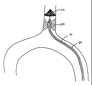
In a first embodiment, a filter system for temporary placement in a
vessel, either an artery or vein, is provided as depicted in Figs. 1A, 1B, 1C,
and 1D.
The filter system includes support wire 10 having a proximal end, distal
region 11, and
expandable filter 20 mounted at the distal region. The filter comprises
expansion frame
22 and mesh 25 which is sonic welded or adhesive bonded to struts 28 of the
expansion
frame. Anticoagulants, such as heparin and heparinoids, may be applied to mesh
25 to
reduce thrombi formation on the mesh. The filter can be collapsed as shown in
Fig. 1A
to facilitate insertion into a vessel, and thereafter expanded as shown in
Fig. 1B. Wire
guide 26 is included in distal region 11 of the support wire. The wire guide
may be
mounted within the filter (as shown in Fig. 1B and Fig. 1C) or at any other-
suitable
position on support wire 10 proximal of the filter (as shown in Fig. 1E), or
on a distal
extension of the support wire which extends beyond the filter (as shown in
Fig. 1F). In
certain embodiments, the support wire may comprise a ring. A cross-sectional
view of
the support wire through section line C-C is depicted in Fig. 1C. The design
and
construction of a variety of filters for use on guidewire is described in
detail in Tsugita
et al., U. S. Patent No. 5,911,734.
In another embodiment, the filter further includes a capture sheath which
covers the filter and is removeable from the filter, the sheath having a port
in its distal
region adapted to receive the guidewire in the manner of a rapid exchange
catheter. In
CA 02345654 2001-03-28
WO 01/08596 PCTIUSOO/40559
Fig. I G, support wire 10 is inserted in lumen 51 of a rapid exchange catheter
50. The
catheter includes side port 60 in its distal region, adapted to receive
guidewire 30. In Fig.
11, the catheter includes skive 61 which receives guidewire 30. In Fig. 1J,
elongate
member 70 carries tubular segment 75 having skive 77 at a distal region of
elongate
member 70. The tubular segment acts as a capture sheath for the filter while
the skive
receives the guidewire. In Fig. 1K, elongate member 70 carries first and
second tubular
segments, 75 and 79, adapted to receive, respectively, the filter and the
guidewire.
When in use, guidewire 30 is first inserted into a vessel and advanced
distal to the region of interest. The catheter, which carries the filter in
lumen 51, is
inserted over the guidewire, the guidewire engaged through side port 60. The
filter is
advanced distally passing atheromatous lesion 100. The guidewire can then be
withdrawn and catheter 50 drawn proximal, leaving the catheter and the filter
inserted in
the vessel as depicted in Fig. 1H. Catheter 50 is then removed from the
vessel.
Expansion frame 22 is expanded to capture embolic materials downstream the
atheromatous lesion. An endovascular device, such as an angioplasty catheter
with or
without a stent, can be inserted over support wire 10 to position adjacent
atheroma 100.
After vascular procedures are performed with the endovascular device(s), the
device(s)
are withdrawn and removed from the vessel. The filter with the captured emboli
is then
contracted and removed.
The filter system also includes guidewire 30 having a proximal end and
distal end 33. The guidewire is slideably received by support wire 10 through
wire guide
26 as depicted in Fig. 1 D. The filter system further includes endovascular
devices, such
as atherectomy catheters, endovascular imaging devices, stent-deployment
catheters,
11
CA 02345654 2007-01-15
angioplasty catheters, pressure monitors, electrophysiology catheters, and
aspirators,
which are adapted to receive guidewire 30 and/or support wire 10 in their
lumens.
Different constructions of distal end 33 of the guidewire are depicted in
Figs. 2A, 2B, and 2C. Distal end 33 may assume a substantially linear
configuration
relative to the proximal end of the guidewire as depicted in Fig. 2A.
Alternatively,
distal end 33 may assume an angular configuration relative to the proximal end
of the
guidewire as depicted in Fig. 2A. Distal end 33 may be shaped like a fishhook
as
depicted in Fig. 2C. The distal region of the guidewire may be constructed of
a flexible
material to facilitate entry through a region of interest, and preferably is
equipped with
an atraumatic tip as is known in the art. The embodiments in Figs. 2B and 2C,
having
a curvilinear design, are particularly useful in achieving access to a complex
lesion in a
tortuous vessel.
Figs. 3A and 3B depict alternative embodiments of expandable filter 20
mounted on the distal region of support wire 10. In Fig. 3A, filter 20
comprises a
parachute frame, and mesh 25 which is welded (e. g., sonic or laser) or
adhesive
bonded to struts 28. Wire guide 26 is included in the distal region of the
support wire
and projects distally from filter 20 for engaging a guidewire. In Fig. 3B,
filter 20
comprises compressible struts 22, and mesh 25. In an expanded condition,
filter 20
assumes the shape of an eggbeater.
By way of example, when the filter system as disclosed herein is
intended for use in the aorta, the area of the mesh required for the device is
calculated
from Bernoulli's equation as described in our earlier applications including
Barbut et
al., U.S. Patent No. 5,650,126, issued July 22, 1997 and Barbut et al. , U.S.
Patent
No. 5,769,816, issued June 23, 1998.
12
CA 02345654 2007-01-15
In an embodiment of the guided filter system that is to be used in the
aorta, mesh with dimensions within the following ranges is desirable: mesh
area is
0.004-5 in2, more preferably 0.007-4 in2, more preferably 0. 010-3 in2, more
preferably 0.015-2 in2, more preferably 0.020-1 in2, more preferably 0.025-
0.076 in2;
mesh thickness is 60-280 m, more preferably 70-270 m, more preferably
80-260 m, more preferably 90-250 m, more preferably 100-250 m, more
preferably 120-230 m, more preferably 140- 210 m; thread diameter is 30-145
m,
more preferably 40-135 m, more preferably 50-125 m, more preferably 60-115
m,
more preferably 70-105 m, and pore size is 500 m or less, more preferably
400 m
or less, more preferably 300 m or less, more preferably 200 m or less, more
preferably 100 m or less, more preferably 50 m, or less and usually larger
than at
least a red blood cell. In a preferred embodiment of the invention, mesh area
is 2-8
in2, mesh thickness is 60-200 m, thread diameter is 30-100 m, and pore size
is
50-300 m, In a further preferred embodiment of the invention, mesh area is 3-
5 in2,
mesh thickness is 60-150 m, thread diameter is 50-80 m, and pore size is
100-250 m. In other embodiments, the filter comprises a thin film laser cut
with holes
to allow blood flow. Typical dimensions include pore size of 20-500 m, a
thickness of
0.0005-0.003 inches, and area approximately same as for meshes described
above.
13
CA 02345654 2007-01-15
In other embodiments, the filter comprises a thin film laser cut with
holes to allow blood flow. Typical dimensions include pore size of 20-500, m,
a
thickness of 0.0005-0.003 inches, and area approximately same as for meshes
described above.
Once appropriate physical characteristics are determined, suitable mesh
can be found among standard meshes known in the art. For example, polyester
meshes
may be used, such as meshes made by Saati Corporations and Tetko Inc. These
are
available in sheet form and can be easily cut and formed into a desired shape.
In a
preferred embodiment, the mesh is welded (e.g. sonic or laser) or sewn into a
cone
shape. Other meshes known in the art, which have the desired physical
characteristics,
are also suitable. Anticoagulants, such as heparin and heparinoids, may be
applied to
the mesh to reduce the chances of blood clotting on the mesh. Anticoagulants
other
than heparinoids also may be used, e. g., monoclonal antibodies such as ReoPro
(Centocor)'. The anticoagulant may be painted or sprayed onto the mesh. A
chemical
dip comprising the anticoagulant also may be used. Other methods known in the
art for
applying chemicals to mesh may be used.
In use, as depicted in Fig. 4A, guidewire 30 is inserted percutenously
through a peripheral artery or vein and advanced typically in the direction of
blood
flow. However, guidewire 30 may be inserted and advanced in a direction
opposite the
blood flow, e.g., retrograde through the descending aorta to reach the
coronary artery.
Distal end 33 of the guidewire is passed through occluding lesion 100,
typically an
atheromatous plaque, and positioned distal to the occlusion. Support wire 10
of Fig. 1A
14
CA 02345654 2007-01-15
is inserted over the proximal end of guidewire 30 through wire guide 26, and
advanced
distally until filter 20 is positioned distal to plaque 100 as depicted in
Fig. 4B. By
having wire guide 26 engage the guidewire, the filter and the support wire can
be
easily steered intravascularly to reach the region of interest. Filter 20 is
expanded to
capture embolic material, such as calcium, thrombi, plaque, and/or tissue
debris.
Guidewire 30 is then withdrawn, leaving support wire 10 in position to direct
an
endovascular device to plaque 100.
Percutaneous translumenal angioplasty has been successful in treating
arterial stenoses as well as occlusive venous thrombosis resistant to
thrombolysis. See
American Heart Journal, 125 (2 Pt 1): 362-6 (1993). Angioplasty catheter 40,
which
has angioplasty balloon 42 mounted on the distal region, is inserted over
support wire
10 as depicted in Fig. 4C. In a deflated state, the angioplasty balloon is
advanced over
support wire 10 to a position adjacent plaque 100. The atheromatous plaque is
compressed by inflating balloon 42, thereby dilating the stenosis in the
vessel. In
certain embodiments, the angioplasty catheter includes infusion port 44
proximal and
perfusion port 45 distal to balloon 42. Infusion port 44 may be used to
administer
pharmaceutical agents, e.g., t-PA, adenosine, or nitroglycerin through the
catheter
lumen (not shown). Oxygenated medium or blood may be infused through port 45
to
maintain perfusion to distal organs during angioplasty. In certain
embodiments, a stent
is closely associated with the angioplasty balloon. The stent is typically
crimped onto
the balloon and is capable of controlled radial expansion in the region of
interest upon
application of a radial, outwardly extending force from the interior of the
stent. The
construction of the catheter system carrying a stent is described in detail in
Jang et al.,
U. S. Pat. No. 5,749,848.
CA 02345654 2007-01-15
The angioplasty catheter or other endovascular instrument is withdrawn
from the vessel after completion of angioplasty. Embolic material generated
during the
angioplasty is captured and retained by filter 20. The filter is then
contracted, and with
captured embolic material, is withdrawn from the vessel and removed from the
patient's body.
Alternatively, after filter 20 is positioned and expanded distal to plaque
100, guidewire 30 and support wire 10 may remain in the vessel across plaque
100 as
depicted in Fig. 4D. Angioplasty catheter 40 is then inserted over both
guidewire 30
and support wire 10 to a position adjacent plaque 100. If an atherectomy
device, for
example, is required to remove plaque remaining after angioplasty, angioplasty
catheter 40 is withdrawn, with or without the guidewire, and an atherectomy
catheter is
inserted over guidewire 30 and/or support wire 10 to a position adjacent the
plaque. In
this way, if the position of guidewire 30 across the plaque is lost during the
removal of
angioplasty catheter 40, support wire 10 is available to direct another
endovascular
device to the region of interest. This method is particularly useful for
exchanging guide
catheters during coronary revascularization.
The length of the guidewire and the support wire will generally be
between 30 and 300 centimeters, preferably approximately between 50 and 180
centimeters. The filter will be capable of expanding to an outer diameter of
at least 0.2
centiineters, more preferably at least 0.5 centimeters, more preferably at
least 1.0
centimeters, more preferably at least 1.5 centimeters, more preferably at
least 2.0
centimeters, more preferably at least 2. 5 centimeters, more preferably at
least 3.0
16
CA 02345654 2001-03-28
WO 01/08596 PCT/US00/40559
centimeters, more preferably at least 3.5 centimeters, more preferably at
least 4.0
centimeters, more preferably at least 4.5 centimeters, more preferably at
least 5.0
centimeters. These ranges cover suitable diameters for both pediatric and
adult use. The
foregoing ranges are set forth solely for the purpose of illustrating typical
device
dimensions. The actual dimensions of a device constructed according to the
principles of
the present invention may obviously vary outside of the listed ranges without
departing
from those basic principles.
Although the foregoing invention has, for the purposes of clarity and
understanding, been described in some detail by way of illustration and
example, it will
be obvious that certain changes and modifications may be practiced which will
still fall
within the scope of the appended claims. Moreover, it will be understood that
each and
every feature described for any given embodiment or in any reference
incorporated
herein, can be combined with any of the other embodiments described herein.
17