Note: Descriptions are shown in the official language in which they were submitted.
CA 02355788 2008-07-17
METHOD AND APPARATUS FOR MAPPING A CHAMBER OF A HEART
FIELD OF THE INVENTION
The invention relates generally to methods and apparatus for mapping a
coiidition or
property of an organ of a subject, and particularly to methods and apparatus
for
mapping the electrical and/or the mechanical activity of one or more chambers
of the
heart.
BACKGROUND OF THE INVENTION
Cardiac arrhythmias, the most common of which is ventricular tachycardia
(VT), are a leading cause of death. In a majority of patients, VT originates
from a
1 mm to 2 mm lesion located close to the inner surface of the heart chamber.
One of
the treatments for VT comprises mapping the electrical pathways of the heart
to
locate the lesion followed by ablation of the active site.
U.S. patent 5,546,951 and U.S. patent 6,690,963 and its
corresponding application filed under the Patent Cooperation Treaty and
published
as WO 96/05768,
disclose methods for sensing an electrical property of the heart tissue, for
example,
local activation time, as a function of the precise location within the heart.
The data
-1-
CA 02355788 2008-07-17
are acquired with one or more catheters that are advanced into the heart, the
catheters having electrical and location sensors in their distal tips. Methods
of
creating a map of the electrical activity of the heart based on these data are
disclosed
in commonly assigned U.S. patent 6,226,542 filed on July 24, 1998 and
in its corresponding published European Patent Application no. EP 974,936, as
well
as in U.S. patent 6,301,496 filed on July 22, 1999:.
As indicated in
these applications, location and electrical activity is preferably initially
measured at
about 10 to about 20 points on the interior surface of the heart. These data
points are
then generally sufficient to generate a preliminary reconstruction or rriap of
the
cardiac surface to a satisfactory quality. The preliminary map may be combined
with data taken at additional points in order to generate a more comprehensive
map
of the heart's electrical activity. The detailed map so obtained may then
serve as the
basis for deciding on a therapeutic course of action, for example, tissue
ablation, to
alter the propagation of the heart's electrical activity and to restore normal
heart
rhythm.
Catheters containing position sensors may be used to determine the trajectory
of points on the cardiac surface. These trajectories may be used to infer the
motion
characteristics such as the contractility of the tissue. As disclosed in U.S.
patents
5,738,096 and 6,066,094, maps
depicting such motion characteristics may be constructed when the trajectory
information is sampled at a sufficient number of points in the heart. A high
quality
-2-
CA 02355788 2001-08-16
preliminary map of motion characteristics is dependent on acquiring a
sufficient
number of points representatively spaced about the heart chamber volume.
In constructing these preliminary maps, it is desirable that the data are
sampled at points sufficiently spaced to outline the entire volume of thc
chamber
under study. If the preliminary map adequately outlines the heart volume,
acquisition of additional points will generally enable the detailed
reconstruction to
permit accurate diagnosis and treatment. Occasionally however, incomplete
sampling, as, for example, by localizing the sample points to only a portion
of the
heart volume, will result in the generation of an incomplete map. Further
sampling
may lead to a more detailed map of the partial cardiac volume, but this may be
inadequate for proper diagnosis and treatment.
In creating maps of the heart using the above-referenced systenls, the initial
data points for the preliminary reconstruction are generally acquired under
the
guidance of an imaging modality such as fluoroscopy that permits the
card;ologist to
observe the placement of the catheter tip within the heart chamber. Once the
preliminary map is generated, subsequent points may then be acquired under the
guidance of the preliminary map and a location system based on, for example,
electromagnetic or acoustic sensors. Unfortunately, unassisted fluoroscopy
provides
relatively poor visualization of topographical features within the heart.
While
contrast-assisted fluoroscopy, in which a contrast agent is injected into the
heart
chamber under examination, significantly improves the observation of
topography,
the contrast agent obscures the observation of the catheter tip. Thus,
fluoroscopy is
insufficient to properly guide the cardiologist to the points on the interior
of the
-3-
CA 02355788 2001-08-16
heart necessary for the generation of a preliminary map of the electrical
activity that
roughly encompasses the complete heart volume. The potentially harmful effects
of
the contrast agent and of ionizing radiation to the patient also limit the
amount of
data that can be collected under fluoroscopy.
Electrical activity at a point in the heart is typically measured by advancing
a
catheter containing an electrical sensor (an electrode) at or near its distal
tip to that
point in the heart, firmly contacting the tissue with the electrode and
acquiring data
at that point. Alternatively, electrical activity may be measured with
catheters
containing multiple electrodes. In the case of catheters with multiple
electrodes, one
or more electrodes are generally present at the catheter tip and other
electrodes may
be present along the catheter body.
It is generally important to maintain good electrical contact between the
electrodes and the tissue in order to obtain a reliable and stable electrical
reading.
Fluoroscopy produces images that are lacking in topographical detail.
Accordingly,
in taking measurements under the guidance of this imaging modality, the
catheter tip
may not actually be in effective contact with the tissue. Alternatively, it
may be
possible to bruise the intracardial tissue by excessive pressure of the
catheter tip
against the tissue while making such measurements.
PCT application WO 98/35720 discloses an x-ray guided surgical location
system with extended mapping volume. The application does not teach or suggest
navigation of a catheter tip for the purpose of mapping a chamber of a heart
guided
by topological information contained in acquired images of the chamber.
-4-
CA 02355788 2001-08-16
U.S. Patent 5,391,199 discloses an apparatus and method for treating cardiac
arrhythmias. The method of the `199 patent comprises obtaining a perspective
image of the organ to be mapped; advancing one or more catheters to sites
adjacent
to or within the organ; sensing the location of each of the catheter's distal
tips with a
non-ionizing field; sensing local information of the organ; processing the
local
information to create one or more data points; and superimposing the one or
more
data points on the perspective image of the organ or structure. The `199
patent does
not teach or suggest the registration of the image with a positional frame of
reference of a position sensor contained in or proximate to the catheter tip.
Furthermore, the '199 patent does not teach or suggest navigating the catheter
tip
under the guidance of topological information contained in acquired images of
the
chamber.
U.S. Patent 5,433,198 discloses an apparatus and method for cardiac
ablation. The apparatus and method of the `198 patent includes a multi-
electrode
catheter introduced percutaneously into a subject's heart and deployable
adjacent to
various endocardial sites. The electrodes are connectable to a mapping unit,
an
ablation power unit and a pacing unit, all of which are under computer
control.
Intracardiac electrogram signals emanated from a tachycardia site of origin
are
detectable by the electrodes. Their arrival times are processed to generate
various
visual maps to purportedly provide real-time guidance for steering the
catheter to the
tachycardia site of origin. In one aspect, the apparatus of the `198 patent
also
includes a physical imaging system which is capable of providing different
imaged
physical views of the catheter and the heart. These physical views are said to
be
-5-
CA 02355788 2001-08-16
incorporated into the various visual maps to provide a more physical
representation.
The `198 patent does not disclose or suggest the use of a catheter having a
sensor
which provides three-dimensional position information of the catheter tip in a
positional frame of reference, nor does it disclose or suggest registering
chamber
images with said frame of reference.
U.S. Patent 6,052,618 discloses a device for mapping electrical activity in
the
heart. The device of the `618 patent has an imaging unit, such as a
fluoroscopic
imaging unit, for generating a physical in vivo image of a patient's heart as
an
anatomical reference image; an electrode catheter with at least one electrode
for
sensing intracardiac electrical activity in a patient's heart; and signal
processing
equipment for determining activation times from sensed electrical activity at
different points in the heart. The device of the `618 patent further includes
means
for generating a graphic image showing the activation times at different
points in the
heart and superimposing this graphic image onto the anatomical image. The `618
patent does not disclose or suggest the use of a catheter having a sensor
which
provides three-dimensional position information of the catheter tip in a
positional
frame of reference, nor does it disclose or suggest registering chamber images
with
said frame of reference. Furthermore, as stated at column 3 lines 34-38 of the
`618
patent, "In this type of image, the heart appears, at best, as a pale shadow.
The heart
is not shown at all in these figures. The body parts seen most clearly in the
radiograph are skeletal parts, such as spinal vertebrae and ribs." Thus, the
`618
patent does not teach or suggest the use of images containing topological
information suitable for guiding the navigation of the catheter tip.
-6-
CA 02355788 2008-07-17
SUMMARY OF THE INVENTION
The present invention is directed to a method for intracardially mapping a
condition of a chamber of a heart of a subject. The method of the invention is
preferably
applied to the mapping of an electrical, mechanical or electromechanical
condition of the
heart chamber. While the method may be applied to any of the heart's chambers,
it is
especially useful for the mapping of the left ventricle. The mapping is
conducted with a
mapping catheter having a distal tip that may be already located in the heart
of the
subject. The catheter distal tip has at least one sensor contained therein or
proximate
thereto that is capable of sensing condition information of the chamber and
providing
three-dimensional position information of the catheter tip in a positional
frame of
reference. The method of the invention involves acquiring a first image of the
chamber
taken from a first projection and a second image of the chamber taken from a
second
projection wherein the second projection is different from the first
projection. The two
projections are preferably separated by an angle of between about 75 degrees
to about
105 degrees, and, more preferably, the two projections are separated by an
angle of about
90 degrees. The first and second images are taken from two perspectives such
as a left
anterior oblique (LAO) and a right anterior oblique(RAO) projection. The two
images
are preferably contrast-assisted fluoroscopic images that depict the chamber
at the same
phase in the cardiac cycle, preferably, at end-diastole. Both the first and
second chamber
images contain topological information of the chamber that include the chamber
contour.
The topological information contained in or derived from the first and second
images
provide guidance to an acquisition point in the chamber. The method further
comprises
registering the first image and the second image with the positional frame of
reference.
-7-
CA 02355788 2008-07-17
The distal tip of the mapping catheter may be advanced into the chamber to the
acquisition point where condition information and position information are to
be
acquired with the at least one sensor. The catheter tip may be navigated to
the acquisition
point in the chamber guided by topological information contained in or derived
from the
first and second images. The topological information used to guide the
navigation of the
catheter is preferably a reconstruction of the chamber, such as a three-
dimensional
reconstruction derived from the topological information contained in the
chamber
images. After the condition and position information are acquired at the first
acquisition
point, the catheter tip is similarly navigated to additional acquisition
points where
additional condition and position information are acquired. The acquisition
points are
sufficient in number and spacing throughout the chamber to permit the
generation of a
map of the condition in the chamber, which is preferably created from the
acquired
condition and position information.
In one embodiment, the at least one sensor comprises a position sensor capable
of
providing both three-dimensional position information as well as mechanical
condition
information. In another embodiment, the at least one sensor comprises a
position sensor
capable of providing three-dimensional position information and an electrode
for sensing
electrical information. The at least one sensor preferably comprises an
electromagnetic
sensor that generates signals responsive to the strength of a magnetic field
generated by
magnetic field radiators external to the patient wherein the signal intensity
is indicative
of the three-dimensional position of the sensor in the frame of reference.
The method of mapping a chamber of the heart of the invention further
preferably
comprises acquiring an image of a scaling object from each of the first and
the second
-8-
CA 02355788 2008-07-17
projections. The images of the scaling object are used to scale the images of
the heart
chamber. The method also preferably further comprises affixing a registration
position
sensor to the patient prior to the acquisition of the first and second images
of the
chamber. The registration position sensor is affixed to the patient so that an
image of the
registration position sensor is included in the chamber images. The three-
dimensional
position coordinates of the registration position sensor are determined and
used to
register the images of the chamber in the frame of reference.
In another embodiment, the invention is directed to a method for
intracardially
mapping a condition of a chamber of a heart of a subject. The method of the
invention is
preferably applied to the mapping of an electrical, mechanical or
electromechanical
condition of the heart chamber. While the method may be applied to any of the
heart's
chambers, it is especially useful for the mapping of the left ventricle. The
mapping is
conducted with a mapping catheter having a distal tip that is already located
in the heart
of the subject. The catheter distal tip has at least one sensor contained
therein or
proximate thereto that is capable of sensing condition information of the
chamber and
providing three-dimensional position information of the catheter tip in a
positional frame
of reference. The catheter distal tip may be advanced into the chamber and the
catheter
tip may be navigated to an acquisition point in the chamber. Navigation of the
catheter
tip is guided by a reconstruction, preferably a three-dimensional
reconstruction of
topological features of the chamber registered in the positional frame of
reference. After
the condition and position information are acquired at the first acquisition
point, the
catheter tip may be similarly navigated to additional acquisition points where
additional
condition and position information are acquired. The acquisition points are
sufficient in
-9-
CA 02355788 2008-07-17
number and spacing throughout the chamber to permit the generation of a map of
the
condition in the chamber, which is preferably created from the acquired
condition and
position information.
The reconstruction of the chamber used to guide the navigation of the catheter
tip
is preferably based on a first image of the chamber taken from a first
projection and a
second image of the chamber taken from a second projection. The first
projection and the
second projection are preferably separated by an angle of about 75 degrees to
about 105
degrees, and more preferably, by an angle of about 90 degrees. The first image
and the
second image are preferably taken from an LAO projection and an RAO
projection. Each
of the first and second images contain topological information of the chamber.
The
topological information contained in the images preferably comprises the
chamber
contour. The first and second chamber images are preferably contrast-assisted
fluoroscopic images. The images preferably depict the chamber at the same
phase of the
cardiac cycle, preferably at end-diastole.
In one embodiment, the at least one sensor comprises a position sensor capable
of
providing both three-dimensional position information as well as mechanical
condition
information. In another embodiment, the at least one sensor comprises a
position sensor
capable of providing three-dimensional position information and an electrode
for sensing
electrical information. The at least one sensor preferably comprises an
electromagnetic
sensor that generates signals
-10-
CA 02355788 2001-08-16
responsive to the strength of a magnetic field generated by magnetic field
radiators
external to the patient, the signal intensity being indicative of the three-
dimensional
position of the sensor in the frame of reference.
The method of mapping a chamber of a heart of the_ invention preferably
further comprises acquiring an image of a scaling object from each of the
first and
second projections. The images of the scaling object are used to scale the
chamber
images. The method of the invention preferably further comprises affixing a
registration position sensor to the patient prior to acquisition of the first
and second
chamber images. The registration position sensor is affixed to the patient so
that an
image of the registration position sensor is included in the chamber images.
The
three-dimensional position coordinates of the registration position sensor are
determined and used to register the images of the chamber in the frame of
reference.
Another aspect of the invention is directed to an apparatus for intracardially
mapping a condition of a chamber of a heart. The apparatus of the invention
comprises a mapping catheter having a distal tip. The catheter distal tip has
at least
one sensor contained therein or proximate thereto. The at least one sensor is
capable
of sensing condition information of the chamber and provides three-dimensional
position information of the catheter tip in a frame of reference. The
apparatus of the
invention fiuther comprises means for registering a plurality of images of the
chamber with the positional frame of reference. The chamber images are taken
from
a plurality of projections relative to the chamber and contain topological
information
of the chamber. The apparatus of the invention also comprises signal
processing
circuits for acquiring condition information and position information at a
plurality of
-11-
CA 02355788 2001-08-16
acquisition points in the chamber with the at least one sensor wherein the
points are
sufficient in number and spacing throughout the chamber to permit the
generation of
a map of the condition in the chamber.
The at least one sensor contained in or proximate to the catheter distal tip
preferably comprises a position sensor capable of providing three-dimensional
position information and an electrode for sensing electrical information. More
preferably, the at least one sensor comprises an electromagnetic sensor that
generates signals responsive to the strength of a magnetic field generated by
magnetic field radiators external to the patient. The intensity of the signals
generated by the sensor is indicative of the three-dimensional position of the
sensor
in the frame of reference.
The apparatus for mapping a chamber of a heart of the invention preferably
further comprises a scaling object. The apparatus also preferably further
comprises
a registration position sensor to register the images with the frame of
reference.
The apparatus of the invention also preferably further comprises image-
processing circuits for constructing a reconstruction, preferably a three-
dimensional
reconstruction of the chamber from topological information contained in the
chamber images. The apparatus also preferably further comprises circuits for
mapping the condition of the chamber using the condition and position
information
acquired with the at least one sensor.
In another embodiment, the invention is directed to an apparatus for
intracardially mapping a condition of a chamber of a heart of a subject. The
apparatus of the invention comprises a mapping catheter having a distal
tip.The
-12-
CA 02355788 2001-08-16
catheter distal tip has at least one sensor contained therein or proximate
thereto. The
at least one sensor is capable of sensing condition information of the chamber
and
provides three-dimensional position information of the catheter tip in a frame
of
reference. The apparatus further comprises image prqcessing circuits for
constructing a topological reconstruction, preferably, a three-dimensional
reconstruction, of the chamber in the frame of reference, as well as signal
processing
circuits for acquiring condition information and position information at a
plurality of
acquisition points in the chamber with the at least one sensor. Condition and
position information is acquired at points sufficient in number and spacing
throughout the chamber to permit the generation of a map of the condition in
the
chamber.
The image processing circuits used in the apparatus of the invention
preferably construct the topological reconstruction from a plurality of images
of the
chamber. The images are taken from a plurality of projections relative to the
chamber wherein each image contains topological information of the chamber.
The at least one sensor contained in or proximate to the catheter distal tip
preferably comprises a position sensor capable of providing three-dimensional
position information and an electrode for sensing electrical information. More
preferably, the at least one sensor comprises an electromagnetic sensor that
generates signals responsive to the strength of a magnetic field generated by
magnetic field radiators external to the patient. The intensity of the signals
generated by the electromagnetic sensor is indicative of the three-dimensional
position of the sensor in the frame of reference.
-13-
CA 02355788 2008-07-17
The apparatus of the invention preferably further comprises a scaling object.
The
apparatus also preferably further comprises a registration position sensor to
register the
images with the frame of reference.
There is also provided a use of the apparatus described above for
intracardially
mapping a condition of a chamber of a heart of a subject.
The features and advantages of the invention will be more readily apparent
from
the detailed description set forth below, taken in conjunction with the
accompanying
drawings.
BRIEF DESCRIPTION OF THE DRAWINGS
Fig. I is an LV-gram image of the left ventricle of a human heart taken from
the
right anterior oblique (RAO) projection;
Fig. 2 is a fluoroscopic image of a catheter located in the heart of Fig. I
taken
from the RAO projection;
Fig. 3 is the LV-gram of Fig. 1 in which a contour image has been created
about
the interior of the left ventricle;
Fig. 4 is the extracted contour image of Fig. 3;
Fig. 5 is a superposition of the contour image of Fig. 4 and the fluoroscopic
image of Fig. 2;
Fig. 6 is the image of Fig. 5 in which the display was marked to indicate
points in
the chamber from which condition information was acquired;
Fig. 7 is equivalent to the image of Fig. 6 taken from the left anterior
oblique
(LAO) projection;
Fig. 8 is a representation of an algorithm used to automatically find a
catheter tip
in a displayed image;
-14-
CA 02355788 2001-08-16
Fig. 9A and Fig. 9B are schematic views of a C-ann taking fluoroscopic
images of a chamber of a heart of a patient from the LAO and RAO projections,
respectively;
Fig. 9C and Fig. 9D are schematic views of a C-artn taking fluoroscopic
images of a chamber of a heart of a patient from the cranial and caudal
projections,
respectively;
Fig. 9E is a schematic view of a patient showing the coordinate systems of a
position sensor location system and a fluoroscopy imaging system.
Fig. 10A and Fig. lOB are contrast-assisted fluorograms of the left ventricle
of a patient taken from the RAO and LAO projections, respectively;
Fig. 11 shows the fluorograms of Fig. 10A and Fig. 10B in registration with
a position sensor location system frame of reference;
Fig. 12 shows the fluorograms of Fig. 11 separated from each other along
directions normal to the respective images;
Fig. 13A - Fig. 13F schematically depict the steps of an algorithm to
reconstruct a chamber of a heart from contour information contained in two
chamber
images;
Fig. 14 shows the reconstruction of the heart chamber using the algorithm
depicted in Fig. 13A - Fig. 13F;
Fig. 15 shows some elements of a position sensor location system for
performing the method of the invention; and
Fig. 16 shows additional elements of a position sensor location system for
performing the method of the invention.
-15-
CA 02355788 2001-08-16
DETAILED DESCRIPTION OF PREFERRED EMBODIMENTS
The present invention is directed to methods and apparatus for intracardially
surveying a condition of a chamber of a heart of a subject. The method and
apparatus of the invention are amenable to surveying the cqndition of any of
the
heart's chambers, but they are particularly useful in surveying the condition
of the
left ventricle of the heart.
The method and apparatus of the invention may be used to survey one or
more conditions or properties of the tissue comprising the chambers of the
heart. As
used herein, the term "condition" refers to either a scalar or a vector
quantity, and
may comprise, for example, an electrical property, a temperature, a pressure,
a pH, a
measure of local heart movement or any other condition or combination thereof.
The method and apparatus of the invention are especially useful for surveying
electrical properties of a heart chamber, including but not limited to
voltage,
impedance, conduction velocity and local activation time (LAT).
As used herein, the term "survey" refers to the collection of data as to the
condition of the chamber at representative points throughout the chamber. The
condition information may be collected individually, or it may be collected
together
with position information so that each data point would reflect the condition
information at a given three-dimensional coordinate within the chamber. If
many
points are sampled during the survey, the survey may be useful in providing a
comprehensive representation of the condition information throughout the heart
chamber. Alternatively, the survey may be preliminary, in which relatively few
points are sampled around the chamber. However, even in the case of a
preliminary
-16
CA 02355788 2008-07-17
survey, if the points are sufficient in number and in distribution around the
chamber,
the resultant data may be used for establishing a "boundary map" of the
chamber,
the detailed state of which may be determined using subsequent more
comprehensive sampling. The method and apparatus of the in~ention are
especially
useful for conducting such preliminary surveys.
Fig. 15 shows elements of a preferred position sensor location system 19 for
carrying out the methods of the invention. The apparatus includes catheter 21
for
insertion into the human body. Distal end 24 of catheter 21 includes a
functional
portion 23 for performing diagnostic and/or therapeutic functions, adjacent to
distal
tip 22. Functional portion 23 preferably comprises electrodes or sensors for
performing electrophysiological measurements, as described, for example, in
U.S.
patent 5,391,199 or in PCT application WO97/24983.,
Altematively or additionally, functional portion 23 may include other
diagnostic apparatus for recording parameter values at points within the body.
Functional portion 23 may also include therapeutic apparatus as known in the
art.
Distal end 22 preferably includes a position sensor 28 that generates signals
used to determine the position, and, preferably, the orientation of the
catheter within
the. body. Position sensor 28 is preferably adjacent to functional portion 23
in a
fixed relation with tip 22. Position sensor 28 preferably comprises three
coils, such
as described in PCT application W096/05768õ
The position sensor 28 enables continuous generation of six
dimensions of position and orientation information with respect to externally
applied
magnetic fields. Altematively, position sensor 28 may comprise other position
-17-
CA 02355788 2008-07-17
and/or coordinate sensors as described in U.S. patent 5,391,199, U.S.
5,443,489 and
PCT application W094/04938, Further,
tip 22 may be coated with an opaque marking material to visualize the tip
under an
imaging apparatus such as a fluoroscope.
Catheter 21 preferably includes a handle 30, having controls 32 that are used
to steer distal end 24 of catheter 21 in a desired direction. Catheter 21
preferably
comprises a steering mechanism in distal end 24 as is known in the art to
facilitate
repositioning of tip 22.
Catheter 21 is coupled via an extension cable 25 to a console 34 which
enables the user to observe and regulate the function of catheter 21. Console
34
preferably includes a computer 36, keyboard 38, signal-processing circuitry
40,
which are typically inside computer 36, and display 42. Signal processing
circuits
40 typically receive, amplify, filter and digitize signals from catheter 21,
including
signals from position sensor 28 and functional portion 23, whereupon these
digitized
signals are used by computer 36 to compute the condition information and the
position and/or orientation of catheter tip 22. Altematively, appropriate
circuitry
may be associated with catheter 21 itself so that circuits 40 receive signals
that are
already amplified, filtered and/or digitized. Preferably, computer 36 includes
a
memory for storing position and condition information. Computer 36 also
comprises image-processing circuits for capturing images from an imaging
modality
either using a video or a DICOM protocol interface, and for rapidly extracting
topographical information from the images. Image processing circuits contained
in
computer 36 also register the images with the position sensor location system
frame
-18-
CA 02355788 2008-07-17
of reference and calculate the chamber reconstruction from the chamber
topological
information contained in the chamber images. Computer 36 preferably further
comprises dedicated graphics circuitry for displaying the chamber
reconstruction
and for superposition of topographical images with other imaps displaying
catheter
tip 22 in the body. Images containing contour information, images showing the
catheter tip 22, images showing chamber reconstruction 160 (Fig. 14) and
superpositions of these images are displayed on display 42. Preferably, the
computer is equipped to receive body surface ECG signals from ECG monitor 73
that is connected to a plurality of ECG body surface leads 52. Altematively,
ECG
monitoring may also be conducted directly by circuits 40.
Additional elements of the position sensor location system used in
connection with the present invention are illustrated schematically in Fig.
16. A
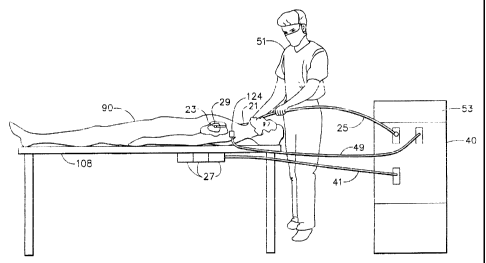
physician 51 inserts catheter 21 through an incision in the vasculature, e.g.,
using an
intravascular approach, into a chamber of a heart 29 of a patient 110, so that
an
electrode contained in functional portion 23 of catheter distal tip 22 and
position
sensor 28 are inside the chamber. In accordance with an exemplary position
sensor
described in PCT patent application number WO 96/05768, filed January 24,
1995,
and in U.S. patent 5,391,199, which are assigned to the assignee of the
present
application
sensor 28 generates signals in response to externally applied magnetic
fields generated by electromagnetic field generator coils 27 fixed to
operating table
108 in proximity to patient 90. The magnitude of the signals generated by
sensor 28
depends on the position and orientation of the sensor in the applied magnetic
field.
-19-
CA 02355788 2008-07-17
Field generator coils 27 are connected via cable 41 to driver circuits which
are part
of signal processing circuits 40. Circuits 40 control the operation of the
generator
coils 27 and the overall position sensor location system.
Alternatively, the system of the invention may employ field generator coils
in the catheter and sensors extemal to the patient.
The method of the invention also uses a registration position sensor
(reference sensor) 124 affixed to the patient during the acquisition of images
of the
heart chamber. Registration position sensor 124 is connected to circuits 40
via cable
49. The two dimensional coordinates of registration position sensor 124 in the
images and the three dimensional coordinates of sensor 124 in the frame of
reference
of the position sensor location system are used to register the images with
the
position sensor location system fi-ame of reference.
While the system and method of the invention are described herein with
reference to electromagnetic sensors, any other location sensor that provides
three-
dimensional position information and, optionally, orientation information, may
be used in the practice of the invention. Illustrative sensors that are also
useful include
acoustic sensors and magnetic sensors. For example, acoustic sensors of the
type
disclosed in U.S. Patent 5,409,000 and in PCT application WO 99/05971,
may be
used in accordance with the system and method of the invention.
As disclosed in U.S. Patent 5, 391,199, mapping the electrical activity of the
heart is performed by positioning the distal tip 22 of catheter 21 at a site
within the
heart, sensing location and electrical information at the site, processing the
sensed
-20-
CA 02355788 2008-07-17
location and electrical information at the site to create a data point, and
repeating
these steps a sufficient number of times to create a map of the heart's
electrical
pathways. For an accurate map of the chamber electrical activity, location and
electrical data are preferably sensed when an electrode at distal tip 22 is in
contact
with the cardiac wall at each site.
Having identified a lesion responsible for an aberrant electrical pathway
from the resultant electrical map of the heart chamber, the aberrant pathway
may be
treated by ablating the cardiac surface at the lesion site. As shown in Figure
16,
ablation is typically performed by supplying RF energy to the site from
ablation
power source 53 via circuits 40 and cable 25 to an electrode contained at
distal tip
22 of catheter 21. Alternatively, therapeutics may be delivered to the site of
the
lesion using a delivery catheter that has position sensing capability as
described for
example in copending U.S. patent 6,309,370.
In this embodiment of the invention, the chamber of the heart is mapped with
the aid of a mapping catheter 21 having distal tip 22. The catheter has at
least one
sensor in or proximate to the catheter distal tip 22, preferably in a
positionally fixed
relationship thereto. The at least one sensor is capable of sensing condition
information of the chamber, and also provides three-dimensional position
information of the catheter tip in a positional frame of reference.
Preferably, the three-dimensional position information is provided by an
electromagnetic position sensor 28 of the type hereinabove described. The
electromagnetic position sensor 28 generates signals responsive to the
strength of a
-21-
CA 02355788 2001-08-16
magnetic field generated by magnetic field radiators 27 external to the
patient, the
signals being indicative of the three-dimensional position of the sensor in
the
magnetic field.
The three-dimensional coordinates of the mapping catheter position sensor
28 are usually determined relative to the position of the reference sensor
124. The
reference sensor 124 is also preferably an electromagnetic sensor that
operates
according to the same principles as the position sensor 28 in the mapping
catheter
21. The reference sensor 124 may be positioned external to the patient, for
example,
as part of an adhesive patch applied to the patient's skin as shown in Fig.
16.
Alternatively, the reference sensor 124 may be positioned internal to the
patient, for
example, as a component of a reference catheter that is positioned at a
particular
point in the heart of the patient during the mapping procedure. Thus, the
position
sensor 28 in the mapping catheter 21 provides the three-dimensional
coordinates of
the mapping catheter tip 22 in the frame of reference of the position sensor
location
system relative to the reference position sensor 124.
As indicated hereinabove, the method of the invention is directed to mapping
a condition such as a mechanical and/or an electrical condition of a heart
chamber.
Mechanical properties of the heart may be mapped, for example, by measuring
the
extent of local heart movement of the tissue as a function of location within
the
heart. Local heart movement at a particular location may be assessed by
positioning
the catheter tip 22 at the location and measuring the coordinates of the
catheter tip
22 during various phases of the cardiac cycle. In this case, the position
sensor 28
-22-
CA 02355788 2001-08-16
described hereinabove may function to supply both the three-dimensional
position
information as well as the mechanical condition information.
Electrical information is typically measured by an electrode contained at the
catheter tip. In the acquisition of information for an electrical map of the
heart
chamber, the catheter 21 includes at least two sensors; a position sensor 28
for
sensing the three-dimensional position of the catheter tip 22 as well as an
electrode
23 (condition sensor) for sensing electrical information.
The invention will now be described in terms of a method and apparatus for
measuring the electrical properties of the heart. However, it will be
understood that
using the appropriate sensors, the method is equally applicable to measuring
any of
the above-enumerated conditions.
As shown in Fig. 2, the catheter 21 has one or more electrical sensors 23 at
distal tip 22 to measure conditions of the heart. The condition of the heart
chamber
is measured by the one or more condition sensors 23 (functional portion)
contained
at or proximate the distal tip 22 of catheter 21 that is advanced into the
chamber
being surveyed. In the case where catheter 21 has a single condition sensor
23, the
condition sensor 23 is preferably contained at the catheter distal tip 22.
Using such a
single condition sensor catheter 21 in the method of the invention, the
condition
information of the tissue in the chamber is sensed and acquired on a point-by-
basis.
The condition at any point in the chamber is determined by advancing the
catheter
21 to that point, preferably contacting the tissue at that point with the
electrical
sensor 23 contained at the catheter distal tip 22, and acquiring the condition
information over some time period. Typically, the data at each point are
acquired as
- 23 -
CA 02355788 2008-07-17
a function of time for one or more cardiac cycles. The data are then stored in
computer memory for future use, as, for example, in the construction of a two-
dimensional or a three-dimensional map that graphically depicts the measured
condition over all or a portion of the chamber.
Catheter 21 used in the method and apparatus of the invention may have
more than one condition sensor 23 contained therein. Catheters containing
multiple
sensors that may be useful in characterizing the electrical properties the
heart tissue
are described, for example in U.S. patents 5,409,000; 5,588,432; 5,931,863;
5,931,835; and 5,921,924, and in U.S. patent 6,892,091.
The use of multi-sensor
catheters in the method and apparatus of the invention permit the simultaneous
measurement of condition information at multiple points in the heart chamber,
which can potentially decrease the time required for assessing the overall
condition
of the heart chamber.
As best illustrated in Fig. 15, the catheter 21 used in the method and
apparatus of the invention preferably further comprises one or more positicn
sensors
28 proximate to distal tip 22 that are used to accurately measure the position
and/or
the. orientation of the catheter tip 22 in the body, particularly, in the
heart of the
subject. The position sensor 28 may, for example, operate by sensing or
transmitting acoustic, magnetic or electromagnetic fields. An electromagnetic
field
sensor is preferred as a position sensor. Preferably, position information is
sensed
by the position sensors 28 and acquired simultaneous with the sensing of
condition
information by the condition sensor 23. Catheters having sensors capable of
use in
-24-
CA 02355788 2008-07-17
measuring both electrical properties of the heart tissue as well as the
location of the
catheter tip are described for example in U.S. patent 6,690,963
and in corresponding PCT application W096/05768,
By way of exampip, the NAVI-STARTm
catheter, available from Biosense-Webster, Inc. of Diamond Bar, Califomia, is
a
catheter having both electrical condition and position sensors contained
therein that
may be useful in practicing the method of the present invention.
The mechanical condition of cardiac tissue may be assessed by measuring
the extent of movement of the tissue at a plurality of points on the
endocardium.
Such movement may be measured by contacting the tissue with a catheter tip 22
containing the position or location sensor 28 at or near its distal tip 22.
The extent
of tissue movement at each point on the endocardium may be assessed by
measuring
the distance traversed by a catheter tip 22 in contact with that point
throughout a
cardiac cycle. A map of the mechanical activity is constructed by collecting
such
mechanical data at a plurality of points on the cardiac surface wherein each
point is
characterized by the three-dimensional coordinates of the catheter tip 22 and
hence
the coordinates of a particular point on the cardiac tissue.
The coordinates of the catheter tip 22 during data acquisition are preferably
referenced to a particular point in the cardiac cycle, for example, to the end
diastole
portion of the cardiac cycle.
When used as described herein to measure the mechanical condition of the
cardiac tissue, the location sensor 28 acts not only to determine the location
of the
tissue at each point, but also as a condition sensor for measurement of
mechanical
- 25 -
CA 02355788 2001-08-16
activity. The mechanical condition may be measured alone, or simultaneous with
electrical properties of the tissue by electrode 23 (condition sensor)
contained at the
catheter tip 22.
The catheter 21 used in the method and apparatus ofz the invention further
include means for effecting therapies to the tissue of the heart chamber. For
example, endocardial ablation is well known in the art as a therapeutic
technique for
correcting cardiac arrhythmia. Such therapy may, for example, be effected by
delivering radiofrequency energy to the diseased tissue from an RF ablation
electrode contained on the catheter distal tip 22.
The method of the invention broadly comprises the following steps:
a) acquiring a first image of the chamber which contains topographical
information
of the chamber,
b) advancing the distal tip 22 of the catheter 21 into the chamber;
c) acquiring a second image comprising a representation of the catheter diatal
tip 22
in the chamber;
d) displaying a superposition of topographical information acquired in step
(a) with
the second image of step (c) to generate a displayed superimposed image
comprising representations of the topographical information and the catheter
distal tip 22;
e) acquiring condition information at an acquisition point on the chamber with
the
condition sensor 23, the acquisition point being selected from points on the
displayed superimposed image of step (d) proximate the topographical
information;
-26-
CA 02355788 2001-08-16
f) repeating step (e) at one or more additional acquisition points, the points
being
sufficient in number and spacing throughout the chamber to permit the
generation of a survey map of the condition in the chamber.
The first step in the method of the invention is to acquiri a first image of
the
heart chamber that contains topographical information. The topographical
features
typically depicted in the image include the boundary or contour of the
interior of the
chamber, although other topographical or pathological features may also be
depicted. Exemplary imaging modalities that may be used to acquire the first
image
include single photon emission computerized tomography (SPECT), positron
emission tomography (PET), two or three dimensional echo cardiography,
magnetic
resonance imaging (MRI), computerized tomography (CT) and fluoroscopy. Some
of these modalities, e.g., fluoroscopy, may require the injection of a
contrast agent
into the blood stream or into the chamber to visualize the topographical
features of
the chamber. Due to the fact that fluoroscopy is a commonly found imaging
modality in catheterization laboratories, contrast-assisted fluoroscopy is the
preferred imaging modality for acquiring the first image containing
topographical
information in the method of the invention.
In the case of contrast-assisted fluoroscopy, and perhaps with other imaging
modalities, the first image of the chamber containing topographical
information is
acquired dynamically, i.e., sequential images are acquired after injection of
the
contrast agent. Sequential images are acquired for at least one and preferably
several cardiac cycles. In effect, a multiple frame "moving picture" of the
chamber
is acquired. In some applications of the method of the invention, it is
preferable to
-27-
CA 02355788 2001-08-16
select a single frame of the dynamically acquired image for subsequent use in
the
method of the invention. For these applications, the single frame
corresponding to
the end-diastole portion of the cardiac cycle is preferred. On the other hand,
any
other frame may be selected, provided that it is used consistently for
extraction of
the contour as well as subsequent display of images containing representations
of the
catheter tip 22.
The end diastole point in the cardiac cycle is the point at which the
ventricles
are maximally dilated immediately prior to contraction. The frame
corresponding to
or depicting the chamber in end diastole may be selected by a variety of
methods.
The frames may be viewed manually and the end diastole frame may be selected
as
the frame just prior to the ventricular contraction. Alternatively, the end
diastole
frame may be determined automatically using image-processing techniques. For
example, the boundary or contour of the chamber in each frame may be extracted
using an algorithm such as snakes. The frame whose contour bounds the maximum
area corresponds to the end diastole frame. Alternatively, the frame
corresponding
to end diastole may be correlated with the body surface electrocardiogram
(ECG).
Specifically, the end diastole frame may be defined by a particular feature of
the
QRS wave of the body surface ECG.
In the case in which the left ventricle (LV) is the object of the study, the
fust
image preferably comprises a contrast-assisted fluoroscopy image of the left
ventricle, commonly referred to as an LV-gram. An LV-gram image of a human
heart showing the ventricle in end diastole, taken from the right anterior
oblique
(RAO) projection, is shown in Fig. 1. As seen in Fig. 1, the dark area 11
depicts the
- 28 -
CA 02355788 2001-08-16
interior of the left ventricle filled with contrast agent. As the ventricle is
completely
filled with contrast agent, the topographical features of the ventricle, i.e.,
the
ventricle border or contour 12, is clearly visible in the LV-gram.
After the catheter 21 including condition sensor 23 is idvanced into the heart
chamber being surveyed, the next step in the method of the invention involves
acquiring a second image of the chamber showing the catheter contained
therein.
The second image may be acquired using one of a variety of imaging modalities,
for
example, fluoroscopy, echocardiography, NII2I or CT. Once again, due to the
ubiquitous nature of fluoroscopy in the catheterization laboratory,
fluoroscopy is the
preferred modality for obtaining the second image in the method of the
invention.
Fig. 2 shows a fluoroscopic image of the heart of Fig. 1 taken from an RAO
projection. The image in Fig. 2 shows the catheter 21 having distal tip 22
with an
electrical sensor 23 contained therein. As shown in Fig. 2, however, the non-
contrast-assisted fluoroscopic image is not particularly helpful in providing
readily
discernible visual guidance as to the internal ventricle walls. Furthermore,
the
fluoroscopic image extends to the epicardium. Accordingly, sampling the
condition
information at the endocardium under fluoroscopic guidance alone may lead to
incomplete sampling in only a portion of the heart chamber and may be less
informative in terms of identifying sampling points on the endocardial wall.
The next step in practicing the method of the invention involves displaying a
superposition of the topographical information from the first image with the
second
image comprising a representation of the catheter distal tip 22. In the
practice of the
method of the invention using dynamically acquired imaging modalities, a
variety of
-29-
CA 02355788 2001-08-16
superpositions may be performed in displaying the topographical infomnation
together with the image showing the catheter tip 22. In the case of contrast-
assisted
fluoroscopy as the modality for acquiring the first image containing
topographical
information of the chamber, the contrast-assisted image is=dynamically
acquired.
Accordingly, either a dynamic moving image of the chamber or a static image at
a
single point in the cardiac cycle may be used in the displayed superposition.
Likewise, non-contrast assisted fluoroscopy used to image the catheter tip 22
in the
chamber is also dynamically acquired, so that either a dynamic or static image
showing the catheter tip 22 may be used.
The purpose of creating the superposed displayed image is two-fold. First, to
facilitate the guidance of the catheter tip 22 to the wall of the chamber
under
examination, and second, to provide a visualization that will permit the
cardiologist
to acquire data at representative points throughout the chamber. Mere
superposition
of the images of Fig. 1 and Fig. 2 would be inadequate to serve these
purposes, since
the dark area of the LV-gram of Fig. 1 showing the interior of the left
ventricle
would completely obscure the image of the catheter tip 22. Accordingly, it is
desirable to extract or derive the contour information from Fig. 1 prior to
superposition with the image of Fig. 2.
Figure 3 is an LV-gram image of the ventricle shown in Fig. 1 in which
contour image 31 has been created about the contour of the interior wall of
ventricle
11. The contour image may be created, for example, in one of three ways:
A. Manual Creation of Contour Image - The contrast-assisted image is
imported into a drawing program and a continuous contour image is manually
traced
-30-
_____
CA 02355788 2001-08-16
around the entire ventricle contour using the drawing program drawing tool by
manually dragging the mouse pointer or a similar pointing device completely
around
the contour. Alternatively, the contrast-assisted image may be manually marked
at
discrete points with the drawing tool and the contour may bk interpolated
between
these points, using splines, for example.
B. Automatic Creation of Contour Image - The contour image is created and
extracted automatically using a contour extraction algorithm such as snakes.
Snakes
were originally proposed as a regularization approach for locating contours
(see M.
Kass, A. Witkin & D. Terzopoulos, "Snakes: Active Contour Models," Proceedings
of First International Conference Vision, 1987, pp. 259-269 and D.
Terzopoulos,
"Regularization of Inverse Visual Problems Involving Discontinuities," IEEE
Trans.
Pat. Anal. Mach. Intell., vol. PAMI-8, no. 4, 1986, pp. 413-424).
The contour V may be represented as an ordered set of points,
V=[võ v=,..., ve ] wherein each v, is defined by a pair of (x, y) coordinates.
A
snake can be either closed or open, depending on whether the end points are
connected. In the present invention, we preferably use closed snakes.
We denote two functionals EIDt and E,,Xt. E;~, (v;) imposes continuity and
smoothness constraints, wherein Ea, (v;) attracts the snake to salient image
features,
for example, the magnitude of the intensity gradient. We seek to minimize both
E;m
and E~xt. Minimizing both functionals via the snake then turns the boundary
extraction problem into the following energy minimization problem:
VA=argminJ.Z,Emt (v,)+(1-R ~)Ear(vr) (1)
-31-
CA 02355788 2001-08-16
wherein A iE [0,1] is a tradeoff parameter. Setting ~ to 0 means that we
minimize only the EeXt component of the equation. Setting k to 1 means
minimizing
only the E;,,t component. Intermediate a.s result in a tradeoff of E;,,t vs.
E,,xt=
The A parameter may be found empirically or by d pazametric selection
strategy based on the minimax criterion (see H. Freeman, "Computer processing
of
Line Drawing Images," Computer Survey 6, 1974, pp. 57-98).
In the original formulation, the internal energy Ei.t was defined by the first
and the second derivatives along the boundary, giving the snake rubber-sheet
and
thin-plate like behavior respectively, and is approximated by:
Emt (vi ) Jv+ - vi-1 112 + uvi-1 - 2v, + vi+l q2 (2)
Alternatively, E;nt(v;) and E,t(v;) may be defined in- different ways, for
example, as described by K. F. Lai & R. T. Chin, in "Deformable Contours:
Modeling and Extraction", PAIVII-17, No. 11, November 1995, pp. 1084-1090.
C. Semiautomatic Creation of Contour Image - In one variation of the semi-
automatic method, the physician is presented with a snakes contour for
acceptance
or rejection. Rejection of the contour results in further processing leading
to the
presentation of another possible contour. This continues until the physician
accepts
the contour image. Alternatively, a modified snakes algorithm may be employed
which forces the contour image to one or more points pre-selected by the user.
The contour image 31 so produced, extracted from the LV-gram, is shown in
Fig. 4. The x, y coordinates of the extracted contour image are preferably
stored in
-32-
CA 02355788 2001-08-16
computer memory for use in displaying the superposition of topographical
information and the image showing the catheter tip 22.
As indicated previously, the contour information and the image showing the
catheter tip 22 may be either dynamic or static. The contour j 1 and catheter
tip 22
information may be superimposed, for example, in the following ways:
A. Static Contour Image On Static Catheter Tip Image
A static contour image is acquired from a dynamic image by one of the
hereinabove described methods, e.g., the end diastole frame is acquired by
synchronization with the body surface ECG signal. The fluoroscopy image
showing
the catheter tip 22 is also gated to show the same frame as that selected for
the
contour image. The superposition of the contour image on the image showing the
catheter tip 22 is effected by changing the color or intensity of the pixels
corresponding to the stored contour image in the image showing the catheter
tip 22.
B. Static Contour Image On Dynamic Catheter Tip Image
The static contour image as hereinabove described is superimposed on a
dynamic image of the catheter tip 22 in the heart. In this case, the pixel
color or
intensity of each frame of the dynamic fluoroscopy image is processed as
described
above to show the contour image 31 of the chamber 11.
C. Dvnamic Contour Ima eg on Dynamic Catheter Tip Image
Rather than selecting a single frame of the contrast-assisted image, the
entire
sequence is processed to extract the contour of each frame. The stored
contours are
then synchronized with the live dynamic images of the chamber 11 and catheter
tip
- 33 -
CA 02355788 2001-08-16
22 and each frame of the live images is processed to adjust pixel color or
intensity
corresponding to the contour at that point in the cardiac cycle.
The resultant processed images showing the contour and the catheter tip 22
are shown on the display. Fig. 5 is a photograph of the displayed
superposition of
contour image 31 of Fig. 4 with the fluoroscopic image of Fig. 2 showing a
portion
of catheter 21 and catheter tip 22.
Since the first image containing the topographical information (Fig. 1) and
the second image showing the catheter tip 22 (Fig. 2) were both acquired using
the
same imaging modality (fluoroscopy) and from the same projection (RAO),
contour
image 31 in the displayed superimposed image represents points on the interior
wall
of the chamber 11. Accordingly, in order to acquire condition information
concerning the tissue of the chamber, the cardiologist advances the catheter
tip 22
under the guidance of the displayed superimposed image of Fig. 5 to an
acquisition
point shown on the displayed image as being on or proximate to boundary image
31.
At this acquisition point, the catheter tip 22 is in contact with or proximate
to the
chamber wall, and condition information, preferably together with location
information, may be acquired. While viewing the displayed superimposed image,
the cardiologist may acquire the condition and/or position information by
activation
of a foot pedal, for example, which instructs the computer to initiate data
acquisition. Condition and/or position information are preferably acquired
repetitively at each point on the wall of the cardiac chamber for at least one
and
preferably more than one complete cardiac cycle. Data are preferably acquired
at a
frequency of at least about 10 per second, more preferably, at a frequency of
at least
-34-
CA 02355788 2001-08-16
about 20 per second, and most preferably, at a frequency of at least about 50
per
second.
After acquiring data at the first acquisition point, the cardiologist acquires
subsequent data by advancing the catheter tip 22 to successive points in the
chamber, such points being shown in the displayed superimposed image as being
on
or proximate to the contour image. The total number of data points acquired is
a
function of the intended purpose of the survey. If only a preliminary survey
is being
conducted in order to define the boundary of the chamber for another guidance
or
navigation technique, at least 3 and preferably at least 5 points should be
acquired
under the guidance of the displayed superimposed image.
As described herein, the first image containing topographical information
and the second image containing a representation of the catheter tip 22 are
preferably acquired using the same imaging modality, i.e., fluoroscopy.
Furthermore, both images are preferably acquired in the same projection, i.e.,
the
images of Fig. 1 and Fig. 2 were both acquired in the RAO projection.
Acquiring
both images using the same modality and using the same projection is prefenred
because this eliminates the need to register the images. Alternatively, the
first and
second 'unages may be acquired using different imaging modalities and/or from
different projections. However, such images would require registration during
superposition if the displayed superposed image is to serve as a guide for the
chamber contour.
To assist the cardiologist in acquiring representative condition information
throughout the entire chamber, the method of the invention preferably
comprises
-35-
CA 02355788 2001-08-16
marking the display at the points at which condition information is acquired.
This
capability provides the cardiologist with a visual indication of all of the
points or
sites on the cardiac wall at which information was acquired, and helps guide
the
cardiologist to sites where sampling is still required.
The display is preferably marked automatically when means such as the foot
pedal is activated to initiate data acquisition. The position of the catheter
tip 22 in
the display is preferably located automatically by the following algorithm.
The
catheter tip location algorithm is based on the following assumptions:
1) The catheter tip 22 is visualized as dark on the image;
2) The greatest contrast in the displayed superimposed image occurs
between the catheter tip 22 and its surroundings; and
3) The size of the catheter tip 22 may be fixed in the analysis of all images.
The algorithm may be understood by reference to Fig. 8, in which the
catheter tip 22 is approximated by a fixed geometric shape of a given size,
for
example square 81 in Fig. 8. Each square is of the same size, between about
ten (10)
to about twenty (20) pixels. To test whether the catheter tip is visualized in
square
81, the average intensity of the pixels comprising square 81 is computed.
Similarly,
the average intensity is evaluated for the pixels comprising the four squares,
82, 83,
84 and 85 surrounding square 81. The contrast between square 81 and its
neighbors
82, 83, 84 and 85 is the difference in average intensity between square 81 and
the
average intensity of squares 82, 83, 84 and 85. This calculation is iterated
about all
pixels in the image. The catheter tip location is attributed to the square
having the
maxiunum contrast or intensity difference with its surroundings.
-36-
CA 02355788 2001-08-16
Marking the display helps the cardiologist to avoid missing regions of the
heart if the objective is to survey the chamber as a whole. Marking the
display to
indicate the data acquisition sites also permits the cardiologist to return to
a visited
site, for example, to confirm previously sampled condition infoimation.
The displayed superimposed image may be marked with a geometric symbol
for example (e.g., a square, circle, etc.) to depict each point at which
condition
information was acquired. Alternatively, the display may be marked with a
number
or color representative of the magnitude of the condition information acquired
at that
point. The display may be marked, for example, by instructing the computer to
mark the display with the position of the catheter tip when the foot pedal,
which
initiates data acquisition, is activated. Alternatively, the cardiologist may
be
provided with marking means which allows the selection of which of the
acquired
points are to be marked on the displayed superimposed image.
Fig. 6 depicts the displayed superimposed image of Fig. 5 in which
geometric symbols 61 have been marked on the displayed image corresponding to
the points in the chamber at which condition information was acquired.
The topographic information used in the method of the in-, ention as
heretofore described is two dimensional in nature. Accordingly, the contour
image
used in the displayed superimposed image only represents points on the
interior wall
of the heart chamber in a single plane. If the objective of the survey is a
more
comprehensive characterization of the heart chamber, it may be preferable to
perform the method of the invention using images acquired from a plurality of
projections. Briefly, the method of the invention in which image and condition
-37-
CA 02355788 2001-08-16
information are acquired from two projections using fluoroscopy, the preferred
imaging modality, comprises the steps of
a) acquiring a first, contrast-assisted fluoroscopic image of the chamber, the
first, contrast-assisted fluoroscopic image being arquired from a first
projection relative to the subject;
b) creating a first contour image of the interior of the chamber from the
first
contrast-assisted fluoroscopic image;
c) acquiring a second, contrast-assisted fluoroscopic image of the chamber,
the
second, contrast-assisted fluoroscopic image being acquired from a second
projection relative to the subject;
d) creating a second contour image of the interior of the chamber from the
second contrast-assisted fluoroscopic image;
e) advancing the distal tip 22 of the catheter 21 into the chamber,
f) acquiring a first non-contrast-assisted fluoroscopic image comprising a
representation of the catheter distal tip 22 in the chamber, wherein the first
non-contrast-assisted fluoroscopic image is acquired from the first projection
relative to the subject;
g) displaying a superposition of the first contour image of step (b) with the
first
non-contrast-assisted fluoroscopic image of step (f) to generate a first
superimposed image;
h) acquiring the condition information at an acquisition point on the chamber
with the condition sensor, wherein the acquisition point is selected from
-38-
CA 02355788 2001-08-16
points on the first superimposed image of step (g) proximate the first contour
image;
i) acquiring a second non-contrast-assisted fluoroscopic image comprising a
representation of the catheter distal tip 22 in the chambef, wherein the
second
non-contrast-assisted fluoroscopic image is acquired from tW, second
projection relative to the subject;
j) displaying a superposition of the second contour image of step (d) with the
second non-contrast-assisted fluoroscopic image of step (i) to generate a
second superimposed image;
k) acquiring the condition information at an acquisition point on the chamber
with the condition sensor, wherein the acquisition point is selected from
points on the second superimposed image of step (j) proximate the second
contour image;
1) repeating steps (h) and (k) at one or more additional acquisition points,
wherein the points are sufficient in number and spacing throughout the
chamber to pennit the generation of a survey map of the condition in the
chamber.
Preferably, all of the data acquired under the guidance of one of the
displayed superimposed images is collected before collecting data under the
guidance of the second displayed superimposed image.
If only a preliminary survey is being conducted in order to define the
boundary of the chamber for another guidance or navigation technique, at least
three
-39-
CA 02355788 2008-07-17
(3) and preferably at least five (5) points should be acquired under the
guidance of
each of the displayed superimposed images.
As hereinabove described, the method of the invention preferably further
comprises marking the points 61 on the superimposed image a which the
condition
information was acquired. Fig. 7 shows a marked superposition of the contour
and
fluoroscopy images of the left ventricle shown in Fig. 1-6 in which the images
were
acquired in a left anterior oblique (LAO) projection. Sampling the condition
of the
chamber from multiple projections is expected to increase the accuracy of a
preliminary map of the heart chamber based on the data.
If the method of the invention is practiced with a catheter containing a
sensor
for obtaining position information, each data point of condition information
obtained
via the condition sensor may be accompanied by a three dimensional coordinate
of
the tissue at which the data point was obtained. The resulting survey data of
condition and position information obtained by the practice of the method of
the
invention is especially useful for the creation of maps, especially 3-
dimensional
maps of the heart. Methods of creating such maps are disclosed in copending
commonly assigned U.S. patents 6,226,542 and 6,301,496 filed on
July 24, 1998 and July 22, 1999, respectively,
The method of the invention further optionally comprises the
step of creating a map of the condition of the heart based on the position and
condition information obtained from the practice of the method of the
invention.
In another embodiment, the present invention is directed to methods and
apparatus for mapping a chamber of a heart. While useful for any of the
heart's
-40-
CA 02355788 2001-08-16
chambers, the invention is especially useful for mapping the left ventricle of
the
heart.
Another embodiment of the method of the invention involves using
topological information of the chamber contained in or derived from images
acquired from at least two projections. The images are preferably contrast-
assisted
fluoroscopic images, preferably taken from LAO and RAO projections. As
described above, the contsast-assisted fluoroscopic images are acquired
dynamically
over one or more cardiac cycles. The images taken from each projection
preferably
depict the chamber at the same phase of the cardiac cycle, preferably at end-
diastole.
The frames of the fluoroscopic images depicting the chamber in end diastole
are
selected as described hereinabove.
In some embodiments of the invention, the chamber iuiages are registered
with the frame of reference of the position sensor location system. One way of
effecting this registration is by:
(1) Obtaining the three-dimensional coordinates of a fiducial object that
is visible in the images; and
(2) Scaling the images to the position sensor frame of reference.
A convenient fiducial object is a position sensor that is affixed to the
patient
for purposes of registration of the chamber images. Prior to the acquisition
of the
images, a registration position sensor 124 (Fig. 16) is affixed to the patient
at a
location in which it will be visible in each of the chamber images. The
registration
position sensor 124 may be affixed to the patient either externally or
internally. If
affixed to the patient externally, it is preferably affixed to the left side
of the
-41-
CA 02355788 2001-08-16
patient's chest. The registration position sensor 124 is also preferably an
electromagnetic sensor of the type hereinabove described. The three-
dimensional
coordinates of the registration position sensor 124 in the position sensor
location
system frame of reference are measured and used together with the two-
dimensional
location of the registration sensor 124 in the chamber images in registering
the
chamber images in the frame of reference of the position sensor location
system.
The other part of the registration procedure involves scaling the chamber
images to the position sensor frame of reference. The chamber images are
scaled by
obtaining images of a scaling object of known dimensions and calculating
scaling
factors for the chamber images from the images of the scaling object. The
scaling
object may be positioned either internal to or external to the patient. The
scaling
object is preferably an x-ray opaque sphere, preferably having a diameter of
about
40 mm, taped under the left arm of the patient at approximately the same
height as
the heart. If the scaling object cannot be seen in the chamber images,
separate
images of the scaling object should be recorded at the same orientation of the
C-arm
(projection angle and distance of source and intensifier relative to patient)
as the
chamber images. The scaling object should preferably occupy the center of the
image in order to minimize image distortions. The size of the scaling sphere
in the
images may be determined automatically by using a region filling algorithm as
described in Computer Graphics - Principles and Practice, J. D. Foley, A. van
Dam,
S. K. Feiner and J. F. Hughes, Addison-Wesley Publishing Company, 1996, pp.
979-
986 and by ellipse fitting as described in Digital Image Processing, K. R.
Castleman,
Prentice Hall, 1996, pp.501-507. The scaling sphere appears as an ellipse in
the
-42-
CA 02355788 2001-08-16
fluorograms due to different scaling of the fluorograms in the vertical and
horizontal
directions. Knowing the true size of the scaling object, both vertical and
horizontal
correction factors may be calculated from the scaling object images. The
chamber
images are then scaled according to these correction factors. ~
Figs. 9A and 9B are schematic drawings showing acquisition of the images
with a fluoroscope C-arm (fluoroscopic device) 100. The figures show the
patient
106 in longitudinal view facing the patient's head. The C-arm 100 connects x-
ray
source 102 and image intensifier 104. Patient 106 is lying face-up on table
108.
Figs. 9A and 9B depict the acquisition of images in the LAO and RAO
projections,
respectively. In each of these projections, axis 110 of C-arm 100 creates an
angle
(al and a2 in Fig. 9A and Fig. 9B, respectively) with vertical axis 112. The
angle
aTOT separating the two projections is the sum of the angles al and a2 of the
individual projections. Preferably, the two projections are separated by an
angle
aToT of between about 75 to about 105 degrees. More preferably, the
projections are
separated by an angle a-roT of about 90 degrees.
The C-arm 100 may also be inclined with the image intensifier 104 facing
the patient's head (cranial projection) or facing the patient's feet (caudal
projection).
Figs. 9C and 9D show the C-arm inclined in the cranial and caudal direction
perspective, respectively. Preferably, the projection angle of the images in
the
cranial-caudal perspective(a3 in the cranial perspective and aa in the caudal
perspective) is less than about 10 degrees. More preferably, the cranial-
caudal
projection angle of the images is about zero degrees.
- 43 -
CA 02355788 2001-08-16
The projection angles of each of the images in the left-right perspective and
in the caudal-cranial perspective are noted for later use in the method of the
invention. Also, the two-dimensional location of the registration position
sensor in
the chamber images is noted for later use in the method ot the invention. The
registration sensor location in the images may be annotated roanually or
automatically as hereinbefore described.
Fig. 9E shows the coordinate systems of both the position sensor location
system and the fluoroscopy imaging system. The X axis 121 and the Y axis 123
of
the position sensor location system are parallel to the respective X axis 125
and the
Y axis 127 of the image intensifier 104 of the fluoroscopy system when the C-
arm
100 is not inclined, i.e., when the C-arm 100 is at an angle of zero degrees
with
respect to both the right-left and the cranial-caudal perspectives. As shown
in Fig.
9E, the Y axes (123 and 127) run from the caudal end (foot) to the cranial end
(head)
of the patient and the X axes (121 and 125) runs from the patient's right to
left,
perpendicular to the Y axis. The Z axis 129 of the position sensor location
system
runs orthogonal to the system's X and Y axes (121 and 123 respectively). The X-
Y
plane 131 of the fluoroscopy image is parallel to the X-Y plane 133 of the
position
sensor location system. Thus, the projection angle of the fluoroscopy images
with
respect to the image intensifier 104 in the uninclined position will be equal
to the
projection angle of the images with respect to the position sensor location
system.
Knowing the projection angles of each of the images relative to the position
sensor location system frame of reference, the two dimensional coordinates of
the
registration position sensor 124 in the images, the three-dimensional
coordinates of
-44-
CA 02355788 2001-08-16
the registration position sensor 124 in the position sensor location system
frame of
reference and the image scaling factors (as described below), the three-
dimensional
coordinates of each point in the images may be computed using standard linear
algebra techniques.
Once the chamber images are acquired and the end diastole frames are
selected, topological information, preferably in the form of the chamber
contour, is
identified and marked in the images as hereinabove described.
Figs. 10A and 10B show RAO and LAO contrast-assisted fluorograms of a
left ventricle 120. Included in these figures is the delineated contour 122 of
the
ventricle 120 as well as an image of registration sensor 124 that is contained
in a
catheter adhered or taped to the chest of a patient.
The scaled chamber images containing the extracted chamber contours are
then merged with respect to the position sensor location system to meet the
following conditions:
(1) The registration position sensor 124 in the images is located at its
measured three-dimensional coordinates; and
(2) The images are oriented with respect to each other according to the
relative orientation of the projections from which the images were
taken.
Fig. 11 shows the two merged images, 130 and 132 of the ventricle of Figs.
10A and lOB taken from the RAO and the LAO projections, respectively. The
chamber contour 122 has been identified in each of the images. The images are
registered such that location of registration position sensor 124 in the
images
-45-
CA 02355788 2001-08-16
coincides with its measured location in the position sensor frame of
reference. The
images are oriented with respect to each other corresponding to the relative
orientation of the projections from which the images were taken.
Topological information contained in or derived from the chamber images is
used to guide the navigation of the mapping catheter 21 to individual points
in the
chamber for purposes of acquiring condition and position information at each
of the
points. The acquisition points are preferably on the wall of the chamber. The
topological information used to guide the navigation is preferably a
reconstruction,
more preferably, a three-dimensional reconstruction of the chamber based on
topological information contained in or derived from the chamber images. A
three
dimensional reconstruction of the chamber is performed as follows:
To aid in visualizing the reconstruction process, the merged images may be
moved apart along vectors (lines) normal to each image emanating from the
coordinates of the registration position sensor. Fig. 12 shows the two chamber
images of Fig. 11 that have been separated in this fashion. The images are
separated
along vectors 140, 142 emanating from the registration position sensor 124 in
each
of the images. A graphic 144 representing the registration position sensor at
its
correct location in the position sensor location system frame of reference is.
shown in
Fig. 12 at the intersection of these lines.
The algorithm for the three dimensional reconstruction of the chamber from
the contours of the individual chamber images is illustrated schematically in
Figs.
13A - 13F, each of which contains two ovals representing the contours of the
chamber in the RAO and LAO projections. In the following process descr;ption,
we
-46-
CA 02355788 2001-08-16
refer to lines emanating from the contours. All of these lines are normal to
the
respective chamber images from which they emanate. Points on the
reconstruction
are formed, in principle, by the intersection of lines emanating from the RAO
and
LAO projections. In practice, these lines will not always intersect due to
errors
associated with the system such as measurement errors and patient movement.
Accordingly, a threshold distance is defined such that lines separated by less
than
the threshold distance are considered to have intersected for purposes of the
reconstruction algorithm.
The magnitude of the threshold distance will depend on the distance between
points on the contour that are sampled by the algorithm. The larger the
distance
between points on the contour, the greater will be the distance separating the
lines
passing through these points. While a small distance between points gives a
more
accurate reconstruction, a small distance between points means processing a
greater
number of points, which is more computationally intensive. Empirically, it was
found that a distance between points of about 1.0 mm and a threshold of about
1.0
mm strike a reasonable balance between reconstruction accuracy and
computational
intensity.
Step 1. Begin by producing a line (line A in Fig. 13A) passing through a
point on the RAO contour.
Step 2. Next, find the line emanating from the LAO contour that is closest to
line A. Fig. 13A shows three lines emanating from the LAO contour, labeled B,
B'
and B". The closest LAO contour line to line A is defined as the LAO contour
line
that has the shortest mathematical distance to line A. The line emanating from
the
-47-
CA 02355788 2001-08-16
LAO contour that is closest to line A is shown as line B in Fig. 13A. The
projection
of line B on line A is the point on line A that is closest to line B. This
point is shown
as point I1 in Fig. 13A.
Step 3. Next, consider all lines emanating from the *AO contour that are
closer to line B then the predefined threshold distance. In other words, find
all RAO
lines that have a mathematical distance to line B below the predefined
threshold
distance. In Fig. 13B, lines C, C' and C" are all below the predefined
threshold
distance. Of these RAO lines, choose a line C whose projection on line B is
furthest
from point 11. The projection of line C on line B is shown in Fig. 13B as the
point
12.
Step 4. Next, consider lines emanating from the LAO contour (lines D, D'
and D" in Fig. 13C) that are closer to line C then the predefined threshold
distance.
Of these, choose a line D whose projection on line C is furthest from point
12. The
projection of line D on line C is shown in Fig. 13C as the point 13.
Step 5. Now, consider lines emanating from the RAO contour (lines E, E'
and E" in Fig. 13D) that are closer to line D then the predefined threshold.
Of these,
choose line E whose projection on line D is furthest from point B. The
projection of
line E on line D is shown in Fig. 13D as point M.
Step 6. Calculate a curve that is circumscribed by the points 11, 12, 13 and
14
as follows: Define point P1 as the center of the segment between points Ii and
12;
point P2 as the center of the segment between points 12 and 13; point P3 as
the center
of the segment between points 13 and I4; and point P4 as the center of the
segment
between points 14 and I1 (Fig. 13E). Then, calculate the interpolation spline
that
- 48 -
CA 02355788 2001-08-16
passes through the points Pl, P2, P3, P4, i.e., the curve 150 in Fig. 13F.
Calculate
the spline through the midpoints between the line intersections to produce a
more
smoothly shaped reconstruction that is ellipsoidal in cross-section and is a
more
accurate representation of the chamber. The points on the spline are saved as
new
points for the three-dimensional reconstruction of the chamber.
Step 7. Each of above steps of the reconstruction is repeated for all lines
passing through the RAO contour.
Step 8. Once all of the RAO contour lines are processed, the above steps are
repeated for all lines emanating from the LAO contour.
The reconstruction 160, from points produced by this algorithm, is shown in
Fig. 14. As shown in Fig. 14, the algorithm provides a reconstruction with a
smooth
shape that can be used to guide the navigation of a catheter to points within
the
chamber for the purpose of acquiring condition and location information at
said
points.
The reconstruction of the cardiac chamber as described above was
accomplished from two chamber images. Similar techniques may be employed for
reconstructions from more than two images. Altern.atively, back projection
techniques, as described in "Fundamentals of Digital Image Processing" by Anil
K.
Jain, Prentice Hall, Englewood Cliffs, NJ 1989, pp. 439-445, may be used in
reconstructing the cardiac chamber from a plurality of chamber images.
Having derived reconstruction 160 of chamber 120 shown in Fig. 14 in the
frame of reference of the position sensor location system, the reconstruction
is used
to guide the navigation of the mapping catheter distal tip to points in the
chamber at
-49-
CA 02355788 2001-08-16
which condition information is desired to be acquired, preferably adjacent to
or in
contact with the chamber wall. Condition information is acquired via at least
one
sensor contained at the catheter distal tip at each of said points. Condition
information is acquired at a sufficient number of points throughout the
chamber to
permit the generation of a map of the condition in the chamber.
The display of the reconstruction preferably contains a graphic to indicate
the
location of the mapping catheter tip in real time during navigation of the
catheter tip
and acquisition of condition and position information. As with the previously
described embodiment, the display may be marked at the points of data
acquisition
to indicate to the operator the chamber locations where condition information
has
been sampled and to guide the operator to additional sampling points in order
to
obtain a map that is completely representative of the chamber. In addition,
the
display may be annotated to indicate values of the condition information
either
during or after information acquisition. The display may be annotated with
numerical values of the condition information at each one of the acquisition
points.
Altematively, the map may be color coded so that the colors are indicative of
the
value of the condition at each point in the map. Condition information between
acquisition points may be interpolated from the values at the acquisition
points, with
the interpolated values likewise displayed according to either of the above-
mentioned methods.
A major advantage of the method and apparatus of the invention is that once
the images of the chamber are acquired and the topological features of the
chamber
have been ascertained, the catheter tip may be navigated to points within the
heart
-50-
CA 02355788 2001-08-16
for the acquisition of condition information entirely under the guidance of
the
topological information contained in or derived from the images without any
additional imaging during the acquisition of condition information.
Consequently,
condition information may be acquired without using fluproscopy during the
acquisition step, resulting in significant reductions in radiation exposure to
the
patient undergoing the procedure.
Although, the invention has been described in the context of mapping the left
ventricle of the heart, the method may be used in mapping any of the heart's
chambers. Furthermore, the chamber reconstruction as described herein may be
decoupled from the chamber mapping. For example, the reconstruction of the
left
ventricle may be used to provide an anatomical reference for mapping other
portions
of the heart such as the right atrium.
Although this invention has been described in connection with its most
preferred embodiments, it will become readily apparent to those reviewing this
detailed specification that numerous additional embodiments fall well within
the
scope and spirit of the claimed invention as set forth in the claims which
appear
below.
-51-
_