Note: Descriptions are shown in the official language in which they were submitted.
CA 02358521 2001-07-12
WO 00/42950 PCT/US00/01855
FLEXIBLE HEART VALVE
Field of the Invention
The present invention relates to prosthetic heart valves, and, more
particularly, to a prosthetic tissue valve having increased flexibility
enabling it
to follow the motions of the annulus and sinus regions.
Background of the Invention
Prosthetic heart valves are used to replace damaged or diseased heart
valves. In vertebrate animals, the heart is a hollow muscular organ having
four
pumping chambers: the left and right atria and the left and right ventricles,
each
provided with its own one-way outflow valve. The natural heart valves are
identified as the aortic, mitral (or bicuspid), tricuspid and pulmonary
valves. The
valves of the heart separate chambers therein, and are each mounted in an
annulus
therebetween. The annuluses comprise dense fibrous rings attached either
directly
or indirectly to the atrial and ventricular muscle fibers. Prosthetic heart
valves can
be used to replace any of these naturally occurring valves, although repair or
replacement of the aortic or mitral valves are most common because they reside
in
the left side of the heart where pressures are the greatest. In a valve
replacement
operation, the damaged leaflets are excised and the annulus sculpted to
receive a
replacement valve.
The four valves separate each ventricle from its associated atrium, or
from the ascending aorta (left ventricle) or pulmonary artery (right
ventricle).
After the valve excision, the annulus generally comprises a ledge extending
into
and defining the orifice between the respective chambers. Prosthetic valves
may attach on the upstream or downstream sides of the annulus ledge, but
outside of the ventricles to avoid interfering with the large contractions
therein.
Thus, for example, in the left ventricle a prosthetic valve is positioned on
the
CA 02358521 2001-07-12
WO 00/42950 PCT/US00/01855
2
inflow side of the mitral valve annulus (in the left atrium), or on the
outflow
side of the aortic valve annulus (in the ascending aorta).
Two primary types of heart valve replacements or prostheses are known.
One is a mechanical-type heart valve that uses a ball and cage arrangement or
a
pivoting mechanical closure to provide unidirectional blood flow. The other is
a
tissue-type or "bioprosthetic" valve which is constructed with natural-tissue
valve
leaflets which function much like a natural human heart valve, imitating the
natural action of the flexible heart valve leaflets which seal against each
other to
ensure the one-way blood flow.
Prosthetic tissue valves comprise a stent having a rigid, annular ring
portion and a plurality of upstanding commissures to which an intact xenograft
valve or separate leaflets of, for example, bovine pericardium are attached.
The
entire stent structure is typically cloth-covered and a sewing ring is
provided
around the periphery for attaching to the natural annulus. Because of the
rigidity of the material used in the stent and/or wireform, conventional
valves
have a diameter that is minimally affected by the natural motion of the heart
orifice. In the aortic position, the commissures extend in the downstream
direction a spaced distance from the walls of the downstream aortic wall.
Movement of the aortic wall or sinuses does not directly affect movement of
the
cantilevered commissures, though fluid flow and pressures generated by
movement of the walls ultimately does cause the commissures to dynamically
flex to some extent (i.e., they are cantilevered downstream in the aorta).
Because of the inherent rigidity in conventional heart valves, the natural
dilatation of the annulus is restricted, imposing an artificial narrowing of
the
orifice, and increasing the pressure drop therethrough.
Accordingly, there is a need for a more flexible heart valve that responds
to the natural motions of the annulus and downstream vessel walls.
CA 02358521 2001-07-12
WO 00/42950 PCTIUSOO/01855
3
Summary of the Invention
The present invention allows the prosthesis to follow the aortic wall
motion as well as that of the annulus during systole and diastole phases, thus
reducing the loss in pressure caused by restriction of such motions. The
solution is a heart valve having a plurality of leaflets, preferably three,
directly
sutured to the aortic wall, replacing the native valve.
The present invention provides a heart valve including a flexible
wireform or stent that allows relative cusp movement or pivoting. The
continuous maintenance of leaflet orientation at the commissures provides
durability and predictability. Though the leaflets are not wholly independent,
they are allowed to move in regions of greatest anatomical motion.
The present invention differs in another respect from bioprosthetic tissue
valves of the prior art because it does not include a conventional sewing ring
with attendant rigid stent. Alternating peripheral cusps and commissures of
the
prosthetic valve are attached to the annulus region and the sinus region of
the
ascending aorta of the host (in the aortic valve version), downstream from the
location of the natural leaflets (typically excised).
In accordance with one aspect of the present invention, a prosthetic heart
valve is provided including a flexible, generally cylindrical stent having
alternating cusps and commissures. A plurality of flexible leaflets is
attached to
the stent so as to form a one-way valve within the cylinder. A flexible band
is
attached along the stent and has a free edge extending away from the stent
along
the alternating cusps and commissures for connecting the heart valve to an
anatomical orifice.
Another aspect of the present invention is a highly flexible heart valve
including a stent/leaflet subassembly having a peripheral stent and a
plurality of
leaflets disposed therewithin. The stent/leaflet subassembly defines
alternating
cusps and the commissures. A connecting band is attached to the stent/leaflet
CA 02358521 2001-07-12
WO 00/42950 PCTIUSOO/01855
4
subassembly and follows the alternating cusps and commissures. The band
includes a free edge extending from the stent for connecting the heart valve
to
an anatomical orifice.
In a still further aspect of present invention, a prosthetic heart valve
comprises a plurality of flexible leaflets, each having an arcuate cusp edge
and a
coapting edge. The heart valve includes a stent with a plurality of cusps
connected to each other at upstanding commissures to generally define a
substantially cylindrical volume therebetween. The leaflets are attached to
the
stent within the cylindrical volume and the cusps are free to move with
respect
to one another about the commissures.
In another embodiment, the present invention provides a prosthetic heart
valve comprising a stent having a plurality of stent members adjacently
disposed generally around a circle to define a substantially cylindrical
volume
therebetween. The stent includes a plurality of alternating cusps and
commissures. Preferably, the stent members each have a cusp and two
commissure regions, with adjacent commissure regions of the stent members
together defining each of the commissures of the stent. The stent members may
be coupled together to pivot or flexibly move with respect to one another. The
coupling may be permanent, or may comprise a bio-resorbable structure that
permits the stent members and associated leaflets to move independently from
one another.
A further aspect of the invention is a heart valve having three leaflets
which are directly sutured to the aortic wall, replacing the native valve. In
one
embodiment, the commissures of adjacent cusps may be connected during
implant, and can become independent thereafter. This gives the cusps and
commissures freedom to move during systole and diastole, thus improving the
pressure gradient. To facilitate implantation, the commissures may be
initially
temporarily connected, such as with a biodegradable material. In a minimally
CA 02358521 2001-07-12
WO 00/42950 PCT/US00/01855
invasive contexts, the valve may be partially collapsible to facilitate
delivery
through a narrow channel to the implantation site.
Alternatively, the invention provides a reduced pressure gradient
prosthetic heart valve that has multiple, preferably three, totally or
partially
5 independent leaflets. The leaflets are directly sutured to the aortic wall,
replacing the native valve. The commissures of adjacent leaflets may be
connected during implant and become independent thereafter, or they may be
linked together at the commissure. For example, adjacent commissures may be
linked by fasteners that are either unique to each leaflet (a commissure clip
or a
clip in conjunction with a cusp support), or comprise a continuous scalloped
wire or frame around all of the leaflets. The fastener must be sufficiently
flexible to preserve the motion of the three leaflets during systole. Because
of
the independent (or flexibly coupled) nature of the leaflets, the commissures
are
free to move during the systole/diastole cycle, and the pressure gradient
through
the valve is therefore significantly reduced.
Still further, the invention contemplates the replacement of a single
leaflet with an independent prosthetic leaflet, while the other native
leaflets
remain and are still functional. That is, if one native leaflet is not
functioning
because of disease or other condition, the entire valve need not be replaced,
just
the damaged leaflet. The present invention thus enables a less invasive
procedure that obviates the removal of healthy leaflets. Still further, a
minimally-invasive implantation device may be used to deliver and implant the
independent leaflet(s).
Desirably, the stent of the prosthetic heart valve of the present invention
is configured to permit the cusps and commissures to move radially in and out.
In one embodiment, the stent comprises a cloth covered rod-like structure. The
cloth covering closely surrounds the stent and includes a flap projecting
therefrom substantially the entire length of the cusps and commissures for
CA 02358521 2001-07-12
WO 00/42950 PCT/US00/01855
6
connecting the stent to both the flexible band and the leaflets. The band
preferably comprises a suture-permeable inner member, such as silicone,
covered by cloth. The cusps of the stent may be pivotally or flexibly coupled
to
each other at the commissures. Preferably, the stent comprises separate cloth-
covered stent members that each define a cusp region and two commissure
regions, adjacent commissure regions of the stent members together defining
each of the commissures of the stent. The commissure regions of the separate
stent members desirably remain spaced apart, with the leaflets extending
therethrough to be attached between the cloth covering and the outer
connecting
band. In this manner, the leaflets are connected to separate stent members,
and
not to each other to facilitate flexing of the valve.
In another aspect of the present invention, a holder is provided for
mounting the flexible heart valve. The holder includes a central hub with a
plurality of radially outward upper legs, and a plurality of lower legs angled
downward and outward. The upper and lower legs are adapted to connect to the
alternating cusps and commissures of a flexible valve so as to maintain the
position of the valve during implantation.
The present invention further provides a combination of a flexible
prosthetic heart valve and a rigid holder. The flexible heart valve includes
alternating cusps and commissures in a generally cylindrical configuration
adapted to move radially in and out with respect to one another. The holder
includes structure for maintaining a fixed shape of the flexible prosthetic
heart
valve during implantation.
In a still further aspect of the present invention, a heart valve leaflet is
provided comprising a flexible, planar body having an arcuate cusp edge
terminating at outer tips. The planar body includes a coapting edge that is
defined by two relatively angled lines joined at an apex directed away from
the
cusp edge midway between the two tips. Desirably, the leaflet is made of
CA 02358521 2007-02-16
7
pericardial tissue.
The present invention further provides a method of implantation of a heart
valve, including
the steps of: providing a flexible heart
valve having alternating cusps and commissures in a generally cylindrical
configuration and
adapted to move radially in out with
respect to one another; attaching a holder to the valve that restricts
relative movement of the
cusps and commissures;
positioning the heart valve in proximity to an anatomical orifice; implanting
the heart valve;
and, disconnecting the holder from
heart valve.
The present invention in a particular aspect provides a prosthetic heart
valve,
comprising:
a flexible, generally cylindrical stent having alternating cusps and
commissures;
a plurality of flexible leaflets attached to the stent so as to form a one-way
valve within the cylindrical stent; and
a flexible band attached along the stent and having a free edge extending
outward from the stent along the alternating cusps and commissures for
connecting the
heart valve to an anatomical orifice,
the stent comprising a plurality of separate members disposed generally in a
circle to define a cylindrical volume, each member including an arcuate cusp
portion
and two upstanding commissure portions, each pair of commissure portions of
adjacent members being juxtaposed to define each stent commissure, said heart
valve
further including means for non-rigidly connecting the stent members together
at the
juxtaposed pairs of commissure portions to enable relative movement of
adjacent stent
members at the commissures of the stent.
BRIEF DESCRIPTION OF THE DRAWINGS
FIG. 1 is a sectional view through the left half of a human heart showing a
systolic phase of
left ventricular contraction;
FIG. 2 is a sectional view through the left half of a human heart showing a
diastolic phase of
left ventricular expansion;
CA 02358521 2007-02-16
7a
FIG. 3 is an exploded perspective view illustrating sub-assemblies of a
prosthetic heart valve
of the present invention;
FIG. 4A is a top plan view of an internal stent of the prosthetic heart valve
of the present
invention;
FIG. 4B is an elevational view of the internal stent of FIG. 4A;
FIG. 5 is an elevational view of a stent assembly of the prosthetic lieart
valve;
FIGS. 6A and 6B are sectional views through two locations of the stent
assembly, taken along
lines 6A--6A and 6B--6B of
FIG. 5;
FIGS. 7A, 7B, and 7C are plan views of leaflets suitable for use in the
prosthetic heart valve
of the present invention;
FIG. 8 is an exploded perspective view of a stent/leaflet sub-assembly and a
connecting band
of the prosthetic heart valve of
the present invention;
FIG. 9 is an elevational view of an inner member of the connecting
..~`
~..
CA 02358521 2001-07-12
WO 00/42950 PCT/US00/01855
8
band;
Figure 10 is a cross-sectional view through a cusp of the connecting
band shown in Figure 8;
Figure 11 is a perspective view of an assembled prosthetic heart valve of
the present invention;
Figure 12A is a cross-sectional view through a cusp region of the
prosthetic heart valve of the present invention, taken along line 12A-12A of
Figure 11, and showing a portion of the host annulus in phantom;
Figure 12B is a cross-sectional view through a commissure region of the
prosthetic heart valve of the present invention, taken along line 12B-12B of
Figure 11, and showing a portion of the host aortic wall in phantom;
Figure 13 is a schematic view showing relative movement of the aortic
and annulus walls during systolic flow;
Figure 14A is a plan view of only the stent members of the prosthetic
valve flexed in accordance with the anatomical motions during systole shown in
Figure 13;
Figure 14B is an elevational view of the stent members flexed in
accordance with the anatomical motions during systole shown in Figure 13;
Figure 15 is a schematic view showing relative movement of the aortic
and annulus walls during diastolic flow;
Figure 16A is a plan view of only the stent members of the prosthetic
valve flexed in accordance with the anatomical motions during diastole shown
in Figure 15;
Figure 16B is an elevational view of the stent members flexed in
accordance with the anatomical motions during diastole shown in Figure 15;
Figure 17 is a perspective view of an alternative stent assembly for use
in a prosthetic heart valve in accordance with the present invention;
Figure 18 is a perspective view of an internal stent of the stent assembly
CA 02358521 2001-07-12
WO 00/42950 PCT/USOO/01855
9
of Figure 17;
Figure 19 is an exploded view of a commissure tip region of the stent
assembly of Figure 17;
Figures 20A-20E are elevational views of alternative stents for use in a
prosthetic heart valve in accordance with the present invention;
Figure 21 is a detailed view of a commissure region of the alternative
stent of Figure 20E;
Figure 22 is a detailed view of a commissure region of a still further
alternative stent accordance with the present invention;
Figure 23 is an exploded perspective view of the prosthetic heart valve
of the present invention and a holder used during implantation of the valve;
Figure 24 is a perspective view of the holder coupled to the valve;
Figure 25 is a top plan view of the holder coupled to the valve;
Figure 26 is a cross-sectional view through the holder and valve, taken
along line 26-26 of Figure 25;
Figures 27A and 27B are perspective views of an alternative holder for
the prosthetic heart valve of the present invention used during implantation
of
the valve;
Figure 28A is a perspective exploded view of an independent leaflet for
use with other such leaflets that are attached directly to an ascending aorta
and
function together as a prosthetic valve;
Figure 28B is a perspective assembled view of the independent leaflet of
Figure 28A;
Figure 29A is an elevational view of one embodiment of an attachment
structure between two of the adjacent independent leaflets of Figure 28B;
Figure 29B is an elevational view of a continuous structure for attaching
the independent leaflets of Figure 28B;
Figure 30 is a perspective view of an exemplary device for measuring
CA 02358521 2001-07-12
WO 00/42950 PCT/US00/01855
the native tissue prior to select the appropriate sized independent leaflet of
Figure 28B for implantation;
Figures 31A-31C are schematic views of a device for automatically
implanting an independent leaflet of the present invention, illustrating a
5 collapsible articulated holder especially suited for minimally invasive
environments;
Figures 32A-32C are schematic views of a further device for
automatically implanting an independent leaflet of the present invention,
illustrating a multi-part holder for installing attachment staples; and
10 Figure 33 is a cross-sectional view through one embodiment of an
attachment structure for the independent leaflet embodiment, or for multi-
leaflet
embodiments for that matter.
Description of the Preferred Embodiments
The present invention provides a highly flexible aortic heart valve that is
attached generally along a scalloped or undulating perimeter downstream from
where the natural leaflets were originally attached. The natural leaflets
include
arcuate cusp portions separated by common commissure portions. If the natural
valve has three leaflets, and has a vertically oriented flow axis, the
leaflets are
evenly distributed circumferentially 120 apart with lower cusp portions and
upstanding commissure portions. The commissure portions are connected
between the cusp portions and are generally axially aligned along the aortic
wall. The annular root of an aortic valve is composed of fibrous tissue and
generally conforms to the undulating perimeter of the valve to support the
leaflets. In this respect, implanting the aortic heart valve of the present
invention involves excising the natural leaflets and attaching the prosthetic
heart
valve proximate the fibrous annulus, but also in part up the aortic wall.
Because
of the particular construction of the present heart valve, as will be
described
CA 02358521 2001-07-12
WO 00/42950 PCTIUSOO/01855
11
below, the attachment means, be it sutures, staples, adhesives, or otherwise,
may
be anchored into the aortic wall itself, adjacent to the fibrous annulus.
Anatomy
To better illustrate the advantages of the flexible heart valve of the
present invention, an understanding of the movement of the annulus and aorta
is
helpful. In this regard, Figures 1 and 2 illustrate the two phases of left
ventricular function; systole and diastole. Systole refers to the pumping
phase
of the left ventricle, while diastole refers to the resting or filling phase.
Figures
1 and 2 illustrate in cross section the left chamber of the heart with the
left
ventricle 20 at the bottom, and the ascending aorta 22 and left atrium 24
diverging upward from the ventricle to the left and right, respectively.
Figure 1 illustrates systole with the left ventricle 20 contracting, while
Figure 2
illustrates diastole with the left ventricle dilating. The aortic valve 28 is
schematically illustrated here as having leaflets 30. Contraction of the
ventricle
causes the mitral valve 26 to close and the aortic valve 28 to open, and
ejects
blood through the ascending aorta 22 to the body's circulatory system, as
indicated in Figure 1 by the arrows 32. Dilation of the ventricle 20 causes
the
aortic valves 28 to close and mitral valve 26 to open, and draws blood into
the
20 ventricle from the left atrium 24, as indicated in Figure 2 by the arrows
33.
The walls of the left chamber of the heart around the aortic valve can be
generally termed the annulus region 34 and the sinus region 36. The annulus
region 34 generally defines an orifice that is the narrowest portion between
the
ventricle 20 and ascending aorta 22, which as noted above is composed of
generally fibrous tissue. The sinus region 36 is that area just downstream
from
the annulus region 34 and includes somewhat elastic, less fibrous tissue.
Specifically, the sinus region 36 typically includes three identifiable,
generally
concave sinuses (formally known as Sinuses of Valsalva) in the aortic wall
CA 02358521 2001-07-12
WO 00/42950 PCT/US00/01855
12
intermediate the upstanding commissures of the valve 28. The sinuses are
relatively elastic and are constrained by the intermediate, more fibrous
commissures of the aortic annulus. Those of skill in the art will understand
that
the annulus region 34 and sinus region 36 are not discretely separated into
either
fibrous or elastic tissue, as the fibrous commissures of the annulus extend
into
the sinus region 36.
The sinuses tend to move in and out to facilitate fluid dynamics of the
blood in conjunction with systole and diastole. During systole, as seen in
Figure 1, the sinus region 36 expands somewhat to a diameter A. This
facilitates blood flow through the ascending aorta 22 to the rest of the body.
In
contrast, during the diastolic phase as seen in Figure 2, the sinus region 36
contracts somewhat to a smaller diameter B. The diameters A and B are
intended to be a measurement of the radial movement of the commissure
regions of the valve 28. In this regard it will be understood that the cross-
sections shown are not taken in a single plane, but instead are taken along
two
planes angled apart 120 with respect one another and meeting at the midpoint
of the aorta 22. The sinus region 36 has a neutral, or relaxed diameter (not
shown) somewhere in between diameters A and B.
The annular region 34 also moves in and out during the systolic and
diastolic phases. As seen in Figure 1, the annular region 34 contracts
somewhat
to a diameter C during systole. In contrast, during the diastolic phase as
seen in
Figure 2, the annular region 34 expands somewhat to a larger diameter D.
Much like the sinus region 36, the annular region 34 has a neutral, or relaxed
diameter (not shown) somewhere in between diameters C and D.
As will be explained more fully below, the prosthetic valve of the
present invention accommodates the in and out movements of both the annular
region 34 and the sinus region 36. That is, alternating peripheral portions of
the
prosthetic valve are attached to the annular region 34 and the sinus region 36
CA 02358521 2001-07-12
WO 00/42950 PCT/US00/01855
13
and move accordingly. It is important to point out that the preceding
discussion
of dynamic movement of the annulus and sinus regions is based on preliminary
understanding of such movement. That is, direct measurements of these
movements are problematic, and thus certain assumptions and predictions must
be made. The actual dynamic movement in any particular human heart may be
different, but the principles of the present invention would still apply. That
is,
relative movement in the annulus and sinus regions during systole and diastole
does exist, and the flexible prosthetic heart valve of the present invention
can
accommodate any such movement.
Valve Subassemblies
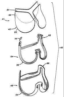
With reference now to Figure 3, the primary sub-assemblies of a
preferred embodiment of the prosthetic heart valve 40 of the present invention
are shown in exploded view. For purposes of discussion, the directions up and
down, upper and lower, or top and bottom, are used with reference to Figure 3,
but of course the valve can be oriented in any direction both prior to and
after
implantation. From top to bottom, the heart valve 40 comprises a group 41 of
three leaflets 42, three angled alignment brackets 44, a stent assembly 46,
and a
connecting band 48. Each of the sub-assemblies seen in Figure 3 is procured
and assembled separately (except for the group of leaflets, as will be
explained),
and then joined with the other sub-assemblies to form the fully assembled
valve
40 as seen in Figure 11.
The prosthetic valve 40 is a trifoliate valve with three leaflets 42.
Although three leaflets are preferred, and mimic the natural aortic valve, the
principles of the present invention can be applied to the construction of a
prosthetic valve with two or more leaflets, depending on the need.
Each of the sub-assemblies seen in Figure 3 include three cusps
separated by three commissures. The leaflets 42 each include an arcuate lower
CA 02358521 2001-07-12
WO 00/42950 PCT/US00/01855
14
cusp edge 50 terminating in upstanding commissure regions 52. Each leaflet 42
includes a coapting or free edge 54 opposite the cusp edge 50. In the
assembled
valve 40, the cusp edges 50 and commissure regions 52 are secured around the
periphery of the valve, with the free edges 54 permitted to meet or "coapt" in
the
middle. The stent assembly 46 also includes three cusps 60 separated by three
upstanding commissures 62. In like manner, the connecting band 48 includes
three cusp portions 64 separated by three upstanding commissure portions 66.
Each of the sub-assemblies will now be described in detail.
Stent Assembly
Various components of a preferred stent assembly 46 are seen in Figures
4-6. The stent assembly 46 comprises an inner stent 70 and an outer cloth
cover
72. More specifically, the inner stent 70 desirably includes three identical
and
separate stent members 74, each of which has a separate cloth covering. As
seen best in Figure 4B, each stent member 74 comprises an arcuate lower cusp
region 76 and upstanding commissure regions 78 each terminating at a tip 80.
The stent members 74 comprise elongate rods or wires, preferably made out of
an elastic biocompatible metal and/or plastic alloy, such as Elgiloy ,
Nitinol,
polypropylene, etc.. The material selected for stent members 74 should be
elastic to permit flexing along their lengths, but should possess a relatively
high
modulus of elasticity to avoid asymmetric deformation of the constructed valve
40. The stent 70 supplies an inner frame for the valve 40 that is relatively
more
rigid than the other components. Therefore, the stent 70 acts to limit total
flexibility of the valve 40.
The stent members 74 are desirably bent into the illustrated shape, using
conventional wire-forming techniques. Each of the stent members 74 is
identical, and terminates in the tips 80 which are bent inward with respect to
the
arcuate cusp regions 76 to nearly form closed circles. As is seen in Figure
4B, a
CA 02358521 2001-07-12
WO 00/42950 PCT/US00/01855
gradual radially outward bend 82 (with respect to the cylindrical stent 70) is
provided in the stent members 74 at a transition between each of the
commissure regions 78 and the intermediate cusp region 76. This bend 82
permits each of the stent members 74 to remain in a circular configuration, as
5 seen from above in Figure 4A. That is, if the cusp regions 76 extended in a
plane between each of the commissure regions 78, the plan view would be
somewhat triangular. Instead, each of the cusp regions 76 includes a lower
apex
84, and the apices of all of the cusps define a circle concentric with and
having
the same diameter as a circle defined by all of the tips 80. The stent 70 thus
10 defines a substantially cylindrical volume therewithin. Of course, other
volumes may be defined by the stent 70 wherein the tips 80 define a circle
that
is smaller or larger than a circle defined by the apices 84. For example, the
apices 84 may be provided outward from the tips 80 so the stent 70 defines a
frusto-conical volume therewithin.
15 As seen in Figure 5, each of the stent members 74 is preferably covered
with a generally tubular cloth 72 from tip to tip 80. The cloth cover 72 is a
biocompatible fabric, such as polyterephthalate, and has a varying cross
sectional shape, as indicated in Figures 6A and 6B. More specifically, the
cloth
cover 72 includes a tubular portion closely conforming around each of the
stent
members 74 and a flap 86 extending radially outward from the stent member
(with respect to the curvature of the cusp regions 76). The cloth cover 72 is
formed by wrapping an elongated sheet of fabric around each of the stent
members 74 and joining the free edges with sutures 88 to form the flaps 86. As
seen in Figure 5, the flap 86 extends from each stent member 74 in a direction
that is generally outward with respect to the cusp region 76, and continues in
the
same general orientation up the commissure regions 78 to the tips 80. The flap
86 has a dimension that is longest at the apex 84 of each cusp region 76 and
shortest at the tips 80. Indeed, the flap 86 is preferably nonexistent at the
tips
CA 02358521 2001-07-12
WO 00/42950 PCTIUSOO/01855
16
80, and gradually increases in size from the tip 80 to the apex 84. Therefore,
the
cross-section of Figure 6A taken through the commissure region 78 shows the
flap 86 having a small dimension dl, and the cross-section of Figure 6B taken
through the apex 84 shows the flap 86 having a longer dimension Q.
The final component of the stent assembly 46 is an attachment means 90
for joining each of a cloth-covered stent members 74. Preferably, the
attachment means 90 comprises threads or sutures sewn through the central
holes in each of the circular tips 80, as shown in Figure 5, although other
suitable attachment means could be used, such as rings, cinches, or the like.
The attachment means 90 may be wrapped around or sewn through the cloth
cover 72. In joining the tips 80, the attachment means 90 are desirably not
wrapped extremely tightly, but are instead provided with some slack to permit
relative movement of the tips, as will be described below. When the stent
members 74 are attached, as seen in Figure 5, the stent 70 exhibits three
cusps
corresponding to the cusp region 76 of each member, and three upstanding
commissures defined by the juxtaposition of adjacent pairs of commissure
regions 78.
In a preferred embodiment of the present invention the attachment
means 90 comprises a non-bioresorbable material to ensure that the individual
stent members 74 are maintained in the shape of the stent 70. In an
alternative
configuration, however, the attachment means 90 comprises a bioresorbable
material that dissolves over a period of time after implantation. In such an
embodiment, the natural host tissues may have grown in and around the porous
portions of the valve 40 to help retain the original shape of the stent 70. In
some instance, however, very little tissue overgrowth may have occurred prior
to the attachment means 90 dissolving, and the individual stent members 74 are
permitted to move radially a great deal with respect to one another. In the
latter
embodiment, wherein the stent members 74 are permitted to spread apart, the
CA 02358521 2001-07-12
WO 00/42950 PCTIUSOO/01855
17
connecting band 48 may be re-configured to be non-continuous at the
commissure portions 66 (see Figure 3). As a consequence, each individual stent
member 74 and associated leaflet 72 moves entirely independently of the
others,
albeit all oscillating with the natural contractions and expansions of the
surrounding aortic wall. Such independent leaflet movement may greatly
reduce any potential pressure drop across the valve. Although one embodiment
is to provide a bioresorbable attachment means 90 such as the sutures shown in
the embodiment of Figure 5, those of skill in the art will understand that any
of
the coupling means connecting the individual stent members 74 disclosed in the
present application could be modified to resorb over time.
The stent assembly 46 provides an inner support frame that is generally
rigid along any one of stent members 74, but which permits the stent members
to move with respect to one another. In this context, "generally rigid" refers
to
the structural strength of the stent members 74 that is sufficient to maintain
the
general shape of the stent 70, but that permits some flexing along the length
of
the stent members. Though the stent members 74 are generally rigid, they are
able to move with respect to one another. More particularly, joining the stent
members 74 with the attachment means 90 creates nodes or pivot points of the
valve 40 at the commissures 62 of the stent assembly 46. As will be more fully
explained below with reference to Figures 13-16, the stent members 74 are
permitted to pivot with respect to one another as they move radially inward
and
outward. Inward pivoting is permitted by spaces 94, seen in Figure 5, defined
between adjacent cloth-covered commissure regions 78 of each stent member
74. These regions 94 are generally triangular and gradually increase in size
from the attached commissure tips to the diverging cusps.
Leaflet Configurations
Figures 7A, 7B, and 7C are plan views of various configurations of
CA 02358521 2001-07-12
WO 00/42950 PCT/US00/01855
18
leaflets 42 suitable for use in the prosthetic heart valve 40. Figure 7A shows
a
leaflet 42 having the aforementioned cusp 50, commissure regions 52, and free
edge 54. It will be noted that the coapting edge 54 comprises two linear
portions extending from an apex 100 to outer tips 102. The two portions of the
free edge 54 are angled with respect to one another and define sides of a
triangular region 104 having as its hypotenuse an imaginary line 106 extending
between the opposed tips 102. The triangular region 104 of each leaflet 42 is
under less tension during dynamic motion of the valve 40, and helps ensure
coaptation of the leaflets. That is, the leaflets 42 are generally secured
along the
cusp 50 and commissure regions 52, and thus the majority of each leaflet 42 is
placed in stress except in the region above imaginary line 106. In this
regard, an
imaginary (dashed) fold line 108 defines an outer margin 110 of the leaflet 42
that is used to secure the leaflets into the valve 40. As will be clear from
the
discussion below, the margins 110 are sutured between the stent assembly 46
and connecting band 48 (Figure 3), and the free edge 54 of the leaflet extends
across the cylindrical region defined within the valve 40, and is generally
free to
move in that region. Because the triangular leaflet region 104 is relatively
stress-free, it tends to roll over under the influence of fluid dynamic
forces, thus
helping the three leaflets to coapt and prevent valve insufficiency.
Figure 7B shows a leaflet 112 that is substantially the same as the leaflet
42 of Figure 7A, and thus like elements will be given the same numbers. The
leaflet 112 includes a pair of generally triangular shaped commissure tabs 114
in
the commissure regions 52. The tips 102 are thus spaced farther apart than in
the version shown in Figure 7A. The commissure tabs 114 are used to more
securely fasten each of the leaflets to the commissures 62 of the stent
assembly
46 (Figure 3). The cloth cover 72 of the stent assembly 46 includes a flap 86
(Figure 5) which diminishes in size in the commissure regions. The tabs 114
are thus wrapped farther around the cloth-covered stent assembly 46 in the
CA 02358521 2001-07-12
WO 00/42950 PCTIUSOO/01855
19
commissure regions and sutured thereto, thus facilitating a more durable
connection.
Figure 7C is a further variation of a leaflet 116 which is, again, the same
in all respects to the leaflets described above, except for somewhat
trapezoidal-
shaped commissure tabs 118. Again, the commissure tabs 118 help to secure
the leaflets 116 in the prosthetic valve 40.
Stent/Leaflet Sub-Assembly
Figure 8 illustrates a stent/leaflet sub-assembly 120 in which the leaflets
42 are secured to the stent assembly 46. Preferably, leaflets 42 are pre-
attached
to align the free edges 54. In this manner, the free edges 54 of each two
adjacent leaflets 42 extend outward in juxtaposition and are received within
the
triangular space 94 defined between the commissure regions 78 of the stent
assembly 46 (Figure 5). The group of leaflets 41 is thus "inserted" underneath
the stent assembly 46 until the juxtaposed free edges 54 of the leaflets 42
are in
close proximity below the attachment means 90. The outer margin 110 of each
leaflet 42 is folded underneath the corresponding cusp 60 of the stent
assembly
46. At this point, sutures or other such means attach the margins I 10 to the
flap
86 of the stent assembly 46. The leaflets 42 can remain attached to one
another
at their adjacent tips 102 (or along the free edges 54 near the tips), or can
be
separated for maximum valve flexibility or when the stent is designed to
separate into individual stent members by bio-resorption of a commissure
couple.
If either the leaflet 112 or leaflet 116 of Figure 7B or 7C are used, the
respective commissure tabs 114 or 118 are wrapped around the adjacent part of
the stent assembly 46 and secured thereto. In a preferred assembly method, the
leaflets 42 are simply retained in position with respect to the stent assembly
46
with temporary sutures or other such means, to permit the stent/leaflet
CA 02358521 2001-07-12
WO 00/42950 PCT/US00/01855
subassembly 120 to be finally joined together with the connecting band 48 of
Figure 8.
Figure 8 also illustrates the three alignment brackets 44 and that each
has a generally L-shaped cross-section and comprises a cloth-covered inner
5 member (not separately numbered). The inner member preferably has minimum
elasticity, but is relatively thin and lightweight. One preferred material for
the
inner member is a polyester film such as Mylar . The brackets 44 are
preferably joined to the valve 40 at the time the stent/leaflet sub-assembly
120
and connecting band 48 are joined, and thus will be described more fully below
10 with respect to Figure 11.
Connecting Band
Figures 9 and 10 illustrate the connecting band 48 in more detail,
comprising an inner member 130 surrounded by a cloth cover 132. As
15 mentioned previously with respect to Figure 3, the connecting band 48
includes
three cusp portions 64 alternating with commissure portions 66, all generally
formed in a tubular configuration. This shape is provided by the inner member
130, with the cloth cover 132 simply draped and sewn thereover. In a preferred
embodiment, the inner member 130 is molded of silicone rubber, and the cloth
20 cover 132 is polyterephthalate.
The inner member 130 has a varying cross sectional shape along the
cusps and commissures. Figure 10 is cross-section through one of the cusp
portions 64 of the connecting band 48, and shows a region of the inner member
130 having an inner ledge 134 and upwardly angled outer free margin 136. The
cloth-covered ledges 134 extend generally radially and define three stent
support regions 138 of the connecting band 48, as seen in Figure 8. The ledge
134 has its greatest radial dimension at the midpoint of each of the cusp
portions
64 and gradually tapers down in size toward the commissure portions 66.
CA 02358521 2001-07-12
WO 00/42950 PCT/US00/01855
21
Likewise, the free margins 136 form their greatest outward angle with respect
to
a central axis of the connecting band 48 at each cusp portion 64, and
gradually
re-align to be parallel to the central axis in the commissure portions 66. The
cross-section of the inner member 130 at the commissure portions 66 is seen in
Figure 12B. A series of triangular shaped ribs 140 projects outward from the
inner member 130. The ribs 140 are formed around the entire inner member
130, along both the cusp and commissure regions. As seen in Figure 8, the
commissure portions 66 of the connecting band 48 define generally axial gaps
142 that help permit flexing of the valve 40. It should be noted that the
connecting band 48 may be discontinuous at the commissure portions 66 if the
valve has bioresorbable commissures and is designed to separate into
individual
"leaflets."
Assembled Valve
Figure 11 illustrates the assembled valve 40 in perspective, while
Figures 12A and 12B show cross-sections through a valve cusp 150 and valve
commissure 152, respectively. The connecting band 48 is sewn or otherwise
attached to the exterior of the stent/leaflet subassembly 120. Actually, as
seen
in Figure 12A, the connecting band 48 is attached underneath the stent/leaflet
subassembly 120 in the cusp 150, but the free margins 136 of the connecting
band are positioned to the outside of the subassembly. In addition, the
alignment brackets 44 are installed with a vertical leg 156 interposed between
the commissures 62 of the stent assembly 46 and the commissure portions 66
(Figure 3) of the connecting band 48. A horizontal leg 154 of each of the
alignment brackets 44 projects radially inward to cover the tips 80 of the
stent
assembly 46. The alignment brackets 44 help hold each two adjacent tips 80 of
the three-piece stent 70 together, especially helping to prevent radial mis-
alignment. The brackets also provide flat surfaces which a holder can contact,
CA 02358521 2001-07-12
WO 00/42950 PCT/US00/01855
22
as seen best in Figure 26.
With reference to the cross-section of Figure 12A, the sandwiched
configuration of the stent assembly 46, leaflet 42, and connecting band 48 can
be seen. More specifically, the cloth flap 86 of the stent assembly 46 aligns
with the leaflet margins 110, which in turn rest on the stent supports 138. A
series of suture stitches 158 are used to secure these elements together.
Preferably, the flap 86 terminates at the same location as the margin 110 of
each
leaflet 42, and at the corner defined in the connecting band 48 between each
ledge 134 and free margin 136. The radially innermost wall of the ledge 134 is
preferably inward from the stent member 74. This construction helps prevent
the stent 70 from migrating downward with respect to the connecting band 48.
The host annulus 162 is seen in phantom with the aortic wall 164
continuing upward therefrom. It can be readily seen that the angled shape of
the
cusp portions 64 of the connecting band 48 conform nicely to the host annulus
region. The triangular ribs 140 provide volume at the free margins 136 of the
connecting band 48 to facilitate connection to the natural tissue; in other
words,
more volume provides more of a "bite" for the surgeon to secure the band 48
with a suture needle. Although the conventional means for attaching the valve
40 to the host tissue is with sutures, which are not shown, the present
invention
should not be construed as limited to being implanted with sutures and other
means such as staples, adhesives, and the like could be used.
Now with reference to Figure 12B, the assembly of the valve
components in the commissure region is seen. The commissure edges 52 of
each of the leaflets 42 are sandwiched in between the stent assembly 46 and
connecting band 48. More particularly, the commissure edges 52 are
sandwiched between the flaps 86 and the generally planar commissure portions
66 of the connecting band 48 (Figure 8). Sutures 170 are provided to join
these
elements together. Again, the commissure edges 52 preferably terminate at the
CA 02358521 2001-07-12
WO 00/42950 PCTIUSOO/01855
23
same location as the flaps 86. Figure 12B also illustrates the gap 142
provided
in the commissure regions of the connecting band 48, and the lack of
structural
connection between the two sides of each valve commissure 152.
Figure 12B shows in phantom a portion of the aortic wall 172 to which
the commissures 152 of the valve 40 are attached. Again, the particular
attachment means is not shown, but the connecting band 48 is traditionally
sutured to the wall 172.
Dynamic Motion of the Prosthetic Heart Valve
Figures 13 and 15 illustrate a conduit portion of a heart in the region of
the aortic valve and relative motions of the conduit walls during systole and
diastole, respectively. In particular, Figure 13 shows an open valve 200 and
systolic blood flow 202, while Figure 15 shows a closed valve 204 and
diastolic
back flow of blood 206. As described with respect to Figures 1 and 2, the
regions around the aortic valve can be generally separated into an annulus
region 208 and a sinus region 210.
As mentioned previously, the annulus region 208 is expected to contract
during the systolic phase, as indicated by the arrows 212 in Figure 13, and
expand during the diastolic phase, as indicated by the arrows 214 in Figure
15.
Conversely, the sinus region 210 is expected to expand during the systolic
phase, as indicated by the arrows 216 in Figure 13, and is expected to
contract
during the diastolic phase, as indicated by the arrows 218 in Figure 15. The
movements of the conduit walls are shown with respect to a neutral or relaxed
position 220, and may be exaggerated from the true movements. Also, as
mentioned above, these movements are educated guesses and may be different
for some, if not most patients. However, the flexible heart valve of the
present
invention accommodates all variations of such movements.
Figures 14 and 16 schematically illustrate the synchronous movement of
CA 02358521 2001-07-12
WO 00/42950 PCT/US00/01855
24
the prosthetic valve 40 of the present invention with respect to the movements
of the host tissue in systolic and diastolic phases as seen in Figures 13 and
15.
To simplify this explanation, Figures 14 and 16 only illustrate the stent 70
of the
present invention, which as previously described acts as a limitation to
movement of the entire valve 40 and fairly represents movement of the entire
valve.
With reference to Figures 14A and 14B, during systole the valve
experiences outward commissure movement, as indicated by the arrows 230. At
the same time, the valve experiences inward movement at the cusps, as
indicated by the arrows 232. During diastole, in contrast, and as seen in
Figures
16A and 16B, the valve experiences inward commissure movement, as indicated
by the arrows 234. At the same time, the valve experiences outward movement
at the cusps, as indicated by the arrows 236.
Alternative Stents
Figures 17-19 illustrate an alternative stent assembly 250 comprising an
inner stent 252 and an outer cloth cover 254. As with the earlier stent
assembly
46, the stent assembly 250 includes alternating cusps 256 and commissures 258.
As best seen in Figure 18, the stent 252 includes three separate stent members
260 having arcuate commissure tips 262 that are curved toward one another. A
generally disk-shaped commissure housing 264 encompasses the adjacent
commissure tips 262, retaining the stent members 260 together while permitting
relative pivoting.
Figure 19 illustrates two adjacent commissure tips 262 and the
commissure housing 264 exploded into a male housing portion 266 and a
female housing portion 268. The housing portions are so named because they
are joined together through interference between a button 270 of the male
housing portion 266 and an aperture 272 on the female housing portion 268.
CA 02358521 2001-07-12
WO 00/42950 PCT/US00/01855
Each portion of the commissure housing 264 includes a circular groove 274 for
receiving the arcuate tips 262. The grooves 274 combined to form a circular
channel having an axis 276 within which the arcuate tips 262 are received and
can slide. When assembled together, the commissure housings 264 thus provide
5 nodes of rotation for each of the stent members 260.
Figure 20A illustrates an alternative stent 280 suitable for use in a heart
valve of the present invention. The stent 280 includes three stent members
282,
each having commissures with a flex region 284 and tips 286. The tips 286 of
adjacent stent members 282 are secured together by sutures or other suitable
10 means (not shown). The flex regions 284 comprise sections of each stent
member 282 which are bent away from each other. The stent members 282 can
thus pivot with respect to one another about the connected tips 286. Upon
inward movement of the stent members 282, a fulcrum 288 is created by
interaction between the stent members at the lower end of the flex region 284.
15 The relative flexibility in inward or outward movement of the stent members
282 can be modified by selection of the cross sectional size and shape of the
stent members, and overall configuration of the flex region 284.
Figure 20B illustrates a second alternative stent 290 suitable for use in a
heart valve of the present invention. The stent 290 includes three wires 292
and
20 has commissure regions 294 formed by bent ends of the wires and a junction
member 296. In this embodiment, the junction member 296 either rigidly holds
the terminal ends of each of the wires 292, or permits the wires to slide or
otherwise flex with respect to one another. If the wires are rigidly attached
to
the junction member 296 the shape of the wires in the commissure region 294
25 reduces stress risers in bending.
Figure 20C illustrates a third alternative stent 300 suitable for use in a
heart valve of the present invention. The stent 300 comprising three separate
wires 302 terminating at circular commissure tips 304. Each of the commissure
CA 02358521 2001-07-12
WO 00/42950 PCTIUSOO/01855
26
tips 304 is rotatably fastened around a pin 306 provided on a junction plate
308
common to adjacent wires 302. In this manner, the tips 304 remained located
close to one another, while the cusps of the wires 302 can pivot in and out.
Figure 20D illustrates a fourth alternative stent 310 suitable for use in a
heart valve of the present invention. The stent 310 is made in one piece with
a
series of alternating cusps 312 and commissures 314. The commissures 314
comprising a nearly 360 bend in the stent 310 which permits each cusp 312 to
easily flex with respect to the other cusps.
Figure 20E illustrates a fifth alternative stent 320 suitable for use in a
heart valve of the present invention. The stent 320 comprises three wire-like
stent members 322, adjacent ones of which are joined together at commissure
regions 324 by a U-shaped coupling 326 and a pair flexible sleeves 328. Figure
21 is a detail of one of the commissure regions 324 showing in hidden lines
the
adjacent ends of the coupling 326 and stent members 322. The couplings 326
are preferably sized with the same diameter as the stent members 322, and the
sleeves 328 are tubular with a constant diameter lumen. The sleeves 328 may
be made of silicone, or a flexible polymer such as polyurethane or the like.
Other flexible interfaces such as sleeves 328 are contemplated, such as, for
example, a single block of silicone into which the commissure regions 324 of
the stent members 322 are molded.
Figure 22 is a detailed view of a commissure region 330 of a still further
alternative stent suitable for use in a heart valve of the present invention.
The
stent is made in one piece with adjacent cusps 332 being joined by a coil
spring
tip 334. Again, great flexibility is provided by the coil spring tips 334 to
enable
relative motion of the cusps 332. The amount of flexibility is selected as in
any
spring by varying the material, cross-sectional size and shape, and number of
turns of the spring.
CA 02358521 2001-07-12
WO 00/42950 PCT/US00/01855
27
Valve Holder
Figures 23-26 illustrate a preferred holder 350 useful for implanting the
flexible heart valve 40 of the present invention. As the heart valve 40 is
relatively flexible, the holder 350 must provide adequate support to insure a
stable platform for the surgeon to position the valve for attachment to the
natural tissue. In other words, because the flexible prosthetic heart valve 40
of
the present invention exhibits alternating cusps and commissures in a
generally
cylindrical configuration that are adapted to move radially in and out with
respect to one another, the holder 350 desirably provides rigid structure for
maintaining a fixed shape of the valve during implantation. In addition, the
holder 350 must include structure to allow quick release from the valve 48
after
the valve is implanted.
As seen in Figure 23, the holder 350 comprises a proximal handle socket
352 having an inner bore 354 for receiving the distal end of a handle (not
shown). The socket 352 may be provided with internal threads, or other such
quick-release coupling structure to facilitate handle connection and
disconnection. The holder 350 has three radially outwardly-directed
commissure legs 356, and three outwardly and downwardly angled cusp legs
358. Consistent with the distribution of the cusps 150 and commissures 152 of
the valve 40, the commissure legs 356 are oriented 120 apart, and the cusp
legs
358 are oriented 120 apart, with the three commissure legs being offset with
respect to the three cusp legs by 60 .
As seen in Figure 24, each of the commissure legs 356 extends outward
from the handle socket 352 into proximity with one of the valve commissures
152 and is secured thereto with an upper suture 360. Likewise, each of the
cusp
legs 358 extends outward and downward from the handle socket 352 into
proximity with a midpoint of one of the valve cusps 150, and is secured
thereto
with a lower suture 362. The lower end of each cusp leg 358 includes a
CA 02358521 2001-07-12
WO 00/42950 PCTIUSOO/01855
28
concavity for mating with the corresponding rod-like stent member 74, as seen
in Figure 26. In this manner, each of the cusps 150 and commissures 152 of the
valve 40 is securely held in relation to the others, thus facilitating
implantation
by the surgeon.
Details of the commissure legs 356 will now being described with
reference to Figures 23 and 26. Each commissure leg 356 extends outward from
the handle socket 352 in a generally rectangular cross-section interrupted by
an
upwardly-facing inner notch 370 oriented cross-wise to the leg. And upwardly-
facing radial channel 372 having a depth of approximately half of each
commissure leg 356 extends from about the inner notch 370 to the outermost
end of the leg. The inner notch 370 is not quite as deep as the channel 372,
as
seen in Figure 26. The radial channel 372 divides the upper portion of each
commissure leg 356 into two walls 374a, 374b. An eyehole 376 is formed in
one of the walls 374a, and a corresponding outer notch 378 is formed in the
other wa11374b aligned with the eyehole. The outer notch 378 is also not quite
as deep as the channel 372.
With reference to Figures 24 and 26, the upper suture 360 is preferably
tied to the eyehole 376 in the first wa11374a. The suture 360 then passes
across
the channe1372, through the outer notch 378, and is passed along the inner
notch 370, again traversing the channe1372. The suture 368 is then passed
through a suture-permeable portion of the valve commissure 152, such as
through the connecting band 48. After passing through the commissure 152, the
suture 360 is again looped through one or both of the notches 370, 378 and re-
tied to the eyehole 376. By proper threading of the upper suture 360, each
commissure 152 can be secured to the commissure leg 356 and easily released
by inserting a scalpel blade into the radial channe1372 to sever the portions
of
the suture therein.
Details of each cusp leg 358 can be seen in Figures 23 and 26. A pair
CA 02358521 2001-07-12
WO 00/42950 PCT/US00/01855
29
of longitudinal rails 380a, 380b are provided on the outer side of each cusp
leg
358. Toward the lower end of the rails 380a,b, a pair of aligned eyeholes 382
provide anchoring locations for the lower suture 362. A scalpel guide or
relief
384 is formed in one of the rails 380b. As seen in Figure 24, the lower suture
362 extends downward from the eyeholes 382, passes through a suture-
permeable portion of the cusp 150, and is then returned and secured to the
eyeholes 382. The relief 384 exposes a portion of the lower suture 362 for
severing by the surgeon using a scalpel blade. It will thus be understood that
the holder 350 can be quickly released from the valve 40 by a series of six
scalpel strokes, with each of the sutures 360, 362 remaining attached to the
holder 350 and being withdrawn from the valve 40 as the holder is withdrawn.
Figures 27A and 27B illustrate an alternative holder 390 useful for
implanting the flexible heart valve 40 of the present invention. The holder
390
is substantially similar to the holder 350 described above, but the ends of
each
of a plurality of rigid legs for attaching to the valve cusps are flared, or,
more
precisely, each lower leg has a width from a hub to a terminal end that is
greatest at the terminal end to provide more surface area to contact the
corresponding valve cusp. That is, the holder 390 includes a plurality of
upper
legs 392 having a generally constant width, and a plurality of lower legs 394
having flared ends 396, the legs extending from a central hub 398. Again, the
upper legs 392 extend radially outward to connect to the valve commissures
152, and the lower legs 394 angle radially outward and downward to connect to
the valve cusps 150. The flared ends 396 impart greater stability to the
flexible
valve 40 during implantation, especially helping to prevent movement of the
cusps 150. In addition, the legs 194 remain fairly narrow until the flared
ends
396 to maintain good visibility through the spaces between the plurality of
legs.
That is, for example, the surgeon can continue to view the valve leaflets 42
between the legs as a check on valve orientation.
CA 02358521 2001-07-12
WO 00/42950 PCT/US00/01855
Independent Leaflets
A further aspect of the invention is a heart valve having three leaflets
directly sutured to the aortic annulus wherein the commissures of adjacent
5 leaflets are totally or partially independent, and may be connected during
implant but will become independent thereafter. This gives the independent
leaflets and juxtaposed commissures full freedom to move during systole and
diastole, thus reducing the pressure gradient. For example, any of the stent
assemblies described herein (such as, e.g., the stent assemblies of Figures 17-
10 19) may include separate stent members initially coupled together with a
permanent or bioresorbable fastener, the latter dissolving after a fairly
short
amount of time to permit independent movement of the leaflets. In addition,
the
present invention discloses an independent leaflet and implantation device
that
can replace a single defective leaflet, with the prosthetic leaflet
functioning in
15 conjunction with the remaining native leaflets.
One example of an independent leaflet 400 seen in Figures 28A and 28B
includes a planar or three-dimensional body 402 defined by a coapting edge 404
and a fixation edge 406. The fixation edge 406 includes an arcuate cusp region
408 and a pair of commissure regions 410. The body 402 is attached along its
20 fixation edge 406 to an arcuate flexible support 412 that preserves the
shape of
the fixation edge and provides a platform for attachment to the native tissue.
Figure 28B shows the leaflet 400 attached to a native annulus 422 adjacent a
native leaflet 423.
The leaflet body 402 may be made from a biological material such as a
25 porcine valve leaflet or a piece of bovine pericardium, chemically or
physically
treated to prevent immunological reaction and fatigue. Alternatively, the
leaflet
body 402 may be formed of a synthetic material such as a polymer or a woven
fabric. The flexible support 412 is desirably made of a synthetic material,
for
CA 02358521 2001-07-12
WO 00/42950 PCTIUSOO/01855
31
example silicone or PTFE.
As mentioned previously, the independent leaflet 400 may be used with
other such leaflets, preferably three total, that are attached directly to an
ascending aorta and function together as a prosthetic valve. That is, as seen
in
Figure 29A, adjacent flexible supports 412 may be attached at their juxtaposed
commissure regions with a separate fastener 414, such as a clip. The clip 414
may be permanent or bioresorbable. Regardless of its longevity, the clips 414,
when securing the leaflets 400 together, permit the leaflets to flex with
respect
to one another so as to allow the assembled valve to move in synch with the
native tissue during the sytole/diastole cycle. This, in turn, reduces the
pressure
gradient through the valve. Figure 29B illustrates an alternative fastener
between independent leaflets in the form of a continuous scalloped wire or
stent
416. Each leaflet 400 (either the leaflet body 402 directly or via the support
412) is attached to the scalloped stent 416 and thus functions together with
the
other leaflets in a prosthetic valve. The embodiment in Figure 29B is similar
to
those previously described, wherein the scalloped stent 416 is highly flexible
and permits both cusp and commissure movement. Again, however, the
commissure regions 418 of the stent 416 may be bioresorbable to result in
completely independent leaflets after a period of time in the body.
Figure 30 is a perspective view of an exemplary independent leaflet
measuring device 420 used to select the appropriate leaflet 400 according to
the
size and geometry of the native annulus 422. The device 420 includes a handle
424 and an arcuate leaflet sizer 426 that is sized and shaped to match the
prosthetic leaflet. Two or more struts 428 may extend between the handle 424
and sizer 426 to improve stability in rotation and manipulation of the sizer.
Figures 31A-31C are schematic views of a device 440 for automatically
implanting an independent leaflet of the present invention, illustrating an
articulated holder especially suited for minimally invasive environments. The
CA 02358521 2001-07-12
WO 00/42950 PCT/US00/01855
32
device 440 includes a handle 442 connected to the independent leaflet 444 via
a
leaflet holder 446, comprising a plurality of movable struts 446. The struts
446
pivot or slide with respect to one another to convert the device from the low-
profile configuration shown in Figure 31A capable of passing through small
apertures, to the full deployment configuration of Figure 31 C. The leaflet
444
may be attached to the leaflet holder 446 with sutures or the like. A
plurality of
staples 448 are provided extending outward from the leaflet fixation edge that
pierce the native tissue and are deformed by a backing tool or assume a bent
shape upon a temperature change. The final configuration is seen in Figure
31 C, and those of skill in the art will appreciate the ease of use of the
separate
leaflet implantation procedure. Once implanted, any structure connecting the
leaflet 444 to the device 440 is severed, and the device removed.
Figures 32A-32C are schematic views of a still further exemplary
implantation device for the independent leaflet embodiment. The device
includes an anvil or backing portion 460 having a handle 462 and a plurality
of
fluid-carrying tubes 464 through which a cold or warrn fluid can circulate, as
seen by the arrows 466. The circulating fluid regulates the temperature of the
tubes 464 during implantation. The device further includes a holder 470,
similar to the holder 440 of Figure 31A, to which an independent leaflet 472
is
temporarily attached, such as with sutures 474. The fluid-carrying tubes 464
of
the backing portion 460 is placed below the native annulus 476 while the
leaflet
holder 470 is maneuvered into position above the annulus. Opposite
displacement of the backing portion 460 and holder 470 causes a plurality of
staples 478 to pierce the annulus 476 and be deflected or otherwise curled by
the
tubes 464. If the staples 478 are shape memory alloy materials like Nitinol,
the
fluid circulating through the tubes 464 can be used to cause a temperature-
induced shape change.
Figure 33 is a cross-sectional view through one embodiment of an
CA 02358521 2001-07-12
WO 00/42950 PCT/US00/01855
33
attachment structure for the independent leaflet embodiment. The structure
includes staples 480 that extend outward from a leaflet fixation edge 482
through the native tissue 484, and curl or bend into a shape that prevents
removal. The curling of the staples 480 may be accomplished using the device
460 (i.e., the backing portion 460 and holder 470) that provide a backing
plate
or anvil, or through a temperature change, such as when a shape memory
material is used. In the illustrated embodiment, the staples 480 may include
adjacent legs that curl at different lengths to accommodate one another.
Exemplary dimension are shown in Figure 33, with the width of the leaflet
fixation edge 482 being about 9 mm, the length of one leg of each staple 480
being about 8 mm, and the length of one leg of each staple 480 being about 10
mm.
The present invention may be embodied in other specific forms without
departing from its spirit or essential characteristics. The described
embodiments
are to be considered in all respects only as illustrative and not restrictive.
In
particular, though the flexible nature of the present heart valve has been
described as being particularly suitable for use in the aortic position, the
advantage of flexibility could equally apply to a valve implanted in other
positions, such as the mitral position. The scope of the invention is,
therefore,
indicated by the appended claims rather than by the foregoing description. All
changes which come within the meaning and range of equivalency of the claims
are to be embraced within their scope.