Note: Descriptions are shown in the official language in which they were submitted.
CA 02362531 2007-01-12
-2-
MEANS AND METHOD FOR LIQUID LEVEL SENSING
This Application is a Division of Canadian Patent
Application Serial No. 2,172,363, filed on September 22,
1994.
Field of Invention
The present invention relates to an automated
analytical system and method for the analysis of liquid
test samples. In another aspect, the invention is related
to a random access system capable of simultaneously
performing a plurality of assays, particularly
heterogeneous and/or homogeneous immunoassays, which
system is also a continuous system in that the system may
be additionally or alternatively loaded at any time with
samples and yet maintain continuous and uninterrupted
assay operations. In yet another aspect, the present
invention relates to the
CA 02362531 2001-11-20
WO 95/08774 PCTIUS94/10850
-3-
various components incorporated into and utilized by
such system.
Background of the Invention
Ihe Automated Analytical System
Although various known clinical analyzers for
chemical, immunochemical and biological testing of
samples are available, clinical technology is rapidly
changing due to increasing demands in the clinical
laboratory to provide new levels of service. These new
levels of service must be more cost effective to
decrease the operating expenditures such as labor cost
and the like, and..must provide shorter turnaround time
of test results to reduce the patient's length of stay
in the hospital as well as improve efficiency of
1-5 outpatient treatment. Modernization of analytical
apparatus and procedures demands consolidation of work
stations to meet the growing challenge placed on
clinical laboratories.
Generally, analysis of a test sample involves the
reaction of test samples with one or more reagents with
respect to one or more analytes wherein it is
frequently desired that the analysis be performed on a
selective basis with respect to each test sample.
However, due to the high demands placed on clinical
laboratories regarding not only volume throughput but
also the number and frequency of various analyses,
CA 02362531 2001-11-20
WO 95/08774 PCT/US94/10850
-4-
there is a need to provide an automated analysis system
which is capable of combining accurate analytical
results, multiple test menu versatility, low reagent
and fluids loss and consumption, and of great benefit
and importance, continuous and high throughput.
The present automated clinical analysis systems
provide much improved accuracy of analytical results in
comparison with accuracies of earlier systems. In the
present systems, analysis of a test sample typically
involves forming a reaction mixture comprising the test
sample and one or more reagents, and the reaction
mixture is then analyzed by an apparatus for one or
more characteristics of. the test sample. Reliance on
automated clinical analyzers has improved the
efficiency of the laboratory procedures, inasmuch as
the technician has fewer tasks to perform. Automated
clinical analyzers provide results much more rapidly
while frequently avoiding operator or technician error,
thus placing emphasis on accuracy and repeatability of
a variety of tests. Automated clinical analyzers
presently available for routine laboratory tests
include a transport or conveyor system designed to
transport containers of sample liquids between various
operating stations. For example, a reaction tube or
cuvette containing a test sample may pass through a
reagent filling station, mixing station, reaction
- - ------ ------
CA 02362531 2001-11-20
WO 95/08774 PCT/US94/10850
-5-
forming station, detection stations, analysis stations,
and the like. Such present transport systems are,
however, not flexible in that transport is in one
direction and the reaction tubes or cuvettes, once
inserted into the apparatus, must pass through without
access before analysis occurs. Even further, the
present transport systems allow only batch-like
operation in that once the system is initially loaded,
testing may only be performed on the initially loaded
samples during a single operation cycle; alternative or
additional samples can not be loaded during the
operation cycle to allow continuing operations for
extended periods.
As for multiple test menu versatility, some of the
presently available automated clinical analyzers, such
as automated immunoassay analyzers like the Abbott IMxm
analyzer and the Abbott TDxm analyzer (Abbott
Laboratories, Abbott Park, Illinois, USA), utilize
procedures involving a variety of different assay
steps. These present systems have typically relied on
detection and measurement of optical changes in a
reaction mixture during the assay process. For example,
a number of well-known techniques using single or
multi-wavelength fluorescence include fluorescent
polarization immunoassays (FPIA) employing homogeneous
immunoassay techniques, microparticle enzyme
CA 02362531 2001-11-20
WO 95/08774 PCTfUS94/10850
-6-
immunoassays (MEIA) employing heterogeneous immunoassay
techniques, and the like. The MEIA technology, such as
that used on the Abbott IMxls analyzer, is used for high
and low molecular weight analytes requiring greater
sensitivity, and FPIA technology, such as that used on
the Abbott TDx6 analyzer, is used primarily for lower
molecular weight analytes. A front surface fluorometer
is used in these systems to quantify a fluorescent
product generated in the MEIA assays, while a
fluorescence polarization optical system is used to
quantify the degree of tracer binding to antibody in
the FPIA assays. -The test samples are automatically
processed in certain of these systems, such as the
Abbott IMx analyzer and Abbott TDx analyzer, by a
robotic arm with a pipetting probe and a rotating
carousel which positions the samples for processing.
These systems are typically compact table-top analyzers
which of f er f ul l y automated, walk-away i mmunoas s ay
testing capabilities for both routine and specialized
immunoassays. These nonisotopic methods eliminate
radioactivity disposal problems and increase reagent
shelf life while meeting the diverse requirements of a
multitude of different assays. Though these presently
available automated clinical analyzers provide a degree
of improved multiple test inenu versatility in
comparison to earlier systems and practices, the
CA 02362531 2001-11-20
WO 95/08774 PCTIUS94/10850
-7-
present analyzers remain limited in that these systems
are one direction only systems, or batch analyzers,
which permit the analysis of multiple samples and
provide for access to the test samples for the
formation of subsequent reaction mixtures, but permit
only one type of analysis at a time. It would, thus, be
an improvement to provide a random access analyzer
which allows for analysis of multiple test samples for
multiple analytes. It would be an even further
improvement if such a random access analyzer allowed
for continuous operations; that is, if additional or
alternative samples could be loaded for analysis during
analysis operations by the system, without interruption
of the analysis operations.
With respect to reagent and fluids consumption and
loss in present automated clinical analyzers, a common
feature of those analyzers is the inclusion of various
reagents within the apparatus itself or placed near the
apparatus for pipetting purposes. In these systems,
liquid reagents, in bulk form, are selected for the
various types of tests which are to be performed on the
test sample, and are stored in or near the apparatus.
Reagent delivery units, such as pumps and the like,
along with valves, control and pipette mechanisins, are
i ncl uded in the present automated anal yz ers so that
different reagents can be mixed according to the type
CA 02362531 2001-11-20
WO 95/08774 PCT/US94/10850
-8-
of test to be performed. In certain of these present
analyzers, for example, the Abbott IMxm analyzer
previously mentioned, all the steps required for
analysis of test samples are automatically performed
and those steps include numerous checks of the
subsystems to insure that assays are run to completion
with valid results. In the Abbott IMxm in particular,
quantification of the fluorescence intensity in the
MEIA method and polarization in the FPIA method, as
well as the final data reduction,'are fully automated
on the analyzer and results are printed by the analyzer
and may be accessed through suitable means for-
automatic data collection by a laboratory computer.
These various aspects of the present automated clinical
analyzers, like the Abbott IMxe, help limit reagent and
fluids consumption and loss, as well as provide other
advantages. Even with those advantages, however,
improvement in reagent and fluids consumption and loss
in an 'analysis system would be desirable. Even further,
such improvement in consumption and loss by these,
combined with benefits of continuous operations,
accuracy of results, and test menu versatility would be
a significant improvement in the art.
With respect to continuous and high throughput in
automated analytical systems; the prior systems have
been unable to provide these desirable characteristics.
CA 02362531 2001-11-20
WO 95/08774 PCT/US94/10850
-9-
In the prior automated analytical systems, the systems
are initially loaded with a plurality of test samples.
The samples are then each tested during a full cycle of
testing by the systems. Though the number of samples
which may be initially loaded in these systems is
fairly large, it is not possible to load additional
test samples in these systems at the same time the
systems are testing the initial load. Additional
samples may only be loaded after testing of the prior
sample load is complete. In order to increase
throughput in these s ys tems then, it would be
advantageous to provide an automated analytical system
which allowed for loading of additional samples at any
time, even while the system is testing other samples.
It would be an even further advantage if such a system
could provide accurate results, multiple test menu
versatility, and low reagent and fluids loss and
consumption while at the same time allowing continuous
access to and testing of samples. The prior systems
have been unable to provide these advantages. The
present automated continuous and random access system
provides, all these advantages. In addition to those
advantages, the present invention also provides
additional improvements directed to specific aspects,
parts, and operations of these systems.
CA 02362531 2001-11-20
WO 95/08774 PCT/US94/10850
-10-
Specific Asnects. Parts, and Ooerations
Other benefits and advantages, in addition to
those previously described (i.e., accurate analytical
results, multiple test menu versatility, low reagent
and fluids consumption and loss, and continuous and
high throughput), directed to specific aspects, parts,
and operations of automated clinical analyzers would
also be improvements in the art.
A. Detection Systems:
For example, an improved analyzer might
incorporate capability to perform both homogeneous and
heterogeneous assays. Automated analytical apparatus
for performing homogeneous assays, the detection of
precipitate formed by reaction between antigens and
antibodies in. a test sample-cell to form light
scattering centers, and methods and apparatus for
detecting immunological agglutination reactions are
known in the art. Such apparatus and methods include,
for example, the steps of measuring light absorption of
the liquid medium with antibody before and after the
antigen-antibody reaction by using light which is
absorbable by the antibody, and calculating the
difference of the absorptions. In this way, the
presence or absence of agglutination is detected based
on the fact that the agglutination reaction reduces the
concentration of antibody, which affects the light
CA 02362531 2001-11-20
WO 95/08774 PCTIUS94/10850
-11-
absorption of the liquid medium. As is typical of
methods and apparatus for performing homogeneous
assays, these procedures do not require separation of
a solid phase from the reaction mixture for further
analysis. Heterogeneous assays are also known through
the use of a sample analyzer for quantitating
relatively small amounts of clinically significant
compounds in a liquid test sample by focusing a light
source onto the sample so that, for example,
fluorescent particles in the sample cause fluorescent
conditions, the intensity of which is the function of
the intensity of-the light beam and the concentration
of fluorescent particles in the sample. A detector
senses photons forming the fluorescent emissions of the
particles when excited by the light beam. The
introduction of a solid phase material into the sample
requires subsequent separation of the solid phase from
the reaction mixture for further analysis and before
the fluorescent emissions can be detected and measured.
It would be advantageous in an automated clinical
analyzer to incorporate apparatus and methods for
performing, selectively on the same sample, various
homogeneous and heterogeneous assays concurrently in a
random access fashion. Such apparatus and methods could
provide for the analysis of a plurality of liquid
samples wherein each sample is analyzed with respect to
CA 02362531 2001-11-20
WO 95/08774 PCT/US94/10850
-12-
at least one analyte utilizing both homogeneous and
heterogeneous assay techniques.
B. Syringe Bubble Flusher:
Another possible benefit and advantage directed to
specific aspects, parts, and operations of automated
clinical analyzers involves the precision and accuracy
of the fluidics within the automated analytical
instrument. That precision and accuracy during assay
procedures is closely related to the precision and
accuracy with which fluids can be aspirated and
dispensed by the instrument. Although a syringe or
similar device within the instrument can provide
aspirating and dispensing steps, performance of prior
syringes is often severely degraded by the presence of
air bubbles in.the syringe. Existing construction and
designs of such syringes have failed to provide
.efficient means of removing such bubbles. For example,
various relatively ineffective and cumbersome manual
techniques and manipulations, such as abruptly tapping
the syringe, and the like, have been used to flush
bubbles out of the syringe. Thus, there remains a need,
and it would be, an improvement in the art, to provide
an automated clinical analyzer with a fluidics system
which includes a syringe or similar device to provide
precise and accurate aspirations, dispensing, and
bubble flushing steps while avoiding problems
CA 02362531 2001-11-20
WO 95/08774 PCTIUS94/10850
-13-
previously encountered in automatic flushing of bubbles
completely from the fluidics system.
C. Reaction Vessel and Loader:
It would also be advantageous in an automated
analytical instrument to provide for ease of loading
and handling of test samples and reagents. Such
l oadi ng and handling can be probl emati c, in parti c ul ar,
when test samples and reagents are contained in
containers of varying shape and size and must be
supplied to the instrument. The present invention
provides improved ease of loading and handling because
of the particular containers used by the system for
handling test samples and reagents, and further
provides for ease of manipulation of those samples and
reagents and reaction processes. In addition, the
invention provides for disposable containers for those
samples and reagents and other advantages, such as
storage free from contaminants. -
D. Reagent Cap Actuator:
Even other benefits and advantages directed to
specific aspects, parts, and operations of autpmated
clinical analyzers are possible. Prior methods for
reducing evaporation of costly reagents from system
containers have utilized manual operations to cap
reagent containers, as well a's use of various other
reclosing container caps which are held open during
CA 02362531 2001-11-20
WO 95/08774 PCTIUS94/10850
-14-
liquid access cycle and then allowed to reseal by
removing the opening force. It would be an improvement
in automated clinical analyzers to provide apparatus
and methods wherein computer-controlled robotic devices
replace the need for manual intervention in these
regards. Those devices could be beneficial and
advantageous if capable of minimizing reagent
evaporation.
E. Cartridge. Feeder and Carton:
Further benefits and advantages of specific
aspects, parts, and operations of automated clinical
anal yz ers are al s er- des i rabl e. The di agnos ti cs i ndus try
has heretofore routinely utilized several types of
systems which each require hand loading of cartridges,
reagent packs, and sample containers into batch and
semi-automatic instruments. In an automated, continuous
and random access analytical system, manual hand
loading of such items is further complicated when
dealing with volume and reliability concerns. It would
clearly be an improvement, then, to incorporate in
these automated clinical analyzers an automatic feeder,
capable of feeding tubular parts, such as cartridges,
and orientating the cartridges with an open end up. An
automatic cartridge feeder hopper of multiple
cartridges could save substantial operator time and
error since multiple cartridges could be loaded into
CA 02362531 2001-11-20
WO 95/08774 PCT/US94/10850
-15-
the hopper feeder system directly from the cartridge
packaging systems, eliminating individual hand feeding
of the cartridges, and assuring reliability within the
automated diagnostic system. Moreover, it would be
advantageous to provide in these systems various
handling and loading means to facilitate the handling
and loading of reaction vessels which are utilized with
systems.
F. Sample Container Segment:
The invention even further provides advantageous
mechanisms for loading particular containers. These
mechanisms are of~ great importance because small, non-
uniform containers are necessary in certain instances.
Where many of these containers must be employed, it
becomes hard, if not impossible, for an operator to
maintain adequate supply to the instrument if loading
and positioning of the containers is largely manual.
The present invention provides improved containers and
loading mechanisms to allow easy and continuous loading
and processing capability.
G. Optics Control System:
A further aspect in which improvement would be
beneficial' is optics and optic controls utilized by
automated analytical systems. Measurements and other
operations performed by thes'e systems often employ
characterization of relative optical properties.
CA 02362531 2001-11-20
WO 95/08774 PCTIUS94/10850
-16-
Because accuracy of these measurements and operations
depend upon aspects of the optical system, the optical
system must function in a reliable manner, particularly
in an automated and continuous system in which a
multitude of simultaneous operations are being
performed in addition to the optical measurements.
H. Smart Wash=
In automated analytical instruments, one
particular area of concern has been carryover by or
cross contamination of the samples, regiments, and
other fluids. Typically, some form of wash is employed
in these devices to wash apparatus between steps when
switching between different fluid substances. The
approach former employed has often been to maximize the
amount of wash.to account for the worst case scenario
of potential contamination. It would be an improvement
to provide a variable wash mechanism that operates
according to the degree of potential contamination to
insure no contamination, but yet conserves wash fluids
and other resources.
I. Chemiluminescent Test:
Chemiluminescent testing is generally known. In
fact, chemiluminescent testing has been employed in
certain automated analytical systems. It appears,
however, that chemiluminescent testing has not been
employed in a continuous and random access system. It
CA 02362531 2001-11-20
WO 95/08774 PCT/US94/10850
-17-
would be an advantage to provide a continuflus and
random access analytical system suitable for
chemiluminescent testing.
J. Sample Segment Container:
The often differing sizes and configurations of
containers employed.with automated analytical systems
presents special problems. Also, keeping track of
those containers and their contents has required
significant resources. It would be advantageous to
provide means in an automated analytical system for
handling and identifying containers.
R. Li cruid .Level Sensor:
Yet another aspect of automated clinical analyzers
which could be advantageously improved is the liquids
measurement systems. In particular, a liquid level
sense mechanism employed for sensing fluid levels in
various sample containers is desired in . those
analyzers. The present invention provides a new fluid
level -sensing system to allow for liquids measurement.
Such system is an improvement in the art.
The present invention, an automated continuous and
random,access analytic'al system and components thereof,
provides for the primary capabilities missing from the
prior art automated clinical analyzers; that is, the
present system combines accurate analytical result
capability, multiple test menu versatility, low reagent
CA 02362531 2005-02-28
-18-
and fluids loss and consumption, and continuous and high
throughput. Because the present invention provides those
capabilities, the invention is a significant improvement in
the art. In addition to those capabilities, however, the
present invention provides other benefits and advantages
over the prior systems. Those benefits and advantages, in
certain instances being directed to specific aspects,
parts, and operations in an automated continuous and random
access analytical system, provide much improvement in that
particular system, as well as in numerous and varied
possible applications.
Summary of the Invention
In accordance with one aspect of the invention, there
is provided an automated liquid level sensing system for
detecting the presence of liquid in a container, said
liquid level sensing system comprising:
a vertically oriented, electrically conductive probe
positioned above said container;
means for vertically moving said probe into and out of
said container;
a signal source electrically connected to said probe,
said signal source energizing said probe with an electrical
signal and causing said probe to transmit said electrical
signal;
a receiving antenna positioned below said container
for receiving said transmitted electrical signal;
CA 02362531 2005-02-28
-18a-
means for analyzing said received electrical signal
for indications that said probe has contacted liquid in
said container;
means for transferring said received electrical signal
from said receiving antenna to said analyzing means; and
means for indicating that liquid has been detected.
In another aspect of the invention, there is provided
a method of automatically detecting the presence of liquid
in a container, said method comprising the steps of:
vertically positioning an electrically conductive
probe above said container;
vertically moving said probe into and out of said
container;
electrically connecting a signal source to said probe,
said signal source energizing said probe with an electrical
signal and causing said probe to transmit said electrical
signal;
positioning a receiving antenna below said container
for receiving said transmitted electrical signal;
analyzing said received electrical signal for
indications that said probe has contacted liquid in said
container;
transferring said received electrical signal from said
receiving antenna to said analyzing means; and
indicating that liquid has been detected.
CA 02362531 2005-02-28
-18b-
The automated liquid level sensing system of the
invention may be employed in an automated analytical system
which also forms part of the invention.
The automated analytical system of the present
invention is capable of simultaneously performing two or
more assays on a plurality of test samples in a continuous
and random access fashion. In particular, the automated
immunoassay analytical system apparatus of the invention
can be viewed as a microprocessor based system of
integrated subassemblies with different groups of assays
being continuously run through separate and changeable
software modules. The microprocessor based system uses
robotic arm pipetters with two degrees of freedom and bi-
directional rotating carousels to process samples.
Critical assay steps such as incubations, washes and
specimen dilution are
CA 02362531 2001-11-20
WO 95/08774 . PCT/US94/10850
-19-
performed automatically by the instrument as scheduled.
The scheduling provided by the system allows for
continued operation as desired, since kitting
operations and processing operations are independent.
Even where continued operation requires addition or
alteration of samples placed in the kitting area, the
scheduling functions to cause the system to process an
optimum throughput in the least amount of time.
According to the invention, ar. automated
continuous and random access analytical system, capable
of simultaneously effecting multiple assays of a
plurality of liquid samples, is provided. The invention
enables performing a method wherein various assays are
scheduled for a plurality of liquid samples. Through
kitting means,. the present system is capable of
creating a unit dose disposable by separately
transferring liquid sample and reagents to a reaction
vessel without initiation of an assay reaction
sequence. From the kitting means, multiple, kitted unit
dose disposables are transferred to a process area,
wherein an aliquot is mixed for each independent sample
with one. or more liquid reagents at different times in
a reaction vessel to form independent reaction
mixtures. Independent scheduling of such kitting and
mixing is achieved during incubation of the multiple
reaction mixtures, simultaneously and independently.
CA 02362531 2001-11-20
PCTIUS94/10850
WO 95/08774
-20-
The system of the present invention is capable of
performing more than one scheduled assay in any order
in which a plurality of scheduled assays is presented.
The incubated reaction mixtures are analyzed
independently and individually by at least two assay
procedures which are previously scheduled.
The automated continuous and random access
analytical system apparatus of this invention is
comprised of a front end carousel assembly inclusive of
a sample cup carousel, a reagent pack carousel and a
reaction vessel carousel mounted concentrically and
serviced by a transfer pipetting means suitable for
kitting and/or mixing reagents with a sample. The
kitted and pipetted reaction vessels are transferred
through a transfer station which provides means for
transferring the kitted and pipetted reaction vessels
to a processing work station which includes a
controlled environment for maintaining temperature and
provides timing for mixing of reagents and incubation.
At least two assay procedural apparatus are provided
which are scheduled for the various samples and kitted
reagents in a unit dose disposable means for analyzing
the incubated reaction mixtures. The unit dose
disposable reaction vessels are removed from the
process carousel by operation of the transfer station,
CA 02362531 2001-11-20
WO 95/08774 PCT/US94/10850
-21-
which includes means for removing the dis.posable
reaction vessel from the system.
Additional advantages and novel features of the
invention will be set forth in part in the description
which follows, and will become apparent to those
skilled in the art upon examination of the following or
may be learned by practice of the invention. The
objects and advantages of the invention may be obtained
by means of the exemplary combinations more
particularly pointed out in the following.specification
and appended claims, including all equivalents thereof.
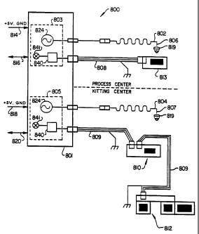
Brief Descrigtion.-of the Drawincs
FIG. 1 is an isometric view of the automated
analytical system illustrating the system cabinetry,
exposed front end carousel, computer screen and
keyboard.
FIG. 2 is an isometric view of the aut.omated
analytical system apparatus frame and cabinet.
FIG. 3 is a top plan view in section of the lower
cabinet of FIGS. 1 and 2 illustrating water and/or
buffer supply station as well as liquid and solid
waster containers of the automated analytical system.
FIG. 4A is a top plan view of the automated
analytical system in section with component covers
removed to show the automated analytical system
apparatus in detail and relative position.
CA 02362531 2001-11-20
WO 95/08774 PCT/US94/10850
-22-
FIG. 4B is a front elevational view of the
automated analytical system in isolation and partial
section of elements of the front end carousel.
FIG. 5 is a- top view in isolation and partial
section of drive and guide elements of the front end
carousel of the automated analytical system being
removed.
FIG. 6 is a cross-sectional side view of a process
carousel of the automated analytical system in
isolation with two reaction vessels in place, one of
which is in position for an FPIA read.
FIG. 7 is an. isometric view of the probe, probe
arm and pipettor of the automated analytical system in
isolation.
FIG. 8 is.a schematic side view of the=probe arm
wiring and sensor means of the automated analytical
system.
FIG. 9A is a sectional side view of the transfer
element of the automated analytical system engaging a
reaction vessel for transfer from the main carousel
into the transfer station.
FIG. 9B is a perspective side elevational view of
the transfer station of the automated analytical
system.
FIG. 10 is a block diagram showing the sequence of
activities to be performed in a first assay.
CA 02362531 2001-11-20
WO 95/08774 PCTIUS94/10850
-23-
FIG. 11 is a block diagram showing the sequence of
activities to be performed in a second assay.
FIG. 12 is a block diagram showing an incubation
period between two activities as comprising a nominal
incubation period and a variable incubation window.
FIG. 13 is a set of six block diagrams each
showing a different combination of activities and
incubation periods reflecting the rules of a flexible
protocol technology.
FIG. 14 is a block diagram showing the timing
protocol for a pipetting activity.
FIG. 15 is... a top plan view of the automated
analytical system in section with component covers
removed to show the automated analytical apparatus in
detail and relative position inclusive of a
chemiluminescent reader for a magnetic particle capture
technology and a chemiluminescent reader for membrane
particle capture technology.
FIG. 16 is a cross sectional view of a detection
head of the detection device for chemiluminescent
detection.
FIG. 17 is a cross sectional view in section of
the detection device light pipe positioned over a
chemiluminescent particle capture container with light
shield in place.
CA 02362531 2001-11-20
WO 95/08774 PCT/US94/10850
-24-
FIG. 18 is a simplified block diagram of the
preferred embodiment of the liquid level sensing device
of the present invention utilized in connection with an
automated analytical system.
FIG. 19 is a more detailed block diagram of the
liquid level sensing system of FIG. 18.
FIG. 20 is a simplified schematic diagram
illustrating the current flow in the fluid level
sensing system of the present invention.
FIG. 21 is an illustration of the geometries
between the probe, its electromagnetic field, a liquid
sample container, and the antenna when the probe is in
ai r.
FIG. 22 is an illustration of the geometries
between the probe, its electromagnetic field, a liquid
sample container, and the antenna when the probe
contacts liquid.
FIG. 23 is an illustration of the geometries
between the probe, its electromagnetic field, a liquid
sample container, and the antenna when the probe has
contacted liquid and the distance from the probe/liquid
combination to the antenna is too great to trigger a
detection.
FIG. 24 is an illustration of a sensing sleeve
which functions to channel the electrical signal from
CA 02362531 2001-11-20
WO 95/08774 PCT/US94/10850
-25-
the probe/liquid combination to the vicinity- of the
receiving antenna.
FIG. 25 is a graphical representation of system
noise level versus signal frequency.
FIG. 26 is a cross-sectional side elevational view
of an automatic bubble flushing syringe of the
automated analytical system.
FIG. 27 is a sectional end view in isolation of
the piston and bore of the automatic bubble flushing
syringe of FIG. 26.
FIG. 28 is a sectional side view in isolation of
the s yri nge bore -end portion of the automatic bubble
flushing syringe with.the reciprocating piston near the
end of travel toward.the bore end portion and a phantom
position within the bore illustrating the piston
withdrawal to the outward extension;
FIGS. 29 and 30 represent a perspective side
elevational view and partial end view of a reagent pack
and reagent pack cover means for use with the automated
analytical system.
FIG. 31 is a top view of a reagent pack having the
reagent containers covered.
FIG. 32 taken along section A-A of FIG. 31
presents a side view in section taken along the line
A-A of FIG. 31 illustrating a cover means in various
open and closed positions.
CA 02362531 2001-11-20
WO 95/08774 PCT/US94/10850
-26-
FIG. 33 is an isometric view of an open reagent
vessel capping means.
FIG. 34 is a perspective side elevational view of
a reagent container lid opening and closing station
with the reagent containers in the reagent pack having
the lids opened.
FIG. 35 presents a different perspective side
elevation view from that of FIG. 34 wherein the reagent
containers of the reagent pack are below elements of
the opening and closing station with the. reagent pack
lids being closed.
FIG. 36 is a. perspective view of a test sample
container segment assembly.
FIG. 37 is a bottom. view of the test sample
container segment assembly of FIG. 36.
FIG. 38 is a cross sectional view in isolation of
the s ampl e carousel with a mounted test s ampl e
container segment assembly also in cross section.
FIG.-39 is a cross sectional view of a modified
sample cup with skirts.
FIG. 40 is a perspective view of a short test
sample Vacutainerm tube segment assembly.
FIG. 41 is a top cross sectional view of the short
test sample Vacutainera tube segment assembly taken
along the line A-A of FIG. 40.
CA 02362531 2001-11-20
WO 95/08774 PCTIUS94/10850
-27-
FIG. 42 is a bottom view of the short test sample
Vacutainere tube segment assembly of FIG. 40.
FIG. 43 is a cross sectional view of a long test
sample cup adaptor with tube in place.
FIG. 44 is a cross sectional view of a short test
sample cup adaptor with tube in place.
FIGS. 45A and 45B represent a top plan view of a
reaction vessel and a side view of the reaction vessel
for use with the automated analytical system,
respectively, with reaction vessel compartments labeled
where appropriate for FPIA processing.
FIG. 45C present an end view of the reaction
vessel of FIG. 45B.
FIG. 46 is an isometric view in section of the
reaction vessel. loading device illustrating the device
holding to vessels and means for mounting other
vessels..
FIG. 47 is a top view of the reaction vessel
loading device presented in an arc which matches the
radius of the reaction vessel carousel, the loading
device having mounted thereon ten reaction vessels.
FIG. 48 is an isomet'ric view in section of the
reaction vessel loading device illustrating the loader
mounted with two reaction vessels and means for
mounting other reaction vessels.
CA 02362531 2001-11-20
WO 95/08774 PCTIUS94/10850
-28-
FIG. 49 is a top view of the reaction vessel
loading device, the reaction vessel loading device
having arced linear dimensions which match the radius
of the reaction vessel carousel, the loader having
mounted thereon two reaction vessels and the capability
of mounting eight additional reaction vessels.
FIG. 50 is a schematic view illustrating the
system control environment airflow and temperature
control of the automated analytical system.
FIG. 51 is an elevational, cross-sectional view of
the process carousel as disposed in the controlled
environmental zone and holding a plurality of reaction
vessels.
FIG. 52 is a perspective view of a heater assembly
for liquid temperature control.
FIG. 53 is a cross-sectional view through the
heater assembly of FIG. 52 showing the heater element
within the block.
FIG. 54 is a partial cross-sectional view of the
heater assembly of FIG. 52 showing liquid tubing, for
example, a tubing coil within the heater assembly.
FIG. 55 is a side elevational view in partial
section of a MEIA cartridge for use with the automated
analytical system.
CA 02362531 2001-11-20
WO 95/08774 PCT/US94/10850
-29-
FIG. 56 is a side elevational view in section of
a MEIA cartridge feeder of the automated analytical
s ys tem.
FIG. 57 is a side sectional view in isolation of
the MEIA cartridge feeder-cartridge orientation pin
mechanism of the cartridge feeder of FIG. 56.
FIG. 58 is a side cross-sectional view in
isolation of a split open cartridge carton shown in
various open positions in phantom as engaged in
cooperation with a cartridge hopper containing multiple
cartridges.
FIG. 59A is an isometric view of the cartridge
carton, taken form the lower side of the cartridge
carton.
FIG. 59B is a partial, isometric view of the
cartridge carton, illustrating the operation of the tab
opening.
FIG. 60 is an isometric view of the cartridge
carton, taken from the upper side of the cartridge
carton.
FIG. 61 is an isometric view of another embodiment
of a free standing cartridge hopper showing the
cartridge hopper in a detached mode suitable for
loading cartridges from a cartridge carton.
FIG. 62 is a schematic of the FPIA optics system
of the automated analytical system.
CA 02362531 2001-11-20
WO 95/08774 PCT/US94/10850
-30-
FIG. 63 is a schematic of the MEIA optics system
of the automated analytical system.
FIG. 64 is a box diagram of the optics control
system of the automated analytical system.
FIG. 65 is a pictorial time graph of the FPIA
reader sequence of the automated analytical system.
FIG. 66 is a pictorial time graph of the MEIA read
sequence of the automated analytical system.
FIG. 67 is a schematic reaction sequenCe of a FPIA
for T4 performed on the automated analytical system.
FIG. 68 is a schematic reaction sequence-of a one-
step sandwich MEIA performed on the automated
analytical system.
FIG. 69 is a schematic reaction sequence of a two-
A 5 step sandwich. MEIA performed on the automated
analytical system.
Detailed Descrintion of the Invention
The following description is divided into separate
sections with headings to more clearly describe the
invention, but should not be considered as limiting the
scope of the invention.
UEFI NI TI ONS
The followi-ng definitions are applicable to the
present invention:
The term "test sample", as used herein, refers to
a material suspected of containing the analyte. The
CA 02362531 2001-11-20
WO 95/08774 PCT/US94/10850
-31-
test sample can be used di rectlv as obtained from the
source or following a pretreatment to modify the
character of the sample. The test sample can be derived
from any biological source, such as a physiological
fluid, including, blood, saliva, ocular lens fluid,
cerebral spinal fluid, sweat, urine, milk, ascites
fluid, raucous, synovial fluid, peritoneal fluid,
amniotic fluid or the like. The test sample can be
pretreated prior to use, such as preparing plasma from
blood, diluting viscous fluids, or the like; methods of
treatment can involve filtration, distillation,
concentration, inactivation of interfering components,
and the addition of reagents. Besides physiological
fluids, other liquid samples can be used such as water,
food products and the like for the performance of
environmental or food production assays. In addition,
a solid material suspected of containing the analyte
can be used as the test sample. In some instances it
may be beneficial to modify a solid test sample to form
a liquid medium or to release the analyte.
The term " analyte" or "analyte of interest", as
used herein, refers to the compound or composition to
be detected or measured and which has at least one
epitope or binding site. The analyte can be any
substance for which there exists a naturally occurring
binding member or for which a binding member can be
CA 02362531 2001-11-20
WO 95/08774 PCTIUS94/10850
-32-
prepared. Analytes include, but are not limited to,
toxins, organic compounds, proteins, peptides,
microorganisms, amino acids, nucleic acids, hormones,
steroids, vitamins, drugs (including those administered
for therapeutic purposes as well as those administered
for illicit purposes), virus particles and metabolites
of or antibodies to any of the above substances. The
term "analyte" also includes any antigenic substances,
haptens, antibodies, macromolecules and combinations
thereof.
The term " analyte-analog", as used herein, refers
to a substance which cross-reacts with an analyte-
specific binding member, although it may do so to a
greater or lesser extent than does the analyte itself.
The analyte-analog can include a modified analyte as
well as a fragmented or synthetic portion of the
analyte molecule, so long as the analyte-analog has at
least one epitopic site in common with the analyte of
interest. An example of an analyte-analog is a
synthetic peptide sequence which duplicates at least
one epitope of the whole-molecule analyte so that the
analyte-analog can bind to an analyte-specific binding
member.
The term "binding member", as used herein, refers
to a member of a binding pair, i. e. , two different
molecules wherein one of the molecules specifically
CA 02362531 2001-11-20
WO 95/08774 PCT/US94/10850
-33-
binds to the second molecule through chemical or
physical means. In addition to antigen and antibody
bi ndi ng pair members, other binding pairs include, as
examples without limitation, biotin and avidin,
carbohydrates and lectins, complementary nucleotide
sequences, complementary peptide sequences, effector
and receptor molecules, enzyme cofactors and enzymes,
enzyme inhibitors and enzymes, a peptide sequence and
an antibody specific for the sequence or the entire
protein, polymeric acids and bases, dyes and protein
binders, peptides and specific protein binders (e. g. ,
ribonuclease, S-peptide and ribonuclease S-protein),
and the like. Furthermore, binding pairs can include
members that are analogs of the original binding
member, for example, an analyte-analog or a binding
member made by recombinant techniques or molecular
engineering. I f the binding member is an immunoreactant
it can be, for example, a monoclonal or polyclonal
antibody, a recombinant protein or recombinant
antibody, a chimeric antibody, a mixture(s) or
fragment(s) of the foregoing, as well as a preparation
of such antibodies, peptides and nucleotides for which
suitability for use as binding members is well known to
those s kill ed in the art.
The term "detectable moiety", as used herein,
refers to any compound or conventional detectable
CA 02362531 2001-11-20
WO 95/08774 PCT/US94/10850
-34-
chemical group having a detectable physical or chemical
property and which can be used to label a binding
member to form a conjugate therewith. Such detectable
chemical group can be, but is not intended to be
limited to, enzymatically active groups such as
enzymes, enzyme substrates, prosthetic groups or
coenzymes; spin labels; fluorescers and fluorogens;
chromophores and chromogens; luminescers such as
chemiluminescers and bioluminescers; specifically
bi ndabl e l i gands such as bi oti n and avi di n;
electroactive species; radioisotopes; toxins; drugs;
haptens; DNA; RNA; polysaccharides; polypeptides;
liposomes; colored particles and colored
microparticles; and the like.
The term ." continuous access", as used herein,
refers to the ability to add additional test samples or
reagents to the automated analytical system of the
present invention without the interruption of assays
which are being performed Yiy the automated analytical
system of the present invention at the time of such
addition.
The term "random access",. as used herein, refers
to the ability of the automated analytical system of
the present invention to simultaneously perform more
than one scheduled assay in any order in which such
--- -------- -
CA 02362531 2001-11-20
WO 95/08774 PCT/US94/10850
-35-
plurality of s chedul ed as s ays are presented into the
automated analytical system of the present invention.
The term "simultaneous", as used herein, refers to
the ability of the automated analytical system of the
present invention to independently perform two or more
scheduled assays at the same time.
The term "kitting", as used herein, refers to the
ability of the automated analytical system of the
present invention to create a unit dose disposable by
separately transferring test samples and.reagents to a
reaction vessel of the present invention without
initiation of an assay reaction sequence.
The term "quat" refers to a polycationic material
solution for assays which use these materials which are
not an antibody.or antigen to capture the analyte from
the sample on the matrix of, for example, MEIA
cartridge. In the present inventive system, quat is
dispensed to the matrix during test processing, prior
to the trarisfer of the reaction mixture from the
reaction vessel.
DETECTION SYSTEMS
The automated analytical system of the present
invention is capable of performing various assays
employing various detection systems known in the art
and include, but are not intended to be limited to,
spectrophotometric absorbance assay such as end-point
CA 02362531 2005-02-28
-36-
reaction analysis and rate of reaction analysis
turbidimetric assays, nephelometric assays, radiative
energy attenuation assays (such as those described in
U.S. Patent No. 4,496,293 and U.S. Patent No. 4,743,561,
ion capture assays, calorimetric assays, fluorometric
assays, electrochemical detection systems, potentiometric
detection systems, amperometric detection systems, and
immunoassays. Immunoassays include, but are not intended
to be limited to, heterogeneous immunoassays such as
competitive immunoassays, sandwich immunoassays,
immunometric immunoassays, and the like, where the amount
of a detectable moiety employed therein can be measured
and correlated to the amount of analyte present in a test
sample.
Generally, in a spectrophotometric assay, such as
those performed on the Abbott Spectrum clinical analyzer
and the Abbott Spectrum Series II clinical analyzer
(Abbott Laboratories, Abbott Park, IL, USA) the
interaction in an assay solution between the analyte to
be determined and a reagent system specific for the
analyte produces a detectable change in the transmittive
properties of the assay solution. The change in the
transmittive properties. refers to the amount of light
absorbed or' scattbred by an assay solution within a
particular wavelength band when a
* trade-maXk
CA 02362531 2001-11-20
WO 95/08774 PCT/US94110850
-37-
beam of light of known intensity is passed through the
assay solution. The change in the transmittive
properties of an assay solution is measured by passing
monochromic light having a known intensity though the
assay solution and determining the ratio of the
intensity of the transmitted or scattered light to the
intensity of the incident light. Nearly all analytes
either absorb energy of a specific wavelength or
interact in an assay solution with a particular reagent
system to produce a detectable change in the
transmittive properties of the assay solution,
characteristics which have resulted in the development
of numerous specific spectrophotometric assays.
Spectrophotometric assays which rely upon the
measurement of the change in the transmittive
properties of an assay solution as a measure of an
analyte in the assay solution include, for example,
assays wherein there is a change in the color of the
assay when there is a change in the turbidity of the
assay solution, that is, turbidimetric or nephelometric
assays.
In a calorimetric assay, the change in the
transmittive properties of an assay solution is
generally referred to as the absorbance of the assay
solution and is dependent upon the change in the color
of the assay solution due to the interaction of the
CA 02362531 2001-11-20
WO 95/08774 PCT/US94/10850
-38-
analyte to be determined and reagent system specific
for the analyte. The absorbance of the assay solution
is related to the concentration of the analyte in the
assay solution. A calorimetric assay utilizes a
chromogenic reagent system capable of interacting in an
assay solution with the particular analyte of interest,
to produce a detectable change in the transmittive
properties, specifically the color, of the assay
solution. Numerous chromogenic reagent systems useful
in the determination of specific analytes have been
developed and are commercially available.
The principle of turbidimetric assays is to
determine the amount of light scattered or blocked by
particulate matter as light passes though an assay
solution. In a turbidimetric assay, the analyte of
interest interacts with a reagent system specific for
the analyte to form a suspension of particles in the
assay solution. As a beam of light having a known
intensity is passed through an assay solution, the
suspension of particles formed by the interaction of
the analyte reagent system blocks or scatters the
incident light, thereby reducing the intensity of the
light transmitted through the assay solution. The
change of the transmittive properties in a
turbidimetric assay refers to the decrease in the
intensity of the light transmitted through an assay
CA 02362531 2001-11-20
WO 95/08774 PCT/US94/10850
-39-
solution, is related to the amount of incident light
that is scattered or blocked by the suspension of
particles, and depends upon the number of particles
present and the cross-sectional area of such particles.
A nephelometric assay is similar to a
turbidimetric assay in that the analyte of interest
interacts with a reagent system specific for the ligand
to form a suspension of particles in the assay
solution. In a nephelometric assay, the change in the
transmittive properties of the assay solution is also
related to the amount of incident light scattered or
blocked by the suspension of particles, but unlike a
turbidimetric assay wherein the intensity of the light
transmitted through the assay solution is measured, the
scattered or =blocked light is measured at an angle to
the light incident to the assay solution. Therefore,' in
a nephelometric assay the change in the transmittive
properties refers to the difference in intensities of
light incident to the assay solution and light
scattered at an angle to the incident light.
Turbidimetric and nephelometric assays are utilized in
the analysis of blood, urine, spinal fluid, and the
like, for the determination of analytes such as
proteins wherein there is no comparable calorimetric
assay due to the lack of an effective chromogenic
reagent system. Yoe and Klimman, Photoelectric Chemical
CA 02362531 2001-11-20
-40-
Analysis. Vol. II: Nephelometry, Wiley & Sons, Inc., New
York, 1929, describe various nephelometric assays.
various reagents and reagent systems which can be
employed for performing spectrophotometric assays on the
automated analytical systems of the present invention
include, but are not intended to be limited to, those for
the simultaneous determination of glucose and urea, such
as described in U.S. Patent No. 5,037,738. The
simultaneous determination of calcium and phosphorous;
the simultaneous determination of cholesterol and
triglycerides; determining isoenzymes; determining blood
ammonia levels, and the like, can be performed on the
apparatus and by the methods of the present invention.
Typically in a fluorometric assay, an analyte in an
assay solution is chemically or immunologically
transformed into a fluorescent complex or conjugate
thereby producing a detectable change in the fluorescent
properties of the assay solution. The change in the
fluorescent properties of the assay solution is measured
by exciting the fluorescent complex or conjugate
properties produced with monochromatic light of a
wavelength within the excitation wavelength band of the
fluorescer, and measuring the intensity of the emitted
light at a
CA 02362531 2001-11-20
WO 95/08774 PCT/1JS94/10850
-41-
wavelength within the emission wavelength band of the
fluorescer. The fluorescent intensity of the emitted
light is related to the concentration of the analyte.
However, the intensity of the fluorescence emitted by
the assay solution may be inhibited when the ligand to
be determined complexes with nonfluorescent
interferences such as protein or phosphates present in
the sample, or when the sample containing the ligand to
be determined has sufficient color so as to act as a
filter and thereby reduce the intensity of the emitted
fluorescence. It is well recognized that in order to
maximize the sensitivity and specificity of a
fluorometric assay, these inhibiting factors, if
present, must be overcome either by removal of the
nonfluorescent interferences or color producing
material prior to the analysis, or by compensating for
the presence of such factors using an internal standard
added to a second aliquot of sample and carrying out
the entire assay procedure using the aliquot containing
the internal standard.
ASSAY FORMATS
Generally, homogeneous and heterogeneous
immunoassays depend upon the ability of a first binding
member of a binding member pair to specifically bind to
a second binding member of a binding member pair
wherein a conjugate, comprising one of such binding
CA 02362531 2001-11-20
WO 95/08774 PCTIUS94/10850
-42-
members labeled with a detectable moiety, is employed
to determine the extent of such binding. For example,
where such binding pair members are an analyte and an
antibody to such analyte, the extent of binding is
determined by the amount of the detectable moiety
present in the conjugate, which either has or has not
participated in a binding reaction with the analyte,
wherein the amount of the detectable moiety detected
and measured can be correlated to the amount of analyte
present in the test sample.
Homogeneous immunoassays typically are performed
in a competitive immunoassay format involving a
competition between an analyte from a test sample and
a tracer for a limited number of receptor binding 'sites
on an antibody to the analyte. The tracer comprises the
analyte or analog thereof labeled with a detectable
moiety wherein the concentration of analyte in the test
sample determines the amount of the tracer that will
specifically bind to the antibody. The amount of the
tracer-antibody conjugate produced by such binding may
be quantitatively measured and is inversely
proportional to the amount of analyte present in the
test sample. For example, fluorescent polarization
techniques for making such determination, such as in
fluorescent polarization immunoassays as described
herein, are based on the principle that a fluorescently
CA 02362531 2001-11-20
-43-
labeled compound when excited by linearly polarized light
will emit fluorescence having a degree of polarization
inversely related to its rate of rotation. When a
molecule such as a tracer-antibody conjugate having a
fluorescent label is excited with a linearly polarized
fluorescent molecule it is constrained from rotating
between the time light is absorbed and emitted. When a
"free" tracer molecule (i.e., unbound to an antibody) is
excited by linearly polarized light, its rotation is much
faster than the corresponding tracer-antibody conjugate
and the molecules are more randomly orientated,
therefore, the emitted light is polarized. Accordingly,
when plane polarized light is passed through a solution
containing the aforementioned reagents, a fluorescent
polarization response is detected and correlated to the
amount of analyte present in the test sample.
Various fluorescent compounds which can be employed
for performing fluorescent polarization assays on the
automated analytical system of the present invention
include, but are not intended to be limited to,
aminofluoresceins, such as described in U.S. Patent No.
4,510,251 and U.S. Patent No. 4,614,823; triazinylamino-
fluoresceins, such as described in U.S. Patent No.
4,420,568 and U. S. Patent No. 4,593,089;
CA 02362531 2001-11-20
-44-
carboxyfluoresceins, such as described in U.S. Patent No.
4,668,640, and the like.
Heterogenous immunoassays typically involve a
labeled reagent or tracer comprising an analyte, an
analog of the analyte, or an antibody thereto, labeled
with a detectable moiety, to form a free species and a
bound species. In order to correlate the amount of tracer
in one of such species to the amount of-analyte present
in the test sample, the free species must first be
separated from the bound species, which can be
accomplished according to methods known in the art
employing solid phase materials for the direct
immobilization of one of the binding participants in the
binding reaction, such as the antibody, analyte or analog
of the analyte, wherein one of the binding participants
is immobilized on a solid phase material, such as a test
tube, beads, particles, microparticles or the matrix of a
fibrous material, and the like, according to methods
known in the art.
Heterogenous immunoassays can be performed in a
competitive immunoassay format as described above
wherein, for example, the antibody can be immobilized to
a solid phase material whereby upon separation, the
amount of the tracer which is bound to such solid phase
material can be detected and correlated to the amount
CA 02362531 2001-11-20
WO 95/08774 PCTIUS94/10850
-45-
of analyte present in the test s ampl e. Another form of
a heterogeneous immunoassay employing a solid phase
material is referred to as a sandwich immunoassay,
which involves contacting a test sample containing, for
example, an antigen with a protein such as an antibody
or another substance capable of binding the antigen,
and which is immobilized on a solid phase material. The
solid phase material typically is treated with a second
antigen or antibody which has been labeled with a
detectable moiety. The second antigen or antibody then
becomes bound to the corresponding antigen or antibody
on the solid phase-material and, following one or more
washing steps to remove any unbound material, an
indicator material such as a chromogenic substance
which reacts with the detectable moiety (e.g., where
the detectable moiety is an enzyme;- a substrate for
such enzyme is added) to produce a color change. The
color change is then detected and correlated to the
amount of antigen or antibody present in the test
sample.
For example, a heterogeneous immunoas s ay which can
be performed by the automated analytical system of the
present invention, in either a competitive or sandwich
immunoassay format, is a microparticle capture enzyme
immunoassay, such as that described in Clinical
CA 02362531 2001-11-20
WO 95/08774 PCT/US94/10850
-46-
Chemistrv, Volume 34, No. 9, pages 1726-1732 (1988),
employing microparticles as the solid phase material.
In addition, the use of sucrose in microparticle
diluent has been found to achieve neutral density of
the microparticles. The methodology entails the
determination of the optimum sucrose concentration
which will eliminate the settling of microparticles.
The sucrose concentration required to achieve neutral
density is assay specific and microparticle lot
specific. The principal involves dissolving sucrose in
solution to increase the density of the diluent. When
the density of the diluent and microparticles are
equivalent, the microparticles will be in a suspended
state. Density neutralization can also be achieved by
using other materials such as metrizamide and/or
metrizoic acid.
Separation of the bound and free species is
accomplished by capture of the microparticles on a
glass fiber matrix of a simple cartridge (herein, the
"MEIA cartridge"), a process that relies on the high
affinity of glass fibers for the microparticles,
wherein the microparticles adhere to the surface of the
fibers irreversibly, and nonspecifically bound material
can be effectively removed by washing the matrix. The
matrix also provides a precisely located mechanical
support for the microparticles during the optical
CA 02362531 2001-11-20
WO 95/08774 PCT/US94/10850
-47-
quantification phase of the assay protocol as described
herein.
When performing a sandwich immunoassay,
microparticles coated with antibody to the analyte in
the test sample are incubated with the test sample
containing the analyte of interest to form a capture
complex with the analyte from the test sample. A
conjugate comprising antibody to the analyte labeled
with a detectable moiety, preferably an enzyme,- is then
incubated with the capture complex to form the second
of a sandwich complex. When performing a competitive
immunoassay, microparticles coated with antibody to the
analyte in,the test sample are incubated with the test
sample containing the analyte of interest and a
conjugate comprising the analyte or analog thereof
labeled with a detectable moiety, preferably an enzyme.
Removal of unbound conjugate is accomplished with the
glass fiber matrix of the MEIA cartridge and, where the
detectable moiety is an enzyme, a substrate for the
enzyme capable of providing a detectable signal is
added and the signal provided thereby is measured and.
correlated to the amount of analyte present in the test
sample. Preferably, the enzyme-substrate system
employed by the competitive and sandwich MEIA formats
is alkaline phosphatase and 4-methylumbelliferyl
CA 02362531 2001-11-20
WO 95/08774 PCTIUS94/10850
-48-
phosphate (MUP), although other enzyme-substrate
systems known in the art can be employed as well.
The MEIA cartridge which is employed by the
automated analytical system ofthe present invention
comprises a reaction well for retaining and
immobilizing microparticle-analyte complexes. The
reaction well has an entrance port and means for
holding a quantity of sample and assay reaction
mixtures positioned over a fibrous matrix which retains
and immobilizes microparticle-analyte complexes as
described above. The fibrous matrix is composed of
fibers having an average spatial separation greater
than the average diameter of the microparticles.
Preferably, the average fiber spatial separation is
greater than 10 microns.
The reaction well further comprises an absorbent
material positioned below the fibrous matrix to enhance
the flow of sample and assay reaction mixtures through
the fibrous matrix. Preferably, the absorbent material
is a fibrous material whose fibers predominantly lie in
a plane perpendicular to the lower surface of the
fibrous matrix. The absorbent material is in fluid
communication with the fibrous matrix. Generally, the
absorbent material is in physical contact with the
lower surface of the fibrous matrix. The interior of
the reaction well, therefore, is generally sized or
CA 02362531 2001-11-20
WO 95/08774 PCTIUS94/10850
-49-
contains positioning means to maintain the fluid
communication between the absorbent material and the
fibrous matrix. Preferably, a spike located at the
bottom of the reaction well can be used to force the
absorbent material into contact with the lower surface
of the fibrous matrix. Additionally, it is preferable
to vent to the atmosphere the gases displaced in the
absorbent material by the liquids absorbed therein
during the performance of an immunoassay. -
According to the immunoassay methodologies
described above, standard solutions of the analyte of
known concentrations covering the clinical
concentration range are typically prepared and assayed
as is the test sample to be assayed. This blank assay
provides a series of signal measurements corresponding
to the known concentrations from which a standard curve
is drawn. The optical signal corresponding to the
unknown sample is correlated in a concentration value
through interpretation from the blank or standard
curve.
ANALYTICAL SYSTEM METHOD
Automated analytical methodology for effecting
analysis of a plurality of test samples according to
the present invention is achieved by introducing
reagent packs, test sample container and reaction
vessels onto concentric carousels of a main carousel.
CA 02362531 2001-11-20
-50 -
The test sample container can be a test tube, cuvette,
Vacutainer* tube, and the like, for holding a test
sample. The reagent packs and test sample containers are
identified and aligned respectively with a reaction
vessel for transfer and kitting of the reaction vessel by
transfer of test sample and specific reagents from the
reagent pack for preparation of a predetermined test. The
reaction vessel containing the test sample and one or
more reagents is transferred to a process carousel
wherein controlled environment conditions exist for
incubation once the sample has been appropriately mixed
with various reagents to form a reaction mixture. When
all assay processing steps have been completed, the
reaction mixture is identified and transferred to at
least, for example, one of a fluorescent polarization
immunoassay reader or a microparticle enzyme immunoassay
cartridge positioned on a separate cartridge wheel or
carousel for further preparation before reading. The
processed test samples are read and the readings are
calculated with the resulting data being recorded and/or
printed.
The methodology of the automated immunoassay
analytical system is achieved through the use of a self-
contained, fully automated, continuous and random access
instrument comprising a main carousel assembly consisting
of the reagent pack carousel, a reaction
* Trade-mark
CA 02362531 2001-11-20
WO 95/08774 PCTIUS94/10850
-51-
vessel carousel and a test sample container carousel
concentrically and independently rotatable. The main
carousel assembly is provided with a transfer pipette
operated by a boom arm for transferring and kitting
test sample and reagents into the reaction vessel
automatically following a predetermined test schedule.
The main carousel assembly is provided with bar code
readers for reagent packs and test sample containers
and has the capability of aligning the reagent pack
carousel and test sample container carousel and a
reaction vessel for pipette transfer operations. Once
the assay to be performed is scheduled, the reaction
vessel carousel, the reagent pack carousel and the test
sample container carousel are rotated until the
reaction vessel, a reagent pack and a test sample
container, respectively, are determined to be in the
transfer pipette access position. The transfer pipette
then transfers the test sample from the test sample
container and, depending upon the assay to be
performed, the reagents from the reagent pack are
transferred to the reaction vessel. The reaction vessel
carousel is then rotated to a transfer station position
which contacts the reaction vessel with a transfer
mechanism and pulls the reaction vessel into the
transfer station. The reaction vessel is then loaded
onto the process carousel by the transfer mechanism.
CA 02362531 2001-11-20
WO 95/08774 PCT/US94/10850
-52-
When performing a fluorescent polarization
immunoassay (FPIA) with the automated analytical system
of the present invention as described in more detail
.below, various pipetting activities are performed by a
second transfer pipette apparatus which is in service
for the process carousel, and the process carousel is
rotated so that the reaction vessel, when properly
pipetted with, for example, FPIA reagents, is at the
read station of the FPIA processing stations* and the
FPIA determination on reading, is made on the reaction
ves s el . The process carousel is then rotated so that
the read reaction- vessel is at the transfer station.
The reaction vessel is again contacted and transferred
by the transfer station. The transfer station is
rotated and pushes the reaction vessel into a release
container opening.
For a microparticle enzyme immunoassay (MEIA)
performed with the automated analytical system of the
present invention as described in more detail below,
after the various pipetting activities for the MEIA,
which can be completed at the main carousel assembly,
the reaction vessel is transferred to the process
carousel as described in the FPIA process. Pipetting
can also be accomplished in the process carousel or
jointly between the two carousels. To complete the
MEIA, the reaction mixture is transferred from the
CA 02362531 2001-11-20
WO 95/08774 PCT/US94/10850
-53-
reaction vessel to a matrix of an MEIA cartridge on a
cartridge carousel with the second transfer pipette.
The matrix is washed with a buffer and a substrate,
such as MUP (defined earlier), or other suitable
substrate known in the art. The cartridge carousel is
then rotated so that the MEIA cartridge is positioned
at an MEIA processing assembly and the MEIA
determination is made. The MEIA reaction vessel is
ejected into "the waste container as described- for the
FPIA reaction vessel. The MEIA cartridge is
independently ejected from the cartridge wheel by an
ejector at an appropriate ejector station into a waste
container.
Preferably, two distinct analytical technologies
as described above, FPIA and MEIA, are incorporated
into the automated analytical system of the present
invention; however, more than two distinct analytical
technologies can be incorporated into the inventive
system. These methods are complimentary and share a
commonality of apparatus and procedural steps, with the
FPIA generally being the method of choice for analytes
of low molecular weight and MEIA for molecules such as
protein hormones, antibodies or analytes of low
molecular weight requiring higher sensitivity. The two
technologies share system components including the
operator control panel, pipetting boom assemblies,
CA 02362531 2001-11-20
-54-
fluidics systems, air and liquid reagent heaters,
printers, bar code reader and stepper motors. Such
commonality of use of system components allows for a
compact instrument despite the dual FPIA and MEIA
capability.
The FPIA optic systems (such as described in U.S.
Patent No. 4,269,511) can utilize a polarizing filter
which is an electrically switched liquid crystal,
maintaining a compact size and avoiding complex and
potentially unreliable moving parts. When performing FPIA
assays utilizing the automated analytical system of the
present invention, the FPIA reagent packs will typically
include a tracer comprising the analyte or analog
thereof, coupled to a detectable moiety, an antibody
specific to that analyte, and a specimen pretreatment
reagent. In a preferred FPIA format, the analyte being
determined competes with the tracer for a limited number
of binding sites on the antibodies specific to the
portion or portions of the analyte and tracer. The
detectable moiety component of the tracer is preferably a
fluorescent moiety selected from the group consisting of
fluoresceins, aminofluoresceins, carboxyfluoresceins,
fluoresceinamines, and the like, more preferably
carboxymethyl-aminomethyl-fluorescein, carboxyethylamino-
methyl-carboxyfluorescein, 6-
CA 02362531 2001-11-20
WO 95/08774 PCTIUS94/10850
-55-
carboxyfluorescein, 5-carboxyfluorescein,
succinylanimomethyl-fluorescein, thiourea-
aminofluorescein, methoxytrianolylaminofluorescein,
aminofluorescein, and the like.
In another embodiment, the FPIA format utilizes a
unique, round, plastic, reaction cuvette suitable for
fluorescence polarization and absorbance assay
technologies which require no orientation other than
top-to-bottom. This plastic reaction cuvette has
physical characteristics of low birefringence
throughout the optical read region as well as stringent
dimensional tolerances which allow reproducible
absorbance readings. Bifringence is defined as the
degree of retardation of the extraordinary ray as it
passes through a material. The greater the degree of
retardation, the greater will be the level of
birefringence. Retardation of the extra-ordinary ray is
dependent on the magnitude and direction of the induced
stress. Therefore, passing a ray of linearly polarized
light through a material with induced stress will
result in depolarization of the ray. In order for a
cuvette to be utilized for fluorescence polarization
measurements, it is important that the cuvette be
prepared under conditions which yield minimum stress.
The geometry of the cuvette has been designed to
utilize the inherent fluidics of automated medical
CA 02362531 2001-11-20
WO 95/08774 PCT/US94/10850
-56-
diagnostic instrumentation to minimize the hydrophobic
effect of plastic.
MEIA results can be determined by quantifying the
rate of fluorescence developed when fluorogenic
substrate is converted by the action of an enzyme
labeled conjugate. For example, when performing either
a competitive MEIA or sandwich MEIA, the specifically
bound alkaline phosphatase on the microparticles is
detected by addition of the fluorogenic substrate MUP
to the matrix. The alkaline phosphatase catalyzes
hydrolysis of the MUP to inorganic phosphate and
fluorescent 4-methylumbelliferone (4-MU). The liberated
4-mu is detected by the MEIA optics assembly front
surface fluorometer which is designed to detect
fluorescence of low concentrations of 4-MU without
interference by fluorescence of 4-MUP at a wavelength
of 367. A system of lenses and optical filters focus
filtered light (wavelength = 365) from a mercury arc
lamp on to the surface of the matrix and focus emitted
fluorescence from 4-MU (wavelength = 448) on to a photo
multiplier tube. Like the FPIA optics assembly, the
MEIA optics system is compact and has no moving parts.
About five percent of the excitation light is detected
by a photodiode, allowing normalization of the
fluorescence data and generation of a control signal
used by the lamp power supply to maintain the intensity
CA 02362531 2001-11-20
WO 95/08774 PCTIUS94/10850
-57-
of the excitation light within five percent over the
useful life of the lamp. The MEIA post-processor uses
linear regression analysis to convert the data from
multiple successive determinations of 4-MU fluorescence
to a rate which is proportional to the concentration of
alkaline phosphatase conjugate specifically bound to
the microparticles.
MEIA formats can be run with a multi-position MEIA
auxiliary carousel and process carousel as well as a
MEIA reagent pack containing microparticle reagent, an
alkaline phosphatase conjugate and, in some cases, a
dilute buffer specific for the assay being performed.
Because the microparticles tend not to settle out of
suspension during the course of the assay, they can
readily be pipetted. The effective surface area of
polystyrene 1=atex microparticles is several fold
greater than that of a large diameter polystyrene bead
(e.g., one quarter inch beads) commonly used in
commercial immunoassays. Because of this large surface
area and the very small diffusion distance between
analyte and the capture molecules on the surface of the
microparticles, the capture phase employed in many of
the MEIA methods being performed reaches equilibrium
within several minutes, allowing for a full carousel of
test samples to be completed in a very short time
frame.
CA 02362531 2001-11-20
WO 95/08774 PCTIUS94/10850
-58-
Unlike an FPIA, the heterogeneous immunoassays,
such as a MEIA, require a separation step as described
above. In particular, after incubation of the
microparticles with a test sample, the microparticles
are separated from the reaction mixture by transfer to
the matrix contained in the MEIA cartridge as described
above. The matrix provides a precisely located
mechanical support for the microparticles during the
subsequent optical read phase-of the assay. This
precisely located mechanical support, i.e. the
cartridge, is fit into the auxiliary carousel at a
predetermined spacing from the reader apparatus by
camming means.
ANALYTICAL SYSTEM APPARATUS
The automated immunoassay analytical system
according to the present invention (hereinafter the
"analytical system" or "system") is both continuous and
random access. The following description of the
analytical system includes a general description of
sufficient scope for those skilled in the relevant
arts,. followed by more detailed descriptions of
critical components and subsystems unique to the
system. The drawings do not illustrate all of the
mechanical and electrical elements for driving and
controlling the various components of the system,
because the structure and operation of such omitted
CA 02362531 2001-11-20
WO 95/08774 PCTIUS94/10850
-59-
elements are known to those of ordinary s ki l l in the
art who having knowledge of the information provided
herein would understand the operation of the system and
the various components and related processes utilized
for treating samples and determining analytical
results.
Referring to the drawings, FIGS. 1 and 2 present
isometric views of the apparatus for the automatic
immunoassay analytical system of the present irivention.
The system apparatus as it appears in FIG. 1 presents
the system apparatus as used by the technician, with
FIG. 2 illustratirig an isometric view of the frame and
cabinetry with component parts removed. The system
apparatus of the present invention is identified
generally as 2 in FIG. 1. The system apparatus 2 has
an exposed front end carousel 4 which is serviced by a
first transfer pipette mechanism 6 for kitting
scheduled tests along with samples into a reaction
vessel. The system provides a computer screen 8 and
computer keyboard 10 along with access panels 12 for
accessing storage and waste compartments. The system
apparatus 2 is provided with rollers 14 for movement of
the system apparatus within a laboratory complex as
required. The freedom of movement of the system
apparatus through rollers 14 is allowed since the
CA 02362531 2001-11-20
WO 95/08774 PCT/US94/10850
-60-
system is fully self-contained but for power
requi rements .
Ref erri ng to FIG. 2, the s ys tem apparatus 2
cabinet frame 16 is illustrated with substantially all
functioning components of the system apparatus removed.
A controlled environment zone 18 is a closed unit
during operation with light shielding and rigid control
of airflow as well as temperature as opposed to the
open front end carousel 4. The front end carousel 4
communicates with the controlled environment zone 18
through a transfer port 20. The front end carousel 4
is mounted to an aluminum base plate which rests on a
support platform 22 and the first transfer pipette
mechanism is mounted on means 24.
Referring to FIG. 3, the top plan view of the
system apparatus 2 shows a portion of the cabinet frame
16 and the front end carousel 4 in partial phantom.
This portion of the cabinet 16 also supports a power
supply 192, a supply bottle 196, a solid waste
container 198, and a liquid waste container_200. The
supply bottle 196 provides buffers for the tests being
performed, while the containers 198 and 200 provide
storage for the processed waste material.
Referring to FIGS. 4A and 4B, components of the
s ys tem apparatus are shown in more detail with rel ati ve
positioning to further illustrate the process flow of
CA 02362531 2001-11-20
WO 95/08774 PCT/US94/10850
-61-
the system apparatus. For example, sample cups 26 are
mounted on a sample cup carousel 28 which is
concentrically fitted within the front end carousel 4
along with reagent pack carousel 32 and reaction vessel
carousel 36. The reagent pack carousel 32 is
concentrically fitted between the sample cup carousel
28 and the reaction vessel carousel 36. The reagent
pack carousel carries reagent packs 30 and the reaction
vessel carousel 36 carries reaction vessels 34. The
front end carousel 4 inclusive of the three front end
carousels, the sample cup carousel 28, reagent pack
carousel 32 and.-reaction vessel carousel 36 can by
example contain the following capacities. The sample
cup carousel 28 can hold 60 blood collection tubes,
such as Vacutainer blood collection tubes, or 90
sample cups which are injection molded as one piece and
can be provided with standalone base mounts.
Standalone base mounts are suitable for technician
handling and pipetting of s ampl es into the s ampl e cups.
The reagent pack carousel 32~provides for 20 different
reagent packs 30. The reaction vessel carousel 36
provides -90 reaction vessels 34.
The front end carousel 4 has an operable bar code
reader 38 for automatically identifying reagent pack
carousel 32 and sample carousel 28. A wash cup 40 is
provided for the first transfer pipette mechanism 6 for
CA 02362531 2001-11-20
WO 95/08774 PCT/US94/10850
-62-
washing as required between transfer of various sample
and reagents. The first transfer pipette mechanism 6
is utilized in kitting the various reagent pack liquid
materials and sample into a reaction vessel 34.- The
reagents and the sample are properly kitted through
means of the first transfer pipette mechanism 6
inclusive of pump means. The various carousels are
rotated and aligned for kitting at the pipetting
station. The kitted reaction vessel 34 is positioned
by reaction vessel carousel 36 into the proper position
for transfer to the transfer station 42. The reaction
vessel 34 is transferred to the transfer station 42
through transfer means described below in more detail
below ( FI G. 9) wherein the trans f er station 42 is then
rotated to move the reaction vessel onto process
carousel 46.
As shown, the process carousel 46 is driven by a
stepper motor 48 and is serviced by a second transfer
pipette mechanism 50. The process carousel 46
supported by three wheels for height control and
control of any radial movement caused by irregularly
shaped carousel elements. Both the FPIA and MEIA
procedures utilize the system apparatus commonly up
through and including the process carousel 46. The
process carousel 46 includes FPIA processing 52 and
FPIA processing lamp 54 for direct reading of FPIA
CA 02362531 2001-11-20
WO 95/08774 PCT/US94/10850
-63-
analysis of kitted, pipetted and properly reacted
reagents sample from the reaction vessel 34. The
process carousel 46 holds, for example, 36 reaction
vessels 34 and has a carousel diameter of about 12.5
inches. The process carousel 46 moves the reaction
vessel 34 between the transfer station 42, the second
transfer pipettor mechanism 50, the point of pipetting,
and the FPIA reader processing 52. The controlled
environment zone 18, which includes the -transfer
station 42 and process carousel 46, provides FPIA
processing with air circulation under temperature
control by cabinet air circulation fan 56.. A wash cup
58 for the second transfer pipette mechanism 50 is
provided. The second transfer pipette 50 is utilized
for adding reagents (pipetting) under conditions of
incubation and timing to the sample in the FPIA test
schedule reaction vessel 34 for FPIA processing.
MEIA processing can also utilize the second
transfer pipette 50 for adding reagents to the sample
before the reaction mix is added to MEIA cartridges 68
which are mounted on the auxiliary carousel 64, also
referred to as the cartridge wheel carousel. The MEIA
reagent mixed sample is transferred to the MEIA
cartridge 68 by the second transfer pipette 50. The
second transfer pipette 50 moves the pipette probe
between the wells in the reaction vessel 34 on the
CA 02362531 2001-11-20
WO 95/08774 PCTIUS94/10850
-64-
process carousel 46 to the MEIA cartridge 68 on the
auxiliary carousel 64 and to the wash cup 58. A rack-
and-pinion drive through two axis stepper motor drives
achieves precision drive on both the R and Z axis.
Travel, for example, on the Z axis can be about 3
inches and on the R axis about 4. 5 to 5.0 inches.
The auxiliary carousel 64 holds, for example, 32
MEIA cartridges 68 and has a diameter of about 9.5
inches. The auxiliary carousel 64 moves the MEIA
cartridges 68 between various stations including the
second transfer pipettor mechanism pipette point, the
MUP dispense station 72, the MEIA washstation 70 and
the MEIA reader 74 and the MEIA cartridge ejection
point 62. The auxiliary carousel 64 is stepper motor
driven and is carried by three wheels with one wheel
located at the Z axis height control at the cartridge
insertion point, the second wheel at the pipette point,
and the third wheel at the MEIA reader in order to
maintain the auxiliary carousel 64 within desired
geometric relationships to these various functions.
MEIA cartridges 68 are loaded into a cartridge
hopper 590 which feeds'the MEIA cartridges 68 into the
auxiliary carousel 64. The automatic feeding of the
MEIA cartridges 68 is provided with a proper height
adjustment of the cartridge- 68 into the auxiliary
carousel 64 as required by MEIA reading. The cartridge
CA 02362531 2001-11-20
WO 95/08774 PCTIUS94/10850
-65-
hopper 590 feeds individual cartridges 68 to the
auxiliary carousel 64 and changes the axis of
orientation of the cartridge 68 from horizontal to
vertical by automatic means. Removal of the MEIA
cartridges 68 is achieved through the use of an ejector
62 which operates through an ejection rod and forces
the MEIA cartridge 68 from the auxiliary carousel 64
which is dropped into the solid waste container 200
(FIG. 3). The auxiliary carousel 64 is - further
serviced by a MEIA buffer heater and dispenser 70, MUP
heater and dispenser probe 72, and MEIA reader 74. The
MEIA cartridges .68 are removed from the auxiliary
carousel 64 by a cartridge ejector 62 after the MEIA
read has been completed.
FIG. 5 provides a top view in isolation and
partial section of elements of the drive and guide
systems of the main .carousel 4 with the various
c arous el s removed. In FIG. 5 a s ampl e cup c arous el
stepper motor 76 is shown mounted with mounting spring
78. The reagent pack carousel motor 80 is also shown
with a mounting spring 82. The reaction vessel
carousel motor 84 and mounting spring 86 are positioned
to the exterior of the two inner carousels, i.e. the
sample cups carousel 28 and the reagent pack carousel
32. Roller guides 88 are provided for the sample cup
carousel 28 and a tensioning spring 90. The reagent
CA 02362531 2001-11-20
WO 95/08774 PCT/US94/10850
-66-
pack carousel is provided with roller guides 92 and
tensioning means 94. The reaction vessel roller guides
96 are also provided with spring elements 98, the
purposes of the guide and these various spring elements
being to maintain very finite tracking of the
concentric carousels when motivated by the individual
stepper motors.
Motion control for the system 2 is performed by 22
stepper motors of various sizes, some of which are
identified herein. The specifications and operation of
the stepper motors are described generally as follows,
such description being sufficient for one skilled in
the art. All the stepper motors are permanent magnet
motors with 200 full steps per shaft revolution which
is equivalent to 1.8 degrees revolution per step. A
single stepping motor control system comprises the
f ol l owi ng:
(1) A step motor connected to a mechanism to
move the mechanism as required.
(2) A driver which applies voltages to the step
motor causing i,t to move in response to 3
control signals from control electronics,
i. e. , an "Indexer".
(3) An Indexer which comprises electronics for
controlling the motor by the driver. It
determines move profiles, which include
CA 02362531 2001-11-20
WO 95/08774 PCT/US94/10850
-67-
direction of rotation, number of steps to
move and acceleration and velocity
parameters.
(4) A home sensor is used for each step motor.
The home sensor is used as a position
reference by the Indexer and can also be
used by the Indexer to check for errors.
(5) Encoders are used by the rotary devices, the
carousels and transfer mechanism to verify
correct movement. At the end of a move, the
encoder count is checked to validate that
the motor moved to the correct position.
The system microprocessor (CPU) is used to determine
the distance, velocity and acceleration of a motor
movement of the steppers. It transfers the information
to the Indexer which then controls the movement. At
the end of the movement, the Indexer then sign4ls the
system microprocessor (CPU) that the movement is
complete. The system microprocessor (CPU) then checks
the encoders to validate the movement if a rotary
mechanism was being moved and checks the Indexer to
verify it had detected no errors.
There are three indexer boards in each system 2.
Each board is identical and can control up to eight
stepper motors. Each board utilizes one slave
microprocessor to provide the eight indexer functions
CA 02362531 2001-11-20
WO 95/08774 PCTIUS94/10850
- -68-
on each board. Such a combination of functions is
referred to as an " 8-axis" indexer. Two of the indexer
axes are not used. The indexer boards communicate to
the system microprocessor (CPU) over a backplane VME
bus. The Indexers have addresses that are modified by
jumpers before installation into the system. This is
the mechanism that allows otherwise identical boards to
reside in the same system backplane VME bus. Each
board is connected via the VME backplane bus to one
cable per board that carries the indexer signals to the
drivers. The In.dexer provides a variety of movement
profiles. Many of the step motor movements require
that the speed of the motor be increased in a
controlled fashion until the final velocity is reached.
At some point in the movement, the speed must then be
decreased in a controlled fashion. This process is
called a "velocity profile" and can be done linearly,
sinusoidally, or parabolically. The type of velocity
profile executed is determined by the system
microprocessor (CPU). The Indexers are available from
vendors as "off the shelf" 8-axis indexers.
There are three PC boards used to provide the 22
separate motor drive circuits. Two of the boards are
identical and referred to as the "Stepper Drive"
boards. Each of the Stepper Drive boards comprises
eight functionally identical stepper driver circuits.
CA 02362531 2001-11-20
WO 95/08774 PCT/US94/10850
-69-
They differ only in the current levels applied to each
stepper motor. The current is controlled by a separate
resistor in each driver circuit. The third board is
called a "Power I/O" board because it contains seven
motor driver circuits and eight solenoid driver
circuits. A single driver receives the following three
inputs from an Indexer which controls its outputs to
the step motor:
(1) Step input - for each step pulse input, the
step motor will be moved one step,
(2) Direction input - constant level signal
which controls the direction of motor
rotation,
(3) Power Hi input - logic level input which
causes the driver to apply maximum power to
the step motor during movement. When Power
Hi is not asserted, a lower power level is
applied to the step motor to reduce heat and
to reduce system power consumption when the
motor is not being moved.
Each driver circuit has a pair of current setting
resistors to set' motor current level for Power High and
to set a different motor current level when Power High
is not asserted. There are two pairs of current
setting resistors for each driver circuit.
Additionally, each board has logic used to identify the
CA 02362531 2001-11-20
WO 95/08774 PCT/US94/10850
-70-
position of the board in the card cage. There are two
pins in the power backplane connectors which are used
to encode each connector slot for the three boards that
drive motors. By grounding or leaving unconnected,
four combinations of two pins are possible. The board
logic decodes the connector position and, through FET
switches, each driver circuit than connects the correct
pair of current setting resistors. The board output is
only enabled if the Stepper Drive is plugged-into one
of the two connectors allocated for Stepper Drive
boards. Each stepper drive circuit is known in the
i ndus try and avai l=abl e from most ci rcui t vendors. The
circuit is known as a "Bipolar chopper half-step
driver". Although there are 200 "full steps" per shaft
revolution, the motor can be driven in such a way as to
cause the shaft to stop midway between the "full step"
position. It can of course also stop at the "full
step" positions, which provides a total of 400 steps
per shaft revolution. This adds to the resolution of
the moving mechanism and aids in reducing motor induced
vibration.
The Power I/O board includes the seven stepper
drive circuits and eight solenoid drivers as indicated
above. Six of the stepper driver circuits are
identical in function to those of the Stepper Drive
boards. The seventh is functionally the same except
CA 02362531 2001-11-20
WO 95/08774 PCT/US94/10850
-71-
that it is provided with less heat sink and therefore
is limited to driving lower power motors. This circuit
is used to drive the small ejector 62. There is only
one pair of current setting resistors per driver
circuit since there is only one Power I/O per system.
Position decoding logic on the Power I/O enables
outputs only when it is plugged into the connector
designated for the Power I/O board.
The Home Sensors are fed into the Indexers and the
Encoder circuits are fed into a VME general purpose
board which provides counters for counting the encoder
pulses and which also makes the counters available to
the system microprocessor (CPU). At the beginning of
a move, the system microprocessor (CPU) sets the
appropriate encoder counter to,zero. It then commands
an Indexer to move the corresponding stepper motor the
required number of steps. At the end-of the move the
system microprocessor (CPU) checks the encoder counter
to verify that the motor did move the correct number of
steps. There is a"window" of acceptability, such that
the counter c,an be off by a few counts. If the counter
is off by more than the permissible number of counts,
an error is declared by the system microprocessor (CPU)
and appropriate action is then taken by the system
microprocessor (CPU).
CA 02362531 2001-11-20
WO 95/08774 PCT/US94/10850
-72-
The Power I/O board provides "chopper drives" to
control various solenoid valves in the system. The
system microprocessor (CPU) sets a bit of one of the
digital I/O boards to energize a valve. The bit is
optically coupled to the Power I/O solenoid drive
circuit. The solenoid drive circuit then provides a
36V turn on voltage for approximately 300msec, after
which the voltage is lowered to about 27 volts to
reduce power dissipation and solenoid temperature rise.
The lower voltage is achieved by applying the 36V in a
chopped fashion such that the time average is about 27
volts, although the actual waveform is comprised only
of 36V and ground signal levels. This is also known as
pulse width modulation.
Referring to FIG. 6, the process carousel 46 is
shown in an isolational cross-sectional side view. One
reaction vessel 34 is at rest or nonoperative position
and a second reaction vessel 34 is in position for FPIA
read. The process carousel 46 is capable of
bidirectional motion for timely movement of the various
reaction vessels 34 to pipettor action, read, or
transfer to and from the carousel. Up to about 36 or
more reaction vessels 34 can be processed at one time
on the process carousel 46 depending on diameter and
sizing of the reaction vessels 34.
CA 02362531 2001-11-20
WO 95/08774 PCTIUS94/10850
-73-
Referring now to FIG. 7, the first transfer
pipette mechanism 6 shown in more detail includes a
transfer pipette Z axis motor 102 which moves the probe
arm 104, probe 106 and probe tip 108 in a vertical
direction while transfer pipette R axis motor 100
drives the probe arm 104, probe adjustment means 106
and probe tip 108 in a horizontal motion. The first
transfer pipette mechanism 6, sometimes labeled "Sample
Probe Arm Mechanism", moves the probe between the
sample cup 26, the reagent pack 30, the reaction vessel
34 and the wash cup 40. The wash cup 40 is used to
wash the interior= and exterior surfaces of the first
transfer pipettor mechanism 6 probe. The drive of the
first transfer pipette mechanism is a rack-and-pinion
drive means along the Z and R axis by two-stepper motor
drivers. A brake- is provided to hold the Z axis
position when power is lost, thus avoiding damage to
the system apparatus. For example, the first transfer
pipette mechanism can be designed to have a Z axis
travel.of about 3 inches and an R axis travel of about
11-1/2 inches.
The first transfer pipette mechanism 6 and the
second transfer pipette mechanism 50 are closely
related in general system apparatus function and
design, with variation on travel and size being the
only substantial differences. Both units have a probe
CA 02362531 2001-11-20
WO 95/08774 PCT/US94/10850
-74-
arm circuit 110 as illustrated by the schematic side
view of FIG. 8. The schematic illustrates the R axis
motor 100 and the Z axis motor 102 in relationship to
an upper PCB 112 and a R axis home sensor 114. A lower
PCB 116 is illustrated in relationship to the Z axis
home sensor 118 with a coil cable 120 connecting the
various elements.
The transfer station 42 plays a key role in
apparatus and process function. Referring to fIGS. 9A
and 9B, the transfer element at the transfer station 42
is shown engaging reaction vessel 34 by means of a
reaction vessel transfer projection 172. The transfer
arm 173 is projected out between reaction vessel
elements of the reaction vessel carousel 36 and, by
rotation of the transfer station 42, engages the
reaction vessel transfer projection 172. By means of
a transfer arm drive gear 174, the transfer arm 173
rack gear 176 moves the transfer arm 173 out and in
relationship to the transfer station 42. The transfer
station 42 has a rotation axis 178. A reaction vessel
34' , shown in phantom, is shown as mounted on the front
end carousel 4, reaction vessel carousel 36 being
engaged by the transfer arm 173 by means of reaction
vessel transfer projection 172. The reaction vessel
34' has a transfer handling means, i.e. transfer
projection 172 which allows the transfer arm 173 of the
_
-- --------
CA 02362531 2001-11-20
WO 95/08774 PCT/US94/10850
-75-
transfer carousel to position an engagement means or
pick 184 for engaging the reaction vessel 34' transfer
projection 172. The reaction vessel 34 is illustrated
onboard the transfer station by reaction transfer
station 42 which moves the reaction vessel 34 between
the front end carousel 4 and the process carousel 46.
The transfer station 42 moves the discarded reaction
vessel 34 from the process carousel 46 to the waste
ejection station (not shown). The transfer station 42
is driven by a stepper motor drive and is. supported by
precision linear ball bearings and axis of rotation
ball bearings.
SYSTEM SCHEDULER
According to the present invention, the analytical
system 2 is controlled by software executed by the
system microprocessor (CPU) which also executes
application software for generating and optimizing the
tests being run on the analytical system 2 (hereinafter
the scheduler"). The scheduler schedules the
activities of assays that have been modelled by using
a flexible. protocol technology which enables the
scheduler to minimize the time during which the
mechanical components of the analytical system 2, i. e. ,
the resources, remain idle by properly sequencing the
activities which comprise the assay. These activities
can be, for example, pipetting (P), optical or other
CA 02362531 2001-11-20
WO 95/08774 PCT/US94/10850
-76-
types of readings (R), cartridge washings (W), -and MUP
dispensing (D), all of which are accomplished using the
system's resources. The resources according to
preferred embodiment of the analytical system 2 include
the primary carousel 46, the auxiliary carousel 64, and
the process pipettor 50. Generally, an activity uses
only one resource, i. e. , a reading (R), washing (W), or
dispensing (D) at one station of a carousel. However,
the pipetting (P) uses more than one resource, i. e. ,
the pipettor 50 and one or both of the carousels 46,
64. The flexible protocol technology is of
developmental software used by a chemist to model
assays, such as the FPIA and MEIA assays, for execution
by instrumentation software on the analytical system 2.
When the chemist is modelling an assay, the flexible
protocol inhibits any sequence of activities for the
assay that will not run on the system 2. Thus, the
system 2 never sees a corrupt assay because the
flexible protocol rules are already imbedded in the
assay protocol.
The flexible protocol technology used to model an
assay specifies (1) what activities are to be performed
for a particular assay and the order. in which the
activities are to be performed, (2) the incubation
periods between the activities, (3) how the activities
are to be performed and their time durations, and (4)
CA 02362531 2001-11-20
WO 95/08774 PCTIUS94/10850
-77-
the equilibration and evaporation constraints for each
assay. With respect to the first specification of the
flexible protocol, the activity protocol, FIGS. 10 and
11 show activities to be performed by an assay and the
order in which the activities are to be performed.
Referring more specifically to FIG. 10, a sequence of
four activities is shown: a first pipetting activity
(Pi), a first reading activity (R1), a second pipetting
activity (P2), and a second reading activity (R2).
This sequence of activities can be, for. example, the
sequence for the FPIA assay as described in more detail
below. Referring to FIG. 11, a second sequence of
activities is shown including two pipetting activities
(P1) and (P2), a washing activity (W), a dispensing
activity (D), and a reading activity (R). This
sequence represents, for example, the MEIA sequence of
activities also described in more detail below.
The second specification of the flexible protocol,
i.e., the incubation schedule, relates to the
incubation periods between the activities as shown in
FIGS. 12 and 13. The incubatior. schedule defines the
time periods between the activities, i. e. ,- the time
dependencies between the activities. More
specifically, the incubation period includes a nominal
time lapse between two activities, i. e. , the nominal
incubation period (NIP), and the amount of time that it
CA 02362531 2001-11-20
WO 95/08774 PCT/US94/10850
-78-
can be varied, i. e. , the incubation window. The
incubation window includes the amounts of time which
the scheduler may add to or subtract from the nominal
incubation period (NIP) to optimize throughout of the
system 2. Referring more specifically to FIG. 12, the
nominal incubation period (NIP) defines the time
between the pipetting activity (P) and the reading
activity (R). The nominal incubation period can be
reduced by the amount of time indicated by the 'negative
portion of the window, in which case.the reading
activity (R) will occur sooner, or increased by the
amount of time indicated the positive portion of the
window, in which case the reading activity (R) will
occur later. Thus, the scheduler has enough
flexibility to vary the incubation period from time Ti
to time T2 to optimize the task being performed on the
system 2.
Referring to FIG. 13, six incubation schedule
rules are shown with respect to time. These rules
20_, describe the proper and improper sequence of activities
associated with incubation periods. Rule (1) specifies
that one activity can initiate more than one incubation
period. More specifically, the first pipetting
activity (P1) initiates a first incubation period (IP1)
constraining the reading activity (R), as well as a
second incubation period (IP2) constraining the
CA 02362531 2001-11-20
WO 95/08774 PCT/US94/10850
-79-
occurrence of a second pipetting activity_ (P2).
However, the converse is not permitted. Referring to
Rule (2), only one incubation period can terminate in
an activity. In other words, one activity cannot be
constrained by more than one incubation period. For
example, the second pipetting activity (P2) cannot be
constrained by the two incubation periods (IP1) and
(IP2) initiated by the first pipetting activity (P1 )
and the reading activity (R), respectively. The
flexible protocol technology would invalidate this
sequence. Referring to Rule (3), the last activity of
an assay must be a termination point for an incubation
period. Thus, the flexible protocol technology would
invalidate the second reading activity (R2) because it
does not terminate an incubation period, unlike the
first reading activity (R1) which terminates the
incubation period (IP) initiated by the pipetting
activity (P). Such "post-incubation" activities are
not permissible. Referring to Rule (4), activities not
constrained by an incubation period that occur prior to
the first incubation period are permissible. These
"pre-incubation" activities such as, for example, 'the
first pipetting activity (P1) and the first reading
activity (R1), are permissible activities in an assay
even though they are not constrained by an intervening
incubation period, as long as they occur prior to the
CA 02362531 2001-11-20
WO 95/08774 PCTIUS94/10850
-80-
first incubation period (IP) which constrains the
second pipetting activity (P2) and the second reading
activity (R2). Although pre-incubation activities are
permissible, Rule (5) specifies that activities
constrained by an incubation period cannot precede a
pair of unrelated activities constrained by a second
incubation period. More specifically, referring to the
specific example for Rule (5), even though the
pipetting activity (P2) and reading activity -(R2) are
constrained with respect to each other by the second
incubation period (IP2), they float in time because
neither are constrained by either the first pipetting
activity (P1) or the first reading activity (R1).
Finally, Rule (6) states that an activity can float
unconstrained =between two other activities constrained
by an incubation period. More specifically, the first
and second pipetting activities (P1) and (P2) are
constrained by the incubation period (IP) which does
not constrain the reading activity (R). The reading
activity (R) is a float activity which is not
constrained by time, but only constrained by its order
with respect to the other two activities, i. e. , it must
occur after the first pipetting activity (P1) and
before the second pipetting activity (P2).
The third specification of the flexible protocol
technology, the activity description, specifies how
CA 02362531 2001-11-20
WO 95/08774 PCT/US94/10850
-81-
activities are to be performed and their time duration,
i. e. , the timing protocol, as indicated above.
Referring more specifically to FIG. 14, the timing
protocol for a pipetting activity (P) is shown. This
particular pipetting activity (P) is similar to the one
used for the MEIA assay which requires three resources
of the analyzing system 2, including the primary
carousel 46, the auxiliary carousel 64, and the process
pipettor 50. The pipetting activity (P) consi-sts of 6
events, commencing with a pre-wash event at time T1
when the application software determines that the
pipettor 50 must :be cleaned of contaminants from a
previous pipetting activity. If, however, the system
software knows that the previous pipetting activity
will not contaminate the current pipetting activity
(P), the pre-wash event will not occur. The pre-wash
event will be described in more detail below.
The duration of the pre-wash period is known to
the system software which commences execution of the
pipetting activity (P) relative to the second event
related to the primary carousel 46. The second event
occurs at time T2 corresponding to the amount of time
that elapses before the reaction vessel 34 is available
on the primary carousel 46 for the pipettor 50. The
reaction vessel 34 will not be available until other
activities have been completed and the primary carousel
CA 02362531 2001-11-20
WO 95/08774 PC1'/US94/10850
-82-
46 has been repositioned if necessary. At time T2, the
pipettor 50 begins aspirating fluid from the reaction
vessel 34. When fully aspirated, the pipettor 50 moves
into position with the auxiliary carousel 64. The
pipetting period for the primary carousel 46, time T2
to time T4, includes the time necessary for the
pipettor 50 to aspirate fluid from the reaction vessel
34 and the amount of time necessary for the pipettor 50
to move clear from the primary carousel 46. The third
event occurs at time T3 representing the amount of time
that elapses before the cartridge 68 is available on
the auxiliary carousel 64 for the process pipettor 50..
At time T3, the auxiliary carousel 64 is in position
for the pipettor 50 to begin dispensing the fluid into
the cartridge 68. Events 4 and 5 occur at times T4 and
T5, respectively, and represent the time after which
the carousels 46, 64 are no longer needed for the
current pipetting activity (P), and are available for
subsequent activities. When the auxiliary carousel 64
becomes available, the pipetting period from time T2
through time T5 is complete. After the pipetting
period, the pipetting 'activity (P) concludes with the
completion of a post-wash cycle at time T6. Whether or
not the post-wash cycle is necessary is dependent on
whether the current pipetting activity (P) would
contaminate the next activity to be performed.
CA 02362531 2001-11-20
WO 95/08774 PCT/US94/10850
-83-
The foregoing description clearly shows that the
flexible protocol technology enables the scheduler to
properly sequence assay activities,' compress incubation
periods and perform other functions so that the
analyzing system 2 is optimized to operate continuously
at high throughput rates. The flexible protocol
technology is to be distinguished from a "fixed"
protocol, such as the one disclosed in European Patent
Application 410,645 published January 30, 199-1, which
describes an analyzer restricted to a fixed cycle that
cannot be optimized. When the scheduler begins the
process of scheduling a test, the process is broken
into two stages: (1) the scheduler reviews the assay
activities just described and the fixed system
activities, such as for example reaction vessel 34
loading and unloading activities and cartridge 68
loading and unloading activities, to ensure that
execution of the test will not clash with the
activities of other tests in process before the test is
kitted, and (2) an attempt to perform each test
activity prior to its original scheduled execution time
within the parameters of the assay protocol to minimize
the amount of time resources are idle and increase the
throughput of tests in the system.
In the first stage, the operator chooses the order
that tests are prepared to run on the system 2 by
CA 02362531 2001-11-20
WO 95/08774 PCT/US94/10850
-84-
selecting the placement of samples 26 on the system 2.
The sample 26 placed closest to the pipette station is
the first sample prepared to run on the system 2. To
guard against evaporation, a test will not be prepared
until the scheduler ensures that all resources used by
the test' s activities will be available at the required
times set forth in the test's assay protocol.
Preparation of the next test in line is postponed when
activities of other tests in progress are using
resources at the time required by an activity of the
next test. The sample preparation area of the system
2 remains idle until the next test is successfully
scheduled without conflict. For example, if a
pipetting activity (P) requiring twenty seconds must be
performed sometimes during a two-minute window within
3-5 minutes after a kitting activity,. preparation is
postponed until the pipetting activity can be
accomplished somewhere in that window. When proper
scheduling of the next test can be achieved, the test
will be prepared and transferred into the process area.
The second stage of the scheduling process is to
optimize the workload for each system resource to
minimize both the resource' s idle time and the time
required to perform the resource' s workload. Once
tests are transferred into the process area, the
scheduler optimizes the existing schedule for each
CA 02362531 2001-11-20
WO 95/08774 PCTIUS94/10850
-85-
resource. At predetermined intervals, the scheduler
examines the next interval of work for each resource.
If there is any idle time in this interval, the
scheduler attempts to minimize the idle time by
rearranging the resource's workload to eliminate idle
time, providing the activities remain within their
allowed incubation windows. When optimization of this
interval is complete, this section of the workload is
performed by the resource at the designated tiines. The
scheduler continues to prepare samples aslong as there
are samples 26 on the system 2 that have tests ordered
to be run. Optimization of the resources' workloads
will continue until all tests transferred into the
system have finished processing.
Another feature of the invention provides a
procedure for interrupting the scheduler's preparation
of samples 26. According to this feature, the operator
of the system 2 identifies a sample 26 for priority
handling (hereinafter the "stat sample") in both the
front-end sample area and the processing area of the
analytical system 2. The operator chooses the order
that tests are prepared to- run on the system 2 by
selecting the placement of samples 26'on the sample
carousel 28. The sample 26 placed closest to the
pipette station is the first sample prepared to run on
the system 2. This pattern of sample 26 preparation is
CA 02362531 2001-11-20
WO 95/08774 PCT1US94/10850
-86-
interrupted whenever the operator places a stat test on
the system 2. Whenever a stat test is ordered, the
system 2 will finish preparing the test on the current
sample, and then move directly to the stat sample to
prepare all its tests. To guard against evaporation,
sample preparation will not begin for a test before
proper scheduling of the test's activities in the
processing area is ensured.
The system scheduling algorithm is also modified
for stat processing. The scheduling algorithm used for
normal tests attempts to maximize the number of tests
processed in the i=nstrument each hour. This occurs by
allowing sufficient time between test activities to
enable other tests' activities to be performed in these
gaps. The scheduling approach used for stat tests
attempts to process this one test in the shortest
amount of time possible. Each activity of a stat test
is scheduled at the earliest possible time of execution
as defined in the test' s assay definition. When all
activities of a test are guaranteed proper scheduling
in the s ys tem 2, s ampl e preparation of the test wi l l
begin. After all tests on the stat sample are
prepared, the s ys tem 2 will return to the s ampl e 26 it
was working on before it serviced the stat sample.
Stat tests receive special consideration in the
processing area when there is idle time in a resource' s
CA 02362531 2001-11-20
WO 95/08774 PCT/US94/10850
-87-
workload. At predetermined intervals, the sqheduler
examines the next interval of work allocated to each
resource in the processing area of the system. if
there is any idle time during this interval, the
scheduler attempts to minimize it by rearranging the
resource's workload as described above in greater
detail. Test activities scheduled for this resource
that can be performed earlier than they are currently
scheduled, as defined by their assay protocols, are
moved forward to fill the idle time. Stat test
activities are the first candidates to be pulled
forward in the workload, thusfurther decreasing the
amount of time-needed to process the stat test in the
instrument. Although stat tests receive special
scheduling treatment, it does so without negatively
affecting the system' s throughout.
SMART WASH
The present invention additionally provides a
method and apparatus for identifying analytical
interactions which are likely to occur between various
steps in a random access analytical system,
particularly steps involving pipetting sequences in
which the likely interactions are carryover by or cross
contamination of test samples or reagents. The method
and apparatus of the present invention determines when
those interactions are likely and allows for random
CA 02362531 2001-11-20
WO 95/08774 PCT/US94/10850
-88-
access processing even in those situations (that is,
the method and apparatus allows the system to react in
a different manner in instances in which those
interactions are likely, than in instances in which the
interactions are less likely). The invention can do
this because the system software (in particular, the
scheduler software) is able to randomly insert and
remove pipetting events from the processing timeline in
order to control carryover or cross-contamination. By
so inserting and removing pipetting events, the system
varies test sample and reagent wash volumes to
correspond with wash volumes necessary for the
particular test samples or reagents being processed in
order to eliminate the possibility of interactions.
The present invention is capable of controlling
carryover or contamination by utilizing a simple
matrix, as described below in detail. The mabrix is
set up in order to relate the particular pipetting
steps performed by the system to the potential of those
steps for carryover and contamination. Based upon
values determined by the system from that matrix, the
system modifies wash volumes between pipetting steps to
minimize wash volumes but to allow sufficient wash
volumes to eliminate contamination or carryover. The
apparatus and method of the invention is particularly
useful when incorporated in the automated analytical
CA 02362531 2001-11-20
WO 95/08774 PCTIUS94/10850
-89-
system particularly described herein, which system is
capable of simultaneously performing two or more assays
on a plurality of test samples in a continuous and
random access fashion.
In order to reduce carryover and contamination,
the present s ys tem and method, in e f f ect, looks at the
sources that create the problem. This can be better
understood in concept by considering the general scheme
of the scheduler software and the pipetting and wash
steps. Since each pipette step can result in carryover
or contamination, as well as possibly be sensitive to
carryover, the -.present invention provides simple
categories for the contaminating potential of each
pipette step and then identifies to which of those
categories each assay step is sensitive. This is where
the aforementioned matrix comes into play. The matrix
is set up to identify when carryover or contamination
is likely, based upon preceding and succeeding
pipetting steps scheduled by the scheduler software.
The apparatus and method, based upon values from the
matrix corresponding to the preceding and succeeding
pipetting steps, causes the analytical system to
respond with appropriate wash characteristics to
eliminate the possibility of undesirable carryover or
contamination when they appear likely. In operation,
the analytical system is automatically cleaned to a
CA 02362531 2001-11-20
WO 95/08774 PC1'/US94/10850
-90-
nominal level, suitable for eliminating carryover or
contamination in the typical instance. In the prior
systems, it was necessary that the system be cleaned at
an extreme level which would eliminate carryover or
contamination in the worst cases. The present
i nventi on, however, provides for extra was hi ng in those
cases in which the system software identifies, based on
the scheduled sequence, the situation of a potentially
contaminating step occurring before a sensitive step.
In the instance of that combination, the software
causes the system to activate a predetermined super
wash that is adequate for controlling the carryover in
those extreme instances.
This approach in the present invention reduces the
amount of washing performed, by the system because
sensitive steps do not necessarily always follow
contaminating steps and so the super wash is not always
employed. In short, the method of the system accounts
for both the s i tuati on where normal wash is required
and the situation where a greater wash is required, and
determines which type wash is necessary in any
instance, even though it is not possible, due to the
random and continuous access nature of the system, to
know a priori when carryover is or is not likely to
occur. The present invention-also allows for pipette
steps to be removed or inserted into the processing
CA 02362531 2001-11-20
WO 95/08774 PCTIUS94/10850
-91-
timeline as required due to the random access nature of
the system, and maintains the system to eliminate the
possibility of a contaminating situation. Even
further, the invention allows the software to adjust
the required washing without having to manipulate other
pipetting steps in the piocessing timeline, even in a
system which allows continuous operation.
The method and apparatus are designed to minimize
wash fluid consumption by the instrument by having the
system software track some basic information relating
to the pipetting steps that immediately precede and
follow any given-step on the timeline. Since they
involve the interaction of all assays with one another,
it is preferred that all assays use the same approach
= 15 to cleaning the pipette within their protocol. Unlike
wash systems and methods previously described, the
method according to the present invention (1) reduces
wash volumes to help the management of onboard liquid
and waste; and (2) reduces washing times to help
improve throughput.
In particular, probe wash control in systems
previously described was provided by recommendations
for post washing after each pipetting block as follows:
Pipetting Post ------> Pipetting Post
Sequence 11 Wash 1 Sequence 2 1 Wash 2
CA 02362531 2001-11-20
WO 95/08774 PCT/US94/10850
-92-
According to the invention, the basic pipette
cleaning is provided as before, i. e. , with a post wash
which should be sufficient to control carryover for
most of assay steps that might follow it. However, if
the recommended post wash is inadequate for controlling
cross-contamination or carryover to the following step,
then a prewash is incorporated for that second step as
follows:
Pipetting Post ---=> Pre Pipetting Post
Sequence 11 Wash 1 Wash 21 Sequence 21 Wash 2
The prewash is variableand has two levels,
nominal and super. The nominal prewash is the volume
that should be used all the time. When carryover is
possible, =the super wash would then be used.
Typically, the nominal wash volume would be zero.
Since the methodology software feature identifies when
carryover is possible, the post wash volumes used
across the system can be reduced in value from what
they were prior to the method, whereby each assay is no
longer required to be. cleaned well enough to control
the worst case carryover situation. Additional wash
needed to control carryover will be added through the
super wash when the software identifies a carryover
potential.
CA 02362531 2001-11-20
WO 95/08774 PCTIUS94/10850
-93-
Parameters, Tables and Terminology for Smart Wash
The method preferably utilizes five parameters to
describe each pipetting step, two index values and
three wash parameters, wherein (i) the two index values
are sus (susceptibility to contamination) and con
(probability to contaminate); and (ii) the three wash
parameters are nom (nominal prewash number), sup (super
prewash number), and pw (post wash number). The wash
parameters are not volumes. The wash parameters are
numbers that identify washes in a wash library as
described below.
Current Wash Library
Wash Total
Number Volume Waste Washcup
0 0 ml - -
1 2 1 ml 1 ml
2 2.5 1 1.5
3 3 1 2
4 3.5 1.5 2'
5 4 2 2
6 4.5 2 2.5
7 5 2 3
8 1 no yes
9 2 no yes
10 3 no yes
11 4 no yes
12 5 no yes
The sus and con parameters are used to flag the
probability for carryover or cross-contamination to
occur. They are related to each other through the
matrix of the present method.
CA 02362531 2001-11-20
WO 95/08774 PCT/US94/10850
-94-
The matrix of the present method contains only 0' s
and 1' s, corresponding to off and on, respectively; 0
= no probability for carryover; 1 = probability for
carryover does exist.
Method Matrix
2_1iB, parameter
1 2 3
none 1 0 0 0
con
w/airgap 2 0 1 1
parameter
w/o airgap 3 0 0 1
con descri t~ion
1 not contaminating (no sample)
2 aspiration of sample or sample mix with airgap
3 aspiration of sample or sample mix without an
airgap
s us descrirtion
1 not susceptible to contamination
2 sensitive to aspiration of sample or sample mix
with an airgap
3 sensitive to aspiration of sample or sample mix
without and with an airgap
For example, a pipette block is susceptible to all
sample pipetting (sus index = 3). For a preceding
pipette step which has a con index of 1 (matrix value
= 0), no super wash is performed. For a preceding
CA 02362531 2001-11-20
WO 95/08774 PCT/US94/10850
-95-
pipette step which has a con index of 2 or 3 (matrix
value = 1), the super wash is performed.
The matrix of the present method provides
information to the software that the probability for
carryover or cross-contamination exists, but it does
not provide information to the software as to what
volumes to use for a wash step, which is instead
provided from the nom, sup and pw parameters. The
matrix of the present method may be expanded should
other contaminating species in addition to sample be
defined.
The con parameter and the pw numbers describe to
the software what state the probe is in prior to the
next pipetting step. The rules established for
identifying these parameters for pipetting steps are
requirements for all assays to foll,bw.
The con and pw parameters are defined as follows:
Description con value nrw number/vol
Not contaminating (no sample) 1 (2 ml)
Asp of sample/sample mix with airgap 2
*<= 50 1 aspirated 1 (2 ml)
*<= 100 1 aspirated 3 (3 ml)
*<= 150 1 aspirated 5 (4 ml)
CA 02362531 2001-11-20
WO 95/08774 PCT/US94/10850
-96-
Aspirating > 150 l of sample or sample mix with an
airgap is discouraged because of the necessity to use
excessive washing.
Asp of sample/sample mix without an airgap 3, use
the same pw values as above.
"*" indicates the level of sample carryover
present when the method of the present invention is not
utilized (post wash only) is 10 ppm or less with the
above recommendations. In all cases, the minimum
allowable pw value is 2 ml wash.
The sus, nom and sup parameters are under the
control of the assay protocol. It is to be understood
that any criteria established for identifying these
parameters are recommendations, and that the assay
protocol- developer will best know which pipetting
sequences are sensitive to carryover, which sequences
create the problem and what wash volume is necessary to
clean the probe.
Nominal and super washes are used for a
susceptible pipette block for control of carryover.
Use 0 for Wash Library numbers 8 through 12, where only
wash to washcup is needed: nom = 0 - no nominal prewash
is preformed; nom = 8 to 12 -- iise Wash Library numbers
8 through 12 (1-5 ml wash-washcup); sup = 0; no super
prewash is performed; sup = 8 to 12 -- use Wash Library
numbers 8 through 12 (1-5 ml wash-washcup).
CA 02362531 2001-11-20
WO 95/08774 PCT/US94/10850
-97-
Because of scheduling constraints, the super wash
volume may not be greater than the mi ni mum post was h( 2
ml), plus the nominal wash; if it is necessary to use
more super wash volume, the nominal wash should be
increased as well. For example, if the nominal wash is
0 ml, super wash may only be 0, 1 or 2 ml. I f the
required super wash is 4 ml, nominal wash must be at
least 2 ml.
The minimum post wash requirement and super wash
volume constraint not only ensures proper scheduling,
but also protects the system from a highly
contaminating step being "hidden" from a susceptible
step because a simple step sits between them on the
timeline that needs only a minimum wash. The minimum
post wash requirement guarantees that the probe will be
properly cleaned when that susceptible step is to be
pipetted.
The kitting center is treated as one pipette
block. Carryover experiments have shown that a post
wash of at least about 2 ml is sufficient to clean the
probe to a carryover level of 1 ppm or less when sample
is kitted first followed by wash and pipetting of
reagents. Total wash following sample should be about
4 ml total wash before next kitting activity.
Contamination of the reagent 'bottle following sample
will come from the outside of the probe. This is
CA 02362531 2001-11-20
WO 95/08774 PCTIUS94/10850
-98-
reduced to insignificant levels by wash to waste cup,
e. g. , 200 to 1, 000 l, followed by from between about
1 ml to about 2 ml wash to the wash cup.
Chemiluminescent Test
According to another embodiment, methods and
apparatus are provided for measuring a chemiluminescent
signal produced by an immune complex formed with
analyte from a test sample such as chemiluminescent
homogeneous immunoassays and chemiluminescent
heterogeneous immunoassays. According to one
embodiment, a chemiluminescent detection signal is
produced by an immobilized immune complex comprising
antibody coated magnetic particles which are separated
by a magnetic field. According to such method, an
optical cuvette containing the immune complex bound to
magnetic particles suspended in solution is utilized
wherein a magnetic field is imposed along the wall of
the cuvette to perform the separation. The particles
which contain the immune complex are washed, a trigger
reagent is added, and the resulting chemiluminescence
from the labeled particles is detected and measured in
the cuvette using a chemiluminescent detection system.
According to another method, analyte is captured in a
liquid phase employing, for example, microparticles,
pol yi oni c capture agents and the like, having a bi ndi ng
affinity for the analyte wherein the capture analyte is
CA 02362531 2001-11-20
Wo 95/08774 PCTIUS94/10850
-99-
subsequentially immobilized by a porous element and a
chemiluminescent signal is then chemically excited and
detected. Accordingly, such method within a continuous
and random access analytical system advantageously
employs fast fusion rates in solution to provide highly
sensitive assays for a wide range of analytes. Such
methods are particularly useful with an automated,
continuous and random access analytical system
apparatus as described herein, and which can further
include fluorescent and chemiluminescent assay
processing on the same platform. Such automated
analytical system:of the present invention is capable
of simultaneously performing two or more assays on a
plurality of test samples in a continuous and random
access fashion. In particular, the automated
immunoassay analytical system apparatus of the
invention can be viewed as a microprocessor based
system of integrated subassemblies with different
groups of assays being run through separate and
changeable software modules. The microprocessor based
system uses robotic arm pipetters with two degrees of
freedom and bidirectional rotating carousels to process
samples. Critical- assay steps such as incubations,
washes and specimen dilution are performed
automatically by the instrument as scheduled.
CA 02362531 2001-11-20
-100-
In particular, the automated, continuous and random
access analytical system apparatus is capable of
performing chemiluminescent assays such as described in
commonly owned U.S. Patent No. 5,089,424. As described,
at least two types of chemiluminescent assays are
possible by the system apparatus, magnetic particle
capture and microparticle membrane capture.
Chemiluminescent assay processes operate by taking
advantage of light scattering properties of certain
conjugate molecules. Those processes may be either
homogeneous or heterogeneous. In the homogeneous assay
processes, light absorption of a liquid medium containing
particular antibodies is measured. The antibodies are
then reacted with antigens to form a precipitate. The
antigen-antibody precipitate solution is then subjected
to the light which is absorbable by the antibody. The
difference in the light absorption measured by the two
light absorption steps is determined and that difference
is indicative of the presence or absence of agglutination
of antibody and antigen. Because the agglutination
reaction reduces the concentration of antibody in
solution, the light absorption by the liquid medium is
reduced in proportion to the degree of antigen-antibody
CA 02362531 2001-11-20
WO 95/08774 PCT/US94/10850
-101-
precipitate formed. As is typical of methods and
apparatus for performing homogeneous assays, these
procedures do not require separation of a solid phase
from the reaction mixture for further.analysis. In
heterogeneous assay processes, on the other hand, solid
phase material must be separated. In these processes,
small amounts of clinically significant compounds in a
liquid test sample are quantitated by focusing a light
source on the sample. For example, a fluorescent light
source might be used. In that case, fluorescent
particles in the sample cause fluorescent conditions,
the intensity of which is related to the intensity of
the light beam and the concentration of fluorescent
particles in the sample. A detector employed in
connection with the light beam senses photons forming
the fluorescent emissions of the particles when excited
by the light beam. Solid phase material in the-sample
must be subsequently separated from the mixture for
further analysis and before the fluorescent emissions
can be detected and measured.
FIG. 15 is a top plan view of the automated
analytical system in'section with component covers
removed to show the automated analytical apparatus in
detail and relative position of the two types of
detection systems utilizing chemiluminescent assay
technology, a magnetic particle capture system and a
CA 02362531 2001-11-20
WO 95/08774 PCT/US94/10850
-102-
microparticle membrane capture system, which may be
employed in the present invention. In one of such
detection systems, the process carousel has two
magnetic separation stations 67 and a chemiluminescent
reader detection module 69 for magnetic particle
capture incorporated thereon for providing
chemiluminescent magnetic particle capture assays. In
the other one of the detection systems, the cartridge
wheel carousel has mounted thereon a chemiluminescent
reader 71 for providing microparticle membrane capture
assays.
A depiction.in cross-sectional view of a signal
detection module 69 for use in the magnetic particle
capture system 67, 69 is shown in FIG. 16. The
detection module 69 comprises a light guide 602. The
module 69 is mounted horizontally in a housing 638 and
positioned near the disposable cuvettes 140 (shown in
phantom) on the reaction carousel 46 (not shown in FIG.
16). In this position, the signal detection module 69
can take readings of the contents of each cuvette 140
as it passes the module 69.
In FIG. 17, a. depiction of a cross-sectional view
of a signal detection module 71 for use in the
microparticle membrane capture system is illustrated.
The detection module 71 includes a light pipe 650. On
either side of the light pipe 650 is positioned
CA 02362531 2001-11-20
WO 95/08774 PCT/US94/10850
-103-
injection conduits 652. The module 71 is mounted
vertically above the fiber cartridges 654 of the
reaction carousel 46 (not shown in FIG. 17) to position
the module 71 for detection of contents of each
cartridge 654 as it passes the module. The module 71
also includes a light shield 660 which serves to shield
environmental light from interfering with the reading.
The cartridge 654 employed in this type system is a
container which has a funnel-shaped aperture 656 to the
cartridge 654 and a solid, porous element 658,
preferably in the form of a fibrous matrix.
It is to be-expressly understood that each of the
magnetic particle capture system 69 and the
microparticle membrane capture system 71 shown in FIG.
16 and 17, respectively, is intended as exemplary of
those type systems. Other systems and arrangements and
configurations are possible. The operation of any such
system will, however, operate in about the same manner.
Chemiluminescent released photons from the contents of
the cuvette 140 or cartridge 654, as the case may be,
are transmitted through a light pipe 602, 650 of the
detection module 69, 71 to a photomultiplier tube (not
shown). The module 69, 71 and photomultiplier tube
assembly measures the chemiluminescent signal of the
contents of the cuvette 140 or cartridge 654, as
applicable.
CA 02362531 2001-11-20
WO 95/08774 PCTIUS94/10850
-104-
LI OUI D LEVEL SENSING
The present invention includes a unique system and
method for sensing fluid levels in the various sample
containers of the automated analytical system. The
fluid level sensing system detects whenever the
automated pipette probe contacts a liquid. The system
detects amplitude changes in a near-radio frequency
(RF) signal which is radiated by the probe and received
by a series of antennas located below the various
sample containers. The- system can be thought of as
detecting a change in capacitance between the probe and
the applicable antenna when the probe contacts liquid.
The system continually monitors the capacitance of the
probe in air and detects a quick change in capacitance
in response to.the probe contacting liquid.
A significant feature of the fluid level sensing
system is that liquid is reported only when both signal
amplitude and rate of signal change indicate that the
probe has contacted liquid. Previous systems which
utilized signal amplitude to detect liquids reported
liquid detections based only on signal amplitudes
exc eedi ng a preset thres hol d. There f ore, these s ys tems
were adversely affected by changes in signal amplitude
induced by changes in temperature, humidity, aging of
parts, parts variation, and most significantly, probe
position in relation to other components in the
CA 02362531 2001-11-20
WO 95/08774 PCT/US94/10850
-105-
automated analytical system. These conditions caused
previous systems at times to falsely indicate the
presence of fluid, or conversely, to fail to detect
fluid presence. Basing liquid detections on both
signal amplitude and rate of change of signal amplitude
greatly reduces the number of such false or failed
detections.
Some previous liquid level detection systems
detected an electrical phase shift of a sinusoidal
signal present on the pipette probe whenever the probe
contacted a liquid. These phase-shift systems were
limited, however, -to analytical systems which utilized
only de-ionized water in the fluid line to the probe.
The present invention's use of signal amplitude for
liquid detection enables the use of a conductive
diluent, such as saline solution, in the fluid line to
the pipette probe.
FIG. 18 is, a simplified block diagram of the
preferred embodiment of the liquid level sensing system
800 of the present invention in connection with an
automated analytical system. A liquid level sensing
circuit board 801 is utilized to monitor pipette probes
806 and 807 when enabled by the automated analytical
system computer (not shown), and to stop probe movement
when the probes have contacted liquid. The liquid
level sensing circuit board 801 mounts in a standard
CA 02362531 2001-11-20
WO 95/08774 PCT/US94/10850
-106-
VME card cage, receiving only about +5V and ground from
the VME bus at connections 814 and 818. DC/DC
converters (not shown) on the board generate local
operating voltages, isolating the board from the VME
bus. Control signals to and from the board are routed
to system I/O boards (not shown) through connections
816 and 820.
The board 801 contains a process liquid level
sensing circuit 803 and a kitting liquid level sensing
circuit 805, each completely independent of the other.
The process liquid level sensing circuit 803 is
dedicated to liquid detections by probe 806 in the
process center, and the kitting liquid level sensing
circuit 805 is dedicated to liquid detections by probe
807 in the kitting center.
The liquid detection systems in the process center
and in the kitting center are essentially identical,
and liquid detections occur in the same manner in each
s ys tem. There f ore, the f ol l owi ng des cripti on, although
describing the liquid detection system in the process
center, is equally applicable to the,kitting center.
Each of the two circuits 803 and 805 is controlled
by a "CALIBRATE" signal from the analytical system
computer, and each circuit provides two output signals,
"READY" and "DETECT". In operation, CALIBRATE is set
except when level sensing is desired. Calibration is
----------- _~
CA 02362531 2001-11-20
WO 95/08774 PCTiUS94/10850
-107-
performed by an auto-zero circuit'811 described in more
detail below. The probe 806 is placed over a liquid
sample container 819, preferably immediately over the
fluid, and the analytical system computer sets desired
gain bits which compensate for the various sizes of
sample containers 819. When the circuit is calibrated
so that its output signal level is zero, CALIBRATE is
de-asserted and READY is asserted. The probe is then
moved toward the sample container 819 until liquid is
encountered, at which time DETECT is set. The
analytical system computer receives the DETECT signal
and, in turn, signals the motor controller (not shown)
to stop vertical probe movement. DETECT remains set as
long as the probe 806 is in liquid. When the probe is
removed from liquid, DETECT is de-asserted, but will
reset if liquid is contacted again. When the probe is
withdrawn from the liquid, and fluid sensing is no
longer required, CALIBRATE is again asserted. In
- CALIBRATE mode DETECTS do not occur, being disabled
logically, regardless of the analog signal received.
A coax cable 802 carries an RF transmit signal
from a low impedance driver signal source 824 on the
sensing system circuit board 801 to the probe 806. A
receiving antenna 813 is mounted in a stationary
position below each rotating carousel, and beneath the
area where liquid sensing is desired. In the kitting
CA 02362531 2001-11-20
WO 95/08774 PCT/US94/10850
-108-
center, liquid detections occur in several locations;
thus antenna 810 and antenna array 812 are mounted
below the reaction vessel 34, the reagent pack 30, and
the test s ampl e segment container 26. The antennas are
connected to the sensing system circuit board 801 by
triax cables 808 and 809.
The RF signal is generated by the low impedance
driver signal source 824, at a frequency of
approximately 125 KHz, and is applied to the probe 806
through the coax cable 802. The RF signal then couples
across the air space between the probe 806 and the
receive antenna 8.13, located below the liquid sample
container 819. In operation, when the probe 806 is
lowered.and contacts liquid, the signal from the probe
to the antenna.increases slightly above that received
when the probe is in air. The signal increases because
the liquid surface, in effect, becomes part of the
transmitting probe, increasing the amplitude of the
transmitted signal and redirecting the probe's
electromagnetic field toward the receiving antenna 813.
The signal is coupled from the probe 806 to the
antenna 813 primarily by an electrical field, which can
be mathematically modeled by capacitance. The
transmission media may be considered as a small
capacitance from the probe 806 to the receiving antenna
813. This type of level sensing may therefore be
CA 02362531 2001-11-20
WO 95/08774 PCT/US94/10850
-109-
referred to as capacitance level sensing. Since the
electrical field is actually part of an electromagnetic
field radiating from the probe, the sensing device may
also be referred to as an RF" (radio frequency)
sensing system, although the actual frequency employed
is several octaves below standard radio frequencies.
FIG. 19 is a more detailed block diagram of the
preferred embodiment of the liquid level sensing system
800 of the present invention in connection with an
automated analytical system. The fluid=level sensing
system 800 includes a synchronous (heterodyne) receiver
which includes an-amplitude detector 841 and a low pass
filter 845. The receiver provides exceptionally
narrow-band reception of the electrical signals
detected by the antennas. In the synchronous receiver,
the amplitude detector 841 multiplies the incoming
signal 830 by a reference signal 843 in order to enable
the extraction of amplitude information. The signal
source 824 also uses the reference signal 843 to
generate the transmitted signal 826, therefore both the
transmitted and received signals are of substantially
the same frequency. The incoming signal must also be
substantially in phase with the reference signal.
After the incoming signal 830 is multiplied by the
reference signal 843, the output of the amplitude
detector 841 is passed through the low pass filter 845
CA 02362531 2001-11-20
WO 95/08774 PCT/US94/10850
-110-
to extract the amplitude information desired. The
filter employed in the receiver of the preferred
embodiment is a Bessel linear phase filter, which
demonstrates minimal or no overshoot, and minimal or no
ri ngi ng.
The liquid detection system 800 also includes an
autozero circuit 811 which enhances signal detection in
the preferred embodiment of the present invention. As
the distance from the probe 806 to the antenna 813
changes, and as the probe approaches components within
the automated analytical system with dielectric
constants higher than the surrounding air, the level of
the signal reaching the antenna 813 slowly changes.
The autozero circuit 811 enables the fluid sensing
system 800 to detect fluid with a very small increase
in the received signal strength (approximately 0. 2 pf)
because the increase when the probe contacts liquid
occurs very rapidly compared to the change that occurs
when moving the probe 806 toward the antenna 813 in
air. The autozero circuit 811 nulls out signals which
sl-owly change in amplitude, and reports only rapidly
changing signals which exceed a predetermined threshold
value.
The autozero circuit timing is such that the
output of the circuit is maintained at about zero when
the probe 806 is stationary or moving vertically.
CA 02362531 2001-11-20
wo 95108774 rcrrtrs94110850
-111-
Changes in signal amplitude which occur slowly, _such as
those caused by the probe approaching other components
of the automated analytical system, are therefore
reduced below the predetermined threshold value by the
autozero circuit, and are not reported as liquid
detections even if the amplitude variations exceed the
threshold value. A rapid signal increase may occur in
less than 200 microseconds due to fluid contact by the
probe. When a rapid signal increase occurs, the
autozero circuitry allows the output from low pass
Bessel filter 844 to increase. The signal then passes
through a second low pass filter 845 and is applied to
a 2-volt simple fixed threshold 846. If the signal
does not exceed the threshold value, the liquid
detection system is maintained in the READY mode at
847. If the increase caused by fluid contact is
sufficient to exceed the threshold 846, then a d-igital
bit is output asserting DETECT at 848. At that time,
the autozero circuit 811 is disabled. The DETECT
signals are routed to the system motor control board at
849, so that when fluid is detected, the motor control
board (not shown) can immediately stop the prabe
movement. .
Still referring to FIG. 19, it may be seen that
the fluid sensing circuit 803 is referenced to system
ground in the immediate vicinity of its associated
CA 02362531 2001-11-20
WO 95/08774 PCTIUS94/10850
-112-
receiving antenna 813. As noted previously, the
circuit 803 is connected to its antenna 813 by the
triax cable 808 which, in the preferred embodiment, is
about ten feet in total length. The outermost
conductor of the triax cable 851, connects to a
grounding plate for the antenna 852, and to a system
baseplate 853, providing the ground reference for the
circuit. The inner shield of the triax cable 854 is a
"driven shield". The inner shield 854 is connected at
one end to a driven shield plate for the antenna 855,
and at the other end to the shield output side of a
signal and driven shield circuit 840. The signal from
the antenna 813 is carried by the inner conductor 856
to the input of the signal and driven shield circuit
840. The signal and driven shield circuit 840 acts as
a buffer which, in turn, drives the inner shield 854.
This reduces the effective capacitance of the cabrle 808
and antenna 813 by a factor of about sixty. The total
capacitance of the antenna and ten-foot cable is
normally about 600 pf. The signal and driven shield
circuit 840 effectively reduces this capacitance to
about 10 pf. This reduction greatly simplifies the
detection of the 0.2 pf increase in signal strength
that occurs when the probe 806 contacts liquid.
Bessel filters are used in the transmit circuits
for repeatability and in the receive circuit for
CA 02362531 2001-11-20
WO 95/08774 PCT/US94/10850
-113-
minimum ringing due to noise spikes, resulting in
minimum noise levels within the system. Sharper
filters have potentially high overshoots and actually
result in a higher noise and ripple level.
FIG. 20 is a simplified schematic diagram showing
the current flow through the fluid level sensing system
800 of the present invention. Total current 826 flows
from the signal source 824 to the probe 806 where it is
divided into two paths. In one path, current 836
leaves the probe and flows through diluent to ground,
returning to. the signal source 824. The diluent is
represented by diluent resistance 834 and diluent
coupling capacitance 838. Separately, a much smaller
current 830 enters the probe 806 and couples through
space to the -receive antenna 813. Capacitor 828
represents the capacitance of the probe 806 in air.
When the probe contacts liquid, additional current
flows through the additional fluid capacitance 832,
added by the increased surface area of the liquid. . The
wavelength of the transmitted signal is small compared
to the geometries within the automated analytical
system, therefore almost all of the coupling from the
probe 806 to the antenna 813 is by the electric field.
By applying a low impedance transmit signal to the
probe and receiving the signal with a separate antenna,
shunting effects of conductive diluent in the probe
CA 02362531 2001-11-20
WO 95/08774 PCT/US94/10850
-114-
plumbing are avoided. It should be noted that the
signal and driven shield circuit 840 (FIG. 19) measures
only current from the antenna 813, not the current
through the diluent.
FIG. 21 is an illustration of the geometries
between the probe 806, its electromagnetic field 815,
a liquid sample container 819, and the antenna 813 when
the probe is in air. The antenna 813 is positioned
directly below the liquid sample container, along the
extension of the longitudinal axis of the essentially
linear probe 806. As shown in FIG. 21, the electrical
signal 815 radiated by the probe in air is strongest on
a plane X-X which is perpendicular to the longitudinal
axis and at the center of the probe' s length. There is
a null Y in the electrical signal along the extension
of the longitudinal axis. Therefore, when the probe
806 is in air, very little signal reaches the antenna
813.
FIG. 22 is an illustration of the geometries
between the probe 806, its electromagnetic field 815,
a liquid sample container 819, and the antenna 813 when
the probe contacts liquid. A greater signal level is
radiated along the extension of the longitudinal axis
than from the probe in air (see FIG. 21). Therefore,
the signal level received by the antenna 813 rises
significantly when the probe 806 contacts liquid.
CA 02362531 2001-11-20
WO 95/08774 PCTIUS94/10850
-115-
FIG. 23 illustrates that even the electromagnetic
field 815 generated by the probe in liquid may be in-
sufficient to generate enough signal at the antenna 813
to trigger a detection if the distance from the liquid
sample container 819 to the antenna 813 is too great.
This condition may arise when short sample cups are
used in the sample segment container 600 (FIG. 36).
Therefore, the sample segment container 600 is equipped
with fluid level sensing sleeves 608 mounted directly
below the positions where short sample cups are
inserted. The sensing sleeves 608 may beconstructed
of aluminum or any other electrically conductive
material.
As shown in FIG. 24, the sensing sleeve 608
functions to channel the electrical signal 815 from the
probe/liquid combination to the vicinity of the
receiving antenna 813. The sleeves 608 are mounted at
a height at which the top of the sleeve approximately
coincides Fiith the top of the liquid in the sample cup.
If the sleeve is mounted too high, it may cause false
fluid detections due to channelling of the signal from
the probe in air. If the sleeve is mounted too low,
fluid detections will be missed because the sleeve will
not adequately function to channel the electrical
signal to the antenna 813.
CA 02362531 2001-11-20
WO 95/08774 PCTIUS94/10850
-116-
FIGS. 43 and 44 are cross sectional, elevational
views of a long test sample cup adaptor sleeve 649 and
a short test sample cup adaptor sleeve 655,
respectively. These adaptor sleeves may be used with
the short test sample Vacutainerm tube segment assembly
of FIG. 4G. Each adaptor sleeve 649 and 655 is
constructed with an electrically conductive core
material 651 which may be, for example, aluminum. A
liquid sample cup 653 is placed in the top of either
type of adaptor sleeve. When the probe contacts liquid
in the sample cup, the core material 651 conducts the
electrical signal=from the probe/liquid combination to
the vicinity of the receiving antenna 813 mounted
below.
FIG. 25 is a graphical representation of system
noise level versus signal frequency. The graph
illustrates the importance of having a high center
frequency (125 KHz) along with a narrow filter band
width (250 Hz). The system noise level peaks at lower
frequencies, and decreases as frequency increases.
Thus, it is advantageous to operate at higher
frequencies with narrow band width to reduce noise.
It is to be understood that the liquid level
sensing system of the present invention can be utilized
in any automated instrument where liquid level sensing
is desired.
- -------------
CA 02362531 2001-11-20
WO 95/08774 PCT/US94/10850
-117-
SYRINGE BUBBLE FLUSHER
The syringe according to the invention can be used
in any automated analytical system or other
applications wherein fluidics are utilized in processes
which are dependent on the precision and accuracy with
which a syringe can aspirate and dispense fluids. A
syringe which has the capacity for automatically
flushing bubbles completely out of the fluidics system
can achieve and maintain such precision and accuracy.
The syringe, according to the present invention, is
configured such that a piston reciprocates through a
seal into a close--~fitting bore. The bore has a closed
end, and the bore and piston define an annulus chamber
in communication with fluid inlet and outlet means.
Fluid is introduced near the seal, through the fluid
inlet means, and into the annulus around the piston,
creating a cross-flow flushing bubbles from the
annulus. While the cross-flow is occurring, the piston
is reciprocated within the bore. The reciprocation
causes high fluid velocities in the annulus between the
piston and the bore. The high fluid velocity dislodges
any bubbles that may be adhering to the piston or bore
wall. When the piston strokes to its full inward
extension, it comes very close to the bore end, thus
dislodging any bubbles stuck on the bore end or piston
end. Bubbles dislodged by the piston movement witzin
CA 02362531 2001-11-20
WO 95/08774 PCT/US94/10850
-118-
the annulus chamber are swept out of the syringe by the
cross-flowing fluid near the seal.
Referring now to FIGS. 26, 27, and 28 in
combination, there is shown a syringe 122 which
aspirates and dispenses fluids to the various pipetting
mechanisms and has the ability to automatically flush
bubbles from the syringe 122. The ability of
diagnostic instrumentation to accurately perform an
assay is critically dependent on the precision and
accuracy with which syringes, i. e. pipetting, can
aspirate and dispense reagents and samples. The
precision and accuracy of a syringe is severely
degraded by the presence of small air bubbles inside a
syringe. Bubbles, unfortunately, are all too common
and are difficult to remove or avoid. Syringe 122
avoids these problems by automatically flushing bubbles
completely out of the fluidics system. The syrir}ge 122
is configured such that a piston 124 reciprocates
through a seal 126 and into a close-fitting bore 128.
The end of the bore 130 is closed. The piston 124 has
a piston end 132 which approximates the geometry of the
closed bore end 130. An annulus 138 exists between the
piston 124 and bore 128. A fluid entry port 134 and a
fluid exit port 136 are positioned 180 degrees apart
and are located near the seal 126. Pressurized fluid
is introduced to the fluid entry port 134. The fluid
CA 02362531 2001-11-20
WO 95/08774 PCTIUS94/10850
-119-
flows into the annulus 138, around both sides of the
piston 124, and then exits through the fluid exit port
136. This crossflow flushes bubbles from the area near
the seal 126.
Still referring to FIGS. 26, 27, and 28 in
combination, while the cross flow through the annulus
138 near the seal 126 is occurring, the piston 124 is
reciprocated inside the bore 128. This reciprocation
causes high fluid flow velocities in the annulus 138
between the piston 124 and the bore 128. The high flow
velocity dislodges any bubbles that may be adhering,to
the piston 124 or--bore wall. The inward stroke of the
piston 124 pushes these dislodged bubbles to the
crossflow area where they are swept out of the syringe
122 by the crossflow. The piston end 132 and the bore
end 130 have similar spherical shapes. When the piston
124 strokes to its full inward extension, it comes very
close to the bore end 130. Any bubble that may be
stuck on the bore end 130 is disrupted and dislodged.
Likewise, the bubbles from the bore end 130 and the
piston end 132 are dislodged and pushed to the
crossflow area where they are swept out of the syringe
122 by the crossflow. The sequence of reciprocating
the piston while crossflowing occurs can be
automatically executed any time by the system
apparatus.
CA 02362531 2001-11-20
WO 95/08774 PCT/US94/10850
-120-
Referring still to FIGS. 26, 27, and 28, crnce the
fluid leaves the fluid exit port 136 of the syringe
122, it must travel through a tube fitting, through a
length of tubing, through another tube fitting, into a
probe 106 and out the probe tip 108. It is at the
probe tip 108 that the aspirating and dispensing of
reagents actually occurs. Any bubbles trapped between
the syringe and the probe tip will also degrade
performance, so there must be no place for the bubbles
flushed out of the syringe to lodge. It is therefore
necessary to use zero dead volume tubing fittings on
the tubing between the syringe and the probe. In
operation of the bubble flushing aspect of the syringe
122 of the present invention, the initial withdrawal
velocity of the piston from the at rest or home
position 1241 is slower than the velocity of the piston
as it approaches a total withdrawal position= 124".
This type of manipulation of the piston action in
relationship to the end of the bore avoids high vacuum
and bubble creation within the bore. On the other
hand, the piston can be withdrawn from the home
position 124' at full speed in order to expedite
removal of preformed bubbles in the end of the bore.
After such bubble flushing procedures, the valves are
closed and aspiration can be perfected. If, however,
the syringe is used for the dispensing aspect, the
CA 02362531 2001-11-20
WO 95/08774 PCT/US94/10850
-121-
valve can be open for metered amounts of liquid to pass
for dispensing purposes.
Referring now to FIG. 26, the operation of the
aspirating aspect of the syringe 122 can now be
explained. Following the bubble flushing operation
explained above, the piston 124 is placed in a home
position 124' and a zero dead volume two way solenoid
valve 135 is closed. With the solenoid valve 135
closed, the fluid in the fluidics system is closed with
the exception of the probe tip 108. The fluid in the
fluidics system is a tryss fluid, or hydraulic fluid
medium, which preferably will not react or mix with the
sample or reagent to be aspirated. Examples of-
hydraulic fluid mediums would include but are not
limited to deionized water, saline solution, and the
like, depending on the properties of the fluid to be
aspirated.
Still referring to FIG. 26, to aspirate a fluid
the probe tip 108 is positioned within the fluid to be
aspirated. The piston 124 is then moved from the home
position 124' to a position representing the amount of
aspirated fluid. The withdrawal of the piston causes
the hydraulic fluid medium to withdraw from the probe
tip 108, thereby drawing into the probe tip 108 the
fluid to be aspirated. The probe tip 108 is then
positioned to a location for expelling the fluid to be
CA 02362531 2001-11-20
WO 95/08774 PCT/US94/10850
-122-
aspirated. The fluid to be aspirated is then expelled
by moving the piston 124 back to the home position
124'. Residual aspirated fluid can occasionally be
flushed from the fluidics system by positioning the
probe tip 108 in a location for disposing fluids,
opening the solenoid valve 135, and forcing the
hydraulic fluid medium through the fluidics system.
Once the fluidics system has been flushed, the solenoid
valve 135 is closed and the syringe 122 can continue to
be used for aspirating fluids.
Re f erri ng again general l y to FIGS. 26, 27, and 28,
in combination, the syringe 122 configuration can be,
but is not intended to be limited to, about 8.3" long,
3. 5" wide and about 2. 7" deep. A linear actuator 125
is mounted to the frame 123. An actuator motor 121
spins.a nut means 127 into which a mating lead screw
137 is screwed. The lead screw 137 is clamped to the
coupler 129 which has a bearing 131 mounted on its
bottom side. The bearing 131 runs in a groove in the
frame 123. Since the coupler 129 is rotationally
cons trai ned by the bearing 131, the coupler 1.29
reciprocates when the linear actuator motor 121 spins
the nut means 127 and the piston 124 which is clamped
into the coupler 129, thus reciprocating the piston 124
through the seal 126. The piston 124 reciprocates
through the seal 126 which is springloaded and carried
CA 02362531 2001-11-20
WO 95/08774 PCT/US94/ 10850
-123-
by a polyethylene wear ring 133, the seal 126 being
comprised of an 0-ring over the polyethylene wear ring.
The clearance between the piston and the bore is small
and, according to a preferred embodiment, from between
about 0.002" and about 0. 008" . When piston 124
reciprocates, very high flow rates are generated in the
annulus 138 between the piston 124 and the bore 128.
These high flow velocities flush bubbles in the bore
128 to the seal area where they are swept out of the
s yri nge 122 by the cross flow. Zero (0) dead volume
fittings are positioned between the syringe 122 and the
tip release means' to ensure that bubbles flushed out of
the syringe 122 have no place to lodge as they are
swept down the communicating tube and out the tip.
It is to -be understood that the syringe of the
present invention can be used in any situation where
the precise manipulation of fluids is desired, whether
the syringe is operated manually or by an automated
instrument, including, but not intended to be limited
to, precision aspirating and dispensing of fluids such
as found in many medical diagnostic instruments and
devices, precision analytical pipetting, and like
situations where precision manipulation of various
volumes of fluid is needed, particularly small volumes
of fluid. In addition, the inclusion of a second valve
__~ --
CA 02362531 2001-11-20
WO 95/08774 PCT/US94/10850
-124-
downstream of the syringe converts the syringe into a
precision positive displacement pump.
The syringe of the present invention is
particularly useful with an automated analytical system
which is capable of simultaneously performing two or
more assays on a plurality of test samples in a
continuous and random access fashion, such as the
system described in greater detail herein. In
particular, the automated immunoassay analytical system
apparatus of the invention can be viewed as a
microprocessor based system of integrated subassemblies
with different groups of assays being run through
separate and changeable software modules. The
microprocessor based system uses robotic arm pipetters
with two degrees of freedom and bidirectional rotating
carousels to process samples. Critical assay steps
such as incubations, washes and specimen dilution are
performed automatically by the instrument as scheduled.
REAGENT CAP AND PACK ACTUATOR
Diagnostic systems utilizing reagents to analyze
fluids depend heavily on the correct concentration of
these fluids or reagents. Since evaporation can affect
reagent concentration, it is important to minimi-ze
evaporation of these fluids from their storage
containers. For example, reagent containers have been
described which utilize self-sealing septum designs to
CA 02362531 2001-11-20
WO 95/08774 PCT/US94/10850
-125-
help control evaporation after, for example, a shipping
seal is broken, removal of reagents, and the like.
However, such designs contribute to cross-contamination
or carryover of reagents from one container to another
with a pipette probe. In addition, manual operation of
presently known container closure systems or use of a
septum cap can result in contamination and evaporation
of costly reagents.
According to the present invention, an apparatus
and method for controlling evaporation of reagents are
provided which facilitate the operation of multiple
containers from an, evaporatively sealed condition to an
open position for access of reagents from the
containers by transfer pipette mechanisms of the
system. As described in greater detail below, the
apparatus is capable of closing multiple containers to
an evaporatively sealed condition employing opening and
closing means. In particular, the reagent containers
are opened and closed by the apparatus in a manner
which minimizes the time the container remains in an
open condition, thus minimizing or eliminating
evaporation while, at the same time, maximizing
accessibility to the liquid reagent contained therein
by a pipette probe or transfer mechanism. The system
methodology for opening and closing the closure and cap
means accommodates variations in container evaporation
CA 02362531 2001-11-20
WO 95/08774 PCT1US94/10850
-126-
differences and will open and close containers Properly
while maintaining an evaporative seal when not in use.
As shown in Figures 34 and 35, an opening and
closing station 464 is provided for opening and closing
closure and cap means 454 of a reagent vessel 450 to
prevent contamination or carryover between different
reagent containers by a common pipette probe while, at
the same time, minimizing evaporation. Although
closure systems having container closures which remain
open with no automatic closing means have been
described, such closure systems are not capable of
opening and closing closure and cap means as described
herein. For example, the opening and closing station
464 of the present invention is capable of opening the
closure and cap means 454 to various. positions, such
as, for example, an evaporatively sealed soft closure
for routine day-to-day usage of opening and closing, or
an evaporatively sealed hard closure for periods of
non-use of reagents or for handling of the reagent
pack.
According to the present invention, a reagent
container or pack 30 (FIGS. .31 and 32) is moved below
the opening and closing station 464 where the station
opens a closure and cap means 454. The fluid in the
container is withdrawn by a pipette probe, and the
opening and closing station closes the container to an
CA 02362531 2001-11-20
WO 95/08774 PCTIUS94/10850
-127-
evaporatively sealed condition. In one embodiment of
the invention shown in FIGS. 29 and 30, a reagent pack
30 has a cover 31 which pivots between an open and
closed position on a pin 37. The pin 37 can include
biased spring means to facilitate movement of the cover
31 between opened and closed positions. For example,
such spring means can be a hinge formed from a
stretched material, such as stretched plastic, to
provide the desired bias.
The apparatus and method of the present invention
is also capable of controlling the opening acceleration
of the reagent container closure systems to reduce or
prevent contamination between reagent containers, and
to prevent the loss of reagents by, for example,
randomly spraye.d or airborne liquid reagents, typically
in the form of droplets, which may otherwise result
from abrupt opening of the containers. The cover 31
extends radially from the other side of the pin 37
forming a tab 33 as a lever arm for the cover 31.
According to this embodiment, the system apparatus
comprises a linear actuator (not shown) positioned at
an opening and closing*station (not shown) on the front
end carousel 4. The linear actuator reciprocates a
plunger 35 having a notched lower end 35' for engaging
the tab 33. When the linear actuator extends the
plunger 35 downwardly, its notched lower end 35'
CA 02362531 2001-11-20
WO 95/08774 PCT/US94/10850
-128-
engages the tab 33 to open the cover 31. When the
actuator withdraws the plunger 35, its notched lower
end 35' pulls the tab 33 up to close the cap 31.
According to a preferred embodiment (FIGS. 31-35),
there is shown, in FIG. 31 a top view of a reagent pack
30 containing reagent containers 450 having reagent
container openings 452 closed by closure and cap means
454. The reagent containers 450 are maintained within
reagent pack walls 456 as well as an open bulk liquid
container 460. The reagent pack walls. 456 give the
reagent pack 30 a configuration which is suitable for
insertion into the reagent pack carousel 32 (FIG. 3) of
the front end carousel 4 (FIG. 3). The reagent
containers 450 and the open bulk liquid container 460
are maintained. within the reagent pack 30 by reagent
pack container mounting and stabilization surfaces 458.
The side sectional view of FIG. 32 is a section taken
along section A-A of FIG: 31 and shows three positions
of the closure and cap means 454. For example, the
closure and cap means 454 is shown in an open position
454' , preferably biased by spring means to a locked
position to prevent closure of the closure and cap
means 454 such as by, for example, movement of the
front end carousel 4, and in an opened but not locked
position 454", and closed position 454. It is to be
understood that open positions 454' and 454" provide
CA 02362531 2001-11-20
WO 95/08774 PCT/US94/10850
-129-
adequate access of a pipette probe to withdraw liquid
reagents from a reagent container 450 through the
container opening 452. In this regard, movement and
positions of the closure and cap means 454 are not
intended to be limited as shown, and other open
positions are contemplated provided that the reagent in
a reagent container 450 is accessible by a pipette
probe. The isometric view of FIG. 33 illustrates a
reagent container 450, closure and cap means 431,
contact surface 433 and reagent container opening 452.
The closure and cap means 454 are shown in an open
position exposing -a cap member 462 which fits inside
the reagent container opening 452 and, together with a
spaced-apart ring member 463, provides an evaporatively
sealed reagent.container 450 when the closure and cap
means 454 are i:n a closed position as described above.
It is to be understood that such evaporatively sealed
closed position can either be a hard seal as described
above, whereby the cap member 462 is securely fixed to
the opening 452 for extended periods of non-use or
handling of the reagent pack 30, or it can be a soft
seal, whereby the cap member 462 is ajar, but
nevertheless in an evaporatively sealed position,
against the opening 452 with no or minimal assistance
from the ring member 463.
CA 02362531 2001-11-20
WO 95/08774 PCT/US94/10850
-130-
An opening and closing station 464 is shown in a
perspective side elevational view in FIG. 34, and a
different perspective side elevational view of the
opening and closing station is shown in FIG. 35. The
opening and closing station 464 has a housing 466 and
a drive motor 468 mounted thereon. The hous i ng 466 has
a mounting means 470 for mounting and fixing the
opening and closing station 464 above the reagent
carousel 32 (FIG. 3). The opening and closing station
464 comprises opening pins 472 which contact a tab
portion 453 of the closure and cap means 454 in a
downward motion to thereby bring the closure and cap
means 454 to a desired open position as shown in FIG.
34. Rotational movement of the reagent carousel 32
positions the desired reagent containers 450 below the
opening and closing station 464 for either opening of
the closure and cap means 454, or for closing the
closure and cap means 454 by the opening and closing
station 464, as previously described.
In operation, the reagent carousel 32 rotates to
position the containers 450 in FIG. 34 away from the
opening pins 472 and under reagent pack closure
activators 474 which are brought into frictional
contact with the open closure and cap means 454 to
thereby push said closure and cap means 454 from the
substantially vertical open position 454' to the open
CA 02362531 2001-11-20
WO 95/08774 PCT/US94/10850
-131-
locked position 454" shown in FIG. 32, wherein the
closure and cap means 454 are locked by internal spring
means (not shown) as described above. For example,
FIG. 34 illustrates the closure and cap means 454 in an.
open position subsequent to contact of the tab 453 of
the closure and cap means 454 by opening pins 472,
whereby said closure and cap 454 pivot about pivot
means 476 to open the closure and cap means 454 to a
substantially vertical position as shown. The opening
and closing station 464 further comprises reagent pack
closure activator members 474 which are in the form of
-three valve-shaped heads and which are in an-inactive
position while the opening pins 472 are pushed down and
against the tab 453 of the closure and cap means 454
for opening the reagent'containers 450. Alternatively,
the reagent pack closure activator members 474 can be
in the form of a single valve-shaped head, having
dimensions similar to the dimensions of the closure and
cap means 31 (FIG. 30) or the closure and cap means 454
(FIG. 34).
In order to close the reagent containers 450, when
in the open position 454' or 454" as shown in FIG. 34,
the reagent container 450 is positioned under the
reagent pack closure actuator members 474 by rotational
movement of the carousel 32, whereby the closure and
cap means 454 are brought into frictional contact with
----- ------- -
CA 02362531 2001-11-20
WO 95/08774 PCT/US94/10850
-132-
either partially lowered opening pins 472, or partially
lowered reagent pack closure actuator member 474, which
drag against the open locked closure and cap means 454
to overcome the spring means and return the closure and
cap means 454 to a partially closed or soft seal
position. Alternatively, the closure and cap means 454
can form a soft seal on the reagent containers 450 by
contact of the reagent pack closure actuator member 474
with the closure and cap means 454, or the actuator
members 474 can be brought into a more.firm contact
with the soft closed closure and cap means 454 to force
the closure and cap means into a hard closed position,
or both. It is to be understood that such soft seal
and hard seal can be similarly accomplished utilizing
the opening pins 472 without assistance from the
reagent pack deserve actuator members 474 whereby in
all instances the reagent container 450 will then be
returned to. a closed evaporatively sealed condition.
It is to be understood that the closure and cap
means 454 can be individual for each reagent container
450 as shown in FIG. 33 or can be of the gang-type as
shown in FIG. 34 and 35. In addition, the opening and
closing station 464 can, for example, have three
opening pins 472 and three reagent pack closure
actuator members 474 which can function independently
and operate for opening either individual reagent
CA 02362531 2001-11-20
WO 95/08774 PCT/US94/10850
-133-
containers or, preferably, simultaneously open all of
the reagent containers 450, whether the closure and cap
means 454 are individual for each reagent container 450
or joined together in presenting an expanded closure
and cap means covering multiple reagent containers 450
as shown in FIGS. 30 and 34.
According to a preferred embodiment, the
acceleration of the opening of the closure and cap
means 454 is controlled by the reagent pack closure
members 474 whereby the reagent pack closure members
474 contact the upper surface of, and reciprocate in an
upward direction -=with, the closure and cap means 454
during the process of opening the closure and cap means
454 as described above. When reciprocally contacting
the upper surface of the closure and cap means 454, the
reagent pack closure members 474 provide downward
resistance onto the closure and cap means 454 to
control the upward or opening acceleration thereof
while, at the same time, allowing the closure and cap
means 454 to open to the desired open position for
access of liquid reagent in the reagent,container 450
by a pipette probe.
It is to be understood that other variations of
the closure and cap means 454 and operation of the
opening the closing station 464 are contemplated
without departing from the teachings of the present
CA 02362531 2001-11-20
WO 95/08774 PCT/US94/10850
-134-
invention. For example, spring means can be associated
with the pivot means 476 of the closure and cap means
454 or with the hinge means 437 of the closure and cap
means 431 (FIG. 33), whereby closing of the closure and
cap means 454 or 431, respectively, can be accomplished
without the assistance of the downward force of the
opening pins 472 or reagent pack actuator members 474.
For example, the closure and cap means 454 or 431 can
be spring biased to the closed position, whereby the
opening pins 472 remain in contact with the tab 453 of
the closure and cap means 454 or the tab 433 of the
closure and cap means 431 subsequent to the opening
operation as described above to maintain the closure
and cap means 454 or 431 in an open position to allow
removal of reagents from a reagent container 450 with
a pipette probe. One pipette has been withdrawn from
the reagent container 450, the opening pins 472 move in
an upward direction away from tabs 453 or 433 to
thereby allow the closure and cap means 454 or 431 to
return to their evaporatively sealed closed positions.
The spring means can be in the form of a stretched
material, sucn as stretched plastic, tension springs,
and the like, whereby the desired bias can be
ascertained by one skilled in the art apprised of the
foregoing considerations. As would be understood by
one skilled in the art, such embodiment can be
CA 02362531 2001-11-20
WO 95/08774 PCT/US94/10850
-135-
employed, for example, in an Abbott IMxm analyzer or
TDx analyzer wherein means for movement of a reagent
pack mounted therein are not provided.
In addition, a pipette probe transfer mechanism
can either be located at a remote location or station
from the open and closing station 464, thereby
requiring movement of a reagent pack to such pipette
probe transfer mechanism for access of reagents in a
reagent container by the pipette probe or can be
associated with the open and closing, station 464,
whereby such movement or positioning of a reagent pack
is not necessary: Moreover, the opening and closing
station 464 is not intended to be limited for use with
the rotational movement of a carousel as described
herein. For example, a reagent pack can be mounted or
otherwise positioned on a non-concentrical or linear
conveyor system, whereby such non=concentrical.system
reciprocates with a' reagent pack to facilitate the
opening and closing of a closure and cap means as
described herein. Similarly, the opening and closing
station can be utilized in conjunction with carousels
and non-concentrical conveyor systems which are not
necessarily in the horizontal plane.
The Kitting Center is treated as one pipette
block. Carryover experiments have shown that a post
wash of at least about 2 ml is sufficient to clean the
---- --------
CA 02362531 2001-11-20
WO 95/08774 PCT/US94/10850
-136-
probe to a carryover level of 1 ppm or less when sample
is kitted first followed by wash and pipetting of
reagents. Total wash following sample should be about
4 ml total wash before next kitting activity.
Contamination of the reagent bottle following sample
will come from the outside of the probe. This is
reduced to insignificant levels by wash to waste cup,
e. g. , 200 to 1, 000 l, followed by from between about
1 ml to about 2 ml wash to the wash cup. -
In order to insure consistent, rapid resuspension
and continued mixing of reagents with minimal operator
involvement, the reagents are mixed automatically each
time a new reagent pack is added to the reagent
carousel, and periodically during instrument operation.
This automated mixing can be accomplished by a back and
forth motion of the reagent carousel with asymmetric
pauses and is complete within approximately 1-2
minutes. The carousel acceleration, velocity, distance
moved, and pause-asymmetry are optimized to yield the
most rapid reagent resuspension without foaming or
bubble formation for the range of fill volumes used on
the instrument.
Automated reagent mixing provides the following
benefits. The operator need not manually mix (e. g. by
inversion or shaking) reagents which have been stored
prior to their placement on the instrument. This allows
CA 02362531 2001-11-20
WO 95/08774 PCTIUS94/10850
-137-
the reagents to be loaded onto the instrument in less
time and with less involvement of the operator. There
is less tendency for reagents to foam or form bubbles
with automatic mixing than with manual mixing such as
inversion. Foam and bubble formations are detrimental
to instrument function and can negatively impact assay
performance. Automated mixing insures that reagents are
always mixed sufficiently and that they are mixed
consistently. Occasional automatic mixing- during
instrument operation keeps reagents in a consistent
suspension, and makes it unnecessary for the operator
to periodically remove reagent packs in order to mix
the reagents. In some circumstances, automated mixing
can dissipate bubbles present at the start of mixing.
A detailed description of kitting and process
activities according to the invention are presented
later herein for FPIA procedures for a phenobarbital
assay; and for MEIA procedures for a CEA assay.
TEST SAMPLE CONTAINER SEGMENT
According to another embodiment, a test sample
container segment assembly adapted to receive a
plurality of test sample containers of varying
dimensions for use with an automated analytical
instrument is provided. The segment assembly
cooperates with a test sample carousel of the automated
analytical instrument to provide variability in test
CA 02362531 2001-11-20
WO 95/08774 PCTIUS94/10850
-138-
sample containers which can be processed by such
automated analytical instrument. Accordingly, the test
sample container segment assembly obviates the need to
transfer test samples from an otherwise incompatible
test sample container to a t=3st sample container which
is required for use with an analytical instrument.
In particular, the test sample carousel comprises
a plurality of positions for receiving the test sample
segments of the present invention. For example, the
carousel is preferably adapted to receive six test
sample segments, with each segment containing receiving
positions for either ten or fifteen test sample
containers. The segment can accommodate, for example,
one or more sample cups and primary tubes, wherein the
number of primary tube sizes and dimensions will vary.
The primary tubes and sample cups may be mixed within
the same s ampl e segment. The di f f erent s ampl e segments
will support the primary tubes and sample cups so that
the aspiration of samples will be accomplished at
substantially the same height to provide the automated
analytical system with a common level for a probe,
which is utilized in combination with pipetting means,
to provide sample transfer and kitting of reagents into
a reaction vessel on a reaction vessel carousel.
Accordingly, since probe pipetting means are in demand
in relationship to time and use, the test sample
CA 02362531 2001-11-20
WO 95/08774 PCT/US94/10850
-139-
segment assembly enables, for example, less travel and
level sense time when such primary tubes and sample
cups are utilized to thereby increase throughput of the
system. Preferably, each sample segment includes an
identifying number and code which is read by an
instrument and associated with sample segments for
kitting and processing within the automated, random
access analytical system instrument. Accordingly, the
test sample carousel, in combination with the test
sample segment assemblies, provide a maximum amount of
accessibility for loading and unloading of sample
containers, such as the primary tubes, sample cups, and
the like containers as described herein.
The test sample container segment assembly 600 is
shown in FIG. 36 in a perspective view. It is to be
understood that test sample containers contemplated
ac cordi ng to the present i nventi on i ncl ude, but are not
intended to be limited to, Vacutainer tubes, test
tubes, cuvettes, vials, sample cups and the like, all
of which can be of varying sizes and dimensions. The
assembly has a frame 601 and a,handling means 603. The
assembly 600 has a test sample container mounting shelf
604 into which test sample containers can be inserted
through container insertion openings 606. Once
inserted into the container insertion opening 606, the
containers are received and surrounded by liquid level
CA 02362531 2001-11-20
WO 95/08774 PCTIUS94/10850
-140-
sense sleeves 608. However, insertion openings 606 of
the assembly can adequately support and fix said test
sample containers without use of sleeves 608. The test
sample container segment assembly 600 has two curved
dimensions which are in a parallel relationship and are
equal to the radius of curvature of the test sample
container carousel. The outer curved portion 610 of
the test sample container segment assembly 600 and the
inner curved portion 612, both being vertical and in
relationship to the container mounting shelf 604,
present an assembly which is mateable with the test
sample container.,carousel. The test sample container
c arous el 28 has pos i ti oni ng and mounting pins whi c h are
receivable within pin receivers 614 and 616 of the test
sample container segment assembly 600. These pin
receiving elements allow an operator to properly
position and mount the test sample container segment
assembly 600 into the test sample container carousel
28. The test s ampl e container carousel 28 has mounting
pins 618 as shown in FIG. 38 which are received by the
.test sample container of assembly 600 receiving
segments 614 and 616. These receiving segments 614 and
616 are shown in FIG. 37, -which is a bottom view of the
test sample container segment assembly of FIG. 36.
A cross sectional view in isolation of the test
sample container carousel with a mounted test sample
CA 02362531 2001-11-20
WO 95/08774 PC1'/US94/10850
-141-
container segment assembly 600 mounted therein is shown
in FIG. 38. The view in FIG. 38 clearly illustrates
the adaptation of the test sample container segment
assembly 600 to the test sample container carousel 28
providing an operator with the handling means 603 and
alignment pins 618 for fitting into test sample
container segment assembly 600 receiving curved
portions 610 and 612.
A cross sectional view of a modified test sample
cup 620 is shown with upper and lower s.kirt portions
624 and 622, respectively, in FIG. 39. Such modified
test sample cup b20 can be utilized within the test
sample container segment assembly for presenting to the
assembly uniform outer dimension which fits routinely
into the test sample container segment assembly 600,
even though the interior of the test sample cup 620 is
buried for various purposes.
A short test sample Vacutainerm tube sample
assembly 626 is shown in perspective view in FIG. 40.
The short test sample Vacutainerm tube segment assembly
626 has a frame 628 and a handling means 630. The
assembly has Vacutainerm tube =mounting shelf 632 in
which Vacutainer tube insertion opening 634 is
provided for guiding and mounting the short test sample
Vacutainer tubes into the short test sample
Vacutainerm tube segment assembly 626.
CA 02362531 2001-11-20
WO 95/08774 PCTIUS94/10850
-142-
A top cross sectional view of the short test
sample Vacutainer tubs segment assembly is shown in
FIG. 41; the view is taken along line A-A of FIG. 40.
Vacutainer@ tube mounting spring means 636 provide a
holding means for the insertable Vacutainerm tube
elements which are of a tubular or test tube
configuration. In addition to the Vacutainerm tube
mounting spring means 636, Vacutainere tube holding
arms 637 are presented which further stabilize and
maintain the Vacutainerm tubes in a specified position
in relationship to the short test sample Vacutainer
tube segment assembly 626 so that when the assembly is
inserted into the test sample carousel 28, the test
sample Vacutainerm tubes will not only be positioned as
to a uniform height, but will also be positioned at a
specific location within the carouseland mounted short
test sample Vacutainer tube segment assembly 626.
A bottom view of the short test sample Vacutainerm
tube segment assembly of FIG. 40 is shown in FIG. 42.
Test sample carousel 28 mounting pin receiving elements
642 and 644 provide assembly mounting positioning
guides within the test sample carousel 28. In FIG. 43
and 44 test sample cup adaptor sleeves of various
lengths are presented. In FIG. 43, a cross sectional
view of a long test sample cup adaptor sleeve 649 is
shown. In FIG. 44, a cross sectional view of a short
CA 02362531 2001-11-20
WO 95/08774 PCT/US94/10850
-143-
test sample cup is shown. FIGS. 43 and 44 allow the
sample cups of FIG. 39 to be used in Vacutainero tube
segments.
The sample acquisition carousel utilized in the
present instrument preferably has, for example, six
positions or sections for mounting test sample
container segment assemblies as illustrated in FIGS. 36
and 40. The test sample segment assemblies are
interchangeable, as the operator desires, with
positioning pins either on the segments or on the
carousel with appropriate receiving means ensuring
proper positioning of the segments to the carousel.
Accordingly, such interchangeable segments allow the
acquisition of a variety of test sample containers
which can reside on the carousel at any given point in
time. It is to be understood, of course, that the
number of sample acquisition carousel positions or
sections may vary, and such number will depend upon the
size of each portion or section and dimensions of the
carousel.
According to the present invention, different
types of segment assemblies can be placed in the test
sample carousel such as, for example, (1) test sample
cup segments which can be used in conjunction with the
instrument sample cup 620, wherein the segment can hold
up to fifteen of such test sample cups; (2) large
CA 02362531 2001-11-20
WO 95/08774 PCT/US94J10850
-144-
Vacutainer tube segments, which can be used with
Vacutainer's tubes from about 0. 400 to about 0. 650
inches in diameter and about 3.000 to 4.000 inches in
length. Such large Vacutainerm tubes can be positioned
in the segments to accommodate up to ten Vacutainerm
tubes; and (3) small Vacutainere tubes, which can be
utilized with the short test Vacutainerm sample tube
segment assembly of FIG. 40, can accommodate
Vacutainere tubes of about 0. 400 to about 0. 650 inches
in diameter and about 2.000 to 3.000 inches in length,
with about ten positions to accommodate up to ten
Vacutainer tubes.
Sample container adapters are also available which
allow sample cup containers to be placed in a
Vacutainerlv tube segment, particularly for sample cups
620. Such adapters.allow sample containers to be used
when a minimum number of sample containers are needed
and space for a sample container is not available.
Level sensing of fluid in any of the test sample
container in the sample segments can be accomplished as
described in detail above. In addition, bar code
identification is provided on the segment assemblies
and adaptor sleeves. Such bar code identifications are
used to identify the segment type and the substitution
of, for example, an adaptor sleeve, as well as, of
course, identifying the test sample, itself.
CA 02362531 2001-11-20
WO 95/08774 PCT/US94/10850
-145-
In one embodiment, an operator can load empty test
sample cups 620 into a test sample segment and pipette
test samples into the sample cups 620. The operator
may choose to configure the test samples according to
a predetermined load list generated via a data entry
process. Vacutainerm tube adaptor sleeves and other
tubes can, of course, be used in place of a sample cup
620 in the appropriate segment. The operator will then
place the test sample segment on the test sample
carousel and indicate to the self-contained scheduling
and computer system that further test samples are to be
processed. The -test sample carousel will scan all
segments at the appropriate time in order to track all
onboard test samples. The instrument will continue, in
sequence, to process test samples unless a "stat" test
is ordered. When a "stat" test is ordered, the
instrument will scan all segments until it finds the
one which contains the "stat" test sample. When the
stat kitting cycle has completed, the front end
carousel will return to a previous sequence. The
operator may choose to brace the instrument into a hold
phase during loading and unloading of test sample
segment assemblies. The kitting process will be
suspended after the next kitting cycle. Once loading
and unloading is complete, a second instruction will
cause the kitting process to resume. The status of
CA 02362531 2001-11-20
WO 95/08774 PCT/US94/10850
-146-
onboard test samples will be subject to audit through
a data entry screen of the instrument. With such
audit, the operator will know which segment assemblies
are completedand may be removed. Single test samples
may be placed into a segment assembly which is residing
on the test sample carousel.
REACTION VESSEL AND LOADER
Unit dose disposable reaction vessels play an
important role in automated, continuous and random
access analytical systems which are. capable of
simultaneously performing at least two different forms
of assays on a.: plurality of test samples in a
continuous and random access fashion, with generally
one reaction vessel required for each assay, wherein
multiple reaction vessels are utilized by such systems.
Means for handling and loading such reaction vessels in
multiple units into the reaction vessel carousel is
particularly useful for the uniform and continuous
operation of the system by the operator.
Referring now to FIGS. 45A-C in combination, there
is illustrated a reaction vessel 34, constructed in
accordance with the principles of the present
invention. The reaction vessel 34 has a top surface
141 with side edges 143 and 145. The side edges 143
and 145 of the reaction vessel 34 taper at one end to
a rounded profile 147. At the opposite end of the top
CA 02362531 2001-11-20
WO 95/08774 PCTIUS94/10850
-147-
141, a vertical tab 151 extends above the top -surface
141. At the end of the top 141 near the rounded
profile 147 is a holding means 149 extending below the
top 141 for securing the cuvette 140 to the reaction
vessel 34. The holding means 149 is typically a press
fit aperture securing the cuvette 140 below the top
141. Extending through, and below, the top 141 are
wells 142, 144, 146, 148, 150, 152, and 154. The wells
142, 144, 146, 148, 150, 152 and 154 can be 'selected
for a specific size, location, and shape -necessary for
containing the reagents, samples, buffers and/or
dilution liquids necessary for the apparatus operation.
At the bottom of well 154 is a reaction vessel tab 153
for use in movement of the reaction vessel 34 by the
transfer station as explained above.
Referring now to FIG. 46, there is shown an
isometric view of the reaction vessel loading strip
175, in section, having two reaction vessels 34 mounted
thereon, illustrating the continuous strip upper
portion handling ledge segments 177 which are separated
by ledge cut-outs 179. Each ledge segment 177
coincides with a reaction vessel mounting means 182,
which means 182 are on a lower portion of the
continuous strip wall 181. The reaction vessel
mounting means 182 includes flexible leg portions 183
having double fin sets 187 on each leg portion 183 for
CA 02362531 2001-11-20
WO 95/08774 PCT/US94/10850
-148-
mounting of each reaction vessel 34. The double fin
sets 187 project perpendicularly from the plane of the
strip continuous wall 181 and leg portions 183. The
leg portions 183 are flexible and in combination with
the double fin sets 187 allow for firm holding of the
reaction vessels 34 when mounted on the reaction vessel
loading device strip 175 and yet release the reaction
vessels 34 when inserted into the reaction vessel
carousel.
Referring now to FIG. 47, there is shown a top
view of the reaction vessel loading device strip 175
having ten reacti-on vessels 34 mounted thereon. -The
reaction vessels 34 are mounted on the reaction vessel
holding device strip 175 through assertion of the
reaction vessAl mounting means 182 into well 152. The
reaction vessel holding device strip is utilized for
loading multiple reaction vessels at one time into the
reaction vessel carousel by arcing the strip continuous
wall 181 to correspond to the radius of curvature of
the reaction vessel carousel. In the embodiment
illustrated, ten reaction vessels 34 are shown attached
to one continuous strip wall 181; however, the reaction
vessel loading device strip 175 can be expanded in
length to accommodate more than ten reaction vessels or
less than ten reaction vessels can be mounted on the
same length strip or reduced strip lengths.
CA 02362531 2001-11-20
WO 95/08774 PCT/US94/10850
-149-
Referring now to FIGS. 46 and 47 in combination
the reaction vessel loading device 175 is comprised of
a semi-rigid plastic strip which holds a plurality of
reaction vessels 34 at one time for loading the
reaction vessels 34 onto the reaction vessel carousel.
An operator would bend the loading device 175 into an
arc which matches the radius of the carousel. Then the
plurality of reaction vessels 34 mounted on the loading
device 175 are inserted into their respective -slots on
10. the carousel. The multiple reaction vessels 34 are
snapped into place on the carousel and then the reagent
vessel loading device 175 is removed for reuse or for
discarding. ,
. Referring now to FIG. 48, there is shown an
isometric view of an alternate reaction vessel loading
device 451, in section, having two reaction vessels 34
mounted thereon. The loading device 451 has a planar
surface 453. Extending below the recessed planar
surface 453 are a plurality of recessed planar surfaces
455 which have a shape generally conforming to the
shape top surface 141 of the reaction vessels 34.
Extending below the recessed planar surfaces 455 are
cuvette plugs 459 which are sized and located for
insertion into the cuvette 140 of the reaction vessel
140. Also extending below the recessed planar surfaces
455 are well plugs 457 which are sized and located for
CA 02362531 2001-11-20
Wo 95/08774 PCT/US94/10850
-150-
insertion into one of the wells 142, 144, 146, 148,
150, 152, or 154 of the reaction vessels 34. The well
plug 457 and cuvette plug 459 have a diminishing size,
or taper, as the plugs progress downward from the
recessed planar surfaces 455, thereby providing ease of
insertion or removal of the well and cuvette 140 of the
reaction vessel 34. Extending upward around the outer
parameter of the planar surface 453 is a continuous
elevated rim 461. On the upper edge of the Elevated
rim 461 is a top surface 462 which is substantially
flat and parallel to the planar surface 453. At either
end of the loading device 451 are handling fins 465
which are parallel to, and extend upward from, the
elevated rim 461.
Referring now to FIG. 49, there is shown a top
view of the alternate reaction vessel loading device
451 of FIG. 48 having ten (10) recessed planar surfaces
455 for holding ten (10) reaction vessels 34. Although
the embodiment illustrated holds ten (10) of the
reaction vessels 34, the loading device 451 can be
configured to have any number of recessed planar
surfaces 455 for holding any number of reaction vessels
34. The well plug 457 and the cuvette plug 459 of the
l oadi ng device 451 i ns ert into, and engage, the well
152 and the cuvette 140, respectively, thereby securing
the reaction vessel 34 to the loading device 451.
CA 02362531 2001-11-20
WO 95/08774 PCTIUS94/10850
-151-
Although the loading device 451 secures the reaction
vessels 34 by engaging the well 152 and the cuvette
140, the loading device could secure the reaction
vessel 34 by engaging singly or in combination any
number of the wells 142, 144, 146, 148, 150, 152, and
154 and the cuvette 140.
Referring now to FIGS. 48 and 49 in combination,
the loading device 451 is preferably manufactured from
a semi-ridged plastic and is generally formed in a
shape corresponding to the radius of curvature of the
reaction vessel carousel. The recessed planar surfaces
455 are spaced to correspond to the locations for
mounting the reaction vessels 34 on the reaction vessel
carousel. The reaction vessels 34 are loaded onto the
loading device 451 by insertion of the well plug 457
and the cuvette plug 459 into the corresponding well
152 and cuvette 140 of the reaction vessel 34. In this
manner, the recessed planar surfaces 455 provide a
cover for the reaction vessels. Also, the well plug
457 and cuvette plug 459 provide a positive seal for
the well 152 and the cuvette 140.
Still referring to FIGS. 48 and 49 in combination,
the loading device 451 with reaction vessels 34 thereon
is positioned on the reaction vessel carousel with the
location of the reaction vessels 34 on the loading
device 34 corresponding to the locations on the
CA 02362531 2001-11-20
WO 95/08774 PCT/US94/10850
-152-
reaction vessel carousel for the reaction vessels 34.
An operator is not required to use extraordinary care
in shaping the loading device 451 since the loading
device 451 is preshaped to fit the dimensions of the
reaction vessel carousel. In this regard, the reaction
vessel loading device is a "drop in" type of loading
device for reaction vessels 34. Once the reaction
vessels 34 on the loading device 451 are aligned, the
reaction vessels 34 are snapped into place on the
reaction vessel carousel using the handling fins 465
and elevated rim 461 of the loading device 451. In
this manner, a plurality of reaction vessels 34 can be
loaded on the reaction vessel carousel at one time
saving the operator time over a method of individually
loading the reaction vessels 34 into the reaction
vessel carousel.
Referring still to FIGS. 48 and 49 in combination,
once the reaction vessels 34 are loaded into the
reaction vessel carousel, the loading device 451 can be
left in place to provide a cover and seal for the
reaction vessels. 34 until used as described herein.
Removal of the loading device 451 from the reaction
vessels 34 is then accomplished by pulling upward on
the loading device, utilizing, for example the handling
fins 465 or the elevated rim 461. Removal of the
loading device 451 does not dislodge the reaction
CA 02362531 2001-11-20
WO 95/08774 PCTIUS94/10850
-153-
vessels 34 from the reaction vessel carousel because
the holding force of the well plug 457 and cuvette plug
459 to the reaction vessels 34 is less than the
reaction vessel carousel holding force on the reaction
vessels 34. This reduced force is due in part to the
tapered profile of the well plug 457 and the cuvette
plug 458, which also eases the insertion of the
reaction vessels 34 onto the loading device 451.
ENVIRONMENT AND TEMPERATURE CONTROL'
A controlled environment zone is necessary for
incubation and chemical reactions within automated
continuous and random access analytical systems.
Temperature control is maintained in the controlled
environment zone for purposes of controlling the
temperature =of disposables, chemicals, tubing,
mechanisms and the like within the incubation and
reaction zone which is optimum to the appropriate
chemical reactions. Temperature control is achieved
utilizing air flow and air temperature as the thermal
dynamic working fluid. Although air or gases do not
transfer heat as rapidly as a liquid bath, air does not
have the associated problems of leakage, evaporation or
contamination.
The controlled environment zone contains carousels
carrying chemistries of different reagents and volumes
thus requiring an unusual temperature control approach
CA 02362531 2001-11-20
WO 95/08774 PCT/US94/10850
-154-
of forcing the heated air to negotiate a pathway with
a substantial pressure drop immediately upstream of the
process carousel. The pressure drop of the pathway is
higher than the pressure drop the air experiences as it
passes under the carousel, whether the carousel is
fully loaded or not. Thus, the heated air distributes
itself evenly about the carousel rather than
preferentially funneling itself into a gap which may
exit at the empty position of the carousel. 'Air flow
control within the controlled environment provides for
minimal air flow above the top $urfaces of the
carousels. Slow-moving air above liquid surfaces
exposed by the open containers on the top portions
cause less evaporation than air moving rapidly.
However, total air flow is relatively high within the
controlled environment zone and air flow along the
carousel under sides can be a combination of turbulent
and l ami nar flow. A reas onabl y high turnover rate and
air flow is necessary to minimize temperature
variation.
Referring now to FIG. 50, there is shown a
schematic view illustrating the environmental airflow
and temperature control system constructed in
accordance with the principles of the present
invention. Temperature control, for reaction and
incubation zones of a continuous analytical system, is
CA 02362531 2001-11-20
WO 95/08774 PCT/US94/10850
-155-
achieved utilizing heated air flow to control a
carousel 19 of the controlled environment zone 18,
which includes, of principle importance, a process
carousel 46. Air flow '202 is motivated by a fan
element 210 and enters through an air inlet 205
including an appropriate air filter system. The air
flow 202 is forced past an air heater element 208 and
through conduit means embodied in the base plate of the
instrument. As the air emerges from the ducting, the
air is directed toward the underside of the carousel 46
which is the carousel environment zone 19 and contains
the samples to be Analyzed, the necessary reagents, and
the disposables used in the processes. As the heated
air passes through the carousel environment zone 19,
its temperature is sampled by a sensor 212. The sensor
212 output is monitored by a controller and when the
controller determines that the system requires
additional amounts of heat, the controller energizes a
solid state relay which applies electrical power to the
heating element 208.
Still referring to FIG. 50, it can be seen that
after leaving the carousel environment zone 19 air is
allowed to circulate within the controlled environment
zone 18. An air exhaust conduit 206 allows air to exit
the controlled environment zone 18 in a controlled
manner through multiple openings. While fan element
CA 02362531 2001-11-20
WO 95/08774 PCT/US94/10850
-156-
210 forces the heated air throughout the system,
ambient air is introduced through air inlet 207
downstream from the most critical areas of temperature
control within the controlled environment zone 18 in
order to provide cooling for fluidics of the system
through provision of cooling fluid to the heater
assembly 501 which are utilized in the fluidics system.
This introduction of ambient air through air inlet 207
is near the air exhaust 206 conduit and the- various
outlets which are in communication with the controlled
environmental zone and the air exhaust conduit 206.
Referring still to FIG. 50, the controlled
environment zone 18 is kept at a desired temperature by
heating air to the correct temperature and appl yi ng air
in large amounts to the most critical areas of the
zone, such as the carousel environment zone 19. Heat
is transferred by convection to the critical areas by
using air which is generally experiencing turbulent
flow, thus in this manner, critical areas may be
brought to temperature as rapidly as possible. The
less critical areas are downstream and are heated under
less forceful conditions, i. e. slow moving air flow.
In addition to having turbulent flow in the critical
areas, the total air flow is relatively high within the
controlled environment zone 18 with the air being
completely expelled. Potential problems which could
CA 02362531 2001-11-20
WO 95/08774 PCT/US94/10850
-157-
occur in dealing with fully loaded carousels versus
partially loaded carousels is solved by forcing the
heated air to negotiate a pathway with a large pressure
drop immediately upstream of the carousel. The
pressure drop of the pathway is higher than the
pressure drop the air experiences as it passes under
the carousel, whether the carousel is fully loaded or
not. Thus, the air distributes itself evenly about the
carousel rather than preferentially funneling into a
gap which may exist at the empty positions on the
carousel. Both FPIA and MEIA procedures utilize the
system apparatus -=commonly through and including the
process carousel 46.
Referring now to FIG. 51, there is shown a cross-
sectional view of the process carousel 46 constructed
in accordance with the principles of the present
invention. The carousel 46 holds a plurality of
reaction vessels 34. The lower portion of the reaction
vessels 34 are disposed within the carousel environment
zone 19 of the controlled environment zone 18. The
carousel 46 includes reacti,on vessel top zones 47 which
are open to the controlled environment zone 18. At the
bottom of the reaction vessel top zones 47, the
carousel 46 and the reaction vessels 34 substantially
seal off the top zones 47 from communicating with
carousel environment zone 19.
CA 02362531 2001-11-20
WO 95/08774 PCTIUS94/10850
-158-
Still referring to FIG. 51, it can be seen that
the walls of the reaction vessel top zones 47 will
isolate the air above the reaction vessels 34 from.the
movement of the air flow 202 in the controlled
environment zone 18. Because the air in the reaction
vessel top zones 47 is not directly subjected to the
air flow 202, there will be less movement of the air
directly over the open containers of the reaction
vessels 34, thereby reducing the amount of evaporation
from the reaction vessels 34. Sealing the top zones 47
from the carousel environment zone 19 allows air flow
202 to pass over.the underside of the reaction vessels
34 without disturbing the air directly over the
reaction vessels 34, thereby transferring heat to, and
from, the -reaction vessels without increasing
evaporation of the fluids in the reaction vessels 34.
Referring now to FIGS. 52-54 in combination, there
is shown the heater assembly 501 from FIG. 50. The
heater assembly generally comprises a heater block or
body 502 having therein a pair of heater elements 505a-
b, and a coiled liquid tubing 521 between the heater
elements 505a-b. The heater body 502 is constructed
of, for example, a metal such as aluminum, silver,
copper, or the like, or any other suitable
thermoconductive material known in the art, with the
various electrical terminals for an electrical
CA 02362531 2001-11-20
WO 95/08774 PCT/US94110850
-159-
resistive heater system, as well as sensing and control
elements for controlling precision.temperature of the
heater assembly for purposes of precision temperature
control heat exchange with liquids which flow through
the heater assembly and maintained at specific
temperatures. Mounting means 516 and 518 are shown as
well as a ground pin 519 imbedded in the metal block
502.
Still referring to FIGS. 52-54 in combination,
liquid enters the coiled liquid tubing 521 through a
liquid inlet 513. After passing through the coiled
liquid tubing 521, the liquid leaves the heater
assembly through a liquid outlet 515 for deposit into
the MEIA cartridge 68 (not shown). A teflon sleeve 517
is secured to the outside of the liquid outlet 515 to
prevent the accumulation of liquid that might cling to
the liquid outlet 515. The heater elements 505a-b are
positioned within the heater body 502 on opposite sides
of the coiled liquid tubing 521. Heater electrical
posts 504 and 506 provide controlled amounts of energy
to the resistance heater elements 505a-b. A thermistor
508 is positioned in the heater body 502 so that it is
centrally located with respect to the heater elements
505a-b and the coiled liquid tubing 521. The
thermistor 508 provides an electrical resistor whose
resistance varies sharply or in a precise manner with
CA 02362531 2001-11-20
WO 95/08774 PCTlUS94/10850
-160-
temperature. The thermistor 508 cooperates _with a
thermostatic connection 509 and a backup thermostatic
connection 511 to power the heater elements 505a-b and
precisely control the temperature of the heater
assembly 501 as well as any liquids contained therein
or passing through.
Referring still to FIGS. 52-54 in combination, the
heater assembly 501 can be sized to accommodate
increased or decreased liquid volume capacity; as well
as heating means for such increased or decreased liquid
capacities. The positioning of the heater assembly 501
immediately above: the use point avoids significant
temperature change during the transfer from the heater
assembly 501 to the receiving materials. Temperature
controls of the liquids within the heater assembly are
controlled from between about 1.0'C and about 'C of
the required liquid temperature. Positioning pf the
heater assembly 501 in relationship to the receiving
means, for example, an MEIA cartridge, allows for about
3/8 inch or less of an air gap transfer from the tip of
the liquid outlet 515 to the point of deposition of a
liquid onto the cartridge, -whereby the liquid is
deposited with little or no temperature change thereof.
MEIA CARTRIDGE. FEEDER. AND CARTON
Referring now to FIG. 55, there is shown an MEIA
cartridge 68 constructed in accordance with the
CA 02362531 2001-11-20
WO 95/08774 PCT/US94/10850
-161-
principles of the present invention. The MEIA
cartridge is generally a cylindrical shape containing
a support matrix material 222 therein. In the top of
the MEIA cartridge 68 is a funnel throat 216 which
tapers down into a cartridge opening 218. The
cartridge opening 218 allows access to the support
matrix material 222 contained therein. The bottom of
the MEIA cartridge 68 is flat and has no funnel throat
or opening. "
Referring now to FIG. 56, there is shown a
cartridge feeder apparatus 500 which will feed MEIA
cartridges 68 singly, upright, and on-demand from a
hopper 590 into a trap door 700. The cartridge hopper
590 contains a plurality of MEIA cartridges 68 which
are positioned horizontally and laterally in the
cartridge hopper 590 with the cartridge opening 218
facing either direction. The cartridge hopper 590 is
removably attached to a bridge 510 that is a stationary
portion of the cartridge feeder apparatus 500. The
bridge 510 has a bridge throat 514 which accepts the
MEIA cartridges 68 from the hopper 590 and allows those
cartridges to pass through the cartridge feeder
apparatus 500. The bridge 510 also has guide rods
512a-b, upon which a shuttle 520 is slidably mounted.
Referring still to FIG. 56, a linear motor 530
moves the shuttle 520 forward and backward on the guide
CA 02362531 2001-11-20
WO 95/08774 PCT/US94/10850
-162-
rods 512a-b of the bridge 510. The shuttle 520 has a
shuttle throat 522 for receiving MEIA cartridges 68
from the bridge throat 514 when the shuttle 520 is in
a home position. The linear motor 530 then slides the
shuttle 520 to a drop position. As the linear motor
530 slides the shuttle 520 from the home position, the
cup'pins 550a-b grasp the MEIA cartridge 68. When the
shuttle 520 reaches the drop position, the cup pins
550a-b release the MEIA cartridge 68 upright into a
chute 560.
Still referring to FIG. 56, the chute 560 has a
tapered inner profile which assists in orienting the
MEIA cartridge 68 in an upright position as the MEIA
cartridge 68 drops into the trap door 700. The chute
560 is rotatably mounted to the cartridge feeder
apparatus 500. A spring 562 helps hold the chute 560
in position for operation of the cartridge feeder
apparatus 500. A dump lever 564 connects to the chute
560, and when pressed rotates the chute 560 against the
force of the spring 562. In this manner, any MEIA
cartridge 68 which lodges in the chute 560 can be
cleared by pressing the dump lever 564, which rotates
the chute 560 and dumps the MEIA cartridge 68. After
the MEIA cartridge 68 has been dumped, the dump lever
564 is released and the spring 562 returns the chute
560 to its normal operational position.
CA 02362531 2001-11-20
WO 95/08774 PCTIUS94/10850
-163-
Referring still to FIG. 56, it can be seen that
mounted to the shuttle 520 are pushers 540a-b. The
pushers 540a-b pass through an opening in the cartridge
hopper 590 and contact the MEIA cartridges 68 in order
to prevent the MEIA cartridges 68 from blocking the
passage through the bridge throat 514 of the cartridge
feeder apparatus 500. In the home position of the
shuttle 520, the pusher 540a passes through an opening
in the hopper 590 and aligns the MEIA cartridges 68
above the bridge throat 514. In the drop position of
the shuttle 520, the pusher 540b passes through an
opposing opening--in the cartridge hopper 590 and also
aligns the MEIA cartridges 68 for passage through the
bridge throat 514.
Referring now to FIG. 57, there is shown the cup
pins 550a-b from FIG. 56. The cup pins 550a-b have a
center profile 552a-b which extends outward and has a
contour matching the funnel throat 216 of the MEIA
cartridges 68. The center profile 552a-b does not have
sufficient height to extend past the funnel throat 216
and come in contact with the cartridge opening 218 or
the support matrix material 222 of the cartridge 68.
The cup pins 550a-b also have an outer lip 554a-b which
extends circumferentially around the center profile
552a-b. The outer lip 554a-b has a inner diameter
sufficient for allowing the MEIA cartridge 68 to fit
CA 02362531 2001-11-20
WO 95/08774 PCTIUS94/10850
-164-
within the outer lip 554a-b. Also, the outer lip 554a-
b does not extend beyond the center section 552a-b.
Referring now to FIGS. 56 and 57 in combination,
it can be seen how the cartridge feeder apparatus 500
can feed cartridges singly in an upright position to a
trap door 700. As previously mentioned, the MEIA
cartridges 68 pass from the bridge throat 514 to the
shuttle throat 522 when the shuttle 520 is in the home
position. As the shuttle progresses from the home
position, the cup pins 550a-b close in on the MEIA
cartridge 68. When the cup pins 550a-b close in on the
cartridge 68, the center profile 552a-b of the cup pin
550a-b facing the cartridge opening 218 will engage and
fit within the funnel throat 216 of the cartridge 68.
In this position, the center profile 552a-b of the cup
pin 550a-b facing the cartridge opening 218 will engage
the funnel throat 216 and will also surround the
outside of the MEIA cartridge 68 with the outer lip
554a-b. The bottom of the MEIA cartridge 68 has no
recess for the center profile 552a-b of the cup pin
550a-b to fit into. As a result, the outer lip 554a-b
of the cup pin 550 engaging the bottom of the cartridge
68 will not surround the bottom of the cartridge 68
with the outer lip 554a-b.
Referring still to FIGS. 56 and 57 in combination,
as the shuttle 520 approaches the drop position, the
CA 02362531 2001-11-20
WO 95/08774 PCTIUS94/10850
-165-
cup pins 550a-b begin to separate for dropping the
cartridge 68 into the chute 560. When the cup pins
550a-b begin to separate, gravity will pull the
cartridge 68 downward. Because the cup pins 550a-b
engaging the bottom of cartridge 68 neither extends
into a funnel throat, nor surrounds the cartridge 68
with an outer lip, the bottom of the cartridge 68 will
be the first portion of the cartridge 68 to begin to
drop toward the chute 560. As the cup pins 550a-b
continue to separate, the center profile 552a-b and
outer lip 554a-b of the cup pins 550a-b engaging the
top of the cartridge 68 will continue to engage the top
portion of the cartridge 68 while the bottom portion of
the cartridge 68 is dropping due to gravitational
forces. Once the cup pins 550a-b have separated by
sufficient distance, the funnel throat 216 of the
cartridge 68 will disengage from the center profile
552a-b, and the outer lip 554a-b of the cup pins 550a-b
engaging the top portion of the cartridge 68 will
disengage, allowing the cartridge 68 to fall in an
upright manner through the chute 560. It can be seen
from FIG. 18, that the design of the cup pins 550a-b
will drop the cartridge 68 in an upright position
regardless of whether the cartridge 68 is inverted or
not in the cup pins 550a-b. In this manner, MEIA
cartridges 68 are dispensed on demand, singly, and in
CA 02362531 2001-11-20
WO 95/08774 PCT/US94/10850
-166-
an upright position, from the hopper 590 through the
cartridge feeder apparatus 500 into a trap door 700.
Referring back to FIG. 56, the trap door 700 has
a trap door body 710 with a cartridge passage 712, and
a semicircular door 720 with a cartridge height
adjustor 722. The MEIA cartridge 68 dropped by the
cartridge feeder apparatus 500, falls into the
cartridge passage. Initially, the semicircular door
720 blocks the cartridge passage 712. When the
carousel under the trap door 700 is positioned to
receive the cartridge 68, the semicircular door 720
rotates to a position that does not block the cartridge
passage 712 and allows the cartridge to pass through to
the carousel. After the cartridge 68 has passed
through the cartridge passage 712, the semicircular
door 720 continues to rotate until the cartridge height
adjuster 722 presses the cartridge into the carousel to
a height sufficient for accurate testing at a later
time.
Referring now to FIG. 58, there is shown a side
cross sectional,view of a cartridge hopper 590 with a
cartridge carton 480 positioned for unloading
cartridges 68 into the cartridge hopper 590. The lower
portion of the cartridge hopper 590 is tapered to a
hopper release opening 486. The upper portion of the
cartridge hopper 590 is large enough to act as a
CA 02362531 2001-11-20
WO 95/08774 PCTIUS94/10850
-167-
reservoir for the cartridges 68 and has two unloading
roller pins 484. The cartridge carton 480 is shown
resting on unloading roller pins 484. The cartridge
carton 480 is also shown in phantom at amaximum open
unloading position 481, again resting and being guided
by the roller pins 484.
Referring now to FIGS. 59A, 59B, and 60 in
combination, there is shown the cartridge carton 480
from FIG. 58. The cartridge carton 480 has break open
or tab opening 482 for ease of opening and-unloading in
combination with the roller pins 484. Perforations 821
in the cartridge- cartons 480 extend from the tab
opening 482, up sides 839a-b of the cartridge cartons
480, and to a top 860 of the cartridge carton 480. The
top 860 of the cartridge carton 480 has a hinge means
880 which connects the two perforations 821. The
cartridge carton 480 can be designed to contain any
number of cartridges; however, a carton capacity of
about 100 is suitable for operation within the
environments of the cartridge hopper 590 and the roller
pins 484 locations. The cartridges 68 are loaded in
the cartridge carton 480 in a lateral orientation with
the cartridge opening 218 facing either direction.
Referring now to FIGS. 58, 59A, 59B, and 60 in
combination, it can be seen how the cartridges 68 in
the cartridge carton 480 are loaded into the cartridge
CA 02362531 2001-11-20
WO 95/08774 PCT/US94/10850
-168-
hopper 590. To load the cartridge hopper 590, the tab
opening 482 is removed from the cartridge carton 480.
The cartridge carton 480 is then positioned on the
roller pins 484 with the hinge means 880 facing upward.
A slight downward force on the hinge means 880 of the
cartridge carton 480 will separate the perforations 821
and open the cartridge carton 480 to the maximum open
position 481as shown in phantom. The cartridges 68
will then be deposited in the cartridge hopper 680 in
the correct laterally horizontal position. Some of the
cartridges 658 will be deposited in an inverted
orientation, however, the cartridge feeder assembly 500
will still be able to drop those inverted cartridges in
an upright position into the feeder means because of
the design of=the cup pins 550a-b.
Referring now to FIG. 61, there is shown an
isometric view of an alternate embodiment of a stand
alone hopper 488 which is detachable from the remainder
of the feed means, the stand alone hopper 488 being
easily ..detached for loading purposes. The hopper
presents cartridge availability indication 494 through
a transparent wall portion for operator inspection.
The stand alone hopper has an attached stand alone base
or platform 492 for supporting the hopper during
loading of multiple cartridges from a carton 480 as
CA 02362531 2001-11-20
WO 95/08774 PCT/US94/10850
-169- -
shown in FIGS. 59A, 59B, and 60, utilizing the. roller
pins 484.
OPTICS CONTROL SYSTEM
The present invention includes an optics control
system shown generally at 248 in FIG. 64 which
simultaneously and continuously manages in real time an
optical system for the FPIA shown generally at 284 in
FIG. 62 (the "FPIA optics system") and an optical
system for the MEIA shown generally at 361 in FIG. 63
(the "MEIA optics system" ), both of which contain
optics used in Abbott' s IMxl& and TDx analyzers which
are well known in the art. The heart of the optics
control system 248 is an optical signal processor 254
("OSP") which is dedicated to the optics systems 284,
361 and communicates with the central processor 255
over a bidirectional bus 257. The scheduler 256
running on the central processor 255 sends macro-
commands to the OSP 254 which interprets them and
generates micro-commands for controlling the FPIA
optics system 284 and the MEIA optics system 361.
Although the scheduler 256 has a priori knowledge of
what both optics systems 284, 361 will read because of
its knowledge of the reagents, the OSP 254 collects
data from both and transmits it back to the central
processor 255 which continues operating the random
access analytical system in real time. The OSP 254 has
CA 02362531 2001-11-20
WO 95/08774 PCT/US94/10850
-170-
no such prior knowledge, but is essential for
controlling, collecting and transmitting the large
volume of data in real time.
To better understand how the optics control s ys tem
248 manages the FPIA and MEIA optics systems 284 and
361, both are defined more specifically as follows.
Referring to FIG. 62, the light source for the FPIA
optics system 284 is a tungsten halogen lamp 286 which
provides a source of light energy for illuminating the
FPIA reaction mixture in the cuvette.140 along an
incident path I. The lamp 286 focuses the light
through an aperture 290, a heat reflector 288, and heat
absorber 292 to a plano convex lens 293 which
collimates the light through an excitation filter 294
at a frequency of 485 nm represented by the fine short
lines fi, i. e. , the incident frequency. The collimated
beam of light is split by a beamsplitter 296, the
reflected portion being focused by a plano convex lens
310 to a reference detector 312, a photodiode, and the
transmitted portion propagating through a transmissive
liquid crystal 298 and focused by another plano concave
lens 301 through the FPIA reaction mixture in the
cuvette 140 which fluoresces at a higher frequency of
535 nm represented by the darker short lines f,, i.e.,
the emitted frequency. A plano convex lens 306
collimates the light emitted from the fluorescing
CA 02362531 2001-11-20
WO 95/08774 PCT/US94/10850
-171-
mixture along an emitted path E through an emission
filter 302 and a polarizer 304 to another plano convex
lens 306 which focuses the emitted light on a photo
multiplier tube ("PMT") 308. Power is supplied to the
lamp 286 and the PMT 308 via inputs 287 and 308(a),
respectively, and control signals are sent to the
liquid crystal 298 via an output 299 which controls the
state of the liquid crystal 298 to be either vertically
or horizontally polarized. The reference detector 312
provides an output 313 to the optical control system
248 which controls the input 287 to the lamp 286. The
PMT 308 also provides an output 308(b) to the optical
control system 248 which transmits data from the PMT
308 to the central processor 255.
Referring to FIG. 63, the light source for the
MEIA optics system 361 is a mercury vapor lamp 364
which provides a source of light energy for
illuminating the contents of the MEIA cartridge 68
along an incident path shown by the double-lined
arrows. The light from the lamp 364 illuminates an
excitation filter 362 which transmits the light at a
frequency of 365nm. Most of that light is reflected by
a chromatic beamsplitter 360 and transmitted through a
plano convex lens 358 that focuses the light into the
open end of the MEIA cartrid'ge 68. The remainder of
the excitation light is transmitted through the
CA 02362531 2001-11-20
WO 95/08774 PCT/US94/10850
-172-
chromatic beamsplitter 360 and illuminates an optical
bandpass filter 368 which transmits 365nm to a
reference detector 366, a photodiode, which provides an
output 367 to the optical control system 248.
As a result of being exposed to excitation light
energy, the contents of the MEIA cartridge 68 fluoresce
at emission wavelengths which include 450nm,
represented by the S-shaped arrows. The emission light
is collected by a lens 358 and, because of the longer
wavelength than the excitation, transmits through the
chromatic beamsplitter 360. The emission proceeds
through emission.:filters 370 and 372, which transmit
light at 450nm, and finally illuminates a PMT 374.
Power is supplied to the lamp 364 and the PMT 374 via
inputs 365 and 374(a), respectively, and the PMT 374
correspondingly provides an output 374(b) to the optics
control system 248 which transmits data from the PMT
374 to the central processor 255.
Another feature of the present invention is the
heater block 363 which maintains the temperature of the
lamp 364 at a minimum temperature of about 70'C during
periods of nonuse. This temperature must be high
enough to ensure that the mercury in the lamp 364
remains in a vapor state to facilitate full brightness
within about one second without adversely affecting the
life of the lamp 364. The normal time period for
CA 02362531 2001-11-20
WO 95/08774 PCT/US94/10850
-173-
changing from cold to full brightness is twenty (20)
seconds. This one-second cycle time for the lamp 364
is necessary for high-speed operation in a continuous
and random-access analytical system, which will be
described in more detail below.
The FPIA and MEIA optics system 284, 361 and the
optics control system 248 are shown in FIG. 64
separated by a dashed line. The output 308(b) from the
PMT 308 and the output 313 from the reference 3etector
312 are analog inputs to a digital signal processor A/D
chip 250 (" DSP" ) which can be, for example, the type
supplied by Crystal Semiconductor. The DSP 250
converts the analog signals to digital signals and
sends them to the OSP 254 via an input bus 252. The
OSP 254 is an=8-bit microcontroller which can be, for
example, an HC 11 s ol d by Mot orol a. A di gi t al output
from the OSP 254 is provided to a digital to analog
converter ("DAC") 269 via a serial output bus 268.
Separate converter modules on the DAC 269 are connected
to separate power supplies 266 and 270 which drive the
PMT 308 and the lamp 286, respectively, via outputs 267
and 271, respectively. 'The OSP 254 cycles the lamp 286
according to macro-commands received from the scheduler
256 and, when turning the lamp 286 on, increases its
intensity to provide sufficient illumination for the
contents of the cuvette 140 based on data stored in the
CA 02362531 2001-11-20
WO 95/08774 PCT/US94/10850
-174-
scheduler 256 and feedback from the reference detector
312. Typically, the illumination is set at about 200
microwatts at a frequency of 485 nm as shown in FIG.
20. The data in the scheduler 256 is part of a table
which prescribes the required sample illumination based
on the reagents known to be used in that particular
FPIA reaction mixture. The OSP 254 simultaneously
adjusts the output gain of the PMT 308 in response to
commands from the scheduler 256 based on the assay
being conducted. The OSP 284 also controls the liquid
crystal 298 via the output 299 by creating and removing
an E-field to swi-tch between vertical and horizontal
polarization based on commands from the scheduler 256.
As irldicated above and throughout this paragraph, all
of the knowledge regarding the assays and the reagents
are resident in the scheduler 256 which relies on the
OSP 254 for real-time execution in response to the
macro-commands.
The same is true when applied to the MEIA optics
system 361. The output 374(b) from the PMT 374 and
output 367 from the reference detector 366 are analog
inputs to another DSP 260 which converts the analog
signals to digital signals for transmission to the OSP
254 via another input bus 262. The OSP 254 provides a
digital output to separate converter modules on the DAC
269 via the serial output bus 268. These converter
CA 02362531 2001-11-20
WO 95/08774 PCTIUS94/10850
- -175-
modules on the DAC 269 are connected to separate power
supplies 276 and 280= which drive the PMT 374 and the
lamp 364, respectively, via outputs 374(a) and 365,
respectively. The OSP 254 cycles the lamp 364
according to micro-commands received the scheduler 256
and, when turning the lamp 364 on, increases its
intensity to provide sufficient illumination for the
contents for the MEIA cartridge 68 based on data stored
in the scheduler 256 and feedback from the photo diode
366. Again, the data in the scheduler 256 is part of
a table which prescribes the required sample
illumination based on the reagents known to be used in
that particular MEIA reaction mixture. The OSP254
simultaneously adjusts the output gain of the PMT 374
in response to commands from the scheduler 256 based on
the assay being conducted.
The operation of the optics control system 248 in
conjunction with the FPIA and MEIA optics systems 284,
361 can best be shown by the pictorial time graphs in
FIGS. 65 and 66, respectively, which illustrate a
simultaneous sequence of. events. Referring to FIG. 65,
time is divided into the following operational periods:
the preread activity period 316, the read sequence
period 314, and the normalization period 352. Each
operational period is initiated by communications
between the scheduler 256 and the OSP 254 as
CA 02362531 2001-11-20
WO 95/08774 PCT/US94/10850
-176-
represented by communication signals 334, 336, 338 on
the time line 332. During the period of each
communication signal 334, 336, 338, the scheduler 256
determines the amount of time necessary to
simultaneously accomplish all the events required for
the corresponding operational period which is initiated
by the trailing edge of the communication signal. More
specifically, when the scheduler 256 determines the
duration of the preread activity period 316, the
trailing edge of the communication signal 334 initiates
the preread activity period 316 during which the
following events, occur: (1) the cuvette 140 is
positioned by the carousel represented symbolically at
319 to be read by the PMT 308, (2) the polarization
state of the liquid crystal 298 is properly set, (3)
the gain of the PMT 308 is set, and (4) the intensity
of the lamp 286 is increased to a level sufficient to
illuminate the FPIA mixture in the cuvette 140.
During the first event, the scheduler 256 allots
enough time 318 for the carousel 319 to rotate the
cuvette 140 to the proper position to be read. When
the carousel 319 stops, the scheduler 256 then allots
a predetermined amount of time 320 for the carousel 319
to stop moving or oscillating as indicated by the
decaying sinusoidal curve 321. During the second
event, the scheduler 256 allots enough time 322 for the
CA 02362531 2001-11-20
WO 95/08774 PCTIUS94/10850
-177-
OSP 254 to transition the liquid crystal 298 from a
vertical state of polarization represented by the
vertically-lined icon to a horizontal polarization
represented by the horizontally-lined icon, the
slanted-line icon therebetween representing the
transition period. During the . third event, the
scheduler 256 allots enough time 324 for the OSP 254 to
adjust the gain of the PMT 308. And finally, during
the fourth event, the scheduler 256 allots enough time
326 for the OSP 254 to increase the intensity of the
tungsten lamp 286 from a standby intensity 328, simmer
state, to a higher full intensity .330, burn state,
sufficient for illuminating the FPIA mixture in the
cuvette 140. Cycling the lamp 286 from off to the full
intensity 330 consumes too much time for a rapid and
continuously operating analytical system and shortens
the life of the lamp 286. The standby intensity 328 is
sufficiently low to extend the life of the lamp 286,
but sufficiently close to its thermal operating point
to facilitate a rapid increase to the full intensity
330 required for illuminating the FPIA mixture within
the allotted period of time 326. This feature is
critical in a continuously operating analytical system
not only because it extends the life of the lamp 286,
but also because it stabilizes the full intensity 330
by maintaining an elevated temperature. Although other
CA 02362531 2001-11-20
WO 95/08774 PCT1US94/10850
-178-
events occur during the preread activity period 316,
those just described are most relevant to the instant
invention.
The scheduler 256 also determines the proper
duration of the read sequence period 314 during the
communication period 336, the trailing edge of which
initiates the read sequence period 314 while holding
the gain of the PMT 308 and the illumination of the
tungsten lamp 286 constant after the preread'activity
period 316. During the read sequence period 314, the
scheduler 256 allots enough time 342 for the PMT 308 to
sense the energy_level of the light emitted from the
fluorescing mixture in the cuvette 140 during
horizontal polarization as represented by the two
horizontally-iined icons and send the corresponding
analog signals to the DSP 250. The scheduler 256 then
allows enough time 346 for the OSP 254 to transition
the liquid crystal 298 from horizontal to vertical
polarization as repr-esented by the slanted-line icon.
At the end of the read sequence period 314, the
scheduler 256 allots enough time 348 for the PMT 308 to
sense the energy level of the light emitted from the
fluorescing mixture in the cuvette 140 during vertical
polarization as shown by the vertically-lined icons and
send the corresponding analog signals to the DPS 250.
After the read sequence period 314 and during the
CA 02362531 2001-11-20
WO 95/08774 PCT/US94/10850
-179-
normalization period 352, the OSP 254 automatically
returns the liquid crystal 298 back to its normal state
as indicated by the icons, reduces the gain of the PMT
308, and -reduces the intensity of the tungsten lamp 286
back to the standby intensity 328. The scheduler 256
is free to initiate another period sequence at any time
during the period of unspecified length 354. The OSP
254 transmits all the data collected during the read
sequence period 314 to the CPU 255 during the scheduler
communication period 338.
The operation of the optics control system 248 in
conjunction with the MEIA optic system 361 is shown in
FIG. 66 wherein time is divided into the following
similar operational periods: the preread activity
period 378, the read sequence period 376, and the
normalization period 417. Each operational period is
initiated by communication between the scheduler 256
and the OSP 254 as represented by communication signals
394, 396, 398 on the time line 392. During the period
of each communication signal 394, 396, 398, the
scheduler 256 determines the amount of time necessary
to simultaneously accomplish all the events required
for the corresponding operational period which is
initiated by the trailing edge of the communication
signal. More specifically, when the scheduler 256
determines the duration of the preread activity period
CA 02362531 2001-11-20
WO 95/08774 PCT/US94/10850
-180-
378, the trailing edge of the communication signal 394
initiates the preread activity period 378 during which
the following events occur: (1) the MEIA cartridge 68
is positioned by the carousel represented symbolically
at 381 to be read by the PMT 374, (2) the gain of the
PMT 374 is set, and (3) the intensity of the mercury
vapor lamp 364 is increased to a level sufficient to
illuminate the MEIA mixture in the MEIA cartridge 68.
During the first event, the scheduler 256 allots
enough time 380 for the carousel 381 to rotate the
cartridge 68 to the proper position to be read. When
the carousel 381 stops, the scheduler 256 then allots
a predetermined time 382 for the carousel 381 to stop
moving or oscillating as indicated by the decaying
sinusoidal curve 383. During the second event, the
scheduler 256 allots enough time 384 for the OSP 254 to
adjust the gain of the PMT 374. During the third
event, the scheduler 256 allots enough time 386 for the
OSP 254 to increase the intensity of the mercury lamp
364 from a standby intensity 388, simmer state, to a
full intensity 390, burn state, sufficient for
illuminating the MEIA mixture in the cartridge 68.
Cycling the lamp 364 from off to the full intensity 390
consumes too much time for a rapid and continuously
operating analytical system and shortens the life of
the lamp 364. In order to extend the life of the lamp
CA 02362531 2001-11-20
WO 95/08774 PCT/US94/10850
-181-
364, a means for maintaining the thermal operating
point of the lamp 364 must be employed for per.iods of
time when the lamp 364 is not needed for illumination.
Either of two methods are used. One method is to
operate the lamp 364 at a current which is sufficiently
low to extend the life of the lamp 364, but
sufficiently close to its thermal operating point to
facilitate a rapid increase to the full intensity 390
required for illuminating the MEIA mixture within the
allotted period of time 386. The other method of
maintaining the lamp 364 close to its thermal operating
point is to encase the lamp 364 in a heater housing
363, which is controlled so as to maintain the lamp 364
at an elevated temperature of approximately 70 degrees
C at all times. This feature is critical to a
continuously operating analytical system not only
because it extends the life of the lamp 364, but also
because its stabilizes the full intensity 390 by
maintaining an elevating temperature. Although other
events occur during the preread activity period 378,
those just described are most relevant to the instant
invention.
The scheduler 256 also determines the proper
direction of the read sequence period 376 during the
communication period 396, the trailing edge of which
initiates the read sequence period 376 while holding
CA 02362531 2001-11-20
WO 95/08774 PCTIUS94/10850
-182-
the gain of the PMT 364 and the illumination of the
mercury vapor lamp 364 constant after the preread
activity period 378. During the read sequence period
376, the scheduler 256 allocates enough time 400 for
the PMT 374 to sense the energy level of light emitted
from the fluorescing mixture in the cartridge 68 during
a sub-read period 402 and send the corresponding analog
signals to the DSP 260 during a dwell period 404. The
read sequence period 376 continues with similar cycles
like cycle 406, including sub-read period 408 and dwell
period 410, as represented by the broken time line 412.
After about eight, (8) of such sub-readings depending
upon the assay being performed, the read sequence
period 376 concludes with a final sub-reading 416.
After the read sequence period 376 and during the
normalization period 417, the OSP 254 automatically
reduces the gain of the PMT 374 and the intensity of
the mercury vapor lamp 364 back to the standby
intensity 388. The scheduler 256 is free to initiate
another preread sequence at any time during the period
of unspecified length 418. The OSP 254 also transmits
all of the data coll'ected during the read sequence
period 376 to the CPU 255 during the scheduler
communication period 398.
~~_ ---
---------
CA 02362531 2001-11-20
WO 95/08774 PCTIUS94/10850
-183-
KITTING AND PROCESS ACTIVITIES
It is to be appreciated that the following
description comprises an outline of the various
functions and steps involved in preferred methods of
the automated analytical system of the invention, in
exemplary kitting and proces's activities, which
functions and methods as will be appreciated by those
skilled in the art, are conducted under computer
control using various types of mathematical algorithms
and associated computer software, depending on the
particular menu of assays being performed on the
instrument.
DESCRI PTI ON OF ACTI VI TI ES FOR FPI A ASSA Y
NI TTI NG AREA
A. ASSUMPTIONS
1. Analyzer is in Standby/Ready mode when sample
is loaded. System has been previously initialized (All
motors are homed, syringe and pumps are purged, all
electronics and sensors are checked.
2. Waste has been emptied, Diluent, MEIA buffer,
MUP, and Quat bulk liquid consumables have been checked
for sufficient volume.
3. All consumable inventory files have been
updated.
----------- ---------
CA 02362531 2001-11-20
WO 95/08774 PCT/US94/10850 -
-184-
B. PREPARATION STEPS
1. User loads empty Reaction Vessel (RV) into RV
carousel.
2. To load a reagent pack(s), the user must
first pause the front end carousels. The system will
complete kitting of the current test and transfer the
test to the process area.
3. User opens the reagent carousel cover, loads
reagent pack(s) into reagent carousel, closes the
reagent carousel cover, then resumes the front-end.
4. Instrument automati call y scans all reagent
packs onboard to verify reagent status.
(a) Each reagent pack is positioned in front
of the reagent pack barcode reader by rotation of the
reagent carousel.
(b) Reagent pack barcode reader reads
barcode to identify assay type and carousel location.
(c) If the barcode is unreadable, the system
will request a barcode override.
(d) If the barcode is good or override
complete, the system will check the system inventory.
The user will be notified if the pack is found to be
empty, invalid or outdated. Once the reagent pack is
found to be good, it is ready to use.
CA 02362531 2001-11-20
WO 95/08774 PCTIUS94/10850
-185-
C. REQUESTING A TEST
1. User has two options for requesting a test or
group of tests on one or more patient samples.
(a) User may download the test request
loadlist from a host computer to create an order list.
(b) User enters test request or creates an
order list on the System directly.
2. If sample cups (no barcode) are used, the
following scenario occurs: -
(a) User refers to order list for segment ID
and position number to place sample.
(b) Use-r loads a sample cup into referenced
position in segment.
(c) User transfers patient sample from blood
collection tube into sample cup.
(d) Segment is placed into sample carousel.
(e) Indication is made to instrument that
samples have been loaded.
(f) Instrument checks consumable
inventories, waste status, cal status, etc.
(g) Sample carousel rotates segment to
segment identificatio.ri reader.
(h) Instrument reads segment identification.
3. If primary tubes (with barcode) are used, the
following scenario occurs (two types of carriers are
CA 02362531 2001-11-20
WO 95/08774 PCT/US94/10850
-186-
used for primary tubes: one for tubes with heights of
75 mm and a second for tubes with heights of 100 mm. ):
(a) User loads primary tube into next
available segment location on sample carousel.
(b) Indication is made to instrument that
samples are available to be run.
(c) I ns trument checks cons umabl e
inventories, waste status, cal status, etc.
D. SCHEDULING A TEST
1. When the sample is presented to the pipettor,
the System attempts to schedule the tests ordered on
that sample for processing. Each test ordered for the
sample will be scheduled separately.
(b) The System checks for adequate inventory
(reagent packs, cartridges, buffer, MUP), system
resources, sample time to complete the test.
(c) The System checks for valid calibration
or orders for them on the order list.
(d) If all test requirements are met, the
test is scheduled for processing.
(e) If all test requirements are not met,
the test request is moved to the exception list. Once
the test requirements have been met, the test request
is moved back to the order list by the user.
CA 02362531 2001-11-20
WO 95/08774 PCT/US94/10850
-187-
2. When a test has been scheduled, the System
moves it to the processing list and attempts to
schedule other tests ordered for that sample.
3. When all tests for the current sample have
been kitted, the System advances to the next sample on
the sample carousel.
E. KI TTI NG A TEST
1. Once a test is scheduled, it is immediately
kitted. (No tests are kitted until the scheduler
ensures that the test can be transferred onto the
process carousel immediately and processed within the
timing requirements of the assay.
2. RV carousel is rotated clockwise until an RV
is detected in pipette axis position.
3. Reagent pack carousel is rotated until
reagent pack for test ordered is at the actuator
position. The actuator opens the reagent cartridge caps
and the reagent pack carousel is then rotated until a
reagent pack for test ordered is in the pipette axis
position. After all pipetting steps have been
completed, the reagent pack carousel is rotated back to
the actuator position where the reagent cartridge caps
are closed.
4. Sample carousel is rotated until sample cup
(or primary tube) is in pipette axis position.
CA 02362531 2001-11-20
WO 95/08774 PCT1US94! 10850
-188-
5. Pipette is always at "HOME" position (Pipette
R-axis is parked over wash station and Pipette Z-axis
is at the Z-clear position) when not in use.
6. Sample kitting.
(a) Sample aspirate.
(i) Syringe aspirates "X" l of air at a
rate of "X" l/sec.
(ii) Pipette R-axis is moved. over sample
cup.
(iii) Pipette Z-axis is moved down to the
Z-above position.
(iv) .:LLS is enabled to ensure that no
liquid is currently detected.
(v) Pipette Z-axis is moved down at
constant speed until fluid is detected or until Z-Asp
limit has been reached (It will be assumed that fluid
is detected)
(vi) Based on the Z-height position at
which fluid is detected and the'Z-height/volume table,
the System calculates the volume of fluid in the well
and compares it to the volume specified in the
pipetting description. If sufficient volume is present
in the well, the aspiration sequence is initiated (If
insufficient volume is present, the test is aborted and
the test request moved to the exception list. The
CA 02362531 2001-11-20
WO 95/08774 PCT/US94/10850
-189-
exception list provides notice to an operator of tests
which cannot be completed).
(vii) The following occur simultaneously
until the total volume of sample required is aspirated:
(1) Pipette Z-axis motor is moved
down at a rate of "X" steps/sec.
(2) Syringe motor aspirates "X" l
at a rate of "X" i/sec.
(3) Liquid Level Sens e-( LLS ) is
checked to ensure probe still in liquid. LLS is
disabled. Pipette Z-axis is moved up to Z-ciear
position.
(4) Pipette R-axis is moved over
the RV sample well.
(5) Pipette Z-axis is moved down
to the dispense-position within the RV sample well.
(6) Syringe dispenses "X" l of
s ampl e at a rate of "X" pl/sec.
(7) Pipette Z-axis is moved up to
Z-clear position.
(b) Probe Post-Wash
The probe is washed to ensure that it is
free from contamination. It is to be understood that
all pipette activities (in both kitting and process
areas) are followed with a probe post-wash to minimize
carryover from one fluid aspirate to another. In some
CA 02362531 2001-11-20
WO 95/08774 PCT/US94/10850
-190-
cases, pipette activities may be preceded with a probe
prewash if necessary to guarantee the validity of the
next fluid aspirate. For this assay description, it
will be assumed that only a post-wash is used.
(i) The inside of the probe is cleaned
first.
(1) Pipette R-axis is moved over
waste area.
(2) Pipette Z-axis is moved down
to appropriate position within the waste area.
(3) The wash valve is opened for
the amount of time specified in the assay protocol.
(4) Wash valve is closed.
(5) Pipette Z-axis is moved up to
the Z-clear position.
(ii) The outside of the probe is cleaned
next.
(1) Pipette R-axis is moved over
wash cun.
(2) Pipette Z-axis is moved down
to wash position within the wash cup.
(3) The wash valve is opened for
the amount of time specified in the assay protocol.
(4) Wash valve is closed.
(iii) Pipette is returned to "HOME"
position.
CA 02362531 2001-11-20
-191-
7. Popper kitting ("Popper" is defined as a
substance which eliminates interfering substances in
assays such as, for example, those discussed and claimed
in U.S. Patent 4,492,762 issued January 8, 1985
(a) Popper aspirate.
(i) Syringe aspirates "X" l of air at
a rate of " X" l /sec.
(ii) Pipette R-Axis is moved over the
popper reagent bottle in the Reagent Pack.
(iii) Pipette Z-axis is moved down to the
Z-above position.
(iv) LLS is enabled to ensure no liquid
currently detected.
(v) Pipette Z-axis is moved down at
constant speed until fluid is detected or until the Z-
aspiration-lower (Z-Asp) limit is reached (it will be
assumed that fluid is detected).
(vi) Based on the Z-height position at
which fluid is detected and the Z-height/volume table,
the System calculates the volume of fluid in the well and
compares it to the volume specified in the pipetting
description. If sufficient volume is present in the well,
the aspiration sequence is initiated (if sufficient
volume is not present, the test is aborted and the test
request moved to the exception list).
CA 02362531 2001-11-20
WO 95/08774 PCT/US94/10850
-192-
(vii) The following occur simultaneously
until the total volume of popper required is aspirated:
(1) Pipette Z-axis motor is moved
down at a rate of "X" steps/sec.
(2) Syringe aspirates "X" l at a
rate of "X" l/sec.
(3) LLS is checked to ensure probe
still in liquid.
(4) LLS is disabled.
(5) Pipette Z-axis is moved up to
Z-clear position.
(6) Pipette R-axis is moved over
the RV reagent 1 well.
(7) Pipette Z-axis is moved down
to the dispense position within the RV reagent 1 well.
(8) Syringe dispenses "X" l of
popper at a rate of "X" pl/sec.
(9) Pipette Z-axis is moved up to
Z-clear position.
(b) Probe post-wash.
The probe is again washed to ensure that
it is free from contamination 'as described in section
6 (Sample Kitting).
8. Antiserum kitting
(a) Antiserum aspirate
CA 02362531 2001-11-20
WO 95/08774 PCT/US94/10850
-193-
(i ) Syringe aspirates "X" l of air at a
rate of "X" l/sec.
(ii) Pipette R-Axis is moved over the
antiserum reagent bottle in the Reagent Pack.
(iii) Pipette Z-axis is moved down to the
Z-above position.
(iv) LLS is enabled to ensure no liquid
currently detected.
(v) Pipette Z-axis is moved down at
constant speed until fluid is detected or until the Z-
Asp limit is reached (it will be assumed that fluid is
detected).
(vi) Based on the Z-height position at
which fluid is detected and the Z-height/volume table,
the System caiculates the volume of fluid in the well
and compares it to the vol-ume specified in the
pipetting description. If sufficient volume is present
in the well, the aspiration sequence is initiated (if
sufficient volume is not present, the test is aborted
and the test request moved to the exception list).
(vii) The following occur simultaneously
until the total voliume of antiserum required is
aspirated:
(1) Pipette Z-axis motor is moved
down at a rate of "X" steps/sec.
CA 02362531 2001-11-20
WO 95/08774 PCTIUS94/10850
-194-
(2) Syringe aspirates "X" micro
liter ( l) at a rate of "X" l/sec. LLS is checked to
ensure p=robe still in liquid.
(3) LLS is disabled.
(4) Pipette Z-axis is moved up to
Z-clear position.
(5) Pipette R-axis is moved over
the RV reagent 2 well.
(6) Pipette Z-axis is moved down
to the dispense position within the RV reagent 2 well.
(7) Syringe dispenses "X" l of
antiserum at a rate of "X" l/sec.
(8) Pipette Z-axis is moved up to
Z-clear position.
(b) Probe post-wash.
The probe is again washed to ensure that
it is free from contamination as described in section
6 (Sample Kitting).
9. Tracer kitting.
(a) Tracer aspirate.
(i ) Syringe aspirates "X" l of air at a
rate of "X" pl/sec.
(ii) Pipette R-Axis is moved over the
tracer reagent bottle in the Reagent Pack.
(iii) Pipette Z-axis is moved down to the
Z-above position.
CA 02362531 2001-11-20
WO 95/08774 PCT/US94/10850
-195-
(iv) LLS is enabled to ensure no liquid
currently detected.
(v) Pipette Z-axis is moved down at
constant speed until fluid is detected or until the Z-
Asp limit is reached (it will be assumed that fluid is
detected).
(vi) Based on the Z-height position at
which fluid is detected and the Z-height/volume table,
the System calculates the volume of fluid in-the well
and compares it to the volume specified in the
pipetting description. If sufficient volume is present
in the well, the-aspiration sequence is initiated. (if
sufficient volume is not present, the test is aborted
and the test request moved to the exception list).
(vii) The following occur simultaneously
until the total volume of tracer required is aspirated:
(1) Pipette Z-axis motor is moved
down at a rate of "X" steps/sec.
(2) Syringe aspirates "X" l at a
rate of "X" l/sec.
(3) LLS is checked to ensure probe
still in liquid.
(4) LLS is disabled.
(5) Pipette Z-axis is moved up to
Z-clear position.
CA 02362531 2001-11-20
WO 95/08774 PCTIUS94/10850
-196-
(6) Pipette R-axis is moved over
the RV reagent 3 well.
(7) Pipette Z-axis is moved down
to the dispense position within the RV reagent 3 well.
(8) Syringe dispenses "X" l of
tracer at a rate of "X" l/sec.
(9) Pipette Z-axis is moved up to
Z-clear position.
(b) Probe post-wash.
The probe is again washed to ensure that
it is free from contamination as described in section
6 (Sample Kitting).
_,.
F. TRANSFER OF REACTION VESSEL ( RV ) INTO PROCESS AREA
1. RV carousel is rotated to transfer station.
2. Process carousel is rotated so that the empty
position is aligned with the transfer station.
3. Transfer mechanism 0-axis is rotated to
sample entry area.
4. Transfer mechanism R-axis grabs the RV and
pulls it into the transfer mechanism.
5. Transfer mechanism 0-axis is rotated so that
RV is aligned with the empty position on the process
carousel.
6. RV is loaded onto process carousel.
CA 02362531 2001-11-20
WO 95/08774 PCTIUS94/10850
-197-
PROCESS AREA
A. Wait for temperature equilibration time and
evaporation window to expire.
B. FIRST PIPETTE ACTIVITY (preparation of sample
blank comprising diluted sample and popper).
1. Incubation timer is set according to assay
file specifications.
2. Precision diluent aspirate. The following
activities are performed simultaneously:
(a) Syringe aspirates "X" l -at a rate of
"X" l/sec.
(b) Was-h valve is opened.
(c) Wait "n" seconds.
(d) Wash valve is 'closed.
3. Sample aspirate.
(a) Pipette R-axis is moved over the RV
s ampl e well.
(b) LLS is enabled to ensure no liquid
currently detected.
(c) Pipette Z-axis is moved down at constant
speed until fluid is detected OR until the Z-Asp limit
is reached (it will be assumed that fluid is detected).
(d) Based on the Z-height position at which
fluid is detected and the Z-height/volume table, the
System calculates the volume of fluid in the well and
compares it to the volume specified in the pipetting
CA 02362531 2001-11-20
WO 95/08774 PCT/US94/10850
-198-
description. If sufficient volume is present, the
aspiration sequence is initiated (if sufficient volume
is not present, the test is aborted, and the test
request moved to the exception list).
(e) The following occur simultaneously until
the total volume of sample required is aspirated:
(i) Pipettor Z-axis motor is moved down
at a rate of "X" steps/sec.
(ii) Syringe aspirates "x" l of sample at
a rate of "X" l/sec.
(iii) LLS is checked to ensure probe still
in liquid.
(iv) LLS is disabled.
(v) Pipette Z-axis is moved up to Z-above
position.
4. Diluent/sample dispensed to the RV predilute
well.
(a) Pipette R-axis is moved over the RV
predilute well.
. (b) Pipette Z-axis is moved down to the
dispense position within the RV predilute well.
(c) Syringe dispenses "X" l of
diluent/sample at- a rate of "X" l/sec.
(d) Pipette Z-axis is moved up to Z-clear
pos i ti on.
CA 02362531 2001-11-20
WO 95/08774 PCT/US94/10850
-199-
5. Probe post-wash.
The probe is again washed to ensure that it
is free from contamination as described in section 6
(Sample kitting).
6. Precision diluent aspirate. The following
activities are performed simultaneously:
(a) Syringe aspirates "X" l at a rate of
"X" l/sec.
(b) Wash valve is opened. -
(c) Wait "n seconds.
(d) Wash valve is closed.
7. Popper aspirate.
(a) Pipette R-axis is moved over the RV
Reagent (popper) well.
(b) LLS is - enabled to ensure no liquid
currently detected.
(c) Pipette Z-axis is moved down at constant
speed until fluid is detected or until the Z-Asp limit
is reached (it will be assumed that fluid is detected).
(d) Based on the Z-height position at which
fluid is detected and the Z-height/volume table, the
System calculates the volume of fluid in the well=and
compares it to the volume specified in the pipetting
description. If sufficient volume is present, the
aspiration sequence is initiated (if sufficient volume
CA 02362531 2001-11-20
WO 95/08774 PCT/US94/10850
-200-
is not present, the test is aborted and the test
request moved to the exception list).
. (e) The following occur simultaneously until
the total volume of popper required is aspirated:
(i) Pipette Z-axis motor is moved down at
a rate of " X" s teps /s ec.
(ii) Syringe aspirates "X" l at a rate of
"x" l/sec.
(iii) LLS is checked to ensure probe still
in liquid.
(iv) LLS is disabled.
(v) Pipette Z-axis is moved up to the Z-
above pos i ti on.
8. Diluted sample aspirate.
(a)= Pipette R-axis is moved over the RV
predilute well.
(b) LLS is enabled to ensure no liquid
currently detected.
(c) Pipette Z-axis is moved down at constant
speed until fluid is detected or until the Z-Asp limit
is reached (it will be assumed that fluid is detected).
(d) Based on the Z-height position at which
fluid is detected and the Z-height/volume table, the
System calculates the volume of fluid in the well and
2~ compares it to the volume specified in the pipetting
description. If sufficient volume is present, the
CA 02362531 2001-11-20
WO 95/08774 PCT/US94/10850
-201-
aspiration sequence is initiated (if sufficient volume
is not present, the test is aborted and the test
request moved to the exception list).
(e) The following occur simultaneously until
the total volume of diluted sample required is
aspirated:
(i) Pipette Z-axis motor is moved down at
a rate of "X" steps/sec.
(ii) Syringe aspirates "X" l at a rate of
"x" l/sec.
(iii) LLS is checked to ensure probe still
in liquid.
(iv) LLS is disabled.
(v) Pipette Z-axis is moved up to the Z-
above.position:
11. Diluted sample/popper diluent dispensed to RV
cuvette.
(a) Pipette R-axis is moved over to the RV
cuvette position.
(b) Pipette Z-axis is moved down to the
dispense position in the RV cuvette.
(c) Syringe dispenses "X" l of diluted
sample/popper/diluent at a rate of X'I 91/sec.
(d) Pipette Z-axis is moved up to the Z-
2 5 above pos i ti on.
CA 02362531 2001-11-20
WO 95/08774 PCTIUS94/10850
-202-
12. Probe post-wash.
The probe is again washed to ensure that it
is free from contamination as described in section 6
(sample kitti-ng) to complete first pipette activity.
C. BLANK PREREAD ACTIVITY PERIOD (allotted time 378).
When the incubation timer expires, the FPIA optics
system 284 simultaneously performs the following
activities in response to the scheduler 256 and the
optics control system 248 as described above_in more
detail:
1. The carousel 46 positions the cuvette 140
for a reading.
2. The polarization state of the liquid crystal
298 is set.
3. The gain of the PMT 308 is set.
4. The intensity of the lamp 286 is increased
from the simmer state to the burn state.
D. BLANK READ SEQUENCE PERIOD (allotted time 314).
The FPIA optics system 284 then performs the following
in response to the scheduler 256 and the optics control
system 248 as described above:
1. The intensity with horizontal polarization
is read during the allotted time period 342.
2. The state of the liquid crystal 298 is
transitioned to vertical polarization,
CA 02362531 2001-11-20
WO 95/08774 PCTIUS94/10850
-203-
allowing enough time for settling, during
the allotted time 246.
3. The intensity with vertical polarization is
read during the allotted time period 348.
4. The OSP 254 converts the digitized analog
signals from the PMT 308 to normalized
readings by comparing the intensity at the
detector 312 to the intensity of the lamp
286 ("background readings" ) .
E. BLANK NORMALI ZATI ON PERIOD.
1. The OSP 254 transmits the background
readings to the CPU 255 to be stored.
2. The intensity of the lamp 286 is decreased
back to the simmer state.
F. SECOND PIPETTE ACTIVITY (for reaction between
diluted sample, popper, tracer and antiserum).
1. Incubation timer is set according to assay
file specifications.
2. Precision diluent aspirate.
(a) The following activities are performed
simultaneously:
(i) Syringe aspirates "X" l at a rate of
"X" l/sec.
(ii) Wash valve is opened.
(iii) Wait "n" seconds.
(iv) Wash valve is closed.
CA 02362531 2001-11-20
WO 95/08774 PCT/US94/10850
-204-
3. Antiserum aspirate.
(i) Pipette R-axis is moved over the RV
Reagent 2 (antiserum) well.
(ii) LLS is enabled to ensure no liquid
currently detected.
(iii) Pipette Z-axis is moved down at
constant speed until fluid is detected OR until the Z-
Asp limit is reached (it will be assumed that fluid is
detected). _
(iv) Based on the Z-height position at
which fluid is detected and the Z-height/volume table,
the System calculates the volume of fluid in the well
and compares it to the volume specified in the
pipetting description. If sufficient volume is present,
the aspiration sequence is -initiated. (If sufficient
volume is not present, the test is aborted and the test
request moved to the exception list.)
(v) The following occur simultanebusly
until the total volume of antiserum required is
aspirated:
(1) Pipette Z-axis motor is moved down
at a rate of "X" steps/sec.
(2) Syringe aspirates "X" l at a rate
of "X" l/sec.
(3) LLS is checked to ensure probe
still in liquid.
CA 02362531 2001-11-20
WO 95/08774 PCT/US94/ 10850
-205-
(4) LLS is disabled.
(5) Pipette Z-axis is moved up to the
Z-above position.
4. Tracer aspirate.
(a) Syringe aspirates "X" l of air at a
rate of "X" l/sec.
(b) Pipette R-axis is moved over the RV
Reagent 3 (tracer) well.
(c) LLS is enabled to ensure no liquid
currently detected.
(d) Pipette Z-axis is moved down at constant
speed until fluid.:is detected OR until the Z-Asp limit
is reached (it will be assumed that fluid is detected).
(e) Based on the Z-height position at which
fluid is detected and the Z-height/volume table, the
System calculates the volume of fluid in the well and
compares it to the volume specified in the pipetting
description. If sufficient volume is present, the
aspiration sequence is initiated (if sufficient volume
is not present, the test is aborted and the test
request moved to the exception list).
(f) The following occur simultaneously until
the total volume of tracer required is aspirated: (i) Pipette Z-axis motor is
moved down at
a rate of "X" steps/sec.
- -
-----
____
_,,,_
CA 02362531 2001-11-20
WO 95/08774 PCT/US94/10850
-206-
(ii) Syringe aspirates "X" l at a rate of
"X" l/sec.
(iii) LLS is checked to ensure probe still,
in l i qui d.
(v) LLS is disabled.
(vi) Pipette Z-axis is moved up to the Z-
above pos i ti on.
5. Diluted sample aspirate.
(a) Pipette R-axis is moved over the RV
predilute well.
(b) LLS is enabled to ensure no liquid
currently detected.
(c) Pipette Z-axis is moved down at constant
speed until fluid is detected OR until the Z-Asp limit
is reached (it will be assumed that fluid is detected).
?=(d) Based on the Z-height position at which
.fluid is detected and the Z-height/volume table, the
System calculates the volume of fluid in the well and
compares it to the volume specified in the pipetting
description. If sufficient volume is present, the
aspiration sequence is initiated (if sufficient volume
is not present, the 'test is aborted and the test
request moved to the exception list.
(e) The following occur simultaneously until
the total volume of diluted sample required is
aspirated:
CA 02362531 2001-11-20
WO 95/08774 PCTIUS94/10850
-207-
(1) Pipette Z-axis motor is moved down
at a rate of "X" s teps /s ec.
(2) Syringe aspirates "' X" l at a rate
of "X" l/sec.
(3) LLS is checked to ensure probe
still in liquid.
(4) LLS is disabled.
(5) Pipette Z-axis is moved up to the
Z-above position.
6. Diluted sample/tracer/aspirate/antiserum/
diluent dispensed to RV cuvette.
(a) Pipette R-axis is moved over to the RV
cuvette position.
(b) Pipette Z-axis is moved down to the
dispense position in the RV cuvette.
(c) Syringe dispenses "X" l of diluted
sample/tracer/air/antiserum/diluent at a rate of "X"
l/sec.
{d) Pipette Z-axis is moved up to the Z-
above position.
7. Probe post-wash.
The probe is again washed to ensure that it
is free from contamination as described in section 6
(Sample kitting) to complete the second pipette
activity.
CA 02362531 2001-11-20
WO 95/08774 PCT/US94/10850
-208-
8. Next activity started when incubation timer
expires.
G. FINAL PREREAD ACTIVITY PERIOD (allotted time 378).
When the incubation timer expires, the FPIA optics
system 284 simultaneously performs the following
activities in response to the scheduler 256 and
the optics control system 248 as described above
in more detail:
1. The carousel 46 positions the cuvette 140
for a reading.=
2. The polarization state of the liquid crystal
298 is set.
3. The gain of the PMT 308 is set.
4. The intensity of the lamp 286 is increased
from the simmer state to the burn state.
H. FINAL READ SEQUENCE PERIOD (allotted time 314).
The FPIA optics system 284 then performs the
following in response to the scheduler 256 and the
optics control system 248 as described above:
1. The intensity with horizontal polarization
is read during the allotted time period 342.
2. The state of the liquid crystal 298 is
transitioned to vertical polarization,'
allowing enough time for settling, during
the allotted time 246.
CA 02362531 2001-11-20
PCTIUS94/10850
WO 95/08774
-209-
3. The intensity with vertical polarization is
read during the allotted time period 348.
4. The OSP 254 converts the digitized analog
signals from the PMT 308 to normalized
readings by comparing the intensity at the
detector 312 to the intensity of the lamp
286 ("final readings").
I. FINAL NORMALI ZATI ON PERIOD:
1. The OSP 254 transmits the final readings to
the CPU 255 to be stored.
2. The CPU 255 is. used to calculate the NET
intensity (I) and the millipolarization (mP)
which is calibrated against a standard curve
to determine a concentration result.
3. The intensity of the lamp 286 is decreased
back to the simmer state.
J. RV UNLOAD (this activity occurs when resources are
not in use). The following are performed
simultaneously:
1. Process carousel is rotated so that the empty
position is at the transfer station. Transfer mechanism
0-axis is moved to process carousel.
2. RV is grabbed with the transfer mechanism R-
axis and pulled into the transfer mechanism.
3. Transfer mechanism 0-axis is rotated so that
RV is aligned with the waste container.
CA 02362531 2001-11-20
WO 95/08774 PCTIUS94/10850
-210-
4. RV is pushed into the waste container.
D,ESCRI PTI ON OF ACTI VI TI ES FOR MEIA A SSA Y
KITTING AREA
A. ASSUMPTIONS
1. Analyzer is in Standby/Ready mode when sample
is loaded. System has been previously initialized (All
motors are homed, syringe and pumps are purged, all
electronics and sensors are checked).
2. Waste has been emptied, dilution, MEIA
buffer, MUP, and Quat bulk liquid consumables have been
checked for sufficient volume.
3. Cartridges have been placed into hopper and
are available for loading onto auxiliary carousel when
needed (for MEIA assays only).
4. All=consumable inventory files have been
updated.
B. PREPARATION STEPS
1. User loads empty RVs into RV carousel.
2. To load a reagent pack(s), the user must
first pause the front end carousels. The system will
complete kitting of the current test and transfer the
test to the process area.
3. User opens the reagent carousel, loads
reagent pack(s) into reagent carousel, closes the
reagent carousel cover, then resumes the front-end.
CA 02362531 2001-11-20
WO 95/08774 PCTIUS94/10850
-211-
4. Instrument automatically scans all reagent
packs onboard to verify reagent status.
5. Each reagent pack is positioned in front of
the reagent pack barcode reader by rotation of the
reagent carousel.
6. Reagent pack barcode reader reads barcode to
identify assay type and carousel location. I f the
barcode is unreadable, the system will request a
barcode override.
7. If the barcode is good or override complete,
the system will check the system inventory. The user
will be notified if the pack is found to be empty,
invalid or outdated. Once the reagent pack is found to
be good, it is ready to use.
C. REQUESTING A TEST
1. User has two options for requesting a test or
group of tests on one or more patient samples.
(a) User may download the test request
loadlist from a host computer to create an order list.
(b) User enters test request or creates an
order list on the System directly.
2. If sample cups (no barcode) are used, the
following scenario occurs:
(a) User refers to order list for segment ID
and position number to place sample.
CA 02362531 2001-11-20
WO 95/08774 PCT/US94/10850
-212-
(b) User loads a sample cup into referenced
position in segment. -
(c) User trans f e rs patient s ampl e from blood
collection tube into sample cup.
(d) Segment is placed into sample carousel.
(e) Indication is made to instrument that
s ampl es have been loaded.
(f) Instrument checks consumable
inventories, waste status, assay calibration, etc.
(g) Sample carousel rotates segment to
segment identification reader.
(h) Instrument reads segment identification.
3 . I f primary tubes (with barcode) are used, the
following scenario occurs:
(a) User loads primary tube into next
available *segment location on sample carousel (two
types of carriers are used for primary tubes: one for
tubes with heights of 75 mm and a second for tubes with
heights of 100 mm. ) .
(b) Indication is made to instrument that
samples are available to be run.
(c) Sample carousel rotates segment to
segment identification reader.
D. SCHEDULING A TEST
1. When the sample is presented to the pipettor,
the System attempts to schedule the tests ordered on
CA 02362531 2001-11-20
WO 95ro8774 PCT/iJS94/10850
-213-
that sample for processing. Each test ordered for the
sample will be scheduled separately.
(a) The System checks for adequate inventory
(reagent packs, cartridges, buffer, MUP), system
resources, sample time to complete the test.
(b) The System checks for valid calibration
or orders for them on the order list.
(c) If all test requirements are met, the
test is scheduled for processing.
(d) If all test requirements are not met,
the test request is moved to the exception list. Once
the test requirements have been met, the test request
is moved back to the order list by the user.
2. When a test has been scheduled, the system
moves it to the processing list and attempts to
schedule other tests ordered for that sample.
3. When all tests for the current sample have
been kitted, the System advances to the next sample on
the sample carousel.
E. KI TTI NG A TEST
1. Once a test is scheduled, it is immediately
kitted. (no tests are kitted until the scheduler
ensures that the test can be transferred ozito the
process carousel immediately and processed within the
timing requirements of the assay).
CA 02362531 2001-11-20
WO 95/08774 PCT/US94/10850
-214-
2. RV carousel is rotated clockwise until an RV
is detected in pipette axis position.
3. Reagent pack carousel is rotated until
reagent pack for test ordered is at the actuator
position. The actuator opens the reagent cartridge caps
and the reagent pack carousel is then rotated until
reagent pack for test ordered is in the pipette axis
position. After all pipetting steps have been
completed, the reagent pack carousel is rotated back to
the actuator position where the reagent cartridge caps
are closed.
4. Samplecarousel is rotated until sample cup
(or primary tube) is in pipette axis position.
5. Pipette is always at HOME position (Pipette
R-axis is parked over wash station and Pipette Z-axis
is at the Z-clear position) when not in use.
6. Sample kitting.
(a) Sample aspirate.
(i) Syringe aspirates "X" l of air at a
rate of "X" l/sec.
(ii) Pipette R-axis is moved over sample
cup.
(iii) Pipette Z-axis is moved down to the
Z-above position.
(iv) Pipette Z-axis is moved down to the
Z-LLS position.
CA 02362531 2001-11-20
WO 95/08774 PCT/US94/10850
-215-
(v) LLS is enabled to ensure that no
liquid is currently detected.
(vi) Pipette Z-axis is moved down at
constant speed until fluid is detected or until Z-Asp
limit has been reached (it will be assumed that fluid
is detected ) .
(vii) Based on the Z-height position at
which fluid is detected and the Z-height/volume table,
the System calculates the volume of fluid in the well
and compares it to the volume specified in the
pipetting description. If sufficient volume is present
in the well, the aspiration sequence is initiated (if
sufficient volume is not present, the test is aborted
and the test request moved to the.exception list).
= (viii) The following occur
simultaneously until the total volume of sample
required is aspirated:
(1) Pipette Z-axis motor is moved
down at a rate of "X" steps/sec.
(2) S'yringe aspirates "X" l at a
rate of "X" l/sec.
(3) LLS is checked-to ensure probe
still in liquid.
(4) LLS is disabled.
(5) Pipette Z-axis is moved up to
Z-clear position.
CA 02362531 2001-11-20
WO 95/08774 PCT/US94/10850
-216-
(6) Pipette R-axis is moved over
the RV sample well.
(7) Pipette Z-axis is moved down
to the dispense position within the RV sample well.
(8) Syringe dispenses X" l of
sample at a rate of "X" l/sec.
(9) Pipette Z-axis is moved up to
Z-clear position.
(b) Probe post-wash.
The probe is washed to ensure that it is
free from contamination. It is to be understood that
pipette activities in both kitting and process areas
are generally followed with a probe post-wash to
minimize carryover from one fluid aspirate to another.
In some cases,pipette activities may be preceded with
a probe prewash if necessary to guarantee the validity
of the next fluid aspirate. For this assay
description, it will be assumed that only a post-wash
is used .
(i) The inside of the probe is cleaned
first.
(1) Pipette-R-axis is moved over
waste area.
(2) Pipette Z-axis is moved down
to appropriate position within the waste area.
CA 02362531 2001-11-20
WO 95/08774 PCT/US94/10850
-217-
(3) The wash valve is opened for
the amount of time specified in the assay protocol.
(4) Wash valve is cl os ed.
(ii) Pipette Z-axis is moved up to the Z-
clear position.
(iii) The outside of the probe is cleaned
next.
(1) Pipette R-axis is moved over
wash cup.
(2) Pipette Z-axis is moved down to
wash position within the wash cup.
(3) The wash valve is opened for the
amount of time specified in the assay protocol.
(4) Wash valve is closed.
(5) Pipette is returned to "HOME"
position.
7. Microparticle kitting.
(a) Microparticle aspirate (microparticles
are pipetted directly into the RV incubation well to
save on volume, as this is the most costly MEIA
reagent).
(i ) Syringe aspirates "X" l of air at a
rate of "X" l/sec. '
(ii) Pipette R-Axis is moved over the
microparticle reagent bottle in the Reagent Pack.
CA 02362531 2001-11-20
WO 95/08774 PCT/US94/10850
-218-
(iii) Pipette Z-axis is moved down to the
Z-above position.
(iv) Pipette Z-axis is moved down to the
Z-LLS position.
(v) LLS is enabled to ensure no-liquid
currently detected.
(vi) Pipette Z-axis is moved down at
constant speed until fluid is detected or until the 2-
Asp limit is reached (it will be assumed that fluid is
detected)
(vii) Based on the Z-height position at
which fluid is detected and the Z-height/volume table,
the System calculates the volume of fluid in the well
and compares it to the volume specified in the
pipetting description. If sufficient volume is present
in the well, the aspiration sequence is initiated (if
sufficient volume is not present, the test is aborted
and the test request moved to the exception list).
(viii) The following occur
simultaneously until the total volume of microparticles
required is aspirated:
(1) Pipette Z-axis motor is moved
down at a rate of " X" s teps /s ec.
(2) Syringe aspirates "X" l at a
rate of "X" l/sec.
CA 02362531 2001-11-20
WO 95/08774 PCTIUS94/10850
-219-
(3) LLS is checked to ensure probe
still in liquid.
(ix) LLS is disabled.
(x) Pipette Z-axis is moved up to Z-clear
position.
(xi) Pipette R-axis is moved over the RV
incubation well.
(xii) Pipette Z-axis is moved down to the
dispense position within the RV incubation well.
(xiii) Syringe dispenses "X" l of
microparticles at a rate of "X" l/sec. Pipette Z-axis
is moved up to Z-clear position.
(b) Probe post-wash.
The probe is again washed to ensure that
it is free from contamination as described in section
6 (Sample kitting).
8. Conjugate kitting.
(a) Conjugate aspirate (conjugate, special
wash fluid, and/or specimen diluent are pipetted into
either RV reagent wells or RV predilution well,
depending on volume requirements).
(i) Syringe as pi rates " X" l of air at a
rate of "X" l/sec.
(ii) Pipette R-Axis is moved over the
conjugate reagent bottle in the Reagent Pack.
CA 02362531 2001-11-20
WO 95/08774 PCT/US94/10850
-220-
(iii) Pipette Z-axis is moved down to the
Z-above position.
(iv) Pipette Z-axis is moved down to the
Z-LLS position.
(v) LLS is enabled to ensure no liquid
currently detected.
(vi) Pipette Z-axis is moved down at
constant speed until fluid is detected or until the Z-
Asp limit is reached (it will be assumed that fluid is
detected).
(vii) Based on the Z-height position at
which fluid is detected and the Z-height/volume table,
the System calculates the volume of fluid in the well
and compares it to the volume specified in the
pipetting description. If sufficient volume is present
in the well, the aspiration sequence is initiated (if
sufficient volume is not present, the test is aborted
and the test request moved to the exception list).
(viii) The following occur
simultaneously until the total volume of conjugate
required is aspirated:
(1) Pipette Z-axis motor is moved
down at a rate of " x" steps /s ec.
(2) Syringe aspirates "X" l at a
rate of "X" l/sec.
CA 02362531 2001-11-20
PCT/US94110850
WO 95/08774
-221-
(3) LLS is checked to ensure probe
still in liquid.
(ix) LLS is disabled.
(x) Pipette Z-axis is moved up to Z-clear
position.
(xi) Pipette R-axis is moved over the RV
reagent well.
(xii) Pipette Z-axis is moved down to the
dispense position within the RV reagent well.
(xiii) Syringe dispenses "X" l of
conjugate at a rate of "X" pl/sec.
(xiv) .:Pipette Z-axis is moved up to Z-clear
position.
(b) Probe post-wash.
The probe is again washed to ens ure that
it is"free from contamination as described in section
6 (Sample kitting). _
9. MEIA Buffer Kitting.
(a) RV Carousel is rotated until RV buffer
well is under the MEIA buffer dispenser at buffer
kitting station.
(b) "X" l 'of MEIA buffer is dispensed into
the buffer well at a rate of "X" l/sec
F. TRANSFERRING RV INTO PROCESS AREA
1. RV carousel is rotated to transfer station.
-__-__-
CA 02362531 2001-11-20
WO 95/08774 PCT/US94/10850
-222-
2. Process carousel is rotated so that the empty
position is aligned with the transfer station.
3. Transfer mechanism 0-axis is rotated to
sample entry area.
4. Transfer mechanism R-axis grabs the RV and
pulls it into the transfer mechanism.
5. Transfer mechanism 0-axis is rotated so that
RV is aligned with the empty position on the process
carous el .
6. RV is loaded onto process carousel.
PROCESS AREA
A. System waits,. for temperature equilibration time
and evaporation window to expire.
B. FIRST PIPETTE ACTIVITY (microparticle/sample
reaction)
1. Incubation timer is set according to assay
file specifications.
2. MEIA buffer aspirate.
(a) The process carousel is moved so that
the RV is at the pipetting station.
(b) Syringe aspirates "X" l of air at a
rate of "X" l/sec.
(c) Pipette R-axis is moved over the RV
buf f er well.
(d) Pipette Z-axis,is moved down to the Z-
above position over the RV buffer well.
CA 02362531 2001-11-20
WO 95/08774 PCTIUS94/10850
-223-
(e) Pipette Z-axis is moved down to the Z-
LLS position.
(f) LLS is enabled to ensure no liquid,
currently detected.
(g) Pipette Z-axis is moved down at constant
speed until fluid is detected or until the Z-Asp limit
is reached (it will be assumed that fluid is detected).
(h) Based on the Z-height position at which
fluid is detected and the Z-height/volume table, the
System calculates the volume of fluid in the well and
compares it to the volume specified in the pipetting
description. If sufficient volume is present, the
aspiration sequence is initiated (if sufficient volume
is not present, the test is aborted and the test
request moved to the exception list).
(i ) The following occur simultaneously until
the total volume of MEIA buffer required is aspirated:
(1) Pipette Z-axis motor is moved down
at a rate of "X" steps/sec.
(2) Syringe aspirates "X" l at a rate
of "X" l/sec.
( j) LLS i s-checked to ensure probe s ti l l in
liquid. -
(k) LLS is disabled.
(1) Pipette Z-axis is moved up to Z-above
pos i ti on.
CA 02362531 2001-11-20
W O 95/08774 PCT/US94/10850
-224-
3. Sample aspirate
(a) Pipette R-axis is moved over the RV
sample well.
(b) Pipette Z-axis.is moved down to the Z-
LLS position.
(c) LLS is enabled to ensure no liquid
currently detected.
(d) Pipette Z-axis is moved down at constant
speed until fluid is detected or until the Z-Asp limit
is reached (it will be assumed that fluid is detected).
(e) Based on the Z-height position at which
fluid is detected and the Z-height/.volume table, the
system calculates the volume of fluid in the well and
compares it to the volume specified in the pipetting
description. If sufficient volume is present, the
aspiration s-equence is initiated (if sufficient volume
is not present, the test is aborted and the test
request moved to the exception list).
(f) The following occur simultaneously until
the total volume of sample required is aspirated:
(1) Pipettor Z-axis motor is moved down
at a rate of "X" steps/sec.
(2) Syringe aspirates "X" l at a rate
of "X" l/sec.
(g) LLS is checked to ensure probe still in
liquid.
CA 02362531 2001-11-20
WO 95/08774 PCTIUS94/10850
-225-
(h) LLS is disabled.
(i) Pipette Z-axis is moved up to Z-above
position.
4. MEIA buffer and sample are added to
microparticles in incubation well.
(a) Pipette Z-axis is moved down to the
dispense position within the RV incubation well.
(b) Syringe dispenses "X" l of MEIA buffer
and sample at a rate of "X" l/sec.
(c) Pipette Z-axis is movedup to Z-clear
position.
5. Probe pAst-wash.
The probe is again washed to ensure that it
is free from contamination as described in section 6
(Sample kitting).
C. CARTRIDGE LOAD (This activity occurs when
resources are not in use)
1. Move the auxiliary carousel so that reserved
position is under feeder. '
2. Cycle trap-door mechanism to load flashlight
into carousel.
3. Cycle shuttle mechanism to place another MEIA
cartridge on trap door (for next tab load).
4. Check incubation timer. When expires start
next pipetting.
CA 02362531 2001-11-20
WO 95/08774 PGT1US94/10850
-226-
D. SECOND PIPETTE ACTIVITY (transfer of reaction
mixture to matrix)
1. Incubation timer is set according to assay
file specifications.
2. Buffer aspirate.
(a) The process carousel is moved so that
the RV is at the pipetting station.
(b) Syringe aspirates "X" l of air at a
rate of "X" l/sec.
(c) Pipette R-axis is moved, over the RV
buffer well.
(d) Pipette Z-axis is moved down to the Z-
above position.
(e) Pipette Z-axis is moved down to the Z-
LLS position.
(f) LLS is enabled to ensure no liquid
currently detected.
(g) Pipette Z-axis is moved down at constant
speed until fluid is detected or until the Z-Asp limit
is reached (it will be assumed that fluid is detected).
(h) Based on the Z-height position at which
fluid is detected and the Z-height/volume table, the
system calculates the volume of fluid in the well and
compares it to the volume specified in the pipetting
description. If sufficient volume is present, the
aspiration sequence is initiated (if sufficient volume
CA 02362531 2001-11-20
WO 95/08774 PCT/US94/10850
-227-
is not present, the test is aborted and the test
request moved to the exception list).
(i) The following occur simultaneously until
the total volume of buffer required is aspirated:
(1) Pipette Z-axis motor is moved down
at a rate of "X" steps/sec.
(2) Syringe aspirates "X" l at a rate
of "X" l/sec.
(j) LLS is checked to ensure probe still in
liquid.
(k) LLS is disabled.
(1) Pipette Z-axis is moved up to the Z-
above position.
3. Reaction mixture aspirate.
(a) Pipette R-axis is moved over the RV
incubation well.
(b) Pipette Z-axis is moved down to the Z-
LLS position.
(c) LLS is enabled to ensure no liquid
currently detected.
(d) Pipette Z-axis is moved down at constant
speed until fluid is cletected or until the Z-Asp limit
is reached (it will be assumed that fluid is detected).
(e) Based on the Z-height position at which
fluid is detected and the Z-height/volume table, the
system calculates the volume of fluid in the well and
CA 02362531 2001-11-20
WO 95/08774 PCT/US94/10850
-228-
compares it to the volume specified in the pipetting
description. If sufficient volume is present, the
aspiration sequence is initiated (if sufficient volume
is not present, the test is aborted and the test
request moved to the exception list).
(f) The following occur simultaneously until
the total volume of reaction mixture required is
aspirated:
(1) Pipette Z-axis motor is moved down
at a rate of " X" s teps /s ec.
(2) Syringe aspirates "X" l at a rate
of "X" l/sec.
(g) LLS is checked to ensure probe still in
licTuid.
(h) LLS is disabled.
(i) Pipette Z-axis is moved up to the Z-
clear position.
4. Reaction mixture dispense onto matrix.
(a) The following are performed
simultaneously and concurrently with the reaction
mixture aspirate (above):
(i) The auxiliary carousel is moved so
that the cartridge is at the pipetting station.
(ii) Pipette R-axis is moved over the
MEIA cartridge (matrix) surface.
CA 02362531 2001-11-20
WO 95/08774 PCTIUS94/10850
-229-
(iii) Pipette Z-axis is moved down
to the matrix dispense position.
(iv) Syringe dispenses "X" l of
reaction mixture at a rate of "X" l/sec.
(v) System delays "X" seconds until
reaction mixture has been absorbed by matrix.
5. Buffer wash of matrix.
(a) Syringe dispenses "X" l of buffer at a
rate of "X" l/sec.
(b) Pipette Z-axis is moved.up to the Z-
clear position.
6. Probe post-wash.
The probe is again washed to ensure that it
is free from contamination as described in section 6
(Sample kitting9.
7. When incubation timer expires, next pipette
activity begins.
E. THIRD PIPETTE ACTIVITY (conjugate addition)
1. Incubation timer is set according to assay
file specifications.
2. Conjugate aspirate.
(a) The process carousel is moved=so that
the RV is at the pipetting station.
(b) Syringe aspirates "X" l of air at a
rate of "X" l/sec.
r: .
CA 02362531 2001-11-20
WO 95/08774 PCT/US94/10850
-230-
(c) Pipette R-axis is moved over the RV
reagent 1 (conjugate) well.
(d) Pipette Z-axis is moved down to the Z-
above position.
(e) LLS is enabled to ensure no liquid
currently detected.
(f) Pipette Z-axis is moved down at constant
speed until fluid is detected or until the Z-Asp limit
is reached (it will be assumed that fluid is detected).
(g) Based on the Z-height position at which
fluid is detected and the Z-height/volume table, the
System calculates the volume of fluid in the well and
compares it to the volume specified in the pipetting
description. If sufficient volume is present, the
aspiration sequence is initiated (if sufficient volume
is not present, the test is aborted and the test
request moved to the exception list).
(h) The following occur simultaneously until
the total volume of conjugate required is aspirated:
(i) Pipette Z-axis motor is moved down at
a rate of "X" steps/sec.
(ii) Syringe aspirates "X" l at a rate of
l/sec.
(i) LLS is checked to ensure probe still in
liquid.
(j ) LLS is disabled.
CA 02362531 2001-11-20
WO 95/08774 PCT/US94/10850
-231-
(k) Pipette Z-axis is moved up to the Z-
clear position.
3. Conjugate dispense (performed
simultaneously).
(a) The auxiliary carousel is moved so that
the cartridge is at the pipetting station.
(b) Pipette R-axis is moved over the
cartridge (matrix) surface.
(c) Pipette Z-axis is moved down to the
matrix dispense position.
(d) Syringe dispenses "X" l of conjugate at
a rate of "X" l/s.ec.
(e) Pipette Z-axis is moved up to the Z-
clear position.
(f) Wait "X" seconds until reaction mixture
has been absorbed by matrix.
4. Probe post-wash.
The probe is again washed to ensure that it
is free from contamination as described in section 6
(Sample kitting).
F. RV UNLOAD (This activity occurs when resources are
not in use)
1. The following are performed simultaneously:
(a) Process carousel is rotated so that the
empty position is at the trans,fer station.
CA 02362531 2001-11-20
WO 95/08774 PCTIUS94/10850
-232-
(b) Transfer mechanism 0-axis is moved to
process carousel.
2. RV is grabbed with the transfer mechanism R-
axis and pulled into the transfer mechanism.
3. Transfer mechanism 0-axis is rotated so that
RV is aligned with the waste container.
4. RV is pushed into the waste container.
5. Check incubation timer. When expires start
next activity.
G. PREREAD ACTIVITY PERIOD (allotted time 378). The
MEIA optics system 361 simultaneously performs the
following activities in response to the scheduler 256
and the optics control system 248 as described above in
more detail:
1. The auxiliary carousel 64 positions the
-cartridge 68 for a reading.
2. The gain of the PMT 374 is set.
3. The intensity of the lamp 364 is increased
from the simmer state to the burn state.
H. MATRIX WASH
1. Auxiliary carousel is rotated so that the
cartridge is at the matrix wash station.
2. The following steps are repeated until all
the buffer specified in the assay file for cartridge
wash has been dispensed.
CA 02362531 2001-11-20
WO 95/08774 PCTIUS94/10850
-233-
(a) "X" l of heated MEIA buffer are
dispensed in 5O 1 cycles at a rate of "X" 91/sec onto
the matrix.
(b) Wait "n" seconds.
I. MUP DISPENSE
1. Auxiliary carousel is rotated so that the
cartridge is at the MUP station.
2. 50 1 of heated MUP are dispensed at a rate of
"X" pl/sec onto the matrix.
3. Wait "n" seconds.
J. READ SEQUENCE PERIOD (allotted time 376). The
MEIA optics system 361 then performs the following in
response to the scheduler 256 and the optics control
system 248 as described above:
1. A predetermined number of sub-reads
specified in the assay file specifications
(usually 8) are sensed by the PMT 374.
2. After each sub-read except the last one, the
optics system 361 pauses during the dwell
time.
3. The OSP 254 converts the digitized analog
signals froin the PMT 374 to normalized
readings by comparing the intensity at the
detector 366 to the intensity of the lamp
364 ("normalized readings").
CA 02362531 2001-11-20
WO 95/08774 pCT/US94/ 10850
-234-
4. The raw reads are converted to normalized
reads (light intensity hitting detector/lamp
intensity) by the optics microprocessor.
5. A rate is calculated by the System from the
normalized read vs time.
6. For quantitative assays, the rate is fitted
to a calibration curve to yield a
concentration result.
7. For qualitative assays, the sample rate is
compared to an index or cutoff rate to
determine if the sample is positive or
negative. (or reactive or nonreactive).
K. CARTRIDGE UNLOAD (This activity occurs when
resources are not in use)
1. Auxiliary carousel is rotated so that.
cartridge"is at the ejector station.
2. Ejector is cycled to place cartridge into
waste container.
Schematic reaction sequences are presented in
FIGS. 26, 27 and 28 which are typical of assays that
can be handled by the automated immunoassay analytical
system of the invention. In FIG. 26, a T4 assay, FPIA
sequence 1420, is presented wherein Step 1, T4 bound by
thyroxine binding protein (TBP) 1424, is reacted with
T4 displacing agent 1426 to yield TBP 1428 plus unbound
T4 (1430). In step 2, the T4 (1430) is added to T4
CA 02362531 2001-11-20
WO 95/08774 PCT/US94/10850
-235-
antibody 1432 which yields a reaction product 1434 (T4
antibody-T4 complex). In Step 3, the T4 antibody-T4
complex 1434 is treated with T4 tracer (fluorescent)
1436 which yields a fluorescent polarization measurable
reaction product 1438.
In FIG. 27, a schematic reaction sequence 1440 for
a 1-step sandwich MEIA determination (ferritin) is
presented. In Steps 1 and 2 an anti-ferritin alkaline
phosphatase conjugate is mixed with ferritin sample
1444 and anti-ferritin microparticles 1446 to yield a
ferritin antibody-antigen-antibody complex 1448. In
step 3, the antibody-antigen-antibody complex 1448 is
reacted with 4-methylumbelliferyl phosphate (MUP) 1450
which yields methylumbelliferone (MU) which is
fluorescent. The rate of MU production is measured.
In FIG., 28, the schematic reaction sequence 1456
for a 2-step sandwich MEIA is provided for HTSH assay.
Anti-hTSH specific microparticles 1458 are added to the
HTSH sample 1460 which provides a reaction product HTSH
antibody-antigen complex 1462. In Steps 2 through 4,
the complex 1462 is combined with an anti-hTSH alkaline
phosphatase 1464 yielding hTSH antibody-antigen-
antibody complex 1466. In step 5, the complex 1466 is
reacted with MUP 1450 to yield MU which is fluorescent.
The rate of MU production is measured.
CA 02362531 2001-11-20
WO 95/08774 PCTIUS94/10850
-236-
In accordance with the embodiments of the present
invention, the automated immunoassay analytical system
provides apparatus, software, hardware and process
technology for performing a multitude of assays
continuously and with random access being available to
the operator. The utilization of carousel pipettor
technology for kitting and pipetting operations at
either the main carousel or the process carousel,
depending on the scheduled test, provides scheduling
flexibilities heretofore unachievable. The inventive
system allows for a commonality of kitting and
pipetting for either immuno precipitation or
competitive immunoassay technologies utilizing a common
main carousel, transfer station, first kitting and
pipetting probe and process carousel as well as a
second pipetting probe before separating into
respective apparatus and process requirements. Also
shared is the commonality of cabinetry disposal and
supply materials as well as a common computer network
for scheduling, testing, kitting and pipetting.
It will be seen that multiple assays can be
performed with a minimum of operator input or handling
on the system and the system can be utilized for other
processes and assays which have not been directly
discussed but will be readily apparent to one practiced
in the art in view of the above invention disclosure
CA 02362531 2001-11-20
WO 95/08774 PCTIUS94/10850
-237-
and the claims. It will also be appreciated that
although particular embodiments of the present
invention have been disclosed, -various changes and
adaptations to the apparatus and methods can be made
without departing from the teachings of the
specification and scope of the invention as set out in
the following claims.