Note: Descriptions are shown in the official language in which they were submitted.
CA 02371884 2001-08-27
WO 00/54678 PCT/USOO/05946
SELF-RETAINING SURGICAL
ACCESS INSTRUMENT
1. Technical Field
The present invention generally relates to surgical instruments for
performing laparoscopic and endoscopic surgical procedures, and, more
particularly,
relates to a self-retaining cannula assembly incorporating a novel retention
mechanism
for securing the cannula within an incision in a patient's body while
preventing over
insertion of the cannula during application.
2. Back2round of the Invention
In laparoscopic and endoscopic surgical procedures, a small incision or
puncture is made in the patient's body to provide access for a tube or a
cannula device
which is inserted into the patient's body to permit for viewing of the
surgical site or for
the insertion of instruments used in performing the surgical procedure.
Typically, a
trocar device is used to penetrate the body wall, whereby a sharpened point or
tip of the
trocar assembly creates the path to the surgical site. A cannula is provided
as part of the
trocar assembly such that when the pointed piercing mechanism is removed, the
cannula
remains in place to maintain access to the surgical site. Several incisions
may be made to
provide numerous access ports to the surgical objective, and once the cannulas
are in
place, various surgical instruments such as scissors, dissectors, retractors
or the like, may
be inserted by a surgeon to perform the surgery. Typically, a scope device is
used to
view the area directly, or a miniature camera is used to display the surgical
site on a video
CA 02371884 2001-08-27
WO 00/54678 PCT/US00/05946
monitor in the operating room.
In order to maintain the cannula within the incision, it has been known to
provide various mechanisms such as external sleeves, expandable members, etc.
which
engage the tissue surrounding the incision to prevent undesired removal of the
cannula.
However, such known mechanisms are generally complex in nature. Moreover,
these
mechanisms often are potentially invasive to the surrounding tissue thereby
increasing the
likelihood of undesired tissue tear which consequently increases patient
trauma and
recovery time. Another deficiency in known cannulas of this type concerns the
lack of
structure to prevent over insertion of the cannula during application within
the surgical
site.
SU111MARY OF THE INVENTION
Accordingly, the present invention is directed to a cannula including a
novel tissue gripping arrangement which supports the cannula in an incision in
the
patient's body to provide access to the abdominal cavity during, for example,
a
laparoscopic or endoscopic surgical procedure. In a preferred embodiment, the
cannula
includes tissue gripping elements which are arranged to facilitate insertion
of the cannula
within the cavity by, for example, minimizing insertion force required to
advance the
cannula relative to the operative site while also restricting removal of the
cannula by
increasing the withdrawal force required to remove the cannula. Furthermore,
several of
the tissue gripping elements are specifically adapted to engage the tissue
upon insertion
of the cannula a predetermined distance to thereby minimize the potential of
over-
insertion of the cannula relative to the operative site thereby avoiding
potential
-2-
CA 02371884 2001-08-27
WO 00/54678 PCT/US00/05946
consequences to underlying tissue, organs, etc. Several embodiments of the
cannula with
the tissue gripping elements are disclosed.
BRIEF DESCRIPTION OF THE DRAWINGS
Various embodiments are described with reference to the drawings,
wherein;
FIG. 1 is a side view of a trocar assembly incorporating a cannula having
external fixation structure provided thereon constructed in accordance with
the present
disclosure;
: 0 FIG. 1A is an isolated view of a fixation member of the cannula of FIG. 1;
FIG. 2 is a partial side view illustrating a second embodiment of an
external fixation structure incorporated within a cannula;
FIG. 3 is a partial side view illustrating a third embodiment of the external
fixation structure;
5 FIG. 4 is a partial side view illustrating a further embodiment of the
external fixation structure;
FIG. 5 is a partial side view of a further embodiment of a cannula having
external fixation structure provided thereon which is similar to that provided
on the
embodiment of FIG. 1;
1_0 FIG. 6 is a partial side view of the distal end portion of a cannula
illustrating another embodiment of a fixation structure pattern;
FIG. 7 is a partial side view of the distal end portion of a cannula having a
further external fixation structure pattern formed thereon;
-3-
CA 02371884 2001-08-27
WO 00/54678 PCTIUSOO/05946
FIG. 8 is a side view of a further embodiment of the external fixation
structure in the form of a helical thread;
FIG. 9 is a side view of a further alternative embodiment of a cannula
which is similar to the embodiment of FIG. 2;
FIG. 10 is a partial cross-sectional view of the fixation member of the
embodiment of FIG. 9;
FIG. 11 is a longitudinal cross-sectional view of a portion of a cannula
sleeve having a further alternative external fixation structure design;
FIG. 12 is a longitudinal cross-sectional view of a further cannula
embodiment shown during insertion of the cannula through a patient's body
wall; and
FIG. 13 is a view similar to FIG. 12, which shows an external fixation
structure deployed to a retaining position.
DETAILED DESCRIPTION OF THE PREFERRED EMBODIII?ENTS
The present invention is particularly suited for use with surgical access
devices including cannulas, catheters, endoscopic tubes, sheaths or the like.
Such access
devices are typically utilized in conjunction with a surgical procedure for
introducing/withdrawing fluids or to permit insertion of additional
instrumentation
required to satisfactorily perform the surgical procedure. The following
description of
the present invention will be focused on its use with a surgical trocar or
cannula
assembly; however, it is appreciated that the present invention has
application in any of
the surgical access devices of the type listed hereinabove.
In the following description, as is traditional, the term "proximal" refers to
-4-
CA 02371884 2007-12-05
the portion of the instrument closest to the operator while the term "distal"
refers to the
portion of the instrument remote from the operator.
Referring to the drawing figures wherein like reference numerals represent
similar or identical elements and initially to FIG. 1, there is illustrated a
surgical trocar
assembly 10 which incorporates the principles of the present invention. One
suitable
trocar assembly is disclosed in commonly assigned U.S. Patent No. 4,601,710
to Moll. The presently disclosed
cannula embodiments may be utilized in virtually any trocar assembly of the
type having
an outer sheath or cannula into which an obturator is inserted to provide
access to a
surgical site, particularly, in minimally invasive surgical procedures such as
those
performed, for example, endoscopically or laparoscopically. Briefly, the
presently
disclosed cannula embodiments provide numerous altemative designs for
providing
integral tissue gripping structure. Such structure is particularly
advantageous in that it
increases the retaining characteristics of a cannula within a body wall and
also minimizes
the potential of over insertion of the cannula. The structure also eliminates
the need for
additional separate anchoring mechanisms.
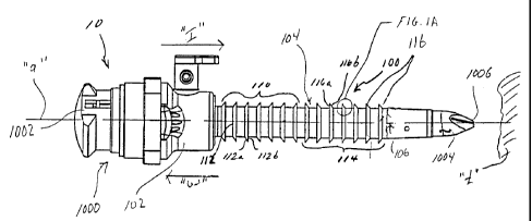
With reference to FIGS. 1 and lA, trocar assembly 10 includes cannula
100 and obturator 1000 which is positionable in the cannula 100. Obturator
1000
includes an obturator housing 1002 and an obturator portion 1004 having
pointed
obturator tip or blade 1006 for penetrating tissue. In use, subsequent to
insertion of trocar
assembly in the tissue site, obturator 1000 is removed from cannula 100
leaving the
cannula 100 in the tissue to serve as a portal for introduction of
instrumentation.
Cannula 100 includes a cannula housing 102 and a cannula sleeve 104
-5-
CA 02371884 2007-12-05
connected to the housing 102 and extending distally therefrom. Cannula sleeve
104
defines longitudinal axis "a" and has an outer wall 106 which defines an inner
longitudinal
opening therein. Cannula 100 includes a first proximal series 110 of external
fixation
members such as generally annular projections or rings 112 which taper
outwardly away
from the surface of cannula 100 in a generally distal direction. In this
manner, a generally
proximal oriented surface 112a is formed at an oblique angle relative to a
longitudinal axis
of cannula 100 and a distal facing planar surface 112b is formed transverse to
a
longitudinal axis of cannula 100. A second distal series 1] 4 of external
fixation members
such as rings 116 are disposed distal of ring series I 10. Rings 116 taper
inwardly towards
the surface of cannula 100 in a generally distal direction and form proximally
facing
planar surface 116a which is preferably transverse to the longitudinal axis
"a" and distally
oriented angled surface 116b as best depicted in FIG. IA.
The above-noted structural arrangement provides for retention of cannula
100 in the body tissue due to the oppositely facing rings 112 and 116.
Additionally, upon
insertion, angled surfaces 116b of distal ring series 114 helps maintain
relative ease of
insertion of cannula 100. Preferably, angled surfaces 116b define an angle "x"
ranging
from about 10 to about 60 relative to the longitudinal axis "a" of cannula
sleeve and,
more preferably, about 45 relative to the longitudinal axis "a". Such
arrangement of
angled surface l 16b minimizes the insertion force needed to advance cannula
100 in an
insertion direction, indicated as arrow I, within the tissue "t".
Cannula 100 is continually advanced to a position whereby proximal ring
series 110 reaches the tissue. At this juncture, planar surfaces 112b of
series 112 engage
the tissue. Planar transverse surfaces 112b effectively substantially increase
the insertion
-6-
CA 02371884 2007-12-05
force required to further insert cannula 100 thereby minimizing the potential
of "over
insertion" of the cannula 100. Moreover, rings 112 provide a tactile
indication to the user
that the cannula 100 has been sufficiently inserted to access the abdominal
cavity and any
further inserting movement may increase the potential of contact of the
cannula with
underlying tissue, organs, etc. When cannula 100 is situated with respect to
the tissue, the
opposing planar surfaces 112b of ring series 1 10 and 116a of ring series 114
serve as
ledges to maintain cannula 100 at a fixed position within the tissue site,
i.e., planar
surfaces 112b resist movement of the cannula 100 in the insertion direction
"I" while
planar surfaces 116a resist movement of the cannula in the withdrawal
direction "W".
Referring to FIG. 2, a proximal portion of cannula 200 features a series 210
of chevron or V-shaped raised surfaces 212, the proximalmost located apexes
214 of
which point proximally. A distal portion of cannula 200 features a series 216
of chevron
shaped raised surfaces 218, the distalmost located apexes 220 of which point
toward the
distal end of cannula 200. With this arrangement, the insertion force is
further reduced
due to the streamline profile presented by the distal series 216. In all other
respects,
cannula 200 is similar to the cannula 100 of FIG. 1.
Referring to FIG. 3, a proximal half of cannula 300 features a series 310
of disjoined chevron shaped raised surfaces formed of disjoined segments 312a
and 312b.
A distal portion of cannula 300 features a series 314 of disjoined chevron
shaped raised
surfaces formed of segments 316a and 316b. This arrangement further reduces
the
insertion force required to insert the cannula by providing a longitudinal
slot 320 as
defined by the open apexes of series 310,314 to which tissue displaced by the
cannula
during insertion may be received and pass.
-7-
CA 02371884 2001-08-27
WO 00/54678 PCT/USOO/05946
Referring to FIG. 4, cannula 400 is the same as cannula 300 except that the
proximal and distal raised surfaces 402,404 do not extend beyond the outer
diameter of
cannula 400, as shown by proximal and distal outer wall portions 406, 408,
respectively,
i.e., the surfaces are confined within the outer boundary of the cannula
sleeve.
Referring to FIG. 5, cannula 500 is very similar to cannula 100 of FIG. 1
with two main exceptions. The first is that proximal and distal series 510,
514 are
respectively displaced a predetermined distance. This provides a central
tissue retaining
region 512 between the two series 510, 514 which receives the proximal and
distal
surfaces of the tissue, such as, for example, the abdominal tissue, i.e.,
inclusive of the
tissue between the epidermis and the peritoneal cavity lining.
Referring to FIG. 6, cannula 600 is similar to cannula 200 except that
raised chevron shaped portions 616 are formed within the boundaries of the
outer
diameter of cannula 600, i.e., the distance "e" across the chevron portions
116 is
substantially equal to or less than the outer diameter of the cannula sleeve
602.
Additionally, cannula 600 is provided with a beveled distal end 615 which has
a chamfer
617 provided thereon. Chamfer 617 facilitates the initial insertion of cannula
within the
tissue site.
Referring to FIGS. 7-11, various alternative embodiments of cannulas are
shown having different external fixation structure. With reference to FIG. 7,
cannula 700
is provided with a series of sinusoidal shaped raised portions 717. Cannula
800 is
provided with helical threads 816 thereon, as shown in FIG. 8. Cannula 900 is
similar to
cannula 200 of FIG. 2, except cannula 900 illustrates that different numbers
of raised
portion elements 912, 916 may be provided in the proximal and distal series
910, 914,
-8-
CA 02371884 2001-08-27
WO 00/54678 PCTIUSOO/05946
respectively. FIG. 10 illustrates the cross-sectional shape of distally
oriented chevron
shaped raised portions 916 of FIG. 9. Raised portions 916 have angled surfaces
916 a
and 916b. Surface 916a is angled such that it forms a greater angle of attack
with the
tissue proximal thereto and surface 916b provides a lesser angle of attack
than surface
916a, with the distal tissue. In this embodiment, the proximal surface 916b is
not
transverse to the axis, but, may range from about 60 - 90 . For proximal
raised surface
912, the opposite relationship to that described in connection with raised
surface 916
would apply.
Referring to FIG. 11, cannula 1000 is provided with a greater number of
series 1002, 1004, 1006, 1008, and 1010 of raised portions wherein adjacent
series have
raised portions oriented in the opposite direction. Additionally, the proximal
and distal
surfaces of each raised portion is curved or arcuate.
Referring to FIGS. 12 and 13, cannula 1100 is provided with a tissue
retention sleeve member 1110 coaxially mounted about the cannula sleeve 1105.
Retention sleeve member 1110 may be fabricated from an elastomeric material
and
preferably formed, e.g., by injection molding techniques to define varying
ridges 1112,
1114 when retention member is in an at rest condition, as shown in FIG. 13.
Preferably,
retention member 1110 is secured at a distal end to cannula 1100 by
conventional means
and is provided with finger levers 1116, 1118 at a proximal end. Finger levers
1116,1118
may be formed of a rigid polymeric material or a suitable metal and secured to
the
proximal end of sleeve member by conventional means, or may be integrally
formed with
the sleeve member. In use, finger levers 1116, 1118 are retracted upon
insertion of
cannula 1100 through the body wall of the patient, as shown in FIG. 12. This
stretches
-9-
CA 02371884 2001-08-27
WO 00/54678 PCT/US00/05946
retention member 1110 thereby smoothing out ridges 1112, 1114. Once the
cannula is
situated in the desired position through the body wall, levers 1116, 1118 are
released
permitting ridges 1112, 1114 to assume their initial unactuated position
thereby acting to
resist proximal or distal movement of cannula 1100 with respect to the body
wall.
Alternatively, instead of being molded from an elastomeric material, retention
member
1110 may be formed from one or more shape memory materials to achieve the
above-
noted results.
While the invention has been particularly shown and described with
reference to certain preferred embodiments, it will be understood by those
skilled in the
art that various modifications in form and detail may be made therein without
departing
from the scope and spirit of the invention. Accordingly, modification to the
preferred
embodiments will be readily apparent to those skilled in the art, and the
generic principles
defined herein nlay be applied to other embodiments without departing from the
spirit
and scope of the invention. Thus, the present invention is not intended to be
limited to
the embodiments shown, but it is to be accorded the widest scope consistent
with the
principles and features disclosed herein.
-10-