Note: Descriptions are shown in the official language in which they were submitted.
CA 02373633 2007-05-10
-1-
SYSTEMS AND METHODS FOR SPINAL FIXATION
BACKGROUND OF THE INVENTION
1. Field of the Invention
The present invention generally relates to methods, systems and
apparatuses for bony fixation, more particularly to methods, systems and
apparatuses adapted for fixing the bones of the spine, and to methods, systems
and apparatuses adapted for securing a prosthetic device within the bones of
the
human body, more specifically securing a prosthetic device within the bones of
the spine.
2. Background of the Invention.
Fixation or fusion of vertebral columns with bone or material, rods or
plates is a common, long practiced surgical method for treating a variety of
conditions. Many of the existing procedures involve the use of components that
protrude outwardly, which may contact and damage a body part, such as the
aorta, the vena cava, the sympathetic nerves, the lungs, the esophagus, the
the
intestine and the ureter. Also, many constructions involve components that may
loosen and cause undesirable problems, often necessitating further surgical
intervention. Additionally, limiting the success of these procedures are the
bio-
mechanical features of the spine itself, whose structure must simultaneously
provide support to regions of the body, protect the vertebral nervous system
and
permit motion in multiple planes.
CA 02373633 2001-11-09
WO 00/67651 PCT/[JS00/12773
As indicated above, spinal surgery for spine fusion generally involves using
implants and instrumentation to provide support to the affected area of the
spine while
allowing the bones thereof to fuse. The technology initially evolved using
bone chips
around and on top of an area of the spine that had been roughened to simulate
a
fracture in its consistency. The area, having encountered the bone chips,
would then
proceed to heal like a fracture, incorporating the bone chips. However,
surgical
procedures dealing with the spine present notable challenges. For example,
bioengineers have been required to identify the various elements of the
complex
motions that the spine performs, and the components of the complex forces it
bears.
This complexity has made it difficult to achieve adequate stability and
effective
healing in surgical procedures directed to the spine.
One surgical technique provided by Cloward, involves cutting a dowel type
hole with a saw across or through the moveable intervertebral disc and
replacing it
with a bone graft that was harvested from the hip bone. This procedure limits
motion
and mobility and results in a fusion of the adjacent vertebral bodies.
However, as a
result of the complex motions of the spine, it is often difficult to secure
the dowel
from displacing. Further, it has become apparent over time, however, that this
particular technique does not always yield a secure fusion.
Other techniques have been developed that involve the placement of various
hardware elements, including rods and hooks, rods and screws and plates and
screws.
The dowel technique also has advanced over the past five years or so, with
dowels
being fabricated from cadaver bone or metals such as titanium or stainless
steel.
These techniques, whether using hardware, dowels or some combination thereof,
have
a common goal to enhance stability by diminishing movement, thereby resulting
in or
enhancing the potential of a fusion of adjacent vertebral bones. For example,
in one
of these other techniques, the disc is removed and adjacent vertebrae are
positioned in
a stable position by placing a plate against and traversing them, which plate
is secured
or anchored to each by means of screws.
CA 02373633 2001-11-09
WO 00/67651 PCT/US00/12773
In another procedure, cages in the form of two parallel circular or
rectangular
devices are made out of a material such as titanium or stainless steel and
these devices
are fenestrated. Bone is packed in the center of the devices that will heal to
adjacent
bone through each fenestration. In this procedure, the disc space is
distracted so all
ligamentous structures are taut and the bones are held in their normal maximal
position of distraction. Because the cages are implanted in spongy bone, they
are
more likely to collapse the surrounding bone, thus resulting in loss of
distraction and
subsequently cage dislodgment.
U.S. Patent 5,591,235 reports a certain spinal fixation device and technique
for
stabilizing vertebrae. In this technique, a hollow screw is inserted into a
hole,
preferably a hole saw recess, in each adjoining vertebrae. A channel is cut
into the
vertebrae, which is lined up with corresponding axial slots in the screw. A
rod is
inserted into the channel and so as to pass through the axial slots in the
screw. The
rod is secured to each of the screws by means of a locking cap. The rod also
is
arranged so as to provide a bridge between the hollow screws in the adjoining
vertebrae. Certain disadvantages have been surmised using such a device and
technique. For example, it has become apparent that the trough in the
vertebral bodies
destabilizes some of the cortex of the vertebrae body wall, which is the
strongest
component.
In addition to fixation or fusion of vertebral columns, the prior art also
describes methods or other spinal repair procedures, such as discectomy
wherein an
artificial disc or prosthetic device is placed within the vertebrae of the
spine. For such
prior art methods and related devices, there have been short comings such as
having
difficulty in securing the prostheses within the vertebral space or resulting
in
significant modification or damage to the load bearing surfaces of the
vertebrae in an
effort to secure the prosthesis.
Thus, it would be desirable to provide a new apparatus, system and methods
for spinal fixation that enhances healing of the bone while providing
structural support
CA 02373633 2001-11-09
WO 00/67651 - 4 - PCT/USOO/12773
to the spine. It would be particularly desirable to provide such an apparatus,
system
and method that would involve the use of open surgical or minimally invasive
surgical
techniques as well as a technique in which the implant burrows in the bone
spine,
traverses across the disk space, and ends in an adjacent or neighboring
vertebrae or
vertebras, providing limited or no protrusions. It also would be desirable to
provide
such an apparatus, system and method where the implant is retained within the
bone
without requiring contour-varying external vertebral wall fixation as compared
to
conventional devices, as such a device would avoid many of the problems
associated
with conventional devices such as blood vessel injury, erosion into organs, as
well as
placement near nerves. Additionally, it would be desirable to provide such an
apparatus, system and method where the implant is retained within the bone and
is
utilized to secure an artificial prosthesis for example within the vertebral
bodies. Such
securing is accomplished with or without the use of the annulus, and without
insult to
portions of the vertebral surfaces bearing significant loading.
SUMMARY OF THE INVENTION
I have now found new methods and apparatus for fixing adjacent vertebrate of
a spine. The methods and apparatus of the invention utilize a new implant
member,
which preferably is arcuate, and avoids the associated problems with prior
cage or
straight rod and screw systems. It is within the scope of the present
invention for the
implant member to have any geometric shape or configuration consistent with
the
intended use including a straight member.
Preferred methods of the invention for stabilizing adjacent vertebrae of the
spine, include the steps of providing a positioning apparatus including two
guide
sleeves, each guide sleeve having a long axis and locating the two guide
sleeves with
respect to the adjacent vertebrae such that a vertex formed by the long axis
of each
guide sleeve is located in the intervertebral space for the adjacent
vertebrae. The
method further includes forming an aperture in each of the adjacent vertebrae
using
the guide sleeves and inserting an implant into the apertures formed in each
of the
CA 02373633 2001-11-09
WO 00/67651 PCT/US00/12773
adjacent vertebrae so that the implant extends between the adjacent vertebrae
and
through the intervertebral space.
Preferably, the aperture formed in the vertebrae is arcuate and the implant
being inserted also is arcuate. The arcuate aperture in each vertebrate can be
suitably
formed by drilling or other ablation. More particularly, an initial aperture
can be
drilled in each of the adjacent vertebrae to create intersecting apertures
with
convergent paths within the intervertebral space; and the initial aperture
then enlarged
to receive the implant. That enlarging of the initial aperture can be suitably
performed
by a variety of procedures, e.g. by using a drill bit, a reamer, an awl,
impaction drill,
shape memory coring device, or curved coring device, or the like.
The step of forming an aperture also can further include inserting a guide
member, after drilling of the initial aperture, into one of the guide sleeves,
down
through the initial aperture in one adjacent vertebrae, through the
intervertebral space
and into the initial aperture in the other adjacent vertebrae; and advancing
an aperture
enlarging device over the guide member so as to enlarge the initial aperture.
In this
case, the aperture enlarging device is suitably a curved reamer or a curved
drill bit,
and the curved reamer or the curved drill bit is advanced over the guide
member so as
to form an arcuate aperture in each of the adjacent vertebrae. It also should
be
appreciated that multiple vertebral holes can be created using the same
methods as
disclosed herein. In that manner, multiple arcuate implants can be placed,
e.g. if
greater mechanical stability is considered desirable.
The positioning apparatus can further include a cross member and an
intervertebral spacer, preferably where the guide sleeves are pivotally
mounted to the
cross member and the intervertebral spacer is spaced from the cross member and
interconnected thereto at about a mid point between the pivot points for the
guide
sleeves. In this case, the stabilizing method can further include locating the
intervertebral spacer in the intervertebral space between the adjacent
vertebrae; and
maintaining alignment of the guide sleeves with respect to the adjacent
vertebrae so
CA 02373633 2001-11-09
WO 00/67651 - 6 - PCT/US00/12773
that a consistent angle is maintained between the guide sleeve and the
vertebrae
during at least a portion of said forming of the aperture. The intervertebral
spacer also
can be configured so as to provide protection to the spine during the drilling
when
disposed in the intervertebral space.
In an alternative embodiment, the positioning system being provided includes
a cutter bracket system and a curved drilling sub-system affixed thereto. The
cutter
bracket system includes a pivot arm whose pivot point is disposed between the
adjacent vertebrae opposite the intervertebral space. More particularly, the
pivot point
is at about the midpoint between the adjacent vertebrae. The curved drilling
sub-
system is affixed to the pivot arm such that as the pivot arm rotates about
the pivot
point the curved drill sub-system follows an established cutting path. In a
more
specific embodiment, the drilling sub-system is affixed proximal or at the
distal end of
the pivot arm. The positioning apparatus according to the alternative
embodiment can
further include a mechanism that temporarily secures the cutter bracket system
to the
adjacent vertebra to be fused and which positions and maintains the pivot
point at the
desired location. Also, the curved drill subsystem can include a curved
cannula, a
flexible member running through the curved cannula and a cutting burr secured
to an
end of the flexible member.
As to the step of forming an aperture using a positioning system according to
the alternative embodiment, this step includes rotating the pivot arm in one
direction
about the pivot point so the curved drilling sub-system forms an aperture in
one of the
adjacent vertebrae and rotating the pivot arm in another direction about the
pivot point
so as to form an aperture in the other of the adjacent vertebrae. In a more
specific
embodiment, the step of forming further includes remounting the curved
drilling
subsystem to the pivot arm before rotating the pivot arm in the another
direction so a
cutting element of the curved drilling subsystem is aligned for the direction
of
movement.
CA 02373633 2001-11-09
WO 00/67651 - ~ - PCT/US00/12773
As to inserting the implant, the method step includes successively drawing a
portion of the implant through the arcuate aperture in one adjacent vertebrae,
through
the intervertebral space and into the arcuate aperture of the other adjacent
vertebrae.
In a specific embodiment, the step of inserting includes securing one end of a
guide
wire to an end of the implant; passing a free end of the guide wire through
the arcuate
aperture in one of the adjacent vertebrae, through the intravertebral space
and through
the arcuate aperture in the other adjacent vertebrae; and pulling on the guide
wire free
end to thereby successively draw the portion of the implant.
In another embodiment, the step of inserting includes inserting a beginning
end of the implant into an entrance opening of one of the adjacent vertebrae;
applying
a force to the portion of the implant extending from the entrance opening so
as to
drive the implant beginning end though the arcuate aperture in the aperture of
said one
of the adjacent vertebrae, through the intervertebral space and into the
arcuate aperture
in the other of the adjacent vertebrae.
The implant being inserted into the final aperture is made from one or more of
a metal (e.g., titanium or stainless steel), bone, morphogenic protein
(including a
combination of bone and bone morphogenic protein), carbon fiber composite,
nitinol
or biodegradable materials such as polyactic acid or polyglycolic acids and
copolymers and other derviatives thereof, or collagen and collagen coated
metal or
bone. The implant also may comprise an in situ-formed plug where the aperture
acts
as a mold for an epoxy or other polymer-based system. Also, the implant can be
solid
or hollow and arranged with or without ingrowth fenestrations and screw holes
for
post-insertion securement. The implant also can be configured so the implant
includes
a first and a second section, where a distal end of each of the first and
second sections
is configured so as to be capable of being secured together. For such an
implant, the
method further includes the steps of inserting the first section into the
aperture in one
of the adjacent vertebrae so that the distal end therefore is disposed in the
intervertebral space; inserting the implant second section into the aperture
in one of
the adjacent vertebrae so that the distal end therefore is disposed in the
intervertebral
CA 02373633 2001-11-09
WO 00/67651 - g - PCT/US00/12773
space; and securing the distal ends of the first and second sections together.
The
implant sections being inserted can be arcuate with a radius substantially the
same as
the arcuate aperture or substantially straight. In particular embodiments, the
distal
ends of the implant sections are secured to each other by e.g. a nut, bolt,
pin,
expansion or press-fit device, or interlocking member on the end of each
section.
Other stabilization methods also can be employed. For instance, a plate can be
applied to the vertrebrae surface with attachments at each end of the tunnel
traversed
by an implant in accordance with the invention.
Another method of the present invention for stabilizing adjacent vertebrae of
the spine includes the step of forming a common channel in and between the
adjacent
vertebrae and inserting a biscuit implant in the common channel so as to
bridge
between the adjacent vertebrae. In more specific embodiments, the step of
forming
includes simultaneously cutting a slot, preferably an arcuate slot, in each of
the
adjacent vertebrae so as to form the common channel and providing a device
configured so as to be capable of simultaneously cutting the slot in each of
the
adjacent vertebrae. Also for said step of inserting, the biscuit implant can
be further
configured so as to include a spacer element that is received in the
intervertebral space
between the adjacent vertebrae when the biscuit is disposed in the common
channel.
In another alternative aspect of the invention, a diskectomy can be performed
and a stabilizing wedge (inner) implant inserted between the vertebrae. The
wedge
(inner tool) establishes lordosis, provides a construction reference, and
carries on it
the stabilizing wedge implant. Retracted stop-cut blades on the inner tool are
then
engaged, cutting into the vertebrae in the vertical plane. A hole saw can be
used to
create a circular cut in the vertebrae to facilitate insertion of the outer
implant. Once
the cut is complete, the bone harvested in the tubular cutter can be
manipulated into
the implant. A circular (outer) implant is then inserted over the inner tool.
The outer
tool then references the position of the inner tool and guides the implant
into place.
After the two implants nest together along a key and groove, the outer tool is
removed. A fenestrated circular member then replaces the outer cutting tool
and the
CA 02373633 2001-11-09
WO 00/67651 - 9 - PCT/US00/12773
inner tool is rotated about 90 degrees and then removed. Working together, the
two
rotated implants capture the vertebral body sections, which are now rotated
about 90
degrees and through their many holes, provide blood exchange with the adjacent
bone
to accomplish fusion.
Also featured is a system and apparatus embodying the described methods or
techniques for internal fixation of the spine.
Other aspects and embodiments of the invention are discussed below.
BRIEF DESCRIPTION OF THE DRAWING
FIG. 1A is a schematic view of a positioning jig according to the present
invention;
FIG. 1B is a front view of the intervertebral spacing member of FIG. lA;
FIG. 2A is a schematic view of the positioning jig of FIG. lA disposed about
two vertebral bodies;
FIG. 2B is a schematic view of an alternative positioning jig according to the
present invention disposed about two vertebral bodies;
FIGS. 3A-E are schematic views that illustrate the various steps of the
process
to form a hole in each vertebral body for implanting a fixating member
therein;
FIGS. 4A and 4B are schematic views that illustrate alternate ways of making
a hole in each vertebral body;
FIG. 4C is a plan view of a Romano device for making a curved hole. Shown
is one of the two opposed curved cutter guides and a flexible cable having a
cutting bit
attached to one end;
FIG. 5A is a schematic view of one device for implanting the fixating member;
FIG. 5B is a schematic view of alternate device for implanting the fixating
member;
FIG. 6A is a schematic view of the vertebral bodies illustrating the
implantation of the fixating member in the holes;
CA 02373633 2001-11-09
WO 00/67651 - 10 - PCT/US00/12773
FIG. 6B is a schematic view of the vertebral bodies illustrating another form
of
implantation of the fixating member in the holes particularly for securing an
intravertebral prosthetic device;
FIG. 6C is a schematic view of the vertebral bodies to illustrate securing of
the
fixating member;
FIGS. 7A-C are schematic views of the implantation of a fixating member
made from nitinol;
FIGS. 8A-B are exemplary cross sectional views of a guide sleeve including a
mechanical guide to guide the nitinol fixating member during insertion;
FIG. 9 is a schematic view of the vertebral bodies with a fixating member
according to a second aspect of the present invention;
FIG. 10 is a schematic view of the vertebral bodies with a fixating member
according to a third aspect of the present invention;
FIG. 11A is a schematic view of a cutter bracket system according to the
present invention;
FIG. 11B is a schematic view of a curved drill used with the cutter bracket
system of FIG. 11 A;
FIG. 12A is a perspective view of a common channel cutting device according
to the present invention;
FIG. 12B is a perspective view of a portion of the channel cutting device of
FIG. 12A with the cutting implement extended;
FIG. 12C is a schematic view of the channel cutting device of FIG. 12A
disposed on two vertebral bodies;
FIG. 12D is a schematic view of the two vertebral bodies to illustrate the
implantation of the biscuit implant in the cut common channel;
FIG. 12E is another view of the two vertebral bodies to illustrate the
implantation of the biscuit implant including a spacing element in the cut
common
channel;
FIG. 12F is a perspective view of the biscuit implant of FIG. 12D;
FIG. 12G is a side view of the biscuit implant with spacing element of FIG.
12E;
CA 02373633 2001-11-09
WO 00/67651 - 11 - PCT/US00/12773
FIGS. 12H-K are perspective views of various exemplary biscuit implants
according to the present invention;
FIGS. 13A-13F illustrate an alternative implant system of the invention; where
FIG. 13A is an isometric view of an inner implant, FIG. 13B is an isometric
view of
an outer implant, FIG. 13C is a lateral view showing a preferred positioning
of the
implant system, FIG. 13D is an anterior view of the outer implant within which
the
inner implant is secured, FIG. 13F is an anterior view of the outer and inner
implant
after rotation, and FIG. 13F is a perspective view of an embodiment of the
implant
system;
FIG. 14A is a schematic view of an inner tool positioned within the
intervertebral disk space;
FIG. 14B is an isomeric view of the inner tool;
FIG. 14C is a cross-sectional view of the inner tool, with retracted and
extended stop-cut blades;
FIG. 15 is a schematic view of the inner and outer tool system positioned in
relation to the vertebral bodies;
FIG. 16 is a schematic view showing bone-to-bone with no gap application;
and
FIGS. 17A-C are schematic views of exemplary implants useable for securing
a prosthetic device.
DETAILED DESCRIPTION OF THE INVENTION
Referring now to the various figures of the drawing wherein like reference
characters refer to like parts, there is shown in FIGS. 1-2 various schematic
views of a
drill guide or positioning jig 100 that positions or aligns the drill bits
before making
the holes in each of the vertebral bodies 2. The positioning jig 100 includes
two guide
sleeves 102, a cross member 104 and an intervertebral spacing member 110. Each
guide sleeve 102 preferably is a hollow tubular member having a lumen or
passage
therein for receiving and guiding the means for forming at least the initial
aperture in
the adjacent vertebrae such as a drill bit 150 (FIG. 3B). As indicated
elsewhere
herein, the aperture may be formed using other techniques such as the ablation
of bone
CA 02373633 2001-11-09
WO 00/67651 - 12 - PCT/US00/12773
by an energy source, e.g., high-pressure water, high-pressure air, ultrasound,
or a
laser. As such, it shall be understood that the internal sizing and
configuration of the
guide sleeves is established to accommodate the particular mechanism used for
forming the aperture.
The guide sleeves 102 are mounted to the cross member 104 in such a way
that they are each pivotal about the cross member and so each can be secured
or
locked in a particular angular position with respect to the cross member.
Although a
single mounting/ pivot point 106 is illustrated, it is within the scope of the
present
invention for the cross member 104 and each guide sleeve 102 to be configured
with a
plurality or more of such pivot/ mounting points. In an exemplary embodiment,
the
cross member 104 and guide sleeves 102 are constructed from stainless steel;
and each
guide sleeve is pivotally secured to the cross member by screws.
The distal end 108 of each guide sleeve 102 is configured for mechanically
engaging a surface, edge, corner or other surface artifact or feature of the
vertebral
body 2. In an exemplary embodiment, and as more clearly illustrated in FIG.
3A, the
guide sleeve distal end 108 is configured or arranged with a cutout that is
designed to
accept the corner of the vertebral body 2. Additionally, the cutout area and
thus the
distal end 108 also are configured with a plurality or more of teeth 107. The
teeth 107
are configured and arranged so the teeth bite into the bony surface of the
vertebral
body when the corner of the vertebral body 2 is received within the cutout
area of the
guide sleeve distal end 108. Each guide sleeve is suitable about 20 cm in
length,
although suitable and preferred guide sleeve lengths can vary depending on the
method of access.
The intervertebral spacing member 110 includes an intervertebral spacer 112
and an interconnecting member 114 that mechanically interconnects the cross
member
104 and the intervertebral spacer 112. The interconnecting member 114 is
secured to
or retained by the cross member 104 so as to be maintained in fixed relation
with
respect to the pivots 106 for both guide sleeves 102. In an exemplary
embodiment,
CA 02373633 2001-11-09
WO 00/67651 - 13 - PCT/US00/12773
the interconnecting member 114 is located at about the midpoint of the cross
member
104 between the pivots 106. The interconnecting member 114 also is secured to
the
cross member 104 so the intervertebral spacer 112 is positioned between the
distal
ends 108 of the guide sleeves 102. More particularly, the interconnecting
member
114 is positioned so the intervertebral spacer 112 is received within the
distended disc
space between the adjacent vertebral bodies 2.
In an exemplary embodiment, the interconnecting member 114 is in the form
of a rod and the cross member 104 is configured with a through aperture 109 in
which
the rod is received. This configuration provides a mechanism by which the
interconnecting member 114 is put into and maintained in fixed relation with
respect
to the pivot points 106. It is within the scope of the present invention for
the cross
member 104 to have any geometric shape, as well as being hollow or solid in
construction, that is otherwise consistent for the intended use of the
positioning jig
100.
The interconnecting member 114 also can be configured so as to prevent
rotational motion of the interconnecting member with respect to the through
aperture
109. For example, the rod and through aperture 109 may be configured so as to
include a flat side in a portion of the circumference for the through aperture
and the
rod. Alternatively, the through aperture and rod may be arranged with a key
and
notch arrangement to prevent rotation.
When the guide sleeves 102 are secured to the cross member 104 and each
guide sleeve distal end 108 mechanically engages the surface of the vertebral
body 2,
the guide sleeves are arranged so they maintain a consistent angle with
respect to the
vertebral body. Additionally, and in combination with the intervertebral
spacer 112,
this arrangement provides a three-point reference that ensures appropriate
angles and
alignment are maintained. Additionally, such a configuration establishes a
condition
whereby the positioning jig 100 locks down on the motion segment of the spine
to be
stabilized.
CA 02373633 2001-11-09
WO 00/67651 - 14 - PCT/US00/12773
The use of the positioning jig 100 in the method of the present invention can
be understood from the following discussion with reference to FIGS. 1-6. It
shall be
understood that as preparation for spinal fixation/ stabilization, the medical
personnel
(e.g., surgeon) obtains access to the motion segment or structures to be
stabilized or
fused using any of a number medical/ surgical procedures known to those
skilled in
the art. In this regard, this would involve such actions as preparing the disc
space and
performing retraction of vessels, muscles and nerves.
In this regard, it should be recognized that the method and positioning jig
100
of the present invention are particularly advantageous when performing a
minimally
invasive surgical procedure. The minimally invasive procedure can be performed
through three holes, each about 1 inch across, in the abdomen and allows for
the
procedure to be executed without visualizing the vertebrae. Thus, and in
contrast to a
number of prior procedures, methods of the invention are not limited to an
anterior
presentation. Such methods of the invention also can be performed through a
posterior, posteriolateral or pedicular approach.
In addition, when using a nitinol implant, the positioning jig 100 allows the
implant to be properly positioned for and during insertion thereof. After
gaining
access, the surgeon also could scrape out the material from the failed disc or
use this
disc or its space as a reference point.
As preparation, the surgical personnel also select an intervertebral spacing
member 110 that is appropriately sized, so it can accommodate the distended
disc
space. The intervertebral spacer 112 portion of the intervertebral spacing
member I 10
is inserted into the intervertebral space 4 between the adjacent vertebrae. In
this way,
the approximate center or mid point of, and the staring point on, the adjacent
vertebrae
to be fused or stabilized is thereby established or defined.
CA 02373633 2001-11-09
WO 00/67651 - 15 - PCTIUSOO/12773
The intervertebral spacer allows the surgeon to maintain extremely accurate
disk spacing. The intervertebral spacer also protects the spinal cord from
accidental
drilling or boring. If desired, the spacer can be made of bone and can be made
with or
without a through hole. The spacer design is suitably based on a construction
that
facilitates the selected technique for creating an arcuate aperture. An
intervertebral
spacer that is comprised of bone offers the advantage of being able to remain
implanted following the procedure.
Other materials also can be suitably employed to form an intervertebral
spacer.
The placement of an implant provides a central axis through which a
compressible,
functional intervertebral disk member can be reliably secured. The artificial
disk
member suitably can be made from a variety of compressible materials,
including e.g.
silicon, elastomeric polymers, polyurethanes and copolymers thereof,
hydrogels,
collagen or bioabsorbables.
Next, the positioning jig 100 is locked down on top of the motion segment to
be immobilized, as more clearly shown in FIG. 2. In this regard, the surgical
personnel slide the interconnecting member 114 of the intervertebral spacing
member
110 into an aperture 109 provided in the cross member 104. In this way, the
aperture
109 in the cross member 104 positions the intervertebral spacing member 110
between the distal and proximal ends of the drilling guides 102. Although
illustrated
as being located in the mid-point, the intervertebral spacing member can be
centrally
located or offset to either side to enable drilling of holes in the vertebrae
laterally
against the spine.
Preferably, the aperture 109 in the cross member 104 is configured so as to
prevent the cross member 104 or intervertebral spacing member 110 from
rotating
with respect to each other. For example, a portion of the aperture 109 and a
portion of
the interconnecting member 114 is flattened so as to pre-define a given
orientation.
Alternatively, the aperture 109 is configured with a notch or keyway and the
CA 02373633 2001-11-09
WO 00/67651 _ 16 _ PCT/US00/12773
interconnecting member 114 is configured with a key or protrusion that is
received in
the keyway.
As provided above, the distal end 108 of each guide sleeve 102 is preferably
configured so each distal end mechanically engages the surface of the
vertebrae 2. In
the illustrated embodiment, the distal end 108 is arranged with a cutout area
that is
designed to accept the corner of the vertebrae 2 as more clearly illustrated
in FIG. 3.
As also shown in FIG. 3, the cutout area is provided with a plurality of teeth
107 that
bite into the bony surface of the vertebrae 2. It is within the scope of the
present
invention for the guide sleeve distal end 108 to be disposed at other
positions on the
surface of the vertebrae 2 such as that illustrated in FIG. 6A.
After locating the positioning jig 100 with respect to the motion segment to
be
fused, the surgical personnel secure the guide sleeves 102 at each of the
pivots 106.
This advantageously ensures that the appropriate angles and alignment of the
guide
sleeves 102 with respect to the vertebrae 2 are maintained as well as locking
the
positioning jig 100 down on the motion segment to be fused.
As noted above, an initial through hole is formed in each vertebrae 2 by any
of
a number of methods, e.g. by a drill, by ablation of the material comprising
the
vertebrae using an energy source such as RF, ultrasonic waves, cryogenics and
water
jets or by any other means known to those skilled in the art and which can be
adapted
for use with the positioning jig 100 of the present invention. For purposes of
describing the present invention, however, the following discussion is
simplified to
describing the method in terms of drilling the initial aperture or initial
through hole 6
in the vertebrae 2. This, however, shall not be inferred as being a limitation
on the
method according to the present invention to only drilling.
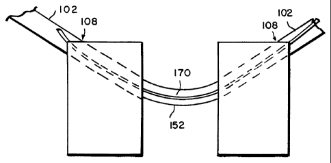
A fixed or flexible drill bit 150 is inserted into and down each drill guide
102
so the ends thereof contact the surface of the vertebrae 2. The surgical
personnel
operate the drill bits in accordance with accepted techniques so as to create
an initial
CA 02373633 2001-11-09
WO 00/67651 - 17 - PCTIUSOO/12773
through hole 6 in each of the vertebrae. Preferably, and as shown in FIG. 3B,
the
through holes 6 being created are intersecting with convergent paths within
the
intervertebral space 4. In other words, the projection of the long axis for
each of these
through holes 6 intersects so the vertex created by intersection of the long
axes is
located within the intervertebral space 4.
The initial through hole 6 initially formed in each vertebrae 2 has a diameter
much less than that of the implant 160 that is to be used to stabilize or fuse
the motion
segment. After forming the initial through hole 6, the surgical personnel
insert a guide
wire 170, such as a 0.093 inch nitinol guide wire, into and down one guide
sleeve 102
and through the through hole in one vertebrae 2. The surgical personnel
continue to
push the guide wire 170 across the intervertebral space 4 and into the through
hole 6
in the other vertebrae as more clearly illustrated in FIGS. 3C-D. In a
particular
embodiment, the guide wire 170 is configured with a slightly curved tip. The
guide
wire 170 is generally in a curved configuration when disposed in the through
hole 6 of
the vertebrae 2.
A flexible/curved drill bit 152 is then passed through one of the guide
sleeves
102 and over the guide wire 170 so as to form a curved through aperture 6a in
each of
the vertebrae as shown in FIG. 3E. The curved or arcuate through aperture 6a
is
formed with a cross-section that complements the cross-sectional shape of the
implant
160. Preferably, the arcuate through aperture is sized to be slightly smaller
than that
of the implant 160 so there is a friction, snug or interference fit between
the implant
160 and the arcuate through aperture 6a.
In this way, when the implant 160 is inserted into the arcuate through
aperture
6a, it will remain therein without further need of screws or other artifacts
or devices
for securing the ends of the implant to each vertebrae 2. It is within the
scope of the
present invention, however, for screws or other devices be provided as an
additional
measure or protection for securing the implant 160 within the vertebrae 2.
CA 02373633 2007-05-10
- 18-
Alternatively, the curved or arcuate through aperture 6a is formed using
any of a number of other techniques as described below. In one case, and as
shown in FIG. 4A, the arcuate through aperture 6a is formed in the vertebrae 2
by using a flexible reamer 200. The flexible reamer is run or passed over the
guide wire 170 to ream or core out the arcuate through aperture 6a. The
cancellous bone of the vertebrae 2 is relatively soft so that it is possible
to use a
reamer to core the hole aperture. Similarly, and as shown in FIG. 4B, a curved
awl or a progressively larger guide wire 170a can be used to punch a curved
hole in the vertebrae. FIG. 4C shows a Romano device suitable for drilling a
curved bore such as that disclosed in USP 5,700,265. A swing arm 830 and
curved guide arm 834 navigate the drill bit 840 through a defined radius of
curvature.
In addition to the mechanical devices for drilling, punching or reaming out
the arcuate through aperture 6a, the discharge end of an energy source, such
as
RF, ultrasonic, cryogenic, laser and water, can be located within the guide
sleeve
102 and passed over the guide wire so as to form the arcuate through aperture.
For example, the nozzle(s) of a high pressure water source can be arranged so
the discharging or ice crystal water impinges on the bony material of the
vertebrae 2 and the material is thereby ablated away to form the arcuate
through aperture 6a. Similarly, laser light, RF waves or ultrasonic waves can
be
focused on the bony material within the vertebrae 2 to form the arcuate
through
aperture 6a.
The foregoing describes the formation of the arcuate through aperture 6a
that receives the implant 160 by passing a mechanism from the entrance to the
exit of the initially formed through hole 6. It is within the scope of the
present
invention, for a guide to be located within the intervertebral space 4 so the
curved through aperture is formed by drilling from the intervertebral space
out,
rather from the outside in.
There is shown in FIG. 2B a schematic view of an alternative positioning
jig 100a that is disposed about two vertebral bodies. This alternative
positioning
jig 100a is similar to the positioning jig 100 of FIG. 2A except for the guide
sleeves. As such
CA 02373633 2001-11-09
WO 00/67651 - 19 - PCTIUSOO/12773
reference shall be made to the foregoing discussion regarding FIGS. 1-2A for
further
details as to the common features for these two positioning jigs 100,100a. In
the
illustrated embodiment, a guide wire 170 is being inserted into one of the
guide
sleeves 102a and is configured so that the proximal end of the guide wire 170
is
arranged so as to include an impact fitting to protect the guide wire about
the proximal
end.
In the alternative embodiment, the guide sleeves 102a are tubular members
that are configured so that at least a portion 103 of each guide sleeve is
arcuate. In the
illustrated embodiment, the arcuate portion 103 of the guide sleeve 102a is
proximal
the vertebral body such that one end of the arcuate portion comprises the
distal end
108 of the guide sleeve that is in contact with the vertebral body 2. It is
contemplated,
however, that the guide sleeve can be configured so as to be substantially
arcuate
between the vertebral body 2 and the cross member 104.
The arcuate shape provides a convenient mechanism that can simplify the
above-described process for making an arcuate through hole 6a in the vertebral
body
2. The arcuate shape also provides a mechanism to orient the tool, device or
apparatus
being inserted into the guide sleeves 102a, for example the drill or high
energy source
for forming the initial through hole, so use of the tool etc. is more
convenient to the
surgical personnel performing the procedure.
After the arcuate through aperture 6a is formed, then the implant 160 is
inserted therein so it is disposed within the through aperture 6a in one
vertebrae 2,
passes or extends across the intervertebral space 4 and disposed within the
through
aperture 6a of the other vertebrae. The implant 160 is made from any one ore
more
suitable materials such as e.g. a metal such as titanium or stainless steel,
bone, bone
with bone morphogenic protein, carbon fiber composite, nitinol. The implant
being
inserted into the final aperture is made from one or more of a metal (e.g.,
titanium or
stainless steel), bone, morphogenic protein (including a combination of bone
and bone
morphogenic protein), carbon fiber composite, nitinol or biodegradable
materials such
CA 02373633 2001-11-09
WO 00/67651 - 20 - PCTIUSOO/12773
as polyactic acid or polyglycolic acids and copolymers and other derivatives
thereof,
or collagen and collagen coated metal or bone. The implant also may comprise
an in
situ-formed plug where the aperture acts as a mold for an epoxy or other
polymer-
based system. The implant, preferably is curved so it generally conforms to
the radius
of the arcuate through apertures 6a in each vertebrae 2, however, other
geometric
shapes are contemplated that are consistent with the intended use including
straight
members.
The implant 160 suitably can be provided with a circular or oval shape. The
diameter or width of the implant can vary over a relatively broad range and
may
depend on the size of the vertebrae and desired implant stiffness. More
specifically,
in preferred embodiments, the implant may suitably range in diameter or width
from
about 5 mm or as small as is mechanically sufficient, to sizes approaching
that of
large intramedullar rods, or about 22 mm. Preferably the implant should have a
diameter or width from about 7 to 12 mm, more preferably about 9 mm. The
implant
also preferably should have an appropriate radius of curvature such that both
vertebrae
are engaged while staying well clear of the spinal cord. That radius
preferably is
about 1.5 inches, as referenced from the arcuate implant's inner radius.
The implant 160 is suitably a solid or hollow (e.g., tubular) member. The
implant can be suitably configured so as to have fenestrations 166 (FIG. 6A)
that
allow biologic elements of bone to traverse through it or across it, thereby
enhancing
potential for stability and for cross-segmental healing. In particular, the
implant 160
can have cutting fenestrations similar to a cheese grater, allowing fragments
of bone
to be pared off as the implant 160 is being inserted into the through
apertures in either
vertebrae. A fenestrated implant 160 that is hollow can be filled with bone
chips or
synthetic or engineered bone healing materials, allowing for bone ingrowth,
and a
cheese grater type of implant with cutting fenestrations can add freshly pared
fragments of bone to the packed bone chips or other materials to enhance bony
ingrowth. Additionally, the fenestrations 166 can be surface dimples,
sharpened
edges, cutting indentations or other alterations in the exterior surface of
the implant
CA 02373633 2001-11-09
WO 00/67651 _ 21 _ PCT/US00/12773
160 to enhance or further ensure the secure fitting of the implant into the
arcuate
through aperture 6a as well as for facilitating bone growth.
The particular technique for inserting the implant 170 into the through
aperture
6a of a vertebrae 2 for fixing of the movable segment is dependent upon the
material
used to make the implant. For an implant 160 made from titanium, and as shown
in
FIG. 5A, a threaded end 162 (e.g., a female threaded end) is provided at one
end of
the titanium implant 160 for threaded engagement with the threaded counterpart
(e.g.,
male counterpart) at one end 172, the distal end of the guide wire 170. This
can be
accomplished for example by removing at least one of the guide sleeves 102
from the
entrance opening of one through aperture 6a so the threaded end 172 of the
guide wire
is exposed. The implant threaded end 162 is then screwed onto the guide wire
threaded end 172 and the so tethered end 162 of the implant 160 is positioned
at the
entrance opening of the through aperture 6a and pulled into place by pulling
on, for
example, the proximal end 174 of the guide wire 170.
Preferably, the distal end 108 of one guide sleeve 102 remains engaged at the
entrance opening for the other through aperture 6a, so as to serve as a
bearing surface
or brace for the guide wire 170 as it is being pulled out of this entrance
opening. This
is done to keep the guide wire 170 from cutting through the cantellous bone
when the
guide wire is under tension because of the pulling action. Alternatively, a
tubular
member with a rounded surface may be advanced over the guide wire and through
the
remaining guide sleeve 102, to ensure that the guide wire pulls from the
appropriate
angle. This technique is suitable for use with metallic and other rigid
material type of
implants.
Alternatively, and as shown in FIG. 513, a pushing mechanism is useable for
inserting or tamping the implant 160 into the arcuate through apertures 6a. In
the
illustrated embodiment, an arcuate pushing mechanism 300 is configured so as
to
rotate about an axis of rotation that corresponds generally to the center of
the circle
subscribed by the arcuate through apertures 6a. The arcuate pushing mechanism
CA 02373633 2001-11-09
WO 00/67651 - 22 - PCTIUSOO/12773
applies a force to the distal end of the implant 160 so as to drive the
proximal end of
the implant through the arcuate through aperture 6a in one vertebrae, across
the
intervertebral space 4 and into the arcuate through aperture 6a of the other
vertebrae 2.
In the illustrated embodiment, the positioning jig 100 is removed except for
the intervertebral spacing member I 10 or bone interverterbral spacer where
the
intervertebral spacer 114 remains disposed in the intervertebral space 4. The
arcuate
pushing mechanism 300 is attached to the end of the interconnecting member 112
by
means of a jig or other member or device so the pushing mechanism can rotate
about
the end of the interconnecting member. In this way, the arcuate arm 302 of the
pushing mechanism 300 can be advanced by having one of the surgical personnel
rotating it about its axis of rotation. Alternatively, or in addition, the
surgical
personnel can strike one end 304 of the arm 302 with a mallet or other
weighted
object so as to drive the implant 160 into the through aperture 6a. For
example,
striking may be required near the end of the insertion process when there is
maximum
friction being developed on the inserted implant. The arm 302 also may be
configured with a curved support sleeve 306 in which the implant is received.
Although the implant 160 and through apertures 6a are sized so that there is
preferably at least a snug-fit therebetween, as an extra measure of
protection, the
implant 160 may be further secured in place at its ends by means of screws 400
as
shown in FIG. 6C. Alternatively, the implant 160 may be secured in place by a
plate,
screw, staple or a combination thereof. Additionally, the implant can be
arranged so
as to include a biting or expansion element(s) that can be driven out in a
lateral
direction so as to engage the bony structure of the vertebrae 2.
As provided above, and as shown in FIGS. 7A-B, the implant 160a can be
made from nitinol. A nitinol implant 160a is advantageous in that a curved
nitinol
implant can be straightened as shown in FIG. 7B prior to insertion into the
arcuate
through apertures 6a. The straightened nitinol implant 160a can be advanced
down
one of the guide sleeves 102 in any of a number of ways, for example, by
pushing or
CA 02373633 2001-11-09
WO 00/67651 - 23 - PCT/USOO/12773
pulling, so it can be driven into the arcuate through apertures 6a. The
nitinol implant
160a also can be inserted into the arcuate through apertures 6a in any of the
other
fashions described above in connection with FIGS. 5A-B.
Additionally, a sharp edge of the nitinol implant can be used like a reamer or
awl to thereby enlarge the initial through hole 6 as the implant is being
inserted or
driven into the initial though aperture. This avoids the intermediate step of
drilling or
otherwise forming the arcuate through aperture 6a before insertion of the
implant.
FIG. 7C depicts an illustrative device 400 for inserting a nitinol implant
160a,
which device includes a guide tube 402 and a pusher 404. The distal end 408 of
the
guide tube 402, similar to the positioning jig guide sleeve distal end 108 is
preferably
configured so as to be capable of releasably mating with a surface, or portion
thereof,
of the vertebrae 2 where the entrance of the arcuate through aperture 6a is
located. In
the illustrated embodiment, the guide tube distal end 408 is configured with a
cut out
so as to receive a corner of the vertebrae 2 therein.
The distal end 408 is disposed on the vertebrae so that the lumen therein is
aligned with the arcuate through aperture 6a. The straightened nitinol implant
160a is
inserted into the guide tube 402 along with the pusher 404 such that the
distal end of
the pusher is in contact with the proximal end of the nitinol implant. The
pusher distal
end 408 mates with the implant proximal end so as to maintain the orientation
and
direction of the nitinol implant 160a within the guide tube 402 so that it
curves in the
proper direction when it exits the guide tube. Alternatively, and as shown in
FIGS.
8A-B, the orientation of the nitinol implant 160a within the guide tube 402 is
maintained with a flat side or with a key and notch type of arrangement.
The pusher 404 includes a stop 406 to limit the travel of the pusher within
the
guide tube 402. This in turn limits the amount of travel by the nitinol
implant 160a so
as to assure that the implant remains buried within the vertebrae and not
exposed
above the surface thereof.
CA 02373633 2001-11-09
WO 00/67651 - 24 - PCTIUSOO/12773
The placement of the implant according to the systems and methods of the
present invention is advantageous in that the inserted implant resides
completely
within the vertebrae and, thus, within the spine, with no protrusion as
compared with
prior art devices. The implant and its placement provide a configuration which
allows
for some compression and cantilever force, but deters rotation and sheer.
Additionally, in the present device, the moment arm is more centrally located
within
the spine as compared to prior devices. This central location also provides
better
stability in the face of torsion as compared to prior art devices.
In general, the placement of an arcuate implant within the arcuate through
apertures as described herein is particularly advantageous because the implant
is
buried to avoid contact with neurovascular structures. The placement provides
load
sharing and thus provides a better healing bio-mechanical environment and also
provides a more advantageous fixation to avoid mechanically sub-optimal
stresses.
Also important, this method allows securement and avoids displacement of a
spinal
fusion or disk replacement device without modification or damage to the
vertebrae's
load bearing surface. Rather, one or two holes placed in or around the center
of a
vertebrae can be sufficient. The method and positioning jig 100 of the present
invention also are advantageous in that the jig can be adapted for use in
minimally
invasive procedures. Additionally, the capability to position implants in
accordance
with the methods described herein enables avoiding blood vessel injury,
erosion into
organs and damage to adjacent nerves. This provides a significant advantage
over
presently existing technologies for disorders of the spine including
fractures, arthritis,
deformity, infections, tumor and mechanical spinal disorders.
Although the foregoing method describes extending a single implant between
adjacent vertebrae this description should not be construed as being a
limitation as one
or more implants can be positioned across each motion segment as described
herein.
CA 02373633 2001-11-09
WO 00/67651 - 25 - PCTIUSOO/12773
In addition, the above described method can be further adapted so as to be
used to secure an intravertebral prosthetic device 500 (i.e., artificial disc)
such as that
shown in FIG. 6A. According to this aspect of the invention, the implant is
made
partly or wholly from a flexible material such as silicon, elastomeric
polymers,
polyurethances and copolymers thereof, hydrogels, collagen, bioabsorbables,
compositions, or a metallic spring or coil, so as to allow continual mobility
between
the vertebral bodies. One or more arcuate implants are provided which pass
through a
partial or complete hole in the prosthesis. This effectively prevents the
prosthesis
from becoming dislodged as well as maintaining its location and orientation
within
the disc space.
There is shown in FIGS. 17A-C exemplary arcuate implants 160b-d for use in
securing the intravertebral prosthetic device 500 within the bones of the
spine.
Referring now to FIG. 17A, there is shown an arcuate implant 160b having a
first
section 163 disposed between two end or second sections 165 that mechanically
engage the first section. The first section 163 is made up of a compressible
material
and the second sections 165 are made up of a material(s), such as metals and
bone,
that is conducive to the attachment of the second sections to the bone thereby
securing
the implant 160b. Alternatively, and as shown in FIG. 17B, the implant 160c
can
comprise a first section 163 that is bonded or otherwise mechanically secured
to the
second sections 165.
The implants 160b,c of either FIGS. 17A,B extends through an aperture or
hole in the prosthetic device 500 and into the vertebral bodies adjacent to
the
prosthetic device similarly to that illustrated in FIG. 16A. Additionally, the
first
section 163 of either of the two implants 160b,c is arranged so as to extend
through
the prosthetic device 500. See also the discussion above for FIG. 16A for
other
aspects of securing the prosthetic device (e.g., one or more apertures in the
prosthetic)
and materials for the implant.
CA 02373633 2001-11-09
WO 00/67651 _ 26 - PCTIUSOO/12773
The above described method also can be further adapted so as to be used to
secure an intravertebral prosthetic device 500 (i.e., artificial disc)
according to another
technique such as that shown in FIG. 6B using an implant, such as the
exemplary
implant 160d shown in FIG. 17C. In this technique any of the mechanisms and
methods described herein are used to form at least an aperture 6, preferably
an arcuate
through aperture, in one adjacent vertebral body 2a. More particularly, the
aperture
forming mechanism or method also forms another aperture 6' in the other
adjacent
vertebral body 2b. This another aperture 6' in the other vertebral body 2b is
not a
through aperture but rather only extends only into a portion of the other
vertebral
body. After forming the apertures 6,6', the prosthetic device 500 is disposed
in the
disc space or intervertebral space 4. The implant 160d is then pressed into
and
through the aperture 6, through the prosthetic device 500 and intervertebral
space 4
and into the aperture 6' of the other adjacent vertebral body 2b.
The exemplary arcuate implant 160d shown in FIG. 17C includes a first
section 163 and a single second section 165 that mechanically engages the
first
section as shown in FIG. 17A or is bonded or other wise mechanically secured
to the
first section as shown in FIG. 17B. Reference shall be made to the above
discussion
regarding FIGS. 17A,B for further details regarding the make up and materials
of the
first and second sections 163, 165 and the discussion for FIG. 16A for other
aspects of
securing the prosthetic device (e.g., one or more apertures in the prosthetic)
and
materials for the implant.
FIG. 9 shows a method for inserting an implant 600 according to a second
aspect of the present invention. A final through aperture 604 is formed in
each of the
vertebrae in accordance with above described techniques such as by drilling.
Except
that the through aperture 604 that receives the implant can be straight as
shown in
FIG. 9 or can be arcuate as shown in any of FIGS. 3-6. As such, reference
should be
made to the foregoing discussion for further details regarding the formation
of the
final through aperture 604.
CA 02373633 2001-11-09
WO 00/67651 _ 27 _ PCTIUSOO/12773
In the method according to the second aspect, the implant is in two sections
602a,b. The proximal ends 608 of the two sections 602a,b are particularly
configured
so they can be mated to each other and interlocked to each other by means of
an
interference fit, a nut and bolt, a screw or a pin 606. Thus, to fix the
moveable
segment, one section 602a is inserted into the through aperture 604 in one
vertebrae 2
and the second section 602b is inserted into the through aperture 604 of the
other
vertebrae. The two sections 602a,b are inserted into their respective through
apertures
until the proximal ends 608 thereof are mated to each other. The pin 606 or
other
securing mechanism is then used to interlock the proximal ends and thus form a
rigid
implant. Although the sections are illustrated as being straight, it is within
the scope
of the present invention for the sections to arcuate so as to form an
interlocking rod
when assembled.
FIG. 10 shows a method for inserting an implant 600 according to a third
aspect of the present invention. According to this method, the apertures 702
in each
vertebrae 2 are formed so they extend from the vertebral space 4 outwards,
penetrating
into the cancellous bone. In this aspect, the apertures 704 formed in the
vertebrae
need not be through apertures. The implant 600 is like that described above
for the
second aspect of the present invention except that it is inverted from that
shown in
FIG. 9.
There is respectively shown in FIGS. 11 A,B a cutter bracket system 1100 and
a curved bit or drill system 1120, the curved drill system being for use with
such a
cutter bracket system. The cutter bracket system 1100 and curved drill system
1120
comprises another embodiment of the present invention for forming arcuate
apertures
6a (FIG. 6A) in each of the adjacent vertebral bodies 2. Referring now to FIG.
11A,
the cutter bracket system includes temporary vertebral screws 1102, pivot
brackets
1104 and a pivot arm 1106. In the illustrated embodiment, there is two
temporary
vertebral screws 1102 that are each secured to the adjacent vertebral body 2
that is to
be fused, however, this shall not be construed as a limitation on the number
of
intervertebral screws. Extending from the temporary vertebral screws 1102 are
the
CA 02373633 2001-11-09
WO 00/67651 _ 28 _ PCT/US00/12773
pivot brackets 1104, which locate the pivot point 1108 with respect to the
adjacent
vertebral bodies 2 and maintian the pivot point in this orientation. The pivot
arm
1106 is rotatably mounted to the pivot brackets 1104 using any of a number of
mechanisms or techniques known in the art so that the pivot arm pivots or
rotates
about the pivot point 1108. In an exemplary embodiment, the temporary
vertebral
screws 1102, the pivot brackets 1104 and the pivot arm 1106 are made from
stainless
steel although other materials are contemplated.
The drill system illustrated in FIG. I 1 B includes a curved cannula 1122, a
flexible cable 1124, a cutting head or burr 1126 and a motor 1130. The
flexible cable
1124 is rotatably disposed with the curved cannula 1122. One end of the
flexible
cable 1124 is attached to the cutting burr 1112 and the other end of the
flexible cable
1124 is attached to the motor 1130, whereby the motor drives the cutting burr
so it to
rotates in the desired manner. In the illustrated embodiment, the motor 1130
also is
mounted to an end of the curved cannula 1122. In an exemplary embodiment, the
curved cannula 1122 is made from stainless steel and the flexible cable 1124
is a
flexible, teflon coated stainless steel cable, the cutting burr 1126 is made
from
stainless steel, although it is within the scope of the present invention for
other
materials to be used.
The motor 1130 includes any of a number of devices known in the art that
develop or provide a rotary output which can be used to rotate the flexible
cable 1124,
such devices include, but are not limited to, electric or pneumatic drills,
DC/AC
electric motors, or, pneumatic, air driven rotary motors. It also is within
the scope of
the present invention for the drill system 1120 to further include a coupling
member,
as is known in the art, that operably and rotatably interconnects the flexible
cable
1124 and the motor 1130 such that the motor is located remote from the curved
cannula 1122. In this way, any of a number of rotary devices such as a drill,
that are
readily available, can be adapted for use in the drill system 1120 of the
present
invention and interconnected to the flexible cable 1124 by means of the
coupling
member.
CA 02373633 2001-11-09
WO 00/67651 - 29 - PCTIUSOO/12773
The drill system 1120 is mounted or attached to the pivot arm 1106, distal
from the pivot point 1108, by means of a connector 1128 on the curved cannula
1122.
The connector 1128 and the corresponding feature on the pivot arm 1106
comprises
any of a number of mechanisms or devices known in the art (e.g., clamp type
mechanism) by which the curved cannula can be removably secured to the pivot
arm
so there is essentially no relative movement therebetween. In a particular
embodiment, the curved cannula 1122 is secured proximal to or at the distal
end of the
pivot arm. In this way when the drill system 1120 is secured to the cutter
bracket
pivot arm 1106 and the cutter bracket pivot arm 1106 is rotated about the
pivot point
1108, the pivot arm guides the curved drill system, in particular the cutting
burr 1126
on a well-defined circular path.
In use, the cutter bracket system 1110 is temporarily secured to the adjacent
vertebral bodies 2 to be fused by the temporary vertebral screws 1102. In
particular,
the cutter bracket system 1110 is secured to the vertebral bodies 2 so that
the pivot
point 1108 is positioned so as to be spaced from a surface of the vertebral
bodies and
so as to be between the adjacent vertebral bodies, more particularly at about
the
midpoint of the intervertebral space 4. After securing the cutter bracket
system to the
vertebral bodies the curved drill system 1120 is mounted to the pivot arm as
described
above.
The pivot arm 1106 is then rotated in one direction, for example a clockwise
direction, about the pivot point 1108 . As the pivot arm 1106 is rotated
thereabout,
the cutting burr 1126 is operated so the drill system 1120 drills an arcuate
hole in the
vertebral body 2 on one side of the pivot point. The curved drill is then
remounted so
the cutting burr 1126 is on the other side of the pivot point 1108 and the
pivot arm is
rotated in a counter clockwise direction so the drill system 1120 drills an
arcuate hole
in the vertebral body 2 on the other side of the pivot point 1108. In an
exemplary
embodiment, the arcuate hole is completely formed when the pivot arm 1106
bottoms
out or contacts the vertebrae being drilled. After forming the arcuate holes,
the curved
CA 02373633 2001-11-09
WO 00/67651 - 30 - PCTIUSOO/12773
drill system 1120 is dismounted from the pivot arm 1106 and the cutter bracket
system 110 is disconnected from the adjacent vertebral bodies 2. In this way,
two
matched arcuate holes are formed in the adjacent vertebral bodies 2 that are
sized and
configured to receive an arcuate implant being inserted therein. Reference
shall be
made to the foregoing discussion for further details regarding such an arcuate
implant
or fixation member.
Although the foregoing describes the formation of the arcuate holes or
apertures 6a in the adjacent vertebral bodies 2 using a curved drill system
1120
mounted to the pivot arm 1106, this shall not be construed as a limitation. As
discussed hereinabove, it is within the scope of the present invention for
other
devices, mechanism or techniques, such as the above-described ablation energy
sources, to be adapted for use with a rotating pivot arm 1106 to form the
through
holes/ apertures. As such these other devices, mechanisms or techniques are
contemplated for use with the above-described cutter bracket system.
In accordance to another method of the present invention, a slot is cut in
each
of the adjacent vertebral bodies and a biscuit implant is inserted into the
slots so as to
also bridge across the intervertebral space 4. Preferably the slots are
simultaneously
cut in the vertebral bodies so a common channel is formed therein. In an
exemplary
embodiment, and with reference to FIGS. 12A,B there is provided a cutting
device
1200 having a cutting implement, for example a circular blade 1206 that is
rotated by
a motor (not shown). The cutting device 1200 also is configured so the blade
1206 is
moveable between a first position, where the blade is disposed within the
device
housing 1202 (FIG. 12A), and a second position, where a portion of the blade
extends
outwardly a predetermined distance from an exterior side 1204 of the housing
(FIG.
12B). Preferably, the exterior side 1204 from which the blade 1206 extends is
configurable so that in one position the exterior side is substantially
parallel to a
tangent at the midpoint of the blade and further includes indicia 1208
representative of
the mid-point of the blade.
CA 02373633 2001-11-09
WO 00/67651 - 31 - PCT/US00/12773
In use, and as shown in FIG. 12C, the cutting device 1200 is positioned so the
device housing exterior side 1204 abuts or is adjacent to the vertebral bodies
2 and so
the indicia 1208 representative of the blade midpoint is pointing towards the
intervertebral space 4, preferably about a midpoint between the adjacent
vertebral
bodies. The rotating circular blade 1206 is then moved from the first to the
second
position so as to simultaneously cut an arcuate slot in each of the adjacent
vertebral
bodies 2. After cutting the slot, the circular blade 1206 is returned to the
first position
with the device housing 1202 and the cutting device 1200 is removed from the
vertebral bodies.
As shown in FIG. 12D, after the arcuate slot 1209 is cut in the adjacent
vertebrtal bodies 2, a biscuit implant 1210a such as that shown in FIG. 12F,
is inserted
into the arcuate slot in each of the adjacent vertebral bodies and so as to
bridge
therebetween. The biscuit implant 120a is secured in the arcuate slot 1209
using any
of the methods described herein for the other implants of the present
invention thereby
fusing and stabilizing the adjacent vertebral bodies. Alternatively, a biscuit
implant
1210b such as that shown in FIG. 12G, is configured so as to include a spacer
element
1212. Thus, when the biscuit implant 1210b is inserted into the arcuate slots
1209 the
spacer element 1209 thereof is received and disposed in the intervertebral
space 4 as
shown in FIG. 12E.
In addition to the exemplary biscuits implants 1210a,b illustrated in FIGS.
12F-G, it is within the scope of the present invention for the biscuit
implant, whether
it is configured with or without a spacer element 1212, to be formed in any of
a
number of geometric shapes that are otherwise consistent with the intended
use. This
includes the biscuit implants 1210c-f shown in FIGS. 12H-K. Reference shall be
made to the foregoing discussion regarding the other implants or fixation
members of
the present invention as to the materials and other features (e.g.,
fenestartions) which
apply equally for a biscuit implant according to the present invention.
CA 02373633 2001-11-09
WO 00/67651 - 32 - PCTIUSOO/12773
There is shown in FIGS. 13A-13F, an implant system according to these
systems and methods. FIG. 13A shows an embodiment of the inner implant 800
adapted for inspection within the outer implant 810 shown in FIG. 13B. The
inner
implant 800 in FIG. 13A is shown as a substantially hollow device equipped
with a
fenestrated wall 802. The inner implant 800 bears on a lateral surface 814 a
key slat
804 adapted to secure and orient the inner implant 800 within the outer
implant 810
shown in FIG. 13B. Specifically, the key slat 804 in the illustrated
embodiment can
slide into a key groove 808 situated on the inner aspect 818 of the outer
implant 810.
In the embodiment shown in FIG. 5B, the outer implant is equipped with a
trough and
trough slit and a fenestrated wall 812 as shown in FIG. 13D. It is understood
that the
devices shown in these figures can be fabricated from a plurality of materials
including both absorbable and non-absorbable biocompatible materials.
Materials
may include metallics, ceramics, plastics, polymers, biological materials and
materials
produced by biotechnology. A variety of suitable materials will be readily
envisioned
by those of ordinary skill in the art for use in the system and methods of the
present
invention.
FIG. 13C shows a lateral view of two vertebral bodies 820 and 822 showing
the general position of the implant system 824. In more detail, the edge of
the outer
implant 828 is shown imbedded and buried in the vertebral bodies 820 and 822.
The
edge of the inner implant 830 is shown positioned within the intervertebral
disc space
834. A set of bone cuts 832 and 836 are made at the buried end of the implant
system
824. FIG. 13D shows an anterior view of the outer implant 838 positioned with
the
inner implant 840 secured within it according to the systems and methods of
the
present invention. FIG. 13E shows an anterior view of the inner implant 844
secured
within the outer implant 842 according to the systems and methods of the
present
invention. In FIG. 13E, however, the entire implant system 845 is shown in the
rotated 90 degrees relative to the angle at which the implant system 848 is
inserted
into the vertebral bodies and disc space (not shown). The inner implant 844 in
this
view assumes a vertical position within the implant system 848, and the outer
implant
is rotated 90 degrees to effect this repositioning.
CA 02373633 2001-11-09
WO 00/67651 _ 33 - PCT/US00/12773
FIG. 13F shows in more detail a perspective view of an embodiment of the
implant system 850 according to the present invention. The inner implant 854
is
shown positioned within the outer implant 858, the entire implant system 850
being
turned vertically. As a consequence of this repositioning, two bone sections
860
contained between the inner implant 854 and the outer implant 858 are turned
to a
vertical position. These bone sections 860 thus provide structural stability
to the
system 850 and to a spine unit (not shown). The vertical repositioning places
cortical
bone in a more supportive position.
In the illustrated embodiment, the outer implant 858 is shown with a
fenestrated wall 852 for facilitating bony ingrowth. These fenestrations are
larger at
the upper and lower confines of the repositioned bone graft sites to enhance
fusion.
Also in the illustratred embodiment, the inner implant 854 is shown with a
hollow
interior section 862 available for containing a solid displacing shim and bone
chips,
bone matrices, growth factors or other agents for encouraging or facilitating
bony
ingrowth and enhancing stable positioning of the verticalized cortical bone
sections.
Other substances useful to the healing process can be provided within this
interior
section 862. For example, antibiotics can be placed in this interior section
862 in a
suitable vehicle. Other substances and matrices can be envisioned by
practitioners of
those arts that will fall within the scope of the present invention.
In more specific embodiments, the outer implant 838,858 is configured so as
to include an axially extending slot or slit 841,864 that is arranged and
configured so
as to permit adjustment of the diameter of the outer implant, for example to
permit the
outer implant to be expanded outwardly. Thus, bone sections can be placed with
as
tight a positioning as possible and the outer implant 838,858 can be placed in
firmer
or closer engagement with the vertebral bodies 820,822. The structure forming
the
adjustment slit 841,864 includes any of a number of configurations, structures
or
arrangements that permit relative movement between the sides of the outer
implant on
either side of the adjustment slit. Such structures, arrangements and
configurations
CA 02373633 2001-11-09
WO 00/67651 - 34 PCTIUSOO/12773
include, but are not limited to an axially extending through aperture or an
axially
extending ship-lap type of joint where portions of the axially extending sides
slidably
overlap each other.
There is generally down in FIGS. 14A - 14C, an inner tool 900 to be used
according to the systems and methods of the present invention. FIG. 14A shows
the
inner too1900 positioned with the intervertebral disc space 906. The inner
too1900
bears on its distal end, a shorter disc end 902 that is adapted for insertion
within the
intervertebral disc space 906 to allow for cutting a segment of the vertebral
bodies 904
and 908 above and below it. FIG. 14B shows a perspective view of an inner tool
910
according to the systems and methods of the present invention. The distal end
912
thereof is adapted for cutting the cortical vertebral end plates that it
abuts. FIG. 14C
shows in more detail an embodiment of the cutting mechanism bone by the inner
tool
916. A cutting end 914 at the distal end of the tool 916 bears a set of stop
cut blades
shown here in the retracted position 920 and in the extended position 918.
Directing
the blades from the retracted position 920 to the extended position 918
effects a cut in
the adjacent bone (vertebral endplate, not shown). While the depicted
embodiment of
a tool can be advantageously employed in conjunction with the implant system
according to these systems and methods other tools and devices can be
envisioned by
skilled practitioners of these arts for cutting bone and for positioning an
implant
system all modification that fall within the scope of the present invention.
Figure 15 shows a lateral view of an embodiment of the tool system 1000
according to the present invention positioned in relation to the vertebral
bodies 1014
and 1018. In this view an inner tool 1002 is shown with its distal end
positioned
between the vertebral bodies 1014 and 1018. An outer tool 1004 is shown in two
positions, a position 1008 before driving it into the vertebral bodies 1014
and 1018
and a position 1020 after driving it into the vertebral bodies 1014 and 1018.
A blade
1022 of the outer tool 1004 is shown positioned at the anterior aspect of the
vertebral
body 1014 before the outer tool 1004 is driven into the vertebral body 1014.
In this
position 1020 after driving the tool 1004 into the vertebral body 1018, the
blade 1024
CA 02373633 2001-11-09
WO 00/67651 - 35 - PCT/USOO/12773
is shown imbedded in the vertebral body 1018, having cut it perpendicular to
its
anterior face. The inner tool 1002 can make bone cuts 1012 at right angles to
the
blade 1024 of the outer tool 1004, thereby creating a bone slab 1010 that can
be
repositioned according to the systems and methods of the present invention.
This
bone slab 1010 (section) can be cut so as to allow for anterior vertebral
distraction by
making these slabs oblong rather than circular.
It should be clear that the methods, systems, and devices of the invention are
not limited to securing a pair of vertebrae, but rather any combination of
multiple
vertebrae segments. It also should be clear that the methods, systems, and
devices are
in no way limited to vertebrae segments. In particular, the invention enables
securing
any solid substrates, particularly bone substrates, without use of protruding
screws or
plates. In this regard, FIG. 16 shows a bone-to-bone application using
techniques of
the invention. It also should be understood that the invention is applicable
to a wide
variety of fixation configurations, including bone-to-bone with a gap; bone-to-
bone
without a gap; bone-to-bone with bony spacers; and bone-to-bone with a non-
bony
spacer such as a metal, polymer, or a biodegradable material.
Although a preferred embodiment of the invention has been described using
specific terms, such description is for illustrative purposes only, and it is
to be
understood that changes and variations may be made without departing from the
spirit
or scope of the following claims.