Note: Descriptions are shown in the official language in which they were submitted.
PCT/AU00/00654
CA 02375962 2001-11-30 Received 19 April 2001
-1-
HEART ASSIST DEVICES. SYSTEMS AND METHODS
FIELD OF THE INVENTION
The present invention relates to heart assist devices, systems and methods.
BACKGROUND OF THE INVENTION
Currently the only real options for improvement of end-stage heart failure are
medical therapy, left ventricular assist devices (LVADs) and transplantation.
ACE
(Angiotensin Converting Enzyme) inhibitors unload the heart and prolong
survival.
LVADs pump blood and significantly improve life style and survival, but are
complicated
to implant, maintain and remove, with relatively high complications relating
to bleeding,
infection, thromboembolism, and device malfunction.
The transplant rate has stabilised at approximately 2,300 per year in the USA,
being limited by organ availability. Transplantation achieves a 75% five year
survival
rate and a 65% ten year survival rate with significant improvements in
functional class.
The number of people awaiting heart transplantation is steadily increasing and
they are a sicker group, with increasing numbers requiring hospitalisation,
intravenous
ionotropes, short-term percutaneous trans-femoral intra-aortic balloon pumping
and/or
LVAD implantation.
The Institute of Medicine has estimated that by the year 2010, up to 70,000
patients will be candidates for permanent mechanical circulatory support
systems.
Over the last ten years, LVADs have been well proven to save lives, acting as
bridges to transplantation for critically ill patients. Recently, LVADs have
been
considered as alternatives to transplantation, and very recently, have been
explanted in a
few patients who have shown recovery. This latest realisation is starting to
gather a lot of
interest as researchers focus on recovery of the failing heart. LVADs totally
unload the
left ventricle and many believe that the heart will then recover. Moreover
there is
evidence beyond the few patients in whom devices have been removed that there
is
reversal in markers of heart failure. On the other hand, others have described
an increase
in myocardial fibrosis which raises a question of whether the heart is being
unloaded too
much.
The intra-aortic balloon pump (IABP) was first proposed in the 1960s as a
method of partial support for the acutelv failing heart, for example, after
heart surgery or
heart attack. It was built as a long thin catheter [10-14 Fr] with an
elongated balloon at its
tip [volume 30-40 ml]. The balloon was inserted via the femoral artery and
inflated and
AMENDED SHEE ~
UEA/AU
~ ----
PCT/AU00/00654
CA 02375962 2001-11-30 Received 19 April 2001
-2-
deflated in counter-pulsation with the heart beat. Inflation in diastole
causes a diastolic
pressure augmentation and increases coronary artery blood flow and deflating
in systole
(triggered by the R wave of the ECG) reduces the afterload, or the pressure
head against
which the left ventricle has to eject blood. Early investigators determined
that the best
and most efficient balloon position was closest to the heart, i.e., in the
ascending aorta.
However, in recent times, the balloon is positioned via the femoral artery in
the
descending aorta for short term (1-10 days) use. There is substantial proof
beyond doubt
that counterpulsation works very well in the short-term to assist hearts to
recover when
drugs (ionotropes etc.) are insufficient or inappropriate to support the
cardiovascular
io system.
Intra-aortic balloon heart pumps operating in counterpulsation assist the
heart
function. When inflated, the balloon propels blood peripherally from within
the aorta to
improve blood circulation in the patient. Moreover, more blood is forced into
the
coronary arteries to help nourish and strengthen the heart muscle. However,
the balloon
comes into direct contact with the blood flowing into the aorta, which can
cause damage
to the blood cells and there is a risk of thromboembolism. In addition,
current intra-aortic
balloon pump systems are inflated by means of a tube passing through the body,
the tube
connecting the balloon to an external compressor. The opening for the tube to
enter the
body provides a possible site of infection or other injury. The tube is
typically inserted
into a groin vessel, the femoral artery, and there is a high risk of
associated leg
complications. Further,_the patient is bedridden and cannot_mobilize.
Additionally, the.
use of a gas to inflate the balloon is not an entirely safe operation since
any leakage of gas
from the balloon into the blood stream could cause an air embolus.
Aortic compression (periaortic diastolic compression) has been described as a
means to increase coronary blood flow. For example, US Patent No. 4,583,523
describes
an implantable heart assist device including an elongated assembly extending
transversely
between the ribs of a patient from the rib cage to the aorta of the heart to
be assisted. The
assembly includes an aorta compressing device at the front end and a mounting
device at
the rear end thereof to support the device from the ribs of the patient. A
motive device
actuafes and deactivates the compressing device alternatively to help pump
blood through
the aorta in a counterpulsation mode of operation. Although this device has
advantages
for many applications, it does require relatively complicated surgery to
implant/explant
the device, particularly in regard to the need to mount the device, including
its motive
means, to the ribs of the patient. Moreover the mounting arrangement and
motive means
AMENDED SHEE i
IPENAU
PCT/AU00/00654
CA 02375962 2001-11-30 Received 19 Apri12001
-3-
of the device have to be positioned outside the rib cage, making the presence
of the device
more noticeable to the patient. There is also substantial risk of infection
with the device
coming through the skin. Furthermore, because the device is attached/mounted
to the
ribs, there may be shear stresses on the aorta as the rib cage moves with
inspiration/expiration. These stresses may cause untoward damage of the aorta.
US Patent No. 4,979,936 discloses an autologous biologic pump in the form of
an apparatus using skeletal muscle formed into a pouch which then surrounds a
collapsible, shape-retaining bladder. The bladder is connected to a second
bladder
enclosed in a sheath around a portion of the aorta. The bladders are filled
with a fluid
io such that when the skeletal muscle contracts in response to an electrical
stimulation, the
fluid is forced from the first bladder into the second bladder sheathed around
the aorta,
expanding that second bladder and forcing the aorta to compress. Although this
approach
may be useful in some circumstances, it is doubtful that it is suitable for
long term in that
the muscle function would probably degrade over time. Furthermore, the muscle
has to
1s be "trained" for many weeks before the device can be relied on to assist
blood circulation.
WO 99/04833 discloses a cardiac ventricle aid device which is implanted in the
abdominal cavity with an aorta sleeve tube placed on, or inserted in, the
descending aorta.
A disadvantage of the disclosed device is it has a separate actuator and
compliance
chamber and its implantation is thus complicated. Another disadvantage is it
is difficult
20 to securely mount the device components to a structure in the abdominal
cavity that is
capable of supporting its weight. A further_.disadvantage is a number of
vertebral arteries
stem from the descending aorta which can be damaged during the implantation of
the
device.
It would be desirable to have a heart assist device that could be quickly and
25 totally implanted in a relatively easy manner and with minimum trauma to
the patient and
to allow ambulation with low risk of complications. Also desirable would be a
heart
assist device that allows partial unloading of the heart longterm, augmenting
the cardiac
output of the native heart, and possibly allowing substantial recovery of the
heart so that
the device could be weaned. Moreover, it would be desirable for such a device
to have no
30 blood contacting surfaces, and not require cardiopulmonary bypass to
implant the device.
In a small proportion of patients however there will exist aortic disease
making a
periaortic device unsuitable. In these patients it would be desirable to be
able to apply the
same aortic counterpulsation, but with a device that replaces the ascending
aorta. For this
reason reference in this specification to "compression of the aorta" includes
compression
AMENDED SHEE,
IPEA/AU
CA 02375962 2009-09-29
-4-
cardiac output of the native heart, and possibly allowing substantial recovery
of the heart
so that the device could be weaned.
It is an object of the present invention to satisfy one or more of the above
desirable criteria.
SUMMARY OF THE INVENTION
Accordingly, in one aspect of the present invention there is provided a heart
assist device when implanted into a patient, the heart assist device
including:
a) an aortic compression means placed so that, when actuated, the aortic
compression means will compress the ascending aorta of the patient, wherein
the aortic
compression means is curved along its length so as to substantially replicate
the curve of
the aorta adjacent to the aortic compression means;
b) a fluid reservoir; and
c) an electrically powered pump means arranged to pump a fluid from the
fluid reservoir to the aortic compression means so as to actuate the aortic
compression
means at least in counterpulsation with the patient's heart,
wherein the fluid reservoir and the pump means are wholly positioned within
the
right chest of the patient.
According to another aspect of the present invention there is provided a heart
assist device implanted wholly into the chest cavity of a patient,
the heart assist device including:
a) an aortic compression means adapted, when actuated, to compress an aorta
of a patient;
b) a fluid reservoir having an external wall which is moveable; and
c) a pump means adapted to pump a fluid from the fluid reservoir to the aortic
compression means so as to actuate the aortic compression means at least
partly in
counterpulsation with the patient's heart and to simultaneously move the
external wall of
the fluid reservoir as the fluid is drawn from, and returned to, the fluid
reservoir,
wherein the moveable wall of the fluid reservoir is positioned in
juxtaposition
with the patient's lung.
CA 02375962 2008-05-13
-5-
According to yet another aspect of the present invention there is provided an
aortic compression means for use in a heart assist device, the aortic
compression means
including:
a) a flexible inflatable cuff placed in contact with the ascending aorta of a
patient;
and
b) a flexible, substantially inelastic, sheath extending around the cuff and
at least
assisting in retaining it in position in contact with the aorta,
wherein the aortic compression means is curved along its length so as to
substantially replicate the curve of the ascending aorta adjacent to the
aortic compression
means.
According to still yet another aspect of the present invention there is
provided an
aortic compression means for use in a heart assist device, the aortic
compression means
including:
a) an elastic inflatable cuff placed in contact with the ascending aorta of a
patient;
and
b) a flexible, substantially inelastic, sheath extending around the cuff and
at least
assisting in retaining it in position in contact with the aorta,
wherein the aortic compression means is substantially C-shaped and includes
two
free ends, one of the free ends includes an elongated tongue adapted for
suturing or
otherwise connected in an overlapping relationship to the other end to retain
the device in
contact with the aorta.
CA 02375962 2008-05-13
-6-
An advantage of the device and system of the present invention is that the
risk of
limb ischemia associated with conventional IAB systems is avoided because
there is no
blood contact with the device whatsoever. Patient ambulation is also possible.
Additionally the implantation technique used for the device of the invention
is less
invasive than those required for other devices. In particular, compared to the
arrangement
taught in US Patent No. 4,593,523, the device of the present invention
provides a better
outcome in term of reduced risk of infection, cosmesis and ease of implant and
explant.
A further advantage of the device and system of the present invention is that
there is little
risk to the patient in the event of device failure. The device has the great
advantage of
being able to be weaned and turned off in the even of cardiac recovery. This
is simply
not possible with known LVADs. Furthermore if the heart shows signs of
relapsing back
into failure, the device can be switched back on.
The compressing means of the device of the present invention preferably
includes a preshaped balloon cuff for wrapping around a portion of the aorta.
Preferably,
the balloon is configured longitudinally to fit the curve, that of a circular
or oval arc, of
the ascending aorta. In a particularly preferred form of the device of the
present
invention, the cross-section of the cuff is C-shaped, allowing wrapping of the
cuff with
some overlap around the aorta. Preferably, the cuff is shaped such that it
does
PCT/AUOO/00654
CA 02375962 2001-11-30 Received 19 April 2001
-7-
In a ninth aspect, the present invention provides an aortic compression means
for
use in a heart assist device, the aortic compression means including:
_: a) an elastic inflatable cuff adapted to be placed about the ascending
aorta
of a patient; and
b) a flexible, substantially inelastic, sheath adapted to extend around the
cuff and at least assist in retaining it in position on the aorta,
wherein the cuff is substantially C-shaped and includes two free ends, one of
the
free ends includes an elongated tongue adapted for suturing or otherwise
connected in an
overlapping relationship to the other end to retain the device adjacent the
aorta.
In a tenth aspect, the present invention provides a heart assist device
including:
a) an aortic compression means adapted by its shape and dimensions to be
placed around the ascending aorta of a patient; and
b) an actuation means to periodically actuate the aortic compression means
in at least partial counterpulsation with the heart,
wherein the aortic compression means and the actuation means are placed wholly
within the right chest cavity of the patient.
In an eleventh aspect, the present invention provides a heart assist device
adapted
for implantation wholly into a bodily cavity of a patient, the device
including:
a) an aortic compression means adapted, when actuated, to compress an
aorta of a patient;
b) a housing with an exterior surface;
c) a fluid reservoir in the housing, the fluid reservoir having a flexible
exterior surface forming part of the housing exterior surface; and
- d) a pump means adapted to pump a fluid from the fluid reservoir to the
aortic compression means so as to actuate the aortic compression means at
least partly in
counterpulsation with the patient's heart,
wherein the fluid reservoir flexible exterior surface is adapted to contract
during
aortic compression and expand in the absence of aortic compression and is
further adapted
to be positioned substantially adjacent a flexible organ in the patient's
bodily cavity.
Preferably, the bodily cavity is the thoracic cavity and the organ is the
lung.
In a further aspect, the present invention provides an implantable system for
assisting the functioning of the heart of a subject, the system including:
AMENDED SHEE'a
N'EA/AU
CA 02375962 2008-10-17
-8-
an implantable device for assisting the functioning of the heart of a subject,
including:
~ means for extemally engaging and compressing the aorta;.
motive means responsive to control signal(s) for actuating and de-activating
the
compressing means cyclically to help blood pump through the aorta, wherein the
compressing means and the motive means are fully implantable within the
thoracic cavity
of the subject and wherein the compressing means and/or motive means include
means
adapted for attachment to the aorta and/or surrounding tissue within the
thoracic cavity of
the subject;
sensing means adapted for sensing the heart and generating sensing signals;
control means responsive to the sensing signals for generating the control
signal(s); and
a power source for providing power to the motive means.
The device of the invention may operate in countersynchronisation to the heart
is (counterpulsation).
An advantage of the device and system of the present invention is that the
risk of
limb ischemia associated with conventional IAB systems is avoided because
there is no
blood contact with the device whatsoever. Patient ambulation is also possible.
Additionally the implantation technique used for the device of the invention
is Iess
invasive than those required for other devices. In particular, compared to the
arrangement
taught in US Patent No. 4,583,523, the device of the present invention
provides a better
outcome in term of reduced risk of infection, cosmesis and ease of implant and
explant.
A further advantage of the device and system of the present invention is that
there is litde risk to the patient in the event of device failure. The device
has the great
advantage of being able to be weaned and turned off in the event of cardiac
recovery.
This is simply not possible with known LVADs. Furthermore if the heart shows
signs of
relapsing back into failure, the device can be switched back on.
The compressing means of the device of the present invention preferably
includes a preshaped balloon cuff for wrapping around a portion of the aorta.
Preferably,
the balloon is configured longitudinally to fit the curve, that of a circular
or oval arc, of
1
the ;ascending aorta. In a pardcularly preferted form of the device of the
present
invention, the cross-section of the cuff is C-shaped, allowing wrapping of the
cuff with
some overlap around the aorta. Preferably, the cuff is shaped such that it
does
concentrically compress the length of enclosed aorta and spreads the
compression forces
PCT/AUOO/00654
CA 02375962 2001-11-30 Received 19 Apri12001
-9-
evenly, reducing any wear or fatigue on any one part of the aorta. The balloon
cuff is
enclosed within a flexible and non-elastic outer sleeve. The sleeve has an
elongated
"tong,ue" on one arm of the C-shaped cuff and this is passed around the aorta
to be
secured by suturing or other means on the outer aspect of the other arm of the
C-shaped
s cuff. This arrangement stops the balloon inflation force from going
outwards.
Furthermore, the preshaped cuff and flexible sleeve are particularly designed
to create a
snug fit and low profile on the aorta, to reduce damage to the aorta and
surrounding
structures, and to create maximum efficiency of the device.
In a preferred form of the invention, the device is adapted for compression of
the
io ascending aorta. An upper mid-line stemotomy provides easy surgical access
to the
ascending aorta and has the further advantage of not being very painful for
the patient. A
minimum incision is required in this procedure. In this mode of use of the
device of the
invention, the compressing means is preferably adapted to squeeze
approximately 15-25
ml of blood from the ascending aorta in each compression cycle.
15 The cuff has a single inlet/outlet port for the fluid to move to
inflate/deflate the
balloon. The fluid used is preferably liquid, such as water or saline, as this
is
noncompressible and less likely to leak compared to gas. Furthermore, using a
liquid
allows a fully implantable device so that the patient can mobilize easily. The
port and
connecting tube to the motive means is of sufficient diameter and length to
allow rapid
20 emptying and filling of the cuff without generating too high compression
pressures. The
fluid must move within 0.15 sec for effective counterpulsation action. The
compressive
force emptying the cuff is the force exerted by the compressed aorta. This
approximately
100 mmHg. A tube lumen of approximately 1 to 1.5 cm with a length of 3 to 8 cm
allows
17 to 25 ml fluid to pass down a gradient of 100 mmHg in less than 0.15 sec.
The
25 compressive force filling the cuff is generated by the motive means, and
this pressure
gradient is approximately the same ie the motive means generates approximately
200
mmHg to allow the fluid to shift into the cuff in less than 0.15 sec.
The port more preferably has a trumpet-shaped or flanged opening into the cuff
to spread the fluid more evenly into the balloon during inflation and to
assist more rapid
30 deflation. There may be a diffuser mounted within the lumen of the port to
reduce the
fluid force on the balloon cuff during inflation.
Preferably, the motive means drives the fluid via a fluid filled sac contained
within the motive means. The motive means of the device of the invention may
be any
means that is capable of cyclically compressing and decompressing the fluid
sac. The
AMENDED SHtE'1
MAU
__.----
___. _._., . . . . ...~~.~.._...__...._..._._,.._....,., --- -
CA 02375962 2008-10-17
-10-
motive means may be a mechanical or an electromechanical device. The motive
means
may be an electric motor/cam arrangement. The motive means may include- spring
mounted amzs driven by a pulse of power to hinged solenoids or the like to
drive the
pressure plates towards each other and thereby compress the aorta. An example
of a
s suitable motive means is an adaptation of the solenoid actuator dcscribed in
US Patent
No. 4,457,673. The motive means may also be based on that used in the Novacor
N100
Left Ventricular Assist System.
Tln+ motive means is preferably enclosed in an air-tight housing. Tbe housing
may have a flexible portion that allows for the fluid shift from the motive
means - the
io flexible portion is presented toward the lung tissue and can thus move back
and forth.
More particularly the motive means is fully implanted within the thoracic
cavity and a
pressure compliance membrane "interfaces" with the lung surface. Altematively
the
housing may be rigid and when the motive means is activated and the fluid sac
compressed, a small vacuum is created within the housing. This vacuum has the
is advantage of increasing the pressure gradient for subsequent emptying of
the cuff, to
make emptying more rapid. The level of vacuum could be adjusted by acc-ssing a
transcutaneous gas reservoir linked to the housing. A final alternative is to
have a
external gas line from the motive means to allow gas exhaust, eliminating the
need for a
compliance chamber, but introducing a percutaneous line that has an increased
risk of
20 infection.
The motive means may be designed so that in the event of failure, it
automatically goes into "off' with the fluid sac filled so that the aorta is
not compressed,
thus minimising risk to the patient.
The motive means may include or be associated with means for detecting speed
zs and completeness of cuff filling and emptying, and of monitoring the fluid
pressure
within the connector tube, means for measuring arterial blood pressure or
flow. The
motive means may also act to record the ECG, having electrodes positioned on
the
housing or as separate wires attached to body tissues.
The means adapted for attachment to the aorta and/or surrounding tissue of the
30 subject may be any suitable means. . For example, the attachment means may
be adapted
for suturing and/or gluing the compressing means or motive means to the aorta
or the
surrounding tissue within the chest cavity: The attachment nieans may be
suturing tabs.
The attachment means may be apertures allowing ingrowth of tissue and/or
surface
PCT/AU00/00654
CA 02375962 2001-11-30 Received 19 April 2001
-11-
portions adapted to promote tissue growth into or onto the compressing means
and/or the
motive means so as to hold the device in position relative to the aorta. For
example, the
cuff may have a plurality of holes through which the cuff may be sutured to
the aorta.
The cuff may also have holes or slits to accommodate coronary artery bypass
grafts to the
s ascending aorta. The motive means will sit within the chest cavity,
preferably the right
thoracic cavity, between the mediastinum and the right lung.
The sensor means may be means detecting a selected physiological event
associated with heartbeat. The sensor means may be any means for producing an
ECG.
Means for detecting the action potentials of the cardiac muscles, for example
electrodes,
are well known to those skilled in the art and will not be described in detail
here.
The control means may be any means capable of providing an output to actuate
the motive means in response to signal(s) providing the sensor means.
The control means may provide signals to the motor means to
countersynchronise compression of the aorta with the heart beat to provide
counterpulsation, for example, aorta compression may commence with aortic
valve
closure (ventricular diastole), whilst aorta release occurs just prior to
contraction/ejection
(ventricular systole).
The power means may be an internal and/or external battery, or TET
(transcutaneous electronic transfer).
De-activation of the compressing means may be timed to the R wave of the ECG
and may be adapted for adjustment either manually or automatically. The
dicrotic notch
on the arterial pressure wave may provide the signal for actuation of the
compressing
means.
In yet a further aspect, the present invention provides a method for improving
blood circulation in a subject, the method including implanting a device in
accordance
with the invention fully within the thoracic cavity of a subject, actuating
the compressing
means periodically in synchrony with the diastole period to compress the
aorta; and
alternating the period of actuation with periods of deactivation of the
compressing means
thereby allowing the aorta to return to its uncompressed shape.
_ The system and device of preferred embodiments of the invention allow
relief/recovery from chronic heart failure whilst allowing the subject to move
around
freely without being constrained by a large external pumping device.
AMENDED SHEET
IPWAU
PCTlAU00/00654
CA 02375962 2001-11-30 Received 19 April 2001
-12-
BRIEF DESCRIPTION OF THE DRAWINGS
Preferred embodiments of the invention will now be described, by way of
examples only, with reference to the accompanying drawings in which:
Fig. la is a schematic drawing of a first embodiment of a heart assist device
according to the invention implanted in the thoracic cavity of a subject;
Fig. lb is an enlarged view of the device shown in Fig. la;
Fig. 2a is an enlarged perspective detailed view of the device shown in Fig. 1
a;
Fig. 2b is a partial top view of the device shown in Fig. 1 a;
Fig. 3 is top view of a second embodiment of a heart assist device according
to
io the invention;
Fig. 4 is a top view of a third embodiment of a heart assist device according
to
the invention;
Fig. 5a is a top view of a fourth embodiment of a heart assist device
according to
the invention;
Fig. 5b is a perspective view of the device shown in Fig. 5a;
Fig. 6 is a block diagram of an embodiment of a cardiac assist system
according
to the invention;
Fig. 7 is a side view of an embodiment of an inflatable cuff;
Fig. 8 is a rear view of the cuff shown in Fig. 7;
Fig. 9a is a top view of the cuff shown in Fig. 7;
Fig. 9b is a top view of the cuff shown in Fig. 7 after application of an
external
sheath;
Fig. 10 is a front view of the cuff shown in Fig. 7;
Fig. 11 is a fifth embodiment of a heart assist device according to the
invention;
Fig. 12 is a schematic side view of a sixth embodiment of a heart assist
device
according to the invention;
Fig. 13 is a schematic side view of a seventh embodiment of a heart assist
device
according to the invention;
Fig. 14 is an indication of an electrical cardiograph (ECG) readout, heart
diastolic pressure (Pr.) and power supply (Po) for the device shown in Fig.
13;
Fig. 15 is a schematic side view of an eighth embodiment of a heart assist
device
according to the invention;
Fig. 16 is an exploded view of the pump housing of the device shown in Fig.
15;
AMENDNVEyD~ EET
~~. . -- - _-.. _ _ __..r._ .._._...._ .._..._ .._ . ....._.... .~_õ__~_
PCT/AU00/00654
CA 02375962 2001-11-30 Received 19 April 2001
-13-
Fig. 17 is a schematic cross sectional view of a ninth embodiment of a heart
assist device according to the invention; and
-- Fig. 18 is a schematic view of a tenth embodiment of a heart assist device
accoWing to the invention.
DETAILED DESCRIPTION OF THE PREFERRED EMBODIMENTS
Fig. 1 a to 2b are schematic drawings showing a first embodiment of a heart
assist
device 10 in accordance with the invention. The device 10 is suitable for
complete
implantation in the thoracic cavity of a subject 99 adjacent the ascending
portion of the
aorta 15, as shown. The device 10 includes an aortic compression means in the
form of a
hinged solenoid 2 (see Figs. 2a and 2b) in a housing 12. The solenoid 2 is
driven by
pulses of electrical power from a controller/battery 14 to actuate wedge-
shaped
compression plates 4 via arms 3. The wedge-shaped plates 4 surround the
ascending
portion of the aorta 15. When the plates 4 are actuated they approach each
other and that
part of the aorta 15 between the plates 4 is compressed. The plates 4 have a
plurality of
holes 6 that provide means for suturing the plates to the aorta 15 and
permitting ingrowth
of tissue therethrough.
Figs. 2a and 2b are detailed schematic drawings of the solenoid 2 which show
that it includes two arcuate plates 26 hinged at 8. The plates 26 are shown in
the de-
activated (resting) position in Fig. 2a and are shown in the actuated position
in Fig. 2b
compressing the aorta 15. The plates 26 are soft form moulded and are actuated
by the
hinged solenoid 4 via arms 23.
Fig. 3 to 5b are schematic drawings of second to fourth embodiments of heart
assist devices in accordance with the present invention.
In the second embodiment shown in Fig. 3, the compression plates 34 are
actuated via arms 33, with each of the arms 33 being acted on by a respective
rod solenoid
38 acting through springs 37 between the rod solenoid 38 and the respective
arm 33.
In the third embodiment shown in Fig. 4, solenoids 48 act on deformable
nitinol
plates 44 connected together at either end 47 to encircle the aorta 15.
In the fourth embodiment shown in Fig. 5a and 5b, wedge-shaped plates 54 are
coniiected together at one end 57 and each plate is actuated by solenoids 58
acting
through arms 53. As best shown in Fig. 5b, the wedge-shaped plates 54
effectively
conform to the shape of the ascending aorta 15.
AMENDED 6iEE'A
ffAIAU
PCT/AU00/00654
CA 02375962 2001-11-30 Received 19 Apri12001
-14-
Fig. 6 is a block diagram of an embodiment of a cardiac assist system
constructed in accordan ce with the invention suitable for use with, for
example, the
cardiac assist device 10.
Initiation of the compression of the aorta 15 by the compression plates 4 is
accomplished by energisation of the solenoid 2. This energisation is under the
control of
a control means 100 which activates the solenoid 2 of the motive means 1 in
response to
signals received from an ECG monitor 102 or systemic arterial blood pressure
103 or the
like. The ECG monitor 102 and/or the control means 1 are preferably implanted
but may
be on the body of the subject 99.
In operation, de-activation of the compression plates 4 draws them apart and
effectively unloads the left ventricle by allowing the aorta 15 to return to
its usual circular
shape. The expansion of the aorta 15 between the de-activated plates causes a
pressure
drop in the aorta 15, facilitating left ventricle ejection (ie unloading of
the heart). After
the heart has finished ejecting blood into the aorta 15 and the aortic valve
closes, the
is plates 4 are activated to move them towards each other and compress the
aorta 15 and
thereby squeeze blood out of the volume of the aorta 15 compressed by the
compression
plates 4 and augment the diastolic pressure. Coronary artery blood flow to the
left
ventricle occurs predominantly in diastole so compression of the aorta 15 also
augments
coronary blood flow.
Figs. 7 to 10 show an aortic compression means in the form of a flexible
hollow
inflatable cuff 60. The cuff 60 is curved along its length so as to
substantially replicate
the curve of the aorta 15 adjacent thereto. The cuff 60 is shown in its de-
activated
(uninflated) state in Fig. 9a, and has two free ends 61 and 62 which are
adapted to overlap
when the cuff 60 is placed around the aorta. As best shown in Fig. 10, the
cuff 60 is
retained adjacent the aorta after implantation by suturing the two free ends
together at 63.
This also ensures that the cuff 60 is a snug fit around the aorta, when the
aorta is in its
usual circular shape.
Further, as best shown in Fig. 9b, a substantially inelastic, flexible sheath
65 is
also preferably placed around the cuff 60. The sheath 65 assists in retaining
the cuff 60
adjaqent the aorta and inwardly concentrates the compression forces generated
by
inflation of the cuff 60, as indicated by arrows 66. The sheath 65 can also
have free ends
sutured together to retain it and the cuff 60 adjacent the aorta in addition
to, or in place of,
the cuff sutures 63. The sheath 65 is preferably made from DACRON (Trade
Mark),
KEVLAR (Trade Mark), TEFLON (Trade Mark), GORE-TEX (Trade Mark),
AMENDED SHEE*s
IPEAIAU
PCT/AUOO/00654
CA 02375962 2001-11-30 Received 19 April 2001
-15-
polyurethane or other flexible inelastic bio-compatible materials. The sheath
65 is
preferably glued, fused or otherwise bonded to the cuff 60.
-- The cuff 60 also has a single inlet/outlet port 64 for the introduction of
fluid to
inflate the cuff 60 and thereby compress the aorta and the removal of fluid
for the
deflation of the cuff and relaxing of the aorta. The fluid is preferably water
or an isotonic
solution of salt or other low-viscosity, non-toxic liquid.
The fluid is actively pumped into the cuff 60 for inflation into the shape
indicated in phantom in Fig. 9b. The cuff 60 can be actively deflated by
suctioning the
fluid from the cuff 60. Alternatively, the cuff 60 can be passively deflated
by the blood
pressure of the constricted aorta re-expanding and returning the cuff 60 to
the state shown
in Fig. 9a, which ejects the fluid from the cuff 60. It is preferable to
actively deflate the
cuff 60 as it gives better presystolic unloading of the heart and counteracts
any high
intrathoracic pressures, such as when the subject coughs. In either case, the
natural
resilience of the cuff 60 also assists in deflation by biasing the cuff 60 to
the shape shown
in Fig 9b.
In another embodiment of heart assist device (not shown), the compression
plates
4 are used to squeeze the cuff 60. This embodiment can be configured to
operate in two
ways. Firstly, the plates 4 can provide a larger aortic compression and the
cuff 60 a
smaller aortic compression, either simultaneously or one after the other. This
reduces the
fluid requirements of the cuff 60. Secondly, the cuff 60 can be set at a fixed
inflation and
provide a cushion between the plates 4 and the aorta.
In other embodiments of cuff (not shown), the sheath is integrally formed with
the cuff, preferably by moulding, or in the form of flexible, inelastic fibres
embedded in
the cuff.
Figs. 11 to 18 are schematic drawings of fifth to tenth embodiments of heart
assist devices in accordance with the present invention that utilise the cuff
60 shown in
Figures 7 to 10.
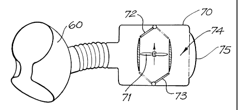
In the fifth embodiment shown in Fig. 11, the cuff 60 is closely coupled to a
fluid-filled air-tight housing 70 that has therein a pump, in the form of
rotatable impeller
71 ar;d a pair of valves 72 and 73 for directing the flow of the impeller 71.
The housing
also includes an inlet/outlet 76 in fluid communication with the inlet/outlet
port 64 of the
cuff 60. A fluid reservoir is also provided in the housing 70 in the form of
an internal
portion 74 of the volume of the housing 70, as is a pressure compliance means,
in the
form of a substantially flexible portion of 75 of the housing 70.
AMENDED SHEk,
UEA/AU
PCT/AUOO/00654
CA 02375962 2001-11-30 Received 19 Apri12001
-16-
In operation, energisation of the impeller 71 with the valves 72 and 73 in the
position shown in Fig. 11 causes fluid to be actively withdrawn from the cuff
60, which
allov3'the aorta to return to its usual circular shape. This fluid is pumped
into the internal
port'ion 74 of the housing 70 and causes the flexible portion 75 to expand to
the position
shown in Fig. 11. When the valves 71 and 73 are in the positions shown in
phantom in
Fig. 11 and the impeller 71 is energised, the fluid in the portion 74 is
pumped into the cuff
60 to expand same and to compress the aorta. The removal of fluid from the
portion 74
causes the flexible portion 75 to retract to the position shown in the phantom
in Fig. 11.
As with earlier embodiments, the control of the impeller and valves is in
response to
signals received from an ECG monitor or systemic arterial blood pressure or
the like.
In the sixth embodiment shown in Fig. 12, the device has only a single valve
76.
The aorta is compressed by positioning the valve 76 as shown in Fig. 12 and
energising
the impeller 71. When the valve 76 is moved to the position shown in phantom
in Fig. 2
and impeller is de-energised the expanding aorta passively ejects the fluid
back into the
portion 74 of the housing 71 and causes the flexible portion 75 to expand to
the position
shown in phantom.
In the seventh embodiment shown in Fig. 13, the impeller 71 is driven in one
direction to cause fluid flow in the direction indicated by the arrow to
deflate the cuff 60
and expand the flexible portion 75. Reversing the direction of the impeller 71
causes the
flexible portion 75 to retract to the position shown in phantom as fluid is
displaced into
the cuff 60 to inflate same. This embodiment requires variable power control
to the motor
driving the impeller 71 and a plot of the motor power requirements (Po)
relative to the
subject's electro cardiograph reading (ECG) and aortic pressure (Pr.) are
shown in Fig.
14.
In the eighth embodiment shown in Figs. 15 and 16, the housing 71 has a rigid
upper portion 71a and a partially rigid lower portion 71b that includes the
flexible portion
75. A motor 77 is mounted in the lower portion 71b that drives a pair of
rollers 78, each
positioned on an end of a common shaft 79. The housing portion 71b also has a
pair of
upstanding guide posts 80 which are slidably received in corresponding holes
in a swash
plater81. The swash plate 81 has a pair of cam formations 82 on its underside.
A fluid-
filled sac 83 is positioned between the swash plate 81 and the housing portion
71a. The
interior of the sac 83 is in fluid communication with the interior of the cuff
60. Power is
supplied to the motor 77 through line 84.
AMENDED SHEE,
IPEAIAU
PCT/AUOO/00654
CA 02375962 2001-11-30 Received 19 April 2001
-17-
In operation, the motor 77 is energised to rotate the rollers 78, which ride
along
the cam formations 82 to drive the swash plate 81 upwards to compress the sac
83 and
eject-the fluid therein into the cuff 60 to inflate same. When the rollers 78
have passed
the cams 82 the swash plate 81 returns to its original position and the
expanding aorta
passively ejects the fluid back into the sac 83. In an alternative embodiment
(not shown),
the rollers 78 are linked to the cam formations 82 to drive the swash plate 81
up and down
and thereby actively inflate and actively deflate the cuff 60. As a further
alternative, (not
shown) a stepper motor(s) can be used to drive the swash plate.
In the ninth embodiment shown in Fig. 17, the housing 71 has a fluid filled
sac
io 83 positioned between a pair of compression plates 84 which are hinged at
85 and driven
by a solenoid 86. Energising the solenoid 86 brings the plates 84 together to
squeeze the
sac 83 and force the liquid therein into the cuff 60 to inflate same. De-
energising the
solenoid 86 draws the plates 84 apart and the expanding aorta passively ejects
the fluid
back into the sac 83. As with earlier embodiments, as the sac 83 inflates the
flexible
portion 75 of the housing 71 expands to accommodate the increase in pressure
in the
housing 71.
In the tenth embodiment shown in Fig. 18, the heart assist device includes a
liquid pressure adjustment means, in the form of remote reservoir 90,
connected between
the cuff 60 and the reservoir 74. Liquid can be added to the heart assist
device, via the
remote reservoir 90, to adjust the liquid retained in the (de-activated) cuff
60 and thereby
adjust the pressure therein. This allows the size of the cuff 60 to be
adjusted to
compensate for changes in the size of the aorta and/or the amount of aortic
compression to
be adjusted to, for example, wean the patient from the heart assist device.
When the
reservoir is positioned near the skin, its volume can be adjusted by using a
needle to inject
or withdraw liquid. When the reservoir is positioned near the heart assist
device, its
volume can be adjusted by adding or withdrawing liquid via a transcutaneous
tube. The
pressure in the reservoir 90 can also be sensed and automatically adjusted so
as to
maintain a predetermined pressure.
It will be appreciated that the system and device of the present invention, in
their
preferred forms, are designed to be simple with no blood contact and a much
lower
morbidity risk compared to LVADs. The device and system allows the heart to
remain
totally un-instrumented, and the device, by effective counterpulsation in the
aorta,
augments the cardiac output up to 15-20%. All natural blood pathways are
maintained.
AMENDED SHEE
MEA/AU
CA 02375962 2001-11-30 PCT/AUOO/00654
Received 19 April 2001
-18-
Pulsatile blood flow is also maintained. The patient is able to ambulate and
there is no
risk of leg ischaemia.
The present invention provides for long term relief and/or stabilization/ of
or
recovery from chronic heart failure. Moreover the present invention may be a
suitable
bridging device for transplantation.
The device and system of the above-described embodiments improve cardiac
work efficiency by reducing the afterload (pressure/resistance to flow which
the heart has
to overcome to eject blood) during systole (ejection phase), by augmenting
diastolic aortic
blood pressure to maintain a greater mean arterial pressure, and by increasing
left
ventricular coronary artery blood flow during diastole.
The preferred embodiments of the heart assist device compress the ascending
aorta. This is advantageous as the ascending aorta is less prone to disease
than the
descending aorta and, being closer to the heart, provides improved pumping
efficiency
and thus a smaller heart assist device.
It will be appreciated by persons skilled in the art that numerous variations
and/or modifications may be made to the invention as shown in the specific
embodiments
without departing from the spirit or scope of the invention as broadly
described. For
example, although the invention has been described in specific reference to
compression
of the aorta, the devices, systems and methods of the present invention can
equally be
used for the compression of the pulmonary artery to effectively act as a right
ventrical
assist device, and the present invention extends to this alternative aspect.
The present
embodiments are, therefore, to be considered in all respects as illustrative
and not
restrictive.
AMENDED SHEE o
IPENAU