Note: Descriptions are shown in the official language in which they were submitted.
CA 02378071 2002-01-09
WO 01/05320 PCT/USOO/19505
ELECTROSURGICAL LESION LOCATION DEVICE
BACKGROUND OF THE INVENTION
The present invention relates generally to the field of electrosurgical
instruments. More specifically, it relates to a device that electrosurgically
fixes
and identifies the location, in situ, of a pathologically suspect tissue mass
in a
patient's body, for facilitating the accurate surgical removal of the mass.
It is often medically desirable to remove a pathologically suspect tissue
mass, such as a suspected tumor or lesion, from a patient's body. For example,
in
treating breast cancer, a suspicious tissue mass is typically identified and
localized
by imaging means, such as mammography or ultrasound. Once localized, the
mass is typically subjected to a biopsy to determine whether or not it is
malignant.
Often, the biopsy will be an "open" biopsy, in which all or part of the
identified
mass is surgically removed, sometimes with a surrounding margin of tissue.
The identification and localization of the suspect mass is usually performed
by a radiologist. The patient is then typically transported to an operating
room for
surgery. To allow the surgeon to be able to locate the identified mass, the
radiologist places one or more localization wires or "Kopan's" wires into the
breast to define and locate the tissue mass to be removed. In using a
localization
wire, a hollow needle or cannula, containing the localization wire, is
inserted into
the breast under local anesthesia, while the breast is under compression
during the
imaging procedure, until the distal end of the localization wire passes
through the
suspect mass. The localization wire is anchored distally beyond the mass by
means such as a barb or hook at the distal end of the wire. The cannula is
then
removed from the body, leaving the wire in place and extending from the body
as
a marker for the surgeon.
The above-described procedure has certain shortcomings, however. One
problem stems from the fact that the localization wire is inserted while the
breast
is under compression during mammography. When the breast is released from
CA 02378071 2002-01-09
WO 01/05320 PCT/USOO/19505
2
compression, the distal end of the wire often migrates and thus shifts
position with
respect to the targeted tissue mass. This may lead to inaccurate placement of
the
incision for the biopsy, with the result that either an excess of tissue
outside of the
target tissue mass is removed, or less than all of the target tissue mass is
removed.
In addition, the wire is sometimes inadvertently shifted, severed, or pulled
out
during surgery, thereby defeating its purpose of accurately guiding the
surgeon to
the target tissue mass. Any inaccuracies in guiding the surgeon can result in
larger than necessary amounts of healthy tissue being removed, with resultant
deformation and scarring of the breast, or in the need to re-enter the
incision site
to remove parts of the target tissue mass that were missed on the first biopsy
attempt.
Another shortcoming associated with prior art localization devices is that,
while the location of the target tissue mass can be marked, no indication is
provided of the dimensions of the mass. Thus, accurate removal of the desired
amount of tissue depends on the surgeon's ability to determine the boundaries
of
the tissue mass during surgery.
It would therefore be advantageous to provide a localization device that
minimizes or eliminates the aforementioned problems associated with the
migration and inadvertent removal of the localization wire. It would be
further
advantageous for such a device to provide an accurate indication of the
dimensions and boundaries of the target tissue mass. Furthermore, such a
device
should be easy to use, and should be compatible with existing imaging
equipment
and surgical methods.
SUMMARY OF THE INVENTION
Broadly, the present invention is a device for localizing a target tissue mass
in a body, comprising a tubular trocar with at least a first plurality of
locator wires
that are movable between a retracted position fully contained within the
trocar and
a deployed position extending radially from the trocar. In a preferred
embodiment, the device includes an electrosurgical cutting element at its
distal
CA 02378071 2002-01-09
WO 01/05320 PCT/US00/19505
3
end, and first and second pluralities of locator wires that, when deployed,
respectively define first and second locating perimeters. The first plurality
of
locator wires is connected to a first tubular wire-carrying member
longitudinally
mounted for axial movement within the trocar between a proximal position
corresponding to the retracted position of the first plurality of locator
wires, and a
distal position corresponding to the deployed position of the first plurality
of
locator wires. The second plurality of locator wires is connected to a second
tubular wire-carrying member longitudinally mounted in the trocar, coaxially
with
the first tubular member, for movement between a distal position corresponding
to
the retracted position of the second plurality of locator wires, and a
proximal
position corresponding to the deployed position of the second plurality of
locator
wires. The trocar has a portion having first and second pluralities of slot-
shaped
apertures through which the first and second pluralities of locator wires
emerge
when moved to their respective deployed positions.
BRIEF DESCRIPTION OF THE DRAWINGS
Figure 1 is a perspective view of an electrosurgical device constructed in
accordance with the present invention;
Figure 2 is a perspective view of the distal end of the tubular trocar of the
device illustrated in Figure 1;
Figure 3 is a longitudinal cross-sectional view of the tubular trocar, taken
along line 3 - 3 of Figure 2, showing the locator wires in their retracted
position;
Figure 4 is a cross-sectional view taken along line 3 - 3 of Figure 2, but
with the tubular members of the trocar arranged to partially deploy the
locator
wires;
Figure 5 is a transverse cross-sectional view of the trocar, taken along line
5 - 5 of Figure 3;
CA 02378071 2002-01-09
WO 01/05320 PCT/USOO/19505
4
Figure 5A is a transverse cross-sectional view of the trocar, taken along
line 5A - 5A of Figure 3;
Figure 6 is a transverse cross-sectional view of the trocar, taken along line
6- 6 of Figure 3;
Figure 6A is a transverse cross-sectional view of the trocar, taken along
line 6A - 6A of Figure 3;
Figure 7 is a top view, partially in cross-section, of the handle portion of
the device illustrated in Figure 1;
Figure 8 is a view of the interior of the handle portion of the device
illustrated in Figure 1, with the outer shell of the handle portion removed,
showing the mechanism arranged for the locator wires of the device to be in
their
retracted position;
Figure 9 is a view similar to that of Figure 8, showing the mechanism
arranged for the locator wires of the device to be in their deployed position;
Figure 10 is a perspective view of the distal portion of the trocar of the
device, with the locator wires in their deployed position;
Figure 11 is a side elevational view of the distal portion of the trocar of
the
device, with the locator wires in their deployed position;
Figure 12 is a side elevational view of a part of the handle portion of the
device, with the trocar removed;
Figure 13 is a perspective view of the attachment fitting by which the
trocar of the device is removably attached to the handle portion;
Figure 14 is a cross-sectional of a modified form of a locator wire that may
be used in the present invention;
CA 02378071 2007-11-22
CA 02378071 2002-01-09
WO 01/05320 PCTIUSOO/19505
Figure 15 is a cross-sectional of another modified form of a locator wire
that may be used in the present invention; and
Figures 16 and 17 are cross-sectional views of a part of the handle portion
of the present invention, showing the electrical switching mechanism and the
5 locking mechanism used in the preferred embodiment.
DETAILED DESCRIPTION OF A PREFERRED EMBODIMENT
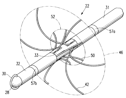
Referring now more particularly to the drawings, Figure I illustrates a
particular embodiment of an electrosurgical lesion location device 20
constructed
in accordance with the present invention. The lesion location device 20
includes
an elongated, tubular trocar portion 22 and a handle portion 24. The proximal
end
of the trocar portion 22 is fixed to an attachment fitting 26, by which the
trocar
portion 22 is removably attached to the handle portion 24. A detailed
description
of the trocar portion 22 will be provided first, followed by descriptions of
the
attachment fitting 26 and the handle portion 24.
The Trocar Portion
The trocar portion 22 (hereinafter referred to as the trocar 22) extends
between the proximal attachment fitting 26 and a hemispherical distal tip 28,
the
latter preferably being formed of high density polyethylene (HDPE) or the
like.
The trocar 22 is advantageously provided with a cutting element 30 that
extends
distally from the distal tip 28 of the trocar 22. The cutting element 30 is
preferably an electrosurgical electrode, such as the type disclosed and
claimed in
PCT Application No. PCT/US99/21416 (published under WO 00/16697 on March
30, 2000) for "Electrosurgical Biopsy Device and Method ".
The cutting element or electrode 30 is
preferably made of 302 stainless steel wire, of approximately 0.014 in.
(approximately 0.36 mm) diameter. As explained below, the electrode 30 is
activated with radio frequency (RF) electrical energy to ablate adjacent
tissue.
CA 02378071 2002-01-09
WO 01/05320 PCT/USOO/19505
6
In the preferred embodiment illustrated, the elongate trocar 22 comprises a
proximal trocar tube 31 and a distal trocar tube 32, connected by an
intermediate
member 33. The trocar tubes 31, 32 and the intermediate member 33 may be
formed of a sturdy, high impact biocompatible material, such as medical grade
polymer (e.g., polycarbonate). In a particular example of the device, the
outside
diameter of the trocar 22 is approximately 0.125 in. (approximately 3.2 mm),
although this dimension is exemplary only.
Electrosurgical techniques have been used in a variety of circumstances. In
electrosurgery, high frequency electrical energy is applied through a primary
electrode to tissue. The electrical energy flows through the tissue to a
return
electrode. The tissue adjacent to the primary electrode is ablated, to form an
opening in the tissue. The return electrode in monopolar electrosurgery may be
an
electrode placed on the exterior of the patient's body at a point remote from
the
primary electrode. In bipolar electrosurgery, the return electrode may be a
smaller
electrode positioned somewhat near the primary electrode. An exemplary biopsy
instrument using electrosurgical techniques is described in International
Application Number PCT/US97/15092 (Applicant Ethicon Endo-Surgery),
published under the Patent Cooperation Treaty on 5 March 1998 with
International Publication Number WO 98/08441. Another electrosurgical biopsy
instrument is disclosed and claimed in above-mentioned PCT Application No.
PCT/US99/21416, published under WO 00/16697 on March 30, 2000.
The illustrated embodiment of the present invention uses monopolar
electrosurgical techniques to cut through subcutaneous tissues to reach a
target
tissue mass. Electrical energy is provided to the electrode 30. The return
electrode (not shown) is attached to the patient's body remote from the point
at
which the trocar 22 is inserted to provide the return electrical path.
Alternatively,
the electrosurgical aspect of the device may be bipolar, in which the return
electrical path is provided by a return electrode (not shown) on the device
itself.
A conductor 34 (Figure 3) extends axially through the interior of the trocar
portion 22 to conduct electrical energy from the handle portion 24 of the
device to
CA 02378071 2002-01-09
WO 01/05320 PCT/USOO/19505
7
the electrosurgical electrode 30. The conductor 34 is insulated to maintain
electrical isolation from the adjacent components, which may be of conductive
metal, as explained below.
Referring now to Figures 3 through 6A, the intermediate member 33 of the
trocar 22 has a proximal portion 35 that is dimensioned to fit snugly into the
open
distal end of the proximal trocar tube 31, and a distal portion 36 that is
similarly
dimensioned to fit snugly into the open proximal end of the distal trocar tube
32.
The proximal portion 35 is provided with a first plurality of longitudinal
grooves
or channels 37 in its outer surface, each of which opens into one of a first
plurality
of axially-elongate, slot-like apertures 38 that are formed in the
intermediate
member 33 of the trocar 22. The distal portion 36 of the intermediate member
33
is formed with a second plurality of longitudinal grooves or channels 39, each
of
which opens into one of a second plurality of axially-elongate, slot-like
apertures
40 that are also formed in the intermediate member 33, distally from the first
plurality 3 8. The axial locations of the first and second pluralities of
apertures are
such that the distal ends of apertures 3 8 in the first plurality overlap
slightly with
the proximal ends of the apertures 40 in the second plurality. The apertures
38 in
the first plurality alternate with the apertures 40 in the second plurality
around the
circumference of the trocar portion 22. In the illustrated embodiment, there
are
six apertures in each of the first and second pluralities, but this number may
be
varied from as few as two to ten or more. The apertures of each plurality are
evenly spaced around the circumference of the trocar.
Figure 3 shows a first plurality of locator wires 42 contained in a retracted
position within the trocar portion 22. Each of the first plurality of locator
wires 42
is associated with a corresponding one of the first plurality of apertures 38.
Each
of the first wires 42 has a proximal end that is attached to the distal end of
a first
tubular wire-carrying member 44 that is mounted for axial movement within the
interior of the proximal trocar tube 3 1. The distal end of each of the first
plurality
of wires 42 is movably journaled in one of the channels 37 in the proximal
portion
35 of the intermediate member 33. In their retracted position, the first
locator
CA 02378071 2002-01-09
WO 01/05320 PCT/US00/19505
8
wires 42 are aligned substantially longitudinally within the proximal trocar
tube
31, and they are ftilly contained therein. The first locator wires 42 are
movable
between their retracted position, shown in Figure 3, and a deployed position,
shown in Figures 10 and 11. In their deployed position, the first locator
wires 42
extend substantially radially from the intermediate member 33 of the trocar
22, to
define a first locating perimeter 42 (Figure 10). The first locator wires 42
may be
tensioned to provide a radius of curvature of about 0.295 in. (about 7.5 mm),
and,
in their deployed position, they form a first locating perimeter 46 with a
defined
diameter of about 1.2 in. (about 30 mm). This diameter is exemplary only; both
small and larger locating perimeters may be defined by using locator wires of
different lengths. The first wire-carrying member 44 is longitudinally mounted
for axial movement within the interior of the proximal trocar tube 3 1,
between a
first or proximal position, and a second or distal position. Figure 3 shows
the first
wire-carrying member 44 in its proximal position, corresponding to the
retracted
position of the first locator wires 42. As shown in Figure 4, as the first
wire-
carrying member 44 moves axially toward its distal position, it moves the
first
locator wires 42 attached to it through the channels 37 of the proximal
portion 35
of the intermediate member 33, and then into and through the slot-shaped
apertures 38 in the first plurality of apertures. When the distal ends of the
first
locator wires 42 encounter the slot-shaped apertures 38, the first locator
wires 42
are allowed to move radially with respect to the trocar 22. Pre-tensioning the
first
locator wires 42 so that they tend to bend in an outward, radial direction
with
respect to the trocar aids in assuring that the first locator wires 42
properly exit the
intermediate member 31 of the trocar 22 through the first slotted apertures 3
8.
Disposed within the distal trocar tube 32 is a second plurality of locator
wires 50. The second locator wires 50 have a retracted position in which they
extend longitudinally within the distal trocar tube 32 and are fully contained
therein. Each of the second plurality of locator wires 50 is associated with a
corresponding one of the second plurality of apertures 40. The second locator
wires 50 are movable between their retracted position, shown in Figure 3, and
a
CA 02378071 2002-01-09
WO 01/05320 PCT/US00/19505
9
deployed position, shown in Figures 10 and 11. In the deployed position, the
second locator wires 50 extend substantially radially from the intermediate
member 33 of the trocar 22, to define a second locating perimeter 52. The
second
wires 50 may also be tensioned to provide a radius of curvature of 0.295 inch
(7.5
mm). When the second locator wires 50 are in their deployed position, the
distal
ends of the second locator wires 50 define a circle having a diameter of
approximately 0.47 in. (approximately 12 mm).
The distal ends of the second locator wires 50 are attached to the distal end
of a second tubular wire-carrying member 54. The proximal end of each of the
second locator wires 50 is movably journaled within one of the channels 39 in
the
distal portion 36 of the intermediate member 33. The second wire-carrying
member 54 is longitudinally mounted for axial movement within the distal
trocar
tube 32. Specifically, the second wire-carrying member 54 is carried co-
axially
within the hollow interior of the first wire-carrying member 44, extending
distally
into the interior of the distal trocar tube 32, after slidably passing through
an axial
bore in the inter-mediate member 33.
The second wire-carrying member 54 is axially movable between a distal
position (Figure 3), corresponding to the retracted position of the second
locator
wires 50, and a proximal position, corresponding to the deployed position of
the
second locator wires 50. As shown in Figure 4, as the second wire-carrying
member 54 moves axially from its distal position to its proximal position, the
second wire-carrying member 54 moves the second locator wires 50 through the
journaling channels 39 in the intermediate member 33 and into the second slot-
shaped apertures 40. When the distal ends of the second locator wires 50
encounter the slot-shaped apertures 40, the second locator wires 50 begin to
move
radially with respect to the trocar. Pretensioning the second locator wires 50
so
that they tend to bend in an outward, radial direction with respect to the
trocar aids
in assuring that the second locator wires 50 properly exit the trocar through
the
second slot-shaped apertures 40.
CA 02378071 2002-01-09
WO 01/05320 PCTIUSOO/19505
The proximal portion 35 of the intermediate member 33 provides a stop to
limit the axial movement of the first wire-carrying member 44 in the distal
direction, away from the handle portion 24 of the device (see Figure 1). When
the
distal end of the first wire-carrying member 44 engages the proximal portion
35 of
5 the intermediate member, the first wire-carrying member 44 is at its distal
position, and the first locator wires 42 are fully deployed.
In the preferred form, the distal end of the second wire-carrying member 54
is diametrically enlarged to provide a flared end 56, to which the second
locator
wires 50 are attached. The diameter of the flared end 56 is greater than the
10 diameter of the axial bore through the intermediate member 33, so that the
distal
portion 36 of the intermediate member 33 provides a stop to limit the axial
movement of the second wire-carrying member 54 in the proximal direction.
When this limit is reached, the second wire-carrying member 54 is at its
proximal
position, which corresponds to the second locator wires 50 being fully
deployed.
Each of the first locating wires 42 and the second locating wires 50
corresponds to one of the first apertures 38 and second apertures 40,
respectively.
In the illustrated embodiment, six first locating wires 42 and six second
locating
wires 50 are uniformly spaced around the trocar. In other embodiments, the
number of first locator wires 42 may differ from the number of second wires
50,
and the number of each may vary from as few as two to ten or more.
Furthermore, the locator wires in each plurality (and especially the first
plurality
42) may be of different lengths to provide locating perimeters of different
shapes
and configurations. For example, the locator wires 42 of the first plurality
may be
dimensioned to provide a hemispherical perimeter to access target tissue
masses
that are near the patient's chest wall, or they may provide an asymmetrical
perimeter if the target tissue mass is near the surface of the patient's skin.
Any
configuration can be provided when it is desired to avoid piercing an adjacent
organ, or penetrating an adjacent cavity.
CA 02378071 2002-01-09
WO 01/05320 PCT/US00/19505
11
When the locating wires 42, 50 that are respectively in the first and second
locator wire pluralities are deployed, their tips define the first and second
locating
perimeters 46, 52, respectively. The curvature of the first locating wires 42
in the
proximal direction and the curvature of the second locator wires 50 in the
distal
direction preferably results in the first and second perimeters 46, 52 being
substantially coplanar (defining a plane that is transverse to the axis of the
trocar
22). Alternatively, the first perimeter 46 may define a plane that lies a
short
distance proximally from the plane defined by the second perimeter 52.
When the trocar 22 is inserted through a target tissue mass, as guided by
mammography, ultrasound, or other techniques, the first and second locator
wires
42, 50 are deployed. When deployed, the first and second locator wires
respectively extend into the tissue surrounding a target tissue mass (such as
a
suspected lesion or tumor) in axially opposite directions, thereby securely
anchoring the trocar in the tissue. Accordingly, the trocar is less prone to
move
within the tissue or to be inadvertently removed therefrom, and thus it
provides a
more accurate guide for subsequent surgery than has previously been possible.
Thus, this anchoring action reduces the possibility that the trocar will shift
position or become dislodged before the surgeon has the opportunity to perform
the appropriate surgery. Furthermore, the first locating perimeter 46 may be
used
to define the periphery of the target tissue mass. Thus, while both
pluralities of
locator wires are secured within the target tissue mass and thus locate and
identify
it, the first locator wires 42 also help identify the outer periphery of the
target
mass, with perhaps an added margin of tissue that is identified for removal
with
the target tissue mass.
In their deployed positions, the distal tips of the first locator wires 42
extend farther from the trocar than the distal tips of the second locator
wires 50.
To provide this capability, the first wire-carrying member 44 has a range of
axial
movement that is greater than the range of axial movement of the second wire-
carrying member 54. Preferably, the range of axial movement of the first wire-
carrying member 44 is about twice the range of axial movement of the second
CA 02378071 2002-01-09
WO 01/05320 PCT/US00/19505
12
wire-carrying member 54. The mechanism that provides these respective ranges
of movement is described below in the description of the handle portion of the
invention.
Furthermore, the first and second locator wires 42, 50 are electrically
energized to provide monopolar electrosurgical tissue penetration with minimal
deployment force. Optionally, the continued electrical energization of the
locator
wires after deployment may result in tissue desiccation that facilitates the
visualization of the target tissue mass by means of color and/or texture
differentiation from surrounding tissue. In this embodiment, the first and/or
second locating wires may be electrically connected to a source (not shown) of
electrical energy to provide for electrosurgical penetration. For this
purpose, the
first and second locating wires 42, 50 may be about 0.009 in. (0.23 mm) in
diameter, and are advantageously formed of 17-7 stainless steel or an
equivalent.
The wires may be coated (except for their distal ends) with a polymer having a
high dielectric strength, so that the tip of each locating wire 42, 50 is the
only part
of the wire that is energized at the time the wires are deployed from the
trocar. In
such an embodiment, the first and second wire-carrying elements 44, 54 are
made
of electrically conductive metal tubing to provide an electrical path along
the
trocar from the handle portion 24 (Figure 1) to the locator wires 42, 50. For
example, the first and second wire-carrying members 44, 54 may be formed of
stainless steel.
Proximal and distal guide marks 57a, 57b may advantageously be provided
on the outer surface of the trocar 22. The proximal mark 57a is spaced a small
distance proximally from the first apertures 38, while the distal mark 57b is
spaced a small distance distally from the second apertures 40.
The distance between the marks 57a, 57b is preferably approximately equal
to the diameter of the first locator wire perimeter 46, thereby defining in
the axial
direction a perimeter that is substantially equal to the first perimeter 46
defined by
the first locator wires 42. The marks 57a, 57b may be made with a material
that is
CA 02378071 2002-01-09
WO 01/05320 PCT/US00/19505
13
easily visible to the surgeon or that is readily detected in the mammography,
x-
ray, ultrasound, or other radiological examination. Alternatively, they can be
illuminated via fiber optic means (not shown).
It may be advantageous to modify the one or more of the first or second
locating wires 42, 50 for better visualization. For example, as shown in
Figure 5
14, a locator wire 42 may be formed with a hollow interior and contain an
optical
fiber 43 that extends out of an open end of the wire. This allows the tip of
the
locating wire to be illuminated for easier visualization of the target tissue
mass
and the surrounding tissue as a guide during surgery. Alternatively, or in
addition,
one or more of the first or second locator wires 42, 50 may be formed as an
open-
ended hollow wire 42", as shown in Figure 15, to provide a passage 47 for the
injection of a dye into the local region of tissue, to assist in guiding the
surgeon in
the subsequent surgery.
The Attachment Fitting
The attachment fitting 26 is best shown in Figure 13. It comprises a
narrow, elongate housing 5 8 having a distal end fixed to the proximal end of
the
proximal trocar tube 3 l, and a proximal end formed into a lip 59. A
transverse
finger 60, the purpose of which will be described below, extends laterally
from
the housing 58 near its distal end. The housing 58 includes a pair of opposed
side
walls 61 that define a channel 63. The first and second wire-carrying members
44, 54, extend into the channel 63 through an opening (not shown) at the
distal
end of the housing 58. The first wire-carrying member 44 has a proximal end to
which is fixed a first attachment lug 62. The second wire-carrying member 54
has
a proximal end, extending outwardly from the open proximal end of the first
wire-
carrying member 44, to which is fixed a second attachment lug 64. The
attachment lugs 62, 64 extend out of the channel opening defined by the ends
of
the side walls 61.
The proximal end of the housing 58 is provided with first and second
electrical contacts 66a, 66b. The first contact 66a is electrically connected
to the
CA 02378071 2002-01-09
WO 01/05320 PCT/US00/19505
14
central conductor 34 that extends proximally out of the proximal end of the
second wire-carrying member 54. The second contact 66b is electrically
connected by a coiled wire to the second wire-carrying member 54. The first
and
second wire-carrying members 44, 54 are formed of an electrically conductive
metal, and the second wire carrying member 54 is contained coaxially within
the
first wire carrying member 44, establishing physical contact between the two
wire-carrying members 44, 54. Thus, an electrical path is established from the
second wire carrying member 54 to the first wire-carrying member 44.
The Handle Portion
The handle portion 24 is described with reference to Figures 1, 7, 8, 9, 12,
16, and 17. As best shown in Figure 1, the handle portion 24 has an outer
housing
70 that i's dimensioned and configured to be held comfortably by the
radiologist or
surgeon operating the device, while being large enough to enclose the internal
electrical and mechanical components that will be described below. The housing
70 is advantageously formed of a rigid, nonconductive polymer material.
The housing 70 includes a longitudinal slot 72 configured and dimensioned
for receiving the attachment fitting 26. The transverse finger 60 of the
attachment
fitting 26 fits into a short transverse slot 73 (Figure 12) that branches off
the
longitudinal slot 72 near its distal end. A latch 74 (best shown in Figures 8
and 9)
extends across the transverse slot 73 to engage the finger 60. When the finger
60
is in the transverse slot 73 and the latch 74 engages the finger 60, the latch
74
holds the attachment fitting 26 in the longitudinal slot 72. The slot 72 has
an
opening 75 (Figure 12) at its proximal end that receives the lip 59 at the
proximal
end of the attachment fitting 26, thereby releasably securing the proximal end
of
the fitting 26 in the slot 72. A spring 77 (Figures 8 and 9) biases the latch
74 in
place across the transverse slot 73. A thumb release 76 attached to the latch
74
permits the user to overcome the bias force of the spring 75 to release the
latch 74,
freeing the finger 60, and thereby permitting the removal of the trocar, 22
and the
attachment fitting 26 from the handle 24. The thumb release 76 is arranged to
CA 02378071 2002-01-09
WO 01/05320 PCT/US00/19505
permit one-handed release of the trocar portion 22 from the handle portion 24
by a
person holding the handle portion 24.
First and second electrical contacts 80, 81 in the longitudinal slot 72
(Figure 12) provide electrical connections between the handle portion 24 and
the
5 first and second contacts 66a, 66b, respectively, in the attachment fitting
26. As
described above, the first and second wire-carrying members 44, 54 may be
electrically conductive to provide such electrical connectivity to the locator
wires
42, 50. The attachment fitting 26 is configured so that when it is inserted
into the
elongate slot 72 of the handle portion 24, appropriate electrical contact is
made
10 between the handle portion contacts 80, 81 and the attachment fitting
contacts
66a, 66b, respectively. For example, the trocar conductor 34 leading to the
electrosurgical electrode 30 may make electrical contact with the first handle
contact 80 through the first attachment fitting contact 66a, and the first and
second
wire-carrying meinbers 44, 54 may make electrical contact with the second
handle
15 contact 81 through the second attachment fitting contact 66b.
Electrical energy is provided to the handle electrical contacts 80, 81 from a
power cord 82 (Figure 1). Power may be supplied to the power cord 82 by any
suitable, commercially available electrosurgical generator (not shown),
preferably
one that generates an output signal having a frequency of about 0.5 MHz to
about
1 MHz. Such generators typically have a foot-pedal operated power switch (not
shown) for turning the electrical power to the handle on and off.
A control lever 83 on the handle portion 24 allows the surgeon selectively
to energize the electrosurgical electrode 30 and the locator wires 42, 50. The
control lever 83 is preferably placed on one side of the handle portion 24 so
that
the surgeon can manipulate the control lever 83 with a thumb or finger of the
same hand the surgeon is using to hold the handle portion 24.
The control lever 83 preferably has two positions: a first position in which
electrical energy is provided to the first handle contact 80 for providing
electrical
energy to the electrosurgical electrode 30; and a second position in which
CA 02378071 2002-01-09
WO 01/05320 PCT/US00/19505
16
electrical energy is provided to the second handle contact 81 for energizing
the
locator wires 42, 50. Specifically referring to Figures 16 and 17, the control
lever
83 is mounted on a shaft 84 that extends into the housing 70. Mounted on the
shaft 84 within the housing is a switch actuator 85 that rotates with the
shaft 84.
The switch actuator 85 is connected to one end of an elongate, flexible
conductive
switching element 86, the other end of which is connected to a terminal 87,
which,
in turn, is electrically connected to wires from the power cord 82. First and
second switch contacts 88a, 88b are provided in the housing, the first contact
88a
being connected by a first wire conductor 89a to the first housing contact 80,
and
the second contact 88b being connected by a second wire conductor 89b to the
second housing contact 81. The switch actuator has a first position (Figure
16),
corresponding to the first position of the control lever 83, in which the
switch
element 86 is brought into contact with the first switch contact 88a, and a
second
position (Figure 17), corresponding to the second position of the control
lever 83,
in which the switch element 86 is brought into contact with the second switch
contact 88b.
Means (not shown) may optionally be included to provide different power
levels to the handle contacts 80, 81. For example, when the switch actuator 85
is
in the first position, power between 60 and 104 watts at 1 MHz may be supplied
to the first handle electrical contact 80. When the switch actuator 85 is in
the
second position, power between 43 and 55 watts at 1 MHz may be supplied to the
second handle electrical contact 81. The electronic circuitry to provide these
dual
power levels is considered to be well within the level of ordinary skill in
the
pertinent arts.
The handle portion 24 includes a deployment mechanism 90 (see Figures 8
and 9) to control movement of the first and second tubular elements 44, 54 of
the
trocar for deploying the first and second locator wires 42, 50 (see Figures 3
and
4). The deployment mechanism 90 includes a first, proximal slider 92, and a
second, distal slider 94. As will be apparent from the following description,
the
first, proximal slider 92 controls the movement of the first tubular element
44 for
CA 02378071 2002-01-09
WO 01/05320 PCT/US00/19505
17
deploying the first locator wires 42. The second, distal slider 94 controls
the
movement of the second tubular element 54 for deploying the second locator
wires 50.
In the embodiment illustrated, the deployment mechanism 90 moves the
first and second sliders 92, 94 simultaneously in opposite directions. The
simultaneous movement of the first and second sliders 92, 94 simultaneously
moves the first and second wire-carrying members 44, 54, thereby also
simultaneously deploying the first and second locator wires 42, 50. A
connecting
element 96 links the first slider 92 and the second slider 94. The connecting
element 96 includes an elongate body that has a first end slot 102 and a
second
end slot 104. The first end slot 102 engages a pin 103 on the first, proximal
slider
92. The second end slot 104 engages a pin 105 on the second, distal slider 94.
The connecting element 96 is pivotally secured to the body of the handle
portion
24 at a pivot point 108.
As noted above, the range of axial movement within the trocar of the first
wire-carrying member 44 that deploys the first locator wires 42 is
approximately
twice the range of axial movement of the second wire-carrying member 54 that
deploys the second locator wires 50. The deployment mechanism 90, and
particularly the connection 96 between the first slider 92 and the second
slider 94,
provides a greater range of movement for the first slider 92 than the second
slider
94. The pivot point 108 is positioned along the body of the connecting element
96
so that the first end of the connecting element 96 (with the first slot 102)
has a
longer range of movement than the second end of the connecting element 96
(with
the second slot 104). The configuration causes the second slider to move about
one half the distance the first slider moves. The first end slot 102 is
approximately twice the length of the second to end slot 104 to accommodate
this
different range of movement.
The connection between each slider 92, 94 and its respective corresponding
wire-carrying member 44, 54 is provided by a transverse extension 110, 112 at
the
CA 02378071 2002-01-09
WO 01/05320 PCT/US00/19505
18
distal end of each slider 92, 94, respectively. As best shown in Figure 12,
the
extension 110 of the first slider 92 has an aperture 111, and the extension
112 of
the second slider 94 has an aperture 113. As shown in Figure 7, the aperture
111
on the first slider 92 receives the attachment lug 62 on the first wire-
carrying
member 44, and the aperture 113 on the second slider 94 receives the
attachment
lug 64 of the second wire-carrying member 54.
A thumb contro198 positioned on the top side of the handle portion 24 is
directly attached to the first, proximal slider 92. Specifically, the thumb
control
98 includes a pin 114 that rides in a slot 116 in the top of the handle
housing 70
(Figure 1). The pin 114 extends into the interior of the housing 70 and is
received
in a recess 118 in the first slider 92, as shown in Figures 8 and 9. As the
thumb
control 98 is moved longitudinally in the slot 116 from a proximal position
(shown in Figure 8) to a distal position (shown in Figure 9) the thumb control
98
moves the first slider 92 longitudinally from its proximal position toward its
distal
position. When the attachment fitting 26 is installed in the handle portion
24, the
first slider 92 is directly connected to the first tubular member 44, so the
movement of the first slider 92 toward its distal position moves the first
tubular
member 44 toward its distal position, deploying the first locator wires 42. As
the
first slider 92 is moving from its proximal position to its distal position,
the
connecting element 96 connecting the first and second sliders 92, 94 causes
the
second slider 94 to simultaneously move from its distal position toward its
proximal position, though at a rate less than (on the order of one-half) the
rate of
movement of the first slider 92. The second slider 94 is connected to the
second
tubular member 54, so that the movement of the second slider 94 directly
corresponds to the movement of the second tubular member 54. Therefore, the
movement of the second slider 94 from its distal position to its proximal
position
moves the second tubular member 54 from its distal position (in which the
second
locator wires 50 are retracted) toward its proximal position (in which the
second
locator wires are deployed).
CA 02378071 2002-01-09
WO 01/05320 PCT/US00/19505
19
It may be advantageous to provide a lock-out mechanism between the
control lever 83 and the locator wire deployment mechanism 90, whereby
deployment of the locator wires 42, 50 is prevented when the lever 83 is
positioned for energizing the electrode 30. An exemplary lock-out mechanism is
shown in Figures 16 and 17. As shown, the switch actuator 85 is formed with a
lobe or finger 120 having a notch 122. When the switch actuator 85 in the
first
position (in which the cutting element 30 is energized, as described above),
the
notch 122 engages a detent 124 at the proximal end of the first slider 92.
This
engagement locks the first slider 92 In its proximal position, thereby
blocking the
movement of both sliders 92, 94 due to their linkage by the linking element
96.
With both sliders 92, 94 inhibited from movement, deployment of the locator
wires 42, 50 is prevented. When deployment of the locator wires 42, 50 is
desired, the switch actuator 85 is rotated by the control lever 83 to its
second
position (Figure 17), releasing the detent 124 from the notch 122, and thereby
unlocking the sliders 92, 94.
Operation
In operation, a target tissue mass is identified through conventional
visualization means, as described above. A small, shallow incision is then
made
(e.g., by a scalpel) at an appropriate place on the patient's body to provide
an
entry site for the trocar 22. To operate the electrosurgical lesion location
device
20 to localize and mark target tissue mass, a surgeon places the control lever
83 its
first position to supply electrical energy to the electrosurgical electrode
30. The
distal tip 28 of the trocar 22 is inserted into the incision and into the
subcutaneous
tissue. The energized cutting electrode 30 cuts into the tissue until the
distal
trocar tip 28 extends through the target tissue mass and the intermediate
portion
33 is located within the target tissue mass, as indicated by mammography or
other
visualization means.
Once the trocar 22 is in place in the tissue, the surgeon moves the control
lever 83 to its second position to activate the electrosurgical tips of the
first and
CA 02378071 2002-01-09
WO 01/05320 PCT/US00/19505
second locator wires 42, 50. The surgeon slides the thumb control 98 from its
proximal position to its distal position. As described above, this movement of
the
thumb control 98 deploys the first and second locator wires 42, 50 into the
tissue,
to anchor the trocar 22 in place, and identify the tissue to be removed in
5 subsequent surgery. Preferably, the trocar 22 is positioned so that the
locator
wires 42, 50 extend to the periphery of the tissue to be removed, as described
above. Once the trocar 22 has been inserted, and anchored with the locator
wires
42, 50, the flow of electrical energy to the trocar 22 is turned off by use of
the foot
pedal switch of the generator (as described above). The thumb release 76 is
10 manipulated to release the finger 60 of the attachment fitting housing 58,
allowing
the trocar 22 to be removed from the handle 24.
The patient can then be removed to the surgical operating room, with the
trocar portion 22 remaining in place, for the surgeon to perform the
appropriate
surgery. The trocar portion 22 is unlikely to shift position in the tissue as
the
15 patient is removed because the locator wires 42, 50 assist in holding the
trocar in
position. This is true even when the tissue is removed from a compressed
condition on a mammography apparatus. When the surgeon opens the tissue
region, the trocar and the deployed locator wires 42, 50 provide the surgeon
direct
indication of the area of tissue to be removed or otherwise operated upon.
20 Those skilled in the art will recognize that various modifications may be
made to the specific embodiment illustrated above without departing from the
spirit of the present invention. For example, numerous modifications may be
made to the handle portion of the device, the mechanism for attaching the
proximal end of the trocar to the handle portion of the device, and to the
specific
mechanism for deploying the locator wires. In addition, while the preferred
embodiment employs an electrosurgical electrode 30 as the incision-making
element, a non-electrical cutting element may work satisfactorily in some
applications. Furthermore, although the preferred embodiment described above
employs two pluralities of locator wires working in opposition (that is, they
are
deployed in radially opposite directions), a device employing only a single
CA 02378071 2002-01-09
WO 01/05320 PCT/US00/19505
21
plurality of locator wires may be suitable in certain procedures. Moreover,
although the preferred embodiment described above employs first and second
pluralities of locator wires that are deployed simultaneously (in unison), it
may be
acceptable to deploy the first and second pluralities of locator wires
sequentially.
These and other modifications that may suggest themselves are considered to be
within the spirit and scope of the invention, as defined in the claims that
follow.