Note: Descriptions are shown in the official language in which they were submitted.
CA 02378438 2002-01-07
WO 01/05374 PCT/CAOO/00843
-1-
Methods for Preparation of Lipid-Encapsulated
Therapeutic Agents
Field of the Invention
This invention relates to a novel method for making particles of lipid-
encapsulated therapeutic agents, and in particular, lipid-encapsulated
therapeutic nucleic acid
particles which may be useful in antisense therapy or gene therapy. .
Background of the Invention
The concept of using lipid particles as carriers for therapeutic agents has
been
considered by numerous people. Formulations have relied on complexation of
therapeutic
agent to the outside of the lipid particle, or actual entrapment of the
therapeutic agent,
although the ability to make formulations of either type depends on a matching
of the
characteristics of the lipids and the therapeutic agent, as well as the
methods employed to
make the particle. In the case of particles with entrapped therapeutic agents,
the entrapment
method may be passive, i.e., the lipid particles are assembled in the presence
of the
therapeutic agent, some of which happens to get trapped; or active, i.e, the
therapeutic agent
is drawn or forced into the interior of a lipid particle as a result of an
induced gradient of
some type. Notwithstanding the many efforts to utilize lipid particles as
carriers, there remain
problems which may limit actual applications of lipid-entrapped therapeutic
agents. These
include low levels of therapeutic agent incorporation on a drug/lipid basis,
low efficiency's of
capture of the therapeutic agent, and lack of a suitable procedure for larger
scale
manufacturing of the lipid-encapsulated therapeutic agent particles.
Large scale manufacturing of fully lipid-encapsulated therapeutic agent
particles has not been achieved where there is a significant electrostatic
interaction between
the lipid and the therapeutic agent. A basic problem is aggregation.
Aggregation normally
results when charged lipid is mixed with oppositely charged therapeutic agent,
resulting in a
solution containing a milky flocculent mass which is not useable for further
processing, let
alone for therapeutic use. The aggregation problem has prevented the
development of
therapeutic compositions which could be of great utility.
Bench scale formulations using charged lipid and oppositely charged
therapeutic agent have been successfully achieved using cationic lipids and
anionic nucleic
o- CA 02378438 2009-05-13
-2-
acids in a passive encapsulation process described in US Pat. No. 5,705,385 to
Bally et al.
(PCT Applic. No. WO 96/40964; See also U.S. issued Patent Nos. 5,981,501;
5,705,385 and 5,976,567) and PCT patent Applic. No. WO 98/51278 to
Semple et al. (See also US Patent Application S.N. 08/856,374) all assigned to
an assignee of
the instant invention. See also Wheeler et al. (1999)
Stabilized plasmid-lipid particles: Construction and characterization. Gen.
Ther. 6:271-281.
These techniques employ an aggregation preventing lipid, such as a PEG-lipid
or ATTA-lipid,
which effectively prevents complex aggregate formation. Resulting fully lipid-
encapsulated therapeutic agent particles have excellent pharmaceutical
characteristics, such as
controlled size (in the 30-250 nm range), full encapsulation (as measured by
nuclease
resistance, for example) and stability in serum.
W098/51278 describes a bench scale procedure for the preparation of the
lipid-encapsulated therapeutic agent particles using passive entrapment. This
known method
employs the two basic steps of lipid hydration and liposome sizing. In the
lipid hydration
step, a cationic lipid solution (95% EtOH solvent) is added dropwise into an
agitated
reservoir containing polynucleotide therapeutic agent in citrate buffer (pH
3.8) to a fmal
composition of 40% EtOH, 9.9 mg/ml lipid and 2.0 mg/ml polynucleotide. Lipid
particles
resulting from this hydration step are typically 400 nm diameter and greater,
which is too
large for general use as a therapeutic. Because of this, extensive post-
formulation processing
such as high temperature extrusion (at 65 C) and optionally freeze-thawing
(from liquid
nitrogen to 65 C waterbath) is required to obtain suitably-sized lipid
particles. The efficiency
of encapsulation using this is fairly high (60-90%) in terms of recovered
final drug:lipid ratio,
but the absolute efficiency of incorporation of starting polynucleotide into
the final particle
formulation is sub-optimal (25-45%).
Commercial large scale manufacturing of these particles is not efficiently
achieved using traditional methods employed in the liposome field. These
problems exist
notwithstanding the great deal of art on the manufacturing of liposome/drug
formulations that
has emerged since the first description of liposome preparation by Bangham,
A.D. et al.
(1965) "The action of steroids and streptolysin S on the permeability of
phospholipid
structures to cations", J. Mol. Biol. 13, 138-147.
CA 02378438 2002-01-07
WO 01/05374 PCT/CAOO/00843
-3-
Known large scale manufacturing techniques for lipid particles can be broadly
classified into the following categories: 1) Lipid Film Hydration (i.e.
Passive entrapment); 2)
Reverse Phase Evaporation; 3) High-Pressure extrusion; 4) and Solvent
injection (dilution)
(see for example US Patent Nos. 4752425 and 4737323 to Martin et al).
Particular
instruments for lipid particle manufacturing disclosed in the art include: US
Patent Nos.
5270053 and 5466468 to Schneider et al; Isele, U. et al. (1994) Large-Scale
Production of
Liposomes Containing Monomeric Zinc Phthalocyanine by Controlled Dilution of
Organic
Solvents. J. Pharma. Sci. vol 83(11) 1608-1616; Kriftner, RW. (1992) Liposome
Production: The Ethanol Injection Technique, in Bruan-Falco et al., eds,
Liposome
Derivatives, Berlin, Springer -Verlag, 1992, pp. 91-100; Kremer et al. (1977)
Vesicles of
Variable Diameter Prepared by a Modified Injection Method. Biochemistry
16(17): 3932-
3935; Batzri, S. and Koin, ED. (1973) Single Bilayer Liposomes Prepared
Without
Sonication, Bioch. Biophys. Acta 298: 1015-1019.
None of the above noted methods or instruments are suitable for scale up of
formulations of charged lipid and oppositely charged therapeutic agents with
the excellent
pharmaceutical characteristics of Bally et al., supra, and Semple et al.,
supra. The
manufacturing techniques set out in Bally et al., supra, and Semple et al.,
supra were
developed only for 1- 100 ml preparations, and are cumbersome and lead to
unsustainable
inefficiencies in large scale manufacturing (i.e. at the scale of 20-200
litres).
The instant invention provides, for the first time, methods for the large-
scale
preparation of fully encapsulated lipid-therapeutic agent particles where the
lipid and
therapeutic agent are oppositely charged. These particles are useful as
therapeutic
compositions and for experimentation and otherwise. It is an object of this
invention to
provide such methods.
Summary of the Invention
In accordance with the present invention, fully lipid-encapsulated therapeutic
agent particles of a charged therapeutic agent are prepared by combining a
lipid composition
comprising preformed lipid vesicles, a charged therapeutic agent, and a
destabilizing agent to
form a mixture of preformed vesicles and therapeutic agent in a destabilizing
solvent. The
destabilizing solvent is effective to destabilize the membrane of the
preformed lipid vesicles
without disrupting the vesicles. The resulting mixture is incubated for a
period of time
CA 02378438 2002-01-07
WO 01/05374 PCT/CAOO/00843
-4-
sufficient to allow the encapsulation of the therapeutic agent within the
preformed lipid
vesicles. The destabilizing agent is then removed to yield fully lipid-
encapsulated therapeutic
agent particles. The preformed lipid vesicles comprise a charged lipid which
has a charge
which is opposite to the charge of the charged therapeutic agent and a
modified lipid having a
steric barrier moiety for control of aggregation. The modified lipid is
present in the preformed
vesicles in an amount effective to retard, but not prevent, aggregation of the
preformed
vesicles. In a preferred embodiment of the invention, effective to provide
efficient formation
of lipid particles on large scale (for example 20-200 liters), a therapeutic
agent solution
comprising nucleic acids (for example antisense oligodeoxynucleotides) is
combined with
preformed lipid vesicles in a 25-40% solution of aqueous ethanol. Incubation
of this mixture
of a period of about 1 hour is sufficient to result in the spontaneous
production of fully
encapsulated therapeutic agent particles.
Brief Description of the Drawings
Fig. 1 shows a possible model of the mechanistic steps of the method of the
invention;
Fig. 2 depicts encapsulation efficiencies as a function of ethanol
concentration
for liposomes containing 10 mol% PEG-Cer;
Fig. 3A depicts the release of calcein entrapped at self-quenching
concentrations in DSPC/Chol/PEG-CerC14/DODAP liposomes as a function of
ethanol
concentration (closed circles) together with the encapsulation efficiencies
obtained using
liposomes of the same lipid composition (open circles);
Fig. 3B illustrates rapid exchange of lipids during the formation of lipid
entrapped nucleic acids using the method of the invention;
Fig 4 shows entrapment efficiencies and calcein leakage data plotted as a
function of temperature;
Figs. 5A, B and C show NMR spectra of lipid-associated oligonucleotides;
Fig. 6 shows a graph of entrapment efficiency plotted as a function of the
initial oligonucleotide-to-lipid ratio; and
Fig. 7 shows encapsulation efficiency for several species of antisense
oligodeoxynucleotides and for plasmid DNA (pDNA).
CA 02378438 2002-01-07
WO 01/05374 PCT/CA00/00843
-5-
Detailed Description of the Invention
Definitions
While the terms used in the application are intended to be interpreted with
the
ordinary meaning as understood by persons skilled in the art, some terms are
expressly
defined to avoid any ambiguity. Thus, as used in the specification and claims
of this
application the term:
charged lipid refers to a lipid species having either a cationic charge or
negative charge or which is a zwitterion which is not net neutrally charged,
and generally
requires reference to the pH of the solution in which the lipid is found.
destabilization refers to modification of the properties of a lipid membrane
as
a result of interaction with a solvent. When the membrane is destabilized, the
fundamental
morphology of the original lipid membrane is preserved. However, the leakage
rate of low
molecular weight solutes increases and lipids can "flip-flop" across the
membrane and
exchange rapidly with other lipid particles. Destabilization of a lipid
membrane is observed
in the invention, for example, at ethanol concentrations of 25-40%. Solvents
which achieve
destabilization but not disruption of lipid vesicles are referred to herein as
destabilizing
solvents.
disruption refers to modification of the properties of a lipid membrane such
that the fundamental morphology of the original membrane is lost. Disruption
of a lipid
membrane is observed, for example, at ethanol concentrations of >60%.
fully encapsulated refers to lipid particles in which the therapeutic agent is
contained in the lumen of a lipid vesicle such as a liposome, or embedded
within a bilayer of
a lipid particle such that no part of the therapeutic agent is directly
accessible to the external
medium surrounding the lipid particle. Lipid particles in which the
therapeutic agent is fully
encapsulated are distinct from particles in which a therapeutic agent is
complexed (for
example by ionic interaction) with the exterior of the particle, or from
particles in which the
therapeutic agent is partially embedded in the lipid and partially exposed to
the exterior
medium. The degree of encapsulation can be determined using methods which
degrade
available therapeutic agent. In the case of a polynucleotide, these methods
include S 1
Nuclease Digestion, Serum Nuclease, and Micrococcal Nuclease analysis.
Alternatively, an
OliGreenTM assay can be employed. In a quantitative sense, a "fully
encapsulated"
therapeutic agent is one where less than 10% of the therapeutic agent, and
preferably less than
CA 02378438 2002-01-07
WO 01/05374 PCT/CA00/00843
-6-
5% of the therapeutic agent in a lipid particle is degraded under conditions
where greater than
90% of therapeutic agent is degraded in the free form. It should further be
noted that
additional therapeutic agent(s) may be associated with the lipid particle by
complexation or
another manner which is not fully encapsulated with out departing from the
present invention.
hydration refers to a common process by which lipid particles, including
liposomes, are formed. In this process, the amount of water in the solvent
surrounding the
lipids is increased from a concentration of around 5% or less (at which
concentration the lipid
molecules are generally individually solvated) to a concentration of 40-60 %
or greater (at
which lipids spontaneously form into membranes, micelles or particles).
lipid refers to a group of organic compounds that are esters of fatty acids
and
are characterized by being insoluble in water but soluble in many organic
solvents. They are
usually divided in at least three classes: (1) "simple lipids" which include
fats and oils as
well as waxes; (2) "compound lipids" which include phospholipids and
glycolipids; and (3)
"derived lipids" such as steroids. A wide variety of lipids may be used with
the invention,
some of which are described below.
preformed vesicle refers to the starting lipid composition used in the method
of the invention which contains lipid vesicles. These vesicles have a self-
closed structure of
generally spherical or oval shape formed from one or more lipid layers and
having an interior
lumen containing a part of the solvent. The vesicles may be unilamellar,
oligolamellar or
multilamellar structures.
The invention disclosed herein relates to a novel method for making lipid-
encapsulated therapeutic agent particles which is particularly applicable to
the large-scale
manufacture of such particles when the lipid and therapeutic agent are
oppositely charged,
such as found in formulations of cationic lipid and anionic polynucleotides.
This invention
relies upon the surprising and unexpected observation that combining preformed
lipid
vesicles with a solution of therapeutic agent can result spontaneously in the
formation of
particles of fully lipid-encapsulated therapeutic agent of a therapeutically
useful size. Thus,
fully lipid-encapsulated therapeutic agent particles are formed in accordance
with the
invention by a method comprising the step of combining a lipid component
comprising
preformed lipid vesicles and a solution of the therapeutic agent and
incubating the resulting
mixture for a period of time to result in the encapsulation of the therapeutic
agent in the lipid
CA 02378438 2002-01-07
WO 01/05374 PCT/CA00/00843
-7-
vesicles. The lipid component further comprises a solvent system which is
effective to
destabilize the membrane of the lipid vesicles without disrupting the
vesicles.
The method of the invention has several important characteristics which make
it of substantial utility to the art. First, it is a large-scale method which
can be used to make
substantial quantities (e.g. >100 g) of the encapsulated therapeutic agent in
a single batch.
Second, the size of the preformed lipid vesicles is substantially maintained,
such that
processing of the lipid particles after introduction of the therapeutic agent
to obtain particles
of therapeutically useful size is not necessary. Third, the efficiency of
encapsulation is high.
Fourth, the amount of therapeutic agent loaded into the particles is high.
The lipid particles used in the present invention are formed from a
combination of several types of lipids, including at least (1) a charged
lipid, having a net
charge which is opposite to the charge of the therapeutic agent; and (2) a
modified lipid
including a modification such as a polyethylene glycol substituent effective
to limit
aggregation. In addition, the formulation may contain a neutral lipid or
sterol. In formulating
the lipid particles using all of the above-mentioned components, the following
amounts of
each lipid components are suitably used: 10 to 40 mol % charged lipid; 25 to
45 mol% neutral
lipid, 35-55 mol% sterol; and 0.5 to 15 mol % modified lipid. Specific lipid
components may
be selected from among the following non-limiting examples.
Charged Lipids
A wide variety of charged lipids and oppositely charged therapeutic agents
may be used with the invention. Examples of such compounds are available and
known to
persons skilled in the art. The following lists are intended to provide
illustrative, non-limiting
examples.
Cationic charged lipids at physiological pH include, but are not limited to,
N,N-dioleyl-N,N-dimethylammonium chloride ("DODAC"); N-(2,3-dioleyloxy)propyl)-
N,N,N-trimethylammonium chloride ("DOTMA"); N,N-distearyl-N,N-dimethylammonium
bromide ("DDAB"); N-(2,3-dioleyloxy)propyl)-N,N,N-trimethylammonium chloride
("DOTAP"); 3(3-(N-(N',N'-dimethylaminoethane)-carbamoyl)cholesterol ("DC-
Chol") and
N-(1,2-dimyristyloxyprop-3-yl)-N,N-dimethyl-N-hydroxyethyl ammonium bromide
("DMRIE"). Additionally, a number of commercial preparations of cationic
lipids are
available which can be used in the present invention. These include, for
example,
CA 02378438 2002-01-07
WO 01/05374 PCT/CAOO/00843
-8-
LipofectinTM (commercially available cationic liposomes comprising DOTMA and
1,2-
dioleoyl-sn-3-phosphoethanolamine ("DOPE"), from GIBCO/BRL, Grand Island, New
York,
USA); LipofectamineTM (commercially available cationic liposomes comprising N-
(1-(2,3-
dioleyloxy)propyl)-N-(2-(sperminecarboxamido)ethyl)-N,N-dimethylammonium
trifluoroacetate ("DOSPA") and DOPE from GIBCO/BRL); and TransfectamTM
(commercially available cationic lipids comprising dioctadecylamidoglycyl
carboxyspermine
("DOGS") in ethanol from Promega Corp., Madison, Wisconsin, USA).
Some cationic charged lipids are titrateable, that is to say they have a pKa
at or
near physiological pH, with the significant consequence for this invention
that they are
strongly cationic in mild acid conditions and weakly (or not) cationic at
physiological pH.
Such cationic charged lipids include, but are not limited to, N-(2,3-
dioleyloxy)propyl)-N,N-
dimethylammonium chloride ("DODMA") and 1,2-Dioleoyl-3-dimethylammonium-
propane
("DODAP").
Anionic charged lipids at physiological pH include, but are not limited to,
phosphatidyl inositol, phosphatidyl serine, phosphatidyl glycerol,
phosphatidic acid,
diphosphatidyl glycerol, poly(ethylene glycol)-phosphatidyl ethanolamine,
dimyristoylphosphatidyl glycerol, dioleoylphosphatidyl glycerol,
dilauryloylphosphatidyl
glycerol, dipalmitoylphosphatidyl glycerol, distearyloylphosphatidyl glycerol,
dimyristoyl
phosphatic acid, dipalmitoyl phosphatic acid, dimyristoyl phosphatidyl serine,
dipalmitoyl
phosphatidyl serine, brain phosphatidyl serine, and the like.
Some anionic charged lipids may be titrateable, that is to say they would have
a pKa at or near physiological pH, with the significant consequence for this
invention that
they are strongly anionic in mild base conditions and weakly (or not) anionic
at physiological
pH. Such anionic charged lipids can be identified by one skilled in the art
based on the
principles disclosed herein.
Neutral Lipids and sterols
The term "neutral lipid" refers to any of a number of lipid species which
exist either in
an uncharged or neutral zwitterionic form a physiological pH. Such lipids
include, for
example, diacylphosphatidylcholine, diacylphosphatidylethanolamine, ceramide,
sphingomyelin, cephalin, cholesterol, cerebrosides and diacylglycerols.
CA 02378438 2009-05-13
-9-
Modified Lipids
Certain preferred formulations used in the invention include aggregation
preventing
lipids such as PEG-lipids or polyamide oligomer-lipids (such as an ATTA-
lipid), and other
steric-barrier or "stealth"-lipids. Such lipids are described in US Patent
Nos. 4320121 to
Sears, 5,820,873 to Choi et al., 5,885,613 to Holland et al., WO 98/51278
(inventors Semple
et al.), and U.S. issued Patent No. 6,320,017 relating to polyamide oligomers.
These lipids prevent precipitation and aggregation of
formulations containing oppositely charged lipids and therapeutic agents.
These lipids may
also be employed to improve circulation lifetime in vivo (see Klibanov et al.
(1990) FEBS
Letters, 268 (1): 235-237), or they may be selected to rapidly exchange out of
the formulation
in vivo (see US Pat. No. 5885613). Particularly useful exchangeable lipids are
PEG-
ceramides having shorter acyl chains (i.e, C14 or C18, referred to herein as
PEG-CerCl4 and
PEG-CerC 18) or PEG-PE having a C 14 acyl chain.
Some lipid particle formulations may employ targeting moieties designed to
encourage localization of liposomes at certain target cells or target tissues.
Targeting
moieties may be linked to the outer bilayer of the lipid particle during
formulation or post-
formulation. These methods are well known in the art. In addition, some lipid
particle
formulations may employ fusogenic polymers such as PEAA, hemagluttinin, other
lipo-
peptides (see US Patent applications SN 08/835,281, and 60/083,294 )
and other features useful for in vivo and/or intracellular delivery.
The preformed lipid vesicles may be prepared in a solution of ethanol or other
organic solvent using a simple lipid hydration step. The percentage of ethanol
or other
organic solvent must be selected such that the lipid particles do not
disassemble or redissolve
into the solvent (generally at >60% ethanol) but provide conditions which
permit the
spontaneous encapsulation process of the invention (approx. 5%-50% ethanol,
more
preferably 25-40% ethanol). Alternatively, additional coniponents such as
detergents may be
included in the lipid vesicle solution which contribute to the destabilization
of the membrane.
For purpose of this specification, "organic solvent" means either a completely
organic solvent
(i.e. 100% ethanol) or a partially organic solvent (such as ethanol in water,
ie. 20% ethanol,
40% ethanol, etc.). A wide variety of water miscible organic solvents may be
used including
ethanol or other alcohols, acetonitrile, dimethylformamide, DMSO, acetone,
other ketones,
and the like. Solvents with greater or lesser polarity may be useful in some
cases. Detergent
CA 02378438 2009-05-13
- 10-
solutions include (3-D-glucopyranoside, TweenTM 20 and those set out in WO
96/40964 and
U.S. issued Patent No. 6,734,171 and any other
detergent or steric barrier compound that can provide the same solubility
features, andJor can
prevent particle aggregation during mixing of oppositely charged lipid and
therapeutic agent.
Preferably all organic solvents or detergent solutions are pharmaceutically
acceptable in trace
amounts, or greater, in order that the fonnulation process does not preclude
patient
administration.
Anionic therapeutic agents include any therapeutic agent with a net negative
charge, or having a negatively charged group that is able to interact with a
cationic lipid
without being blocked by other cationic charge groups of the therapeutic
agent. Such
therapeutic agents include any known or potential therapeutic agent, including
all drugs and
compounds such as, but not limited to, oligonucleotides, nucleic acids,
modified nucleic acids
(including protein-nucleic acids and the like), proteins and peptides with
negative charge
groups, conventional drugs such as plant alkaloids and analogues having
negative charge
groups, and the like. Therapeutic agents which are not inherently anionic may
be derivatized
with anionic groups to facilitate their use in the invention. For example,
paclitaxel can be
derivatized with a polyglutamic acid group linked to the 2' carbon.
Cationic therapeutic agents include any therapeutic agent with a net,positive
charge, or having a positively charged group that is able to interact with a
negative lipid
without being blocked by other negative charge groups of the therapeutic
agent. Such
therapeutic agents include any known or potential therapeutic agent, including
all drugs and
compounds such as, but not limited to modified nucleic acids linked to
cationic charges,
proteins and peptides with positive charge groups, conventional drugs such as
plant alkaloids
and analogues having positive charge groups, and the like. Therapeutic agents
which are'
not
inherently cationic may be derivatized with cationic groups to facilitate
their use in the
invention.
Typically, charged therapeutic agents are initially provided in buffered
aqueous solution, generally containing some amount of ethanol or other organic
solvent. Salt
concentration can strongly effect the self assembly process (see U.S. issued
Patent No. 6,734,171)
employed in the invention, so the buffered salts
employed need to be carefully selected. Further, all buffers must be
pharmaceutically
acceptable, as traces may remain in the final formulation. A suitable buffer
is 300 mM
CA 02378438 2002-01-07
WO 01/05374 PCT/CAOO/00843
-11-
citrate buffer for phosphorothioate oligodeoxynucleotides. For phosphodiester-
based
oligodeoxynucleotides and plasmid DNA which have lower binding affinities, a
buffer of
lower ionic strength is appropriate. For example, typical citarte
concentrations are between
25 and 150 mM, with maximum entrapment occurring at around 50 mM. The amount
of
ethanol or other organic solvent which may be included is controlled by the
solubility of the
therapeutic agent in the aqueous organic mixture, and also by the desired
characteristics of the
final mixture of therapeutic agent and preformed lipid vesicles.
The selection of lipids, destabilizing solvent and therapeutic agents are made
to work in concert to provide fully lipid-encapsulated compositions. Thus, if
the therapeutic
agent is a polyanionic oligonucleotide, the lipid components should be
selected to include
lipids which are cationic under conditions in the stabilizing solvent.
Conversely, if the
therapeutic agent is cationic, the lipids components should be selected to
include lipids which
are anionic under the conditions in the destabilizing solvent. This does not
mean that all of
the lipids included in the lipid solution must be charged, nor does it exclude
the incorporation
of some quantity of like-charged lipids or of zwiterrionic lipids. It merely
means that the
lipid solution should include lipids which have a net charge which is opposite
to the net
charge of the therapeutic agent.
The method of the invention employs relatively dilute solutions of lipid
particles and therapeutic agent. In general, the therapeutic agent solution
will have a
concentration of 1 to 1000 mg/ml, preferably 10-50 mg/ml of the therapeutic
agent, to yield a
final concentration (after mixing with the preformed lipid vesicles) in the
range of 0.2 - 10
mg/ml, preferably about 1-2 mg/ml. Preformed lipid vesicles are combined with
the
therapeutic agent solution such that the resulting lipid concentration (after
mixing with
therapeutic agent solution) is about 1.5 - 30 mg/ml (about 2-40 mM),
preferably 10 mg/ml.
A preferred composition for preformed vesicles for use with polynucleotide
therapeutic agent
is made at the standard lipid ratios (PEG-cerC14: DODAP: DSPC:Chol (molar
ratios
5:25:25:45). This solution, in 100% ethanol, is diluted to 5-50% ethanol,
preferably 40%
ethanol by mixing with aqueous buffer, for example 300 mM citrate, pH 4Ø
Encapsulation results upon stirring the lipid solution and the oligonucleotide
solution together until well mixed, and then incubating with no mixing or
gentle mixing for a
period of from about 1 to 2 hours. The resulting solution is then dialyzed to
remove ethanol
or other material which destabilizes the lipid particle membrane. pH
adjustments may be
M _ _ .. .. ,
CA 02378438 2009-05-13
-12-
used to neutralize surface charges (in the case that the charged lipid is
titratable) in order to
release therapeutic agent which may be complexed with the exterior of the
particle.
At the end of the incubation, the method of the invention results in
spontaneously-formed fully-encapsulated therapeutic agents particles having a
size which is
acceptable for therapeutic use and which can be predicted based on the
starting side of the
preformed lipid vesicles. Thus, in general, a sizing step of the type known in
the art is not
necessary after the addition of the therapeutic agent. This is advantageous
because there is no
requirement for application of mechanical stress to the lipid vesicles after
incorporation of the
therapeutic agent, and thus no risk of loss of or damage to the therapeutic
agent. Should
further sizing of the product particles be desired, however, an optional step
for sizing of the
resulting lipid particles may be employed. Further, a sizing step may be
employed as part of
the preparation of the preformed vesicles prior to the introduction of the
therapeutic agent in
order to obtain starting vesicles of the desired size.
There are several methods for the sizing of lipid particles, and any of these
methods may generally be employed when sizing is used as part of the
invention. The
extrusion method is a preferred method of liposome sizing. see Hope, MJ et al.
Reduction of
Liposome Size and Preparation of Unilamellar Vesicles by Extrusion Techniques.
In:
Liposome Technoloev (G. Gregoriadis, Ed.) Vol. 1. p 123 (1993). The method
consists of
extruding liposomes through a small-pore polycarbonate membrane or an
asymmetric
ceramic membrane to reduce liposome sizes to a relatively well-defined size
distribution.
Typically, the suspension is cycled through the membrane one or more times
until the desired
liposome size distribution is achieved. The liposomes may be extruded through
successively
smaller pore membranes to achieve gradual reduction in liposome size.
A variety of alternative methods imown in the art are available for reducing
the size of a population of liposomes ("sizing liposomes"). One sizing method
is described in
U.S. Patent No. 4,737,323. Sonicating a liposome
suspension either by bath or probe sonication produces a progressive size
reduction down to
small unilamellar vesicles less than about 0.05 microns in diameter.
Homogenization is
another method; it relies on shearing energy to fragment large liposomes into
smaller ones.
In a typical homogenization procedure, multilamellar vesicles are recirculated
through a
standard emulsion homogenizer until selected liposome sizes, typically between
about 0.1
and 0.5 microns, are observed. The size of the liposomal vesicles may be
determined by
CA 02378438 2002-01-07
WO 01/05374 PCT/CAOO/00843
-13-
quasi-electric light scattering (QELS) as described in Bloomfield, Ann. Rev.
Biophys.
Bioeng., 10:421-450 (1981), incorporated herein by reference. Average liposome
diameter
may be reduced by sonication of formed liposomes. Intermittent sonication
cycles may be
alternated with QELS assessment to guide efficient liposome synthesis.
Preferred sizes for liposomes made by the various liposome sizing methods
will depend to some extent on the application for which the liposome is being
made, but will
in general fall within the range of 25 to 250 nm. Specific examples of
suitable sizes are set
out in the Examples below.
In studying the lipid particles made in accordance with the invention, it was
surprisingly found that large empty unilamellar vesicles (LUV), were converted
into
multilamellar vesicles with entrapped therapeutic agent. While not intending
to be bound by
any particular mechanism, it is believed that the process which is occurring
is as shown in
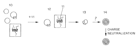
Fig. 1, where a cationic charged lipid and an anionic therapeutic agent are
assumed. The
process starts with a unilamellar vesicle 10 which as a result of the
inclusion of cationic lipids
has positive surface charges on the inside and outside surfaces of the bilayer
wall. Addition
of anionic therapeutic agent, such as antisense oligodeoxynucleotides 11
results in the
formation of an intermediate complex 12 in which the therapeutic agent
molecules 11 are
bound by an ionic/electrostatic mechanism to the oppositely charged lipids on
the surface of
the LUV.
The next step in the process appears to be an aggregation step, in which
aggregates 13 of the LUV/therapeutic agent complexes are formed. This
aggregation step is
very complex and is apparently dependent on the amount of destabilizing agent
(for example
ethanol) and the amount of modified lipid in the preformed vesicles, as well
as being
mediated by the charged therapeutic agent. Some limited knowledge is provided
in the art
about these processes, but they neither predict nor explain the phenomenon
which is the basis
of the present invention. It is known that cationic liposome/DNA complexes
exhibit a large
variety of different structures including clusters of aggregated liposomes
with flat double-
bilayer diaphragms in the areas of contact, liposomes coated with DNA and
(aggregated)
multilamellar structures, where DNA is sandwiched between lipid bilayers
(Gustafsson et al.,
1995; Lasic, 1997; Lasic et al., 1997; Huebner et al., 1999; Xu et al., 1999).
The latter
structures can be flat stacks of bilayers or liposomes, which frequently
exhibit open bilayer
segments on their outer surface. Similar structures have been observed
following binding of
CA 02378438 2002-01-07
WO 01/05374 PCT/CA00/00843
-14-
Ca2+ to negatively charged liposomes (Papahadjopoulos, 1975; Miller and Dahl,
1982; Rand
et al., 1985; Kachar et al., 1986). The structural transformations occurring
in these systems
were attributed to adhesion-mediated processes such as bilayer rupture and
fusion (Rand et
al., 1985; Kachar et al., 1986; Huebner et al., 1999). First, liposomes
aggregate crosslinked
by DNA or Ca2+. Rapid spreading of the contact area deforms the liposomes as
they flatten
against each other. This places the bilayer under increased tension. If the
tension (adhesion
energy) is high enough, the stress imposed on the lipid membrane can be
relieved either by
fusion (increase in area/volume ratio) and/or rupture (volume loss). Most
bilayers rupture
when the area is increased by about 3% (Evans and Parsegian, 1983). Upon
bilayer rupture,
vesicles collapse flattening against each other to form multilamellar stacks.
Membrane-
destabilizing agents such as ethanol can modulate the structural
rearrangements occurring
upon interaction of cationic liposomes with DNA or oligonucleotides.
In the method of the present invention, the formation of multilamellar
liposomes from unilamellar vesicles in the presence of ethanol also points to
an adhesion-
mediated process for their formation. However, the process differs in some way
from the
complexes with their terminated membranes, since the product in this case is
concentric
bilayer shells. While ethanol or a comparable destabilizing agent is required
for the latter
structures to form it is not clear how it affects these structural
rearrangements. These
rearrangements correlate with the loss of the membrane permeability barrier fr
smaller
moeclues and rapid lipid exchange, as well as lipid flip-flop (which
correlates with alcohol
concentration). In addition, the exchange out of the modified lipid from the
LUV may be a
significant factor in to reorganization of the lipid vesicles. In any event,
by some mechanism,
the aggregates 13 rearrange to form multilamellar vesicles 14 with the
therapeutic agent
entrapped between the lamellae and on the inside of the vesicle. This
rearrangement is
dependent not only on the nature of the aggregates formed, but also on the
temperature at
which the aggregates are incubated. Some of the therapeutic agent may also
remain
associated with charges on the exterior of the multilamellar vesicle, and,
these may be
removed by charge neutralization (for example with acid or base in the case of
a titratable
charged lipid), or by ion exchange.
Several characteristics of the lipid vesicles and the destabilizing solvent
were
found experimentally to be of importance to the characteristics of the final
products, and the
CA 02378438 2009-05-13
-15-
selection of these characteristics can be used to control the characteristics
of the product
multilamellar vesicles. These characteristics include:
(1) the inclusion of a charged lipid in the preformed lipid vesicles with a
charge
opposite that of the therapeutic agent;
(2) the inclusion of a modified lipid in an amount sufficient to retard
aggregation,
but not enough to prevent aggregation._ In the case of PEG-CerC14, this amount
was found to
be on the order of 2.5 to 10%;
(3) the inclusion in the destabilizing solvent of a destabilizing agent (such
as
ethanol or detergent) in an amount that destabilizes but does not disrupt the
preformed lipid
vesicles; and
(4) performing the assembly of the fully lipid-encapsulated therapeutic agent
particles at a temperature where the aggregation and the entrapment step are
not decoupled.
In general this will require operation in a temperature range of room
temperature (-20 C) or
above, depending on the concentration of destabilizing agent and the lipid
composition.
The method of the invention can be practiced using conventional mixing
apparatus. For large scale manufacture, however, it may be desirable to use a
specifically
adapted apparatus which is described in a concurrently filed PCT application
No. WO 2001/005373.
The method of the invention will now be further described with reference to
the following, non-limiting examples.
Examples
Materials used in the followina examples are supi2lied as follows:
The phosphorothioate antisense oligodeoxynucleotides and plasmid DNA used in
this study
were provided by Inex Pharmaceuticals (Burnaby, BC, Canada). The rnRNA targets
and
sequences of the oligonucleotides are as follows:
human c-myc, 5'-TAACGTTGAGGGGCAT-3' (Seq ID No. 1);
human ICAM-1, 5'-GCCCAAGCTGGCATCCGTCA-3' (SEQ ID No. 2); and
FITC-labeled human EGFR, 5'-CCGTGGTCATGCTCC-3' (SEQ ID No. 3).
1,2-Distearoyl-sn-glycero-3-phosphocholine (DSPC) was purchased from
Northern Lipids (Vancouver, BC, Canada) and 1,2-dioleoyl-3-
dimethylammoniumpropane
CA 02378438 2009-05-13
-16-
(DODAP), 1,2-dioleoyl-sn-glycero-3-phosphoserine-N-(7-nitro-2-1,3-
benzoxadiazol-4-yl)
(NBD-PS), 1,2-dioleoyl-sn-glycero-3-phosphoethanolamane (DOPE), 1,2-dioleoyl-
sn-
glycero-3-phosphoethanolamine-N-(lissamine rhodamine b sulfonyl) (LRh-PE) as
well as
1,2-dioleoyl-sn-glycero-3-phosphoethanolamine-N-(7-nitro-2-1,3-benzoxadiazol-4-
yl) (NBD-
PE) from Avanti Polar Lipids (Alabaster, AL). 1-Hexadecanoyl-2-(1-
pyrenedecanoyl)-sn-
glycero-3-phosphocholine (Py-HPC) and the oligonucleotide-binding dye OliGreen
were
obtained from Molecular Probes (Eugene, OR). 1-0-(2'-(to-methoxypolyethylene-
glycol)succinoyl)-2-N-myristoylsphingosine (PEG-CerC14), radioactively labeled
[3H]-PEG-
CerCõ as well as 1-0-(2'-(co-methoxypolyethylene-glycol)succinoyl)-2-N-
dodecanoylsphingosine (PEG-CerC20) were provided by INEX Pharmaceuticals
(Burnaby,
BC, Canada). Cholesterol (chol), n-octyl P-D-glucopyranoside (OGP), Triton X-
100, calcein,
dichlorodimethylsilane, sodium hydrosulfite (dithionite), 2-p-
toluidinylnaphthalene-6-
sulfonate (TNS) and polyanetholesulfonic acid (PASA) were obtained from Sigma
(Oakville,
ON, Canada). All materials for transmission electron microscopy including
osmium tetroxide,
lead citrate, maleic acid, sodium cacodylate and the embedding resin Embed 812
were
purchased from Electron Microscopy Sciences (Fort Washington, PA) and low
melting point
(L.M.P.) agarose from Life Technologies (Burlington, Ontario). Cholesterol
(CHOL) was
purchased from Sigma Chemical Company (St. Louis, Missouri, USA). PEG-
ceramides were
synthesized by Dr. Zhao Wang at Inex Pharmaceuticals Corp. using procedures
described in
PCT WO 96/40964. [3H] or [`"C]-CHE was purchased from
NEN (Boston, Massachusetts, USA). All lipids were > 99% pure. Ethanol (95%),
methanol,
chloroform, citric acid, HEPES and NaCI were all purchased from commercial
suppliers.
Analytical Methods: Assays employed to determine if a lipid-therapeutic agent
is
"encapsulated" such as being "fully encapsulated" are set out in WO 98/51278.
Such methods include S1 Nuclease Digestion, Serum
Nuclease, and Micrococcal Nuclease analysis.
The Oligreen Assay was used to quantify the amount of oligonucleotide loaded
into the vesicles. A fluorescent dye binding assay for guantifying single
stranded
oligonucleotide in aqueous solutions was established using a BioluminTM 960
fluorescent
plate reader (Molecular Dynamics, Sunnyvale, California, USA). Briefly,
aliquots of
encapsulated oligonucleotide were diluted in HEPES buffered saline (HBS; 20mM
HEPES,
145mM NaCI, pH 7.5) . A 10 uL aliquot of the diluted sample was added to 100
L of a
CA 02378438 2002-01-07
WO 01/05374 PCT/CAOO/00843
-17-
1:200 dilution of OligreenTM reagent, both with and without 0.1% of Triton X-
100 detergent.
An oligo standard curve was prepared with and without 0.1% Triton X- 100 for
quantification
of encapsulated oligo. Fluorescence of the OligreenTM -antisense complex was
measured
using excitation and emission wavelengths of 485nm and 520nm, respectively.
Surface
associated antisense was determined by comparing the fluorescence measurements
in the
absence and presence of detergent.
Dynamic light scattering. Sizes were determined by dynamic light scattering
using a NICOMP 370 particle sizer (Nicomp Particle Sizing Inc., Santa Barbara,
California).
Throughout the application, number-averaged sizes are presented, which were
obtained by a
cumulant fit from the experimental correlation functions. The polydispersity
is expressed as
the half-width at half-height of a monomodal Gaussian size distribution. The
viscosity of the
ethanol/citrate buffer was determined using an Ubelohde-type viscometer
(Cannon 50). The
viscosity of ethanol/300 mM citrate buffer (40/60 (v/v)) at 23 C measured
relative to water at
the same temperature was found to be 2.674 mPa*s. The zeta potential was
determined by
electrophoretic light scattering using a Coulter light scattering instrument
(DELSA, Coulter
Electronics Inc., FL).
Lipid flip-flop. Lipid flip-flop was determined by chemical reduction of the
fluorescent lipid, NBD-PS, to a nonfluorescent compound with sodium dithionite
(McIntyre
and Sleight, 1991; Lentz et al., 1997). Liposomes were prepared at 20 mM lipid
by extrusion
in the presence of 1 mol% NBD-PS. Only NBD-PS located in the outer monolayer
is
accessible to the reducing agent, dithionite, added to the external medium.
Its redistribution
from the inner monolayer to the outer can be followed after reduction of NBD-
PS in the outer
membrane leaflet. A 1 M sodium dithionite solution was freshly prepared in 1 M
TRIS.
NBD-PS in the outer monolayer was reduced by addition of a 100-fold molar
excess of
sodium dithionite relative to NBD and incubation for 10 min. The completion of
the reaction
was checked by measuring the dithionite fluorescence at 520 nm before and
after reduction
exciting at 465 nm. Excess dithionite was subsequently removed by size
exclusion
chromatography on a Sephadex G50 column. The liposomes were incubated in the
presence
of 40% ethanol and aliquots corresponding to a final lipid concentration of
150 M removed
for measurement at different time points.
Leakage experiments. Ethanol-induced permeabilization of LUVs was measured
at different temperatures and as a function of the size (MW) of the entrapped
solute. Calcein was
CA 02378438 2002-01-07
WO 01/05374 PCT/CA00/00843
-18-
used as a low molecular weight marker for leakage and FITC-dextran (MW 19500)
as a high
molecular weight marker. Leakage of calcein entrapped at self-quenching
concentrations was
followed by monitoring the dequenching of the calcein fluorescence. LUVs were
prepared by
hydration of a lipid film with an aqueous solution containing 75 mM calcein
and 5 mM HEPES
adjusted to pH 7.5 by addition of sodium hydroxide, followed by 5 freeze/thaw
cycles and
extrusion through 2 stacked 100 nm filters (10 passes). In the case of
DSPC/chol/PEG-
CerC14/DODAP extrusion was performed at 60 C. Unentrapped calcein was
exchanged against an
isoosmotic HBS buffer by anion exchange chromatography on a DEAE Sepharose
CL6B column.
The liposome stock solution was diluted to a lipid concentration of 3 M in
HBS containing
varying amounts of ethanol pre-equilibrated at 25, 40 or 60 C. The
fluorescence at 520 nm was
measured (excitation wavelength 488 nm, long-pass filter at 430 nm) with a
Perkin Elmer LS50
Fluorimeter (Perkin Elmer) after 5 min of incubation at the corresponding
temperature. The value
for 100% leakage (maximum dequenching) was obtained by addition of a 10%
Triton X-100
solution to a final concentration of 0.05%. Calcein leakage was calculated
according to
%leakage=(FS Fb)/(FT, Fb)* 100, where Fs is the fluorescence of the sample, Fb
the background
corresponding to calcein containing liposomes in the absence of ethanol and
FTX the Triton X-100
value.
FITC-dextran (MW 19500) was entrapped in DSPC/Chol/PEG-CerC14/DODAP
liposomes incorporating 0.5 mol% LRh-PE at a final concentration of 45 mg/ml.
Entrapment was
performed by addition of the lipids dissolved in ethanol to the FITC-dextran
solution in HBS
followed by extrusion (2 stacked 100 nm filters, 2 passes) and subsequent
removal of ethanol by
dialysis. Unentrapped FITC-dextran was removed by size exclusion
chromatography on a
Sepharose CL4B colurnn (1.5x15 cm). The loss of FITC-dextran from liposomes
exposed to 40%
ethanol was determined after removal of released FITC-dextran by size
exclusion chromatography
on a Sepharose CL4B column (1.5x15 cm). The FITC/LRh-PE ratio was measured
before and
after addition of ethanol. FITC and LRh-PE fluorescence were measured at 515
nm and 590 nm
with the excitation wavelength set to 485 nm and 560 nm, respectively.
Lipid mixing. Ethanol-induced lipid mixing/exchange was followed by the loss
of
resonance energy transfer, occuring between a donor, NBD-PE, and an acceptor,
LRh-PE, which
are in close proximity, upon dilution of the probes into an unlabeled target
membrane (Struck et
al., 1981). LUVs contained 0.75 mol% of both NBD-PE and LRh-PE. Labeled and
unlabeled
liposomes were prepared in HBS pH 7.5 by extrusion at lipid concentrations of
20 mM. Ethanol
CA 02378438 2002-01-07
WO 01/05374 PCT/CAOO/00843
-19-
was added to labeled and unlabeled liposomes to a final concentration of 40%
(v/v). Subse-
quently, the ethanolic dispersions of labeled and unlabeled liposomes were
mixed at a molar lipid
ratio of 1:5 and incubated at the appropriate temperatures. Aliquots were
withdrawn at given time-
points and added to 2 ml of HBS to give a final lipid concentration of 150 M.
Emission spectra
of NBD and LRh were measured in the region from 505 to 650 nm with the
excitation wavelength
set to 465 nm (430 nm emission long-pass filter). After background subtraction
(unlabeled
liposomes at 150 M lipid) the loss of resonance energy transfer was expressed
as the increase in
NBD/LRh ratio.
Pyrene-HPC assay. Pyrene-HPC forms excited state dimers at high concen-
trations, which fluoresce at a different wavelength than the monomers. Excimer
formation is a
diffusion-controlled process and requires two molecules to come together to
form a dimer. Lipid
mixing (target membrane) as well as a decrease in the lateral mobility of
pyrene-HPC in the
membrane can result in a decrease in pyrene excimer fluorescence (Hoekstra,
1990; Duportail and
Lianos, 1996). Lateral phase separation usually results in an increase in
pyrene excimer
fluorescence (Duportail and Lianos, 1996). The rationale of this experiment
was to look at the
effect of oligonucleotide binding on the liposomal membrane. The pyrene-HPC
fluorescence of
liposomes entrapping oligonucleotide was compared to empty control liposomes
before and after
depletion of the transmembrane pH gradient. Increasing the intemal pH to 7.5
results in the release
of membrane-bound oligonucleotides. Liposomes incorporating pyrene-HPC at a
concentration of
7 mol% were prepared by addition of lipids dissolved in ethanol to pH 4
citrate buffer. An aliquot
was removed and oligonucleotide entrapped as described above. The remaining
initial liposomes
were treated the same way in all the subsequent steps (see under entrapment).
The pH gradient
was dissipated with ammonium acetate adjusted to pH 7.5. Liposomes were
diluted into the
appropriate buffer, HBS pH 7.5 or 150 mM ammonium acetate pH 7.5, to a final
lipid
concentration of 2 M. Pyrene-HPC emission spectra were recorded in the
wavelength region
from 365-550 nm with excitation at 345 nm and an emission cut-off filter at
350 nm. The intensity
ratio of monomer fluorescence at 397 nm to dimer fluorescence at 478 nm was
plotted for the
initial liposomes as well as for the oligonucleotide containing liposomes
before and after depletion
of the pH gradient.
31P NMR Spectroscopy. 31P NMR spectra were obtained with a Bruker
MSL200 spectrometer operating at 81 MHz. Free induction decays (FIDs)
corresponding to
800 or 2400 scans were collected by using a 2.8 s 50 pulse with a 3 sec
interpulse delay
CA 02378438 2002-01-07
WO 01/05374 PCT/CAOO/00843
-20-
and a spectral width of 20000 Hz on a 2.0 ml sample in a 10 mm probe. No
proton
decoupling was employed. An exponential multiplication corresponding to 25 Hz
of line
broadening was applied to the FIDs prior to Fourier transformation. The
chemical shift was
referenced to external 85% phoshoric acid (H3P04). The spin-lattice relaxation
times (T,) of
free and encapsulated oligonucleotides at pH 7.5 are essentially the same with
T,free=l.7 sec
and T,eDC,=2 1 sec. The T,-values were measured by an inversion-recovery pulse
sequence.
The interpulse delay of 3 sec for 50 pulses allows for complete relaxation of
all antisense
resonances.
Ultracentrifugation. Liposomes with and without entrapped oligonucleotides
were fractionated by ultracentrifugation on a sucrose step gradient consisting
of 1%, 2.5%,
10% and 15% (w/v) sucrose in HBS pH 7.5 with a step volume of 3.5, 3.5, 2.5
and 1.5 ml,
respectively. Samples were centrifuged for 2 hrs at 36000 rpm (RCFRõaX
221000xg) using a
Beckmann L8-70 ultracentrifuge in combination with a SW41Ti rotor. The
gradient was
either fractionated from the top or individual bands were removed with a
syringe after
puncturing the tube with a needle.
Cryo-Transmission Electron Microscopy (cryo-TEM). A drop of sample was
applied to a standard electron microscopy grid with a perforated carbon film.
Excess liquid
was removed by blotting with filter paper leaving a thin layer of water
covering the holes of
the carbon film. The grid was rapidly frozen in liquid ethane, resulting in
vesicles embedded
in a thin film of amorphous ice. Images of the vesicles in ice were obtained
under cryogenic
conditions at a magnification of 66000 and a defocus of -1.5 micron using a
Gatan cryo-
holder in a Philips CM200 FEG electron microscope.
Freeze-Fracture Electron Microscopy. Samples were cryofixed in the presence
of 25% glycerol by plunging them into liquid Freon 22 cooled by liquid N2. The
fractured
surface was shadowed unidirectionally with platinum (45 ) and coated with
carbon (90 )
employing a Balzers Freeze-Etching system BAF 400D (Balzers, Liechtenstein).
Replicas
were analyzed using a JEOL Model JEM 1200 EX electron microscope (Soquelec,
Montreal,
QC, Canada).
Transmission Electron Microscopy (TEM). Vesicles were fixed by the addition
of 1 volume of 2% osmium tetroxide to 0.5 volumes of vesicles in HBS followed
by
centrifugation at 17000 x g and 4 C for 45 min. The resulting pellet was mixed
with an equal
volume of 3% agarose/PBS, pipetted onto a microscope slide and allowed to cool
to 4 C. The
CA 02378438 2002-01-07
WO 01/05374 PCT/CAOO/00843
-21 -
solidified agarose containing the vesicles was cut into lmm pieces and
transferred to a glass
tube for further processing. The blocks were washed for 3 x 5 min with 0.05 M
maleic acid
pH 5.2 before staining in 2% uranyl acetate for lh. The tissue pieces were
dehydrated through
a graded series of alcohols (50-100%), infiltrated with increasing ratios of
epoxy resin
(EMbed 812):propylene oxide and embedded in 100% EMbed 812 at 60 C for 24h.
Ultrathin
sections were stained with 2% lead citrate and examined using a Zeiss EM 10C
transmission
electron microscope (Oberkochen, Germany)
Phase contrast and fluorescence microscopy. Phase contrast and fluorescence
microscopy were performed on a Zeiss Axiovert 100 microscope using a Plan
Apochromat
63x/1.4NA oil immersion objective in combination with a 1.6x optovar lens and
a XF 100
filter set from Omega Optical (Brattleboro, Vermont) with the following
optical
specifications: excitation 475 20/dichroic 500/emission 535 22.5. Images were
recorded on
Kodak Ektachrome P 1600 color reversal film at 1600 ISO with a Zeiss MC80 DX
microscope camera. Slides and coverglasses were siliconized with
dichlorodimethylsilane to
neutralize the otherwise negatively charged glass surface.
Example 1
Empty preformed vesicles were prepared from a lipid mixture containing
PEG-CerCl4, DODAP, DPSC and CHOL in a molar ratio of 5:25:25:45. The four
lipids
were dissolved in a 100% ethanol to a total lipid concentration of 25 mg/ml
(33 mM). The
ethanolic lipid was then introduced through an injection port with an orifice
diameter of 0.25
mm into a reservoir containing 300 mM citrate buffer, pH 4Ø The reservoir
and all solutions
were at room temperature. The total volume of ethanolic lipid was 6 liters,
and the flow rate
for lipid introduction was 200-300 ml/min. The total volume of citrate buffer
was 9 liters.
The resulting 15 liter mixture had an ethanol concentration of 40% and 180 mM
citrate.
Vesicles of 170 20 nm median diameter were generated. The empty preformed
vesicles
were sized to 90-120 nm median diameter by 1-3 passes through the extrusion
circuit (65 C)
at low pressure (100 p.s.i., reduced from classical 500-1000 p.s.i.) using two
stacked 80 nm
membranes. The empty preformed vesicles were then pooled in a reservoir 20 and
maintained at 40 C until addition of therapeutic agent solution.
Example 2
CA 02378438 2002-01-07
WO 01/05374 PCT/CAOO/00843
-22-
Preformed vesicles of example 1 were used to make fully lipid-encapsulated
therapeutic agent particles using oligonucleotide INX-6295 (Seq. ID No. 1) as
the therapeutic
agent. Oligonucleotide INX-6295 in distilled water was diluted by the addition
of 100 %
ethanol to form a various solutions of 10, 20, 30 40 or 50 mg/ml
oligonucleotide in 40%
ethanol. The ethanolic oligonucleotide was added to the preformed vesicles in
reservoir 20 at
40 C with gentle mixing. The amount and volume of ethanolic oligonucleotide
was
calculated to provide a final drug:lipid ratio of 0.1 to 0.25 by weight. The
mixture was then
incubated at 40 C with gentle and periodic mixing for 1 hour. After
incubation, the solution
was processed by diafiltration to strip free or excess associated
oligonucleotide, remove
ethanol and exchange the buffer system to phosphate buffered saline (PBS), pH
7.4.
Concentration, sterile filtration and packaging complete the preparation of a
commercial
product.
Example 3
The procedure of Example 2 was repeated with changes to various parameters
to determine which might be critical to the preparation of fully lipid-
encapsulated therapeutic
agent particles in accordance with the invention. In these experiments, the
total
oligonucleotide recovery (yield), the total lipid recovery (yield) and the
encapsulation
efficiency were considered as indications of the quality of the product and
the process.
Total oligonucleotide recovery was calculated using the formula :
final oligo concentration (mg / ml) X final volume (ml)
X100%
initial oligo concentration (mg / ml) X initial volume (ml)
Total lipid recovery was calculated using the formula :
final lipid concentration (mg / ml) X final volume (ml)
X100%
initial lipid concentration (mg / ml) X initial volume (ml)
Encapsulation Efficiency (E.E.) was calculated using the formula:
initial oligo (mg / ml) / initial lipid (mg / ml)
X 100%
final oligo (mg / ml) / final lipid (mg / ml)
CA 02378438 2002-01-07
WO 01/05374 PCT/CAOO/00843
- 23 -
The percentage of oligo that is encapsulated (i.e., incorporated in bilayers
or entrapped in the
interior of the lipid particle) was determined with the OliGreen assay
described above.
To assess the significance of the initial drug to lipid ratio, the experiment
was
conducted with two different starting ratios. The results are summarized in
Table 1. No
change in the size of the vesicles was observed in the process of loading the
oligonucleotide.
Table 1
Initial Oligo Lipid Encap Vesicle Final E.E. %
Drug/Lipid Yield % Yield % Oligo % size (nm) Drug/Lipid
Ratio Ratio
0.1 80-90 70-80 _90 106 0.1 100
0.2 60-78 70-75 80 119 0.17-0.2 85-100
To assess the significance of incubation temperature, the experiment was
conducted at room temperatures and at two elevated temperatures for 1 hour.
The results are
summarized in Table 2. As shown, the higher temperature of 60 C begins to
impair the
efficiency of the process, and to lead to an increase in particle size. Thus,
lower temperatures
are preferred.
Table 2
Incubation Temp ( C) Oligo Yield % Encaps Oligo % Vesicle Size (nm)
RT (20-22) 73 90 114
40 84 91 109
60 52 83 140
To assess the significance of incubation time, the experiment was conducted at
three incubation times and an incubation temperature of 40 C. The results are
summarized
in Table 3. As shown, the yield improves between 0.5 hours and 1 hour, but
increased
incubation time beyond an hour does not result in a substantial improvement.
Thus, the most
efficient process in the apparatus used will employ an incubation time of
about 1 hour.
CA 02378438 2002-01-07
WO 01/05374 PCT/CAOO/00843
-24-
Table 3
Incubation time (hr) Oligo Yield % Encapsulated Oligo %
0.5 22 92
1 60 94
2 56 95
To assess the significance of buffer concentration in the oligonucleotide
solution, the experiment was conducted at four different concentrations of
citrate buffer and
an initial drug/lipid ratio of 0.1. The results are summarized in Table 4.
Table 4
Citrate Buffer Conc Oligo Yield % Encapsulated Oligo Vesicle Size (nm)
(mM) %
50 100 94 80
100 88 90 90
200 89 91 93
300 80-90 92 106
To assess the significance of the initial ethanol concentration during the
mixing step, the experiment was conducted with 3 different initial ethanol
concentrations at
each of two initial drug to lipid ratios. The results are summarized in Table
5. There appears
to be an optimum ethanol concentration which is different for each starting
oligo/lipid ratio.
In an addition experiment not reported in the Table, an initial ethanol
concentration of 50 %
was used with an oligo/lipid ratio of 0.1. Significant problems of unknown
cause were
encountered in this experiment and no yield of product was obtained.
CA 02378438 2002-01-07
WO 01/05374 PCT/CA00/00843
- 25 -
Table 5
Initial Initial Oligo Encaps Vesicle Final E.E. %
EtOH % Drug/Lipid Yield % Oligo % size (nm) Drug/Lipid
33 0.2 42-47 88 115 0.12 60
40 0.2 70 82 114 0.15 75
43 0.2 64 62 105 0.19 95
36 0.1 52-66 85-89 110 nd nd
43 0.1 90-100 84-89 116 nd nd
45 0.1 90-100 90-92 108 nd nd
To assess the significance of initial oligonucleotide concentration (and thus
of
the volume of therapeutic agent solution to obtain the same initial drug to
lipid ratio), stock
solutions at four different concentrations of oligonucleotide were used. The
results are
sununarized in Table 6. As shown, this parameter does not appear to be
critical to the results
obtained using the method of the invention.
Table 6
Oligo Stock Initial Oligo Yield % Encaps Oligo % Vesicle Size
mg/ml Drug/Lipid (nm)
10 0.1 85 90 106
20 0.1 80 88 112
0.1 87 90 110
40-50 0.1 80-90 88-94 106
25 Example 4
To demonstrate the applicability of the invention to larger therapeutic
agents,
plasmid pINEX L1018, a 5.5 kb plasmid encoding the luciferase gene linked to a
CMV
promoter, and also carrying SV40 enhancer elements and an AmpL gene was loaded
into
preformed lipid vesicles.
30 Preformed lipid vesicles were prepared by slowly adding 10 mg of lipids
(DSPC/Chol/.DODAP/PEG-CerCl4 in a 20/45/25/10 mol % ratio) dissolved in 100%
ethanol
CA 02378438 2002-01-07
WO 01/05374 PCT/CAOO/00843
-26-
to 25 mM citrate buffer (25 mM citric acid, 255 mM sucrose, adjusted to pH 4
with sodium
hydroxide). Both solutions were prewarmed to 40 C before mixing. The final
ethanol
concentration was 40% (v/v). The ethanolic dispersion of lipid vesicles was
extruded 2X
through 2 stacked 100 nm polycarbonate filters at room temperature. 0.25 mg of
plasmid
DNA in 40% ethanol was added to the lipid vesicles at room temperature,
followed by a 1
hour incubation of the sample at 40 C. The initial plasmidllipid ratio was
0.025.
Subsequently, the sample was dialyzed against 2L of 25 mM saline, pH 7.5 (20
mM HEPES,
150 mM NaCI) for a total of 18-20 hours.
Trapping efficiency was determined after removing remaining external plasmid
DNA
by anion exchange chromatography on a DEAE Sepharose CL6B column. Plasmid DNA
was
quantified using the DNA Binding System PicoGreen lipid by inorganic phosphate
assay
according to Fiske and Subbarrow after separation from the plasmid by a Bligh
Dyer
extraction. In addition, the final lipid concentration was determined by
incorporating 0.25
mol% of the fluorescently-labeled lipid Lissamine rhodamine-PE in the lipid
vesicles.
The final plasmid lipid ratio was 0.022, which corresponds to 88%
entrapment. The resulting lipid-encapsulated therapeutic agent particles had
an average size
of 100 nm and a very small size distribution.
EXAMPLE 5
Liposome preparation: Large unilamellar liposomes in ethanol/buffer
solutions were either prepared by addition of ethanol to extruded liposomes or
by addition of
lipids dissolved in ethanol to an aqueous buffer solution and subsequent
extrusion. Both
methods give the same entrapment results and will be described in greater
detail in the
following:
1. After hydration of a lipid film in pH 4 citrate buffer and 5 freeze/thaw
cycles
LUVs were generated by extrusion through 2 stacked 100 nm filters (10 passes).
In the case
of DODAP/DSPC/Chol/PEG-CerCl4 liposomes the extrusion was performed at 60 C.
Ethanol was subsequently slowly added under rapid mixing. Typical liposome
sizes
determined after removal of ethanol by dynamic light scattering were 90 20
nm for the
DODAP/DSPC/Chol/PEG-CerC14 system. Slow addition of ethanol and rapid mixing
are
important as liposomes become unstable and coalesce into large lipid
structures as soon as the
ethanol concentration exceeds a certain upper limit. The latter depends on the
lipid
CA 02378438 2002-01-07
WO 01/05374 PCT/CA00/00843
-27-
composition. For example, an initially translucent DSPC/Chol/PEG-CerC14/DODAP
liposome dispersion becomes milky white if the ethanol concentration exceeds
50% (v/v).
2. LUVs were prepared by slow addition of the lipids dissolved in ethanol (0.4
ml) to
citrate buffer at pH 4 (0.6 ml) followed by extrusion through 2 stacked 100 nm
filters (2
passes) at RT. Dynamic light scattering measurements performed in ethanol and
after
removal of ethanol by dialysis show no significant differences in size, which
is typically 75
18 nm. The extrusion step can be omitted if ethanol is added very slowly under
vigorous
mixing to avoid locally high ethanol concentrations.
Entrapment procedure. An oligonucleotide solution was slowly added under
vortexing to the ethanolic liposome dispersion, which was subsequently
incubated at the
appropriate temperature for 1 hr, dialyzed for 2 hrs against citrate buffer to
remove most of
the ethanol and twice against HBS (20 mM HEPES/145 mM NaCl, pH 7.5). At pH 7.5
DODAP becomes charge-neutral and oligonucleotides bound to the external
membrane
surface are released from their association with the cationic lipid.
Unencapsulated
oligonucleotides were subsequently removed by anion exchange chromatography on
DEAE-
sepharose CL-6B columns equilibrated in HBS pH 7.5. When octylglucoside was
used in
place of ethanol the detergent was added to liposomes (1:1 v/v) to final
concentrations
ranging from 30-40 mM. All the subsequent steps were performed as described
above except
for the initial dialysis step against pH 4 citrate buffer, which was extended
to 5 hrs. In the
following examples, if not otherwise mentioned, DSPC/ChoUPEG-CerC14/DODAP
liposomes (20:45:10:25 mol%), c-myc (Seq. ID No. 1), 40% (v/v) ethanol, 300 mM
citrate
buffer and incubation at 40 C were used.
Determination of trapping efficiencies: Trapping efficiencies were determined
after removal of external oligonucleotides by anion exchange chromatography.
Oligonucleotide concentrations were determined by UV-spectroscopy on a
Shimadzu
UV 160U spectrophotometer. The absorbance at 260 nm was measured after
solubilization of
the samples in chloroform/methanol at a volume ratio of 1:2.1:1
chloroform/methanol/
aqueous phase (sample/HBS). If the solution was not completely clear after
mixing an
additional 50-100 l of methanol was added. Alternatively, absorbance was read
after
solubilization of the samples in 100 mM octylglucoside. The antisense
concentrations were
calculated according to: c [ g/ l]=A260* 1 OD260 unit [ g/ml] *dilution factor
[ml/ l], where
the dilution factor is given by the total assay volume [ml] divided by the
sample volume [ l].
CA 02378438 2002-01-07
WO 01/05374 PCT/CA00/00843
-28-
OD260 units were calculated from pairwise extinction coefficients for
individual deoxy-
nucleotides, which take into account nearest neighbor interactions. IOD
corresponds to 30.97
g/ml c-myc (Seq. ID. No 1), 33.37 g/ml h-ICAM-1 (Seq. ID No. 2) and 34 g/ml
EGFR
(Seq. ID No. 3). Lipid concentrations were determined by the inorganic
phosphorus assay
after separation of the lipids from the oligonucleotides by a Bligh and Dyer
extraction (Bligh
and Dyer, 1959). Briefly, to 250 l of aqueous phase (sample/HBS) 525 l
methanol and 250
1 chloroform were added to form a clear single phase (aqueous
phase/methanol/chlorofrom
1:2.1:1 vol). If the solution was not clear a small amount of methanol was
added. Subse-
quently, 250 l HBS and an equal volume of chloroform were added. The samples
were
mixed and centrifuged for 5-10 min at 3000 rpm. This resulted in a clear two-
phase system.
The chloroform phase was assayed for phospholipid content according to the
method of Fiske
and Subbarrow (1925). If not otherwise mentioned, trapping efficiencies were
expressed as
oligonucleotide-to-lipid weight ratios [w/w].
EXAMPLE 6
Following the procedures of Example 5, increasing amounts of ethanol were
added to 100 nm DSPC/Chol/DODAP liposomes (no modified lipid component)
prepared by
extrusion. All samples became milky white immediately upon oligonucleotide
addition,
indicating oligonucleotide-induced aggregation. Following incubation with
antisense
oligonucleotides at a molar ODN/lipid ratio of 0.035 at pH 4, ethanol and
unentrapped
oligonucleotides were removed. Table 7 lists encapsulation efficiencies as
determined by
dynamic light scattering, together with the final sizes of the resulting
multilamellar vesicles.
Increasingly more antisense oligonucleotide becomes entrapped as the ethanol
concentration
is increased. The concomitant increase in size and polydispersity reflects a
progressive
reorganization of the LUVs into larger lipid structures, which appear to be
predominantly
large multilamellar liposomes. It should be noted that due to the size of
these systems some of
the lipid is lost on the anion exchange column used to remove external
unentrapped
oligonucleotides. The eluted fraction corresponds to roughly 50-60% of total
lipid. At ethanol
concentrations of 40% and higher the initial liposomes become unstable and
fuse to form a
milky white dispersion.
CA 02378438 2002-01-07
WO 01/05374 PCT/CAOO/00843
-29-
Table 7
% EtOH [v/v] % encapsulation Average size [nm]
0 4.4 148 56
20 20.5 226 104
30 32.5 470 244
after extrusion (no antisense) 99 22
These results demonstrate that ethanol makes the lipid membranes susceptible
to structural
rearrangements which lead lead to entrapment of the oligonucleotide between
the concentric
lamellae of large multilamellar liposomes. However, the size of resulting
liposomes cannot
be readily controlled.
EXAMPLE 7
Liposomes were made containing 2.5 to 10 mol% of modified lipid (PEG-Cer)
and tested using the protocols of Examples 5 and 6. In each case, the decrease
in PEG-Cer
concentration was made up with an increase in DPSC levels. It was found that
the
incorporation of the modified lipid into the liposomes allows the final size
of the antisense-
containing liposomes to be regulated. Liposomes were stable at higher ethanol
concentrations in the presence of PEG-Cer than in its absence. The dispersions
remained
optically translucent in 40% ethanol, although a slight increase in turbidity
was noted for the
sample containing 2.5 mol% PEG-Cer. The increased stability is also reflected
in the higher
amounts of ethanol required for entrapment to occur (Table 8, Fig. 2). Fig. 2
depicts
encapsulation efficiencies as a function of ethanol concentration for
liposomes containing 10
mol% PEG-Cer. Maximum entrapment was reached at 40 % ethanol and ethanol
concentrations in excess of 25% (v/v) (>4.3 M) were required for entrapment to
occur. No
entrapment was found in the absence of ethanol. Table 8 lists trapping
efficiencies and sizes
determined by dynamic light scattering as a function of PEG-Cer content (2.5-
10 mol%) at
the minimum and maximum ethanol concentrations determined from Fig. 2. The
sizes of the
CA 02378438 2002-01-07
WO 01/05374 PCT/CA00/00843
-30-
initial extruded liposomes are given in brackets. The amount of ethanol
required for
entrapment to occur depends on the PEG-Cer content of the liposomes. Liposomes
containing
2.5 mol% PEG-Cer entrapped approximately 15% of the oligonucleotides at 25%
ethanol and
45% in the presence of 40% ethanol. In contrast, at 10 mol% PEG-Cer entrapment
was
virtually abolished in the presence of 25% ethanol (<_5%) and was 60% in 40%
ethanol. In all
cases the initial oligonucleotide-to-lipid ratio was 0.037 (mol/mol).
Entrapment levels
increased from 45% to almost 60% in 40% ethanol when the PEG-Cer content was
increased
from 2.5 to 10 mol%. Liposome size and polydispersity decreased from 131 40nm
to
100 26nm.
Table 8
PEG-CerC14 [mol%] % encapsulation Average size [nm]
25% ethanol
2.5 14.1 125 35(108 26)
10 5 92 18(93 18)
40% ethanol
2.5 45.7 131 40(108 26)
5 50.9 126 36(107 22)
10 56.5 100 26(93 18)
EXAMPLE 8
The perturbing effect of ethanol on lipid membranes has been mainly studied
at low ethanol concentrations (<15% v/v) in relation to changes in lipid
hydration, acyl chain
order, membrane permeability to small ions and induction of chain
interdigitation in DPPC
systems (Slater and Huang, 1988; Barchfeld and Deamer, 1988; Schwichtenhovel
et al. 1992;
Slater et al., 1993; Barry and Gawrisch, 1994; Vierl et al., 1994; Lobbecke
and Cevc, 1995;
Komatsu and Okada, 1996; Holte and Gawrisch, 1997). It was logical to ask
whether
liposomes are still intact at the high ethanol concentrations required for
entrapment. Fig. 3A
CA 02378438 2002-01-07
WO 01/05374 PCT/CAOO/00843
-31 -
depicts the release of calcein entrapped at self-quenching concentrations in
DSPC/Chol/PEG-
CerC14/DODAP liposomes as a function of ethanol concentration (closed circles)
together
with the encapsulation efficiencies obtained using liposomes of the same lipid
composition
(open circles). Both the encapsulation as well as the leakage experiments were
performed at
40 C. Leakage of calcein, a small molecule with a MW of 623, starts at >_30%
ethanol and
reaches a maximum around 40% ethanol. The oligonucleotide entrapment shows a
similar
ethanol dependence indicating that the entrapment is highly correlated with
the
destabilization of the liposomal membrane permeability barrier. In contrast to
calcein, the
release of FITC-dextran (MW 19500) was less than 10% in 40% ethanol. This
shows that the
loss of the permeability barrier is MW dependent, as has also been reported
for detergents
such as octylglucoside (Almog et al., 1990). The liposomes maintained their
morphology in
the presence of 40% ethanol. Phase contrast microscopy of giant liposomes in
40% ethanol
also revealed intact liposomal structures.
Lipids are also able to exchange rapidly between liposomes and between the
inner and outer monolayers of the lipid bilayers comprising the liposomes. As
shown in Fig.
3B, lipid mixing as detected by the NBD-PE/LRh-PE FRET assay is effectively
immediate in
40% ethanol. No increase in vesicle size was observed indicating the lipid
mixing is arising
from rapid lipid exchange between liposomes rather than liposome fusion. The
results shown
in Fig.3B also demonstrate that lipids are able to rapidly migrate (flip-flop)
from one side of
the liposomal lipid bilayer to the other, as shown by the of loss in
fluorescence of NBD-PS
located in the outer lipid monolayer upon chemical reduction with sodium
dithionite.
EXAMPLE 9
The increase in turbidity upon encapsulation indicates that entrapment is
preceded by an initial aggregation step (formation of microaggregates). The
aggregation step
and the entrapment can be decoupled at low temperatures. Samples become turbid
upon or
shortly after addition of oligonucleotide and the turbidity increases over
time. In the absence
of ethanol there is only a slight increase in turbidity following which light
transmission
remains constant. In contrast to samples prepared at 40 C, samples incubated
at 4 C become
translucent again when ethanol is removed and liposomes do not entrap
oligonucleotide.
Entrapment efficiencies are plotted as a function of temperature in Fig. 4
together with
calcein leakage data. Leakage data are presented as the ethanol concentrations
required to
CA 02378438 2002-01-07
WO 01/05374 PCT/CAOO/00843
-32-
induce 50% calcein release. Again there is a qualitative correlation between
the
destabilization of the liposomal membrane and the entrapment efficiency.
EXAMPLE 10
31P-NMR can be used to assay for oligonucleotide entrapment. Figs. 5A and
5B show 31P NMR spectra of c-myc in solution (Fig. 5A) and entrapped in
DODAP/DSPC/Chol/ PEG-CerC14 liposomes (Fig. 5B). Initially, the liposomes
exhibited a
transmembrane pH gradient, where the internal pH is 4 and the external pH 7.5.
Under these
conditions the entrapped oligonucleotides are tightly associated with the
positively charged
liposomal membrane. This immobilization results in the disappearance
(broadening out) of
the NMR signal (Fig. 5B). Upon dissipation of the pH gradient by addition of
ammonium
acetate and adjustment of the external pH to 7.5, DODAP is deprotonated and
the
oligonucleotides dissociate from the liposomal membrane. This is demonstrated
by the
recovery of the NMR signal in Fig. 5C. However, the recovery is incomplete,
about 50% of
the initial signal. The signal attenuation is not due to NMR resonance
saturation. It may be
attributed to two possibilities: either the amount of encapsulated antisense
exceeds its
solubility so that a portion of it precipitates, or the mobility of the
antisense molecules is
spatially constrained e.g. by immobilization between two closely apposing
bilayers (see Fig.
3A). To confirm that the oligonucleotides were encapsulated and localized in
the aqueous
interior of the liposomes, 5 mM MnSO4 was added to the external solution (Fig.
5D). Mn2+ is
a membrane impermeable paramagnetic line broadening agent and will quench the
signals of
all accessible phosphate groups, phospholipids as well as oligonucleotides.
However, the
oligonucleotide signal remained unaffected and disappeared only upon
solubilization of the
liposomes with OGP (Fig. 5E). The whole oligonucleotide signal is recovered
when the initial
liposomes (Fig. 5B) are solubilized with OGP in the absence of Mn2+ (Fig. 5F).
These data
clearly demonstrate that the oligonucleotide is entrapped in the liposomes and
not simply
associated with the external membrane. It should also be noted that entrapped
oligonucleotides were not accessible to the oligonucleotide-binding dye
OliGreen.
The NMR studies describe the interaction between oligonucleotides and
liposomes as seen from the perspective of the oligonucleotides. Changes in
lipid dynamics
and membrane organization can be probed with pyrene-labeled lipids (Duportail
and Lianos,
1996). Pyrene-labeled lipids form excited state dimers at high concentrations,
which fluoresce at a
CA 02378438 2002-01-07
WO 01/05374 PCT/CAOO/00843
-33-
different wavelength than the monomers. Excimer formation is a diffusion-
controlled process and
requires two molecules to come together to form a dimer. The binding of the
oligonucleotides
results in a dramatic reduction of the lateral mobility of all lipid species
relative to control
liposomes, which do not contain oligonucleotides. The membrane is laterally
compressed.
This follows from the observed decrease in excimer fluorescence of pyrene-HPC.
The
depletion of the transmembrane pH gradient results in an increase of the
excimer fluorescence
and restoration of lipid mobility.
EXAMPLE 11
Both the size of the liposomes entrapping antisense as well as the entrapment
efficiency depend on the initial antisense-to-lipid ratio. Fig. 6 shows that
oligonucleotides
can be efficiently entrapped at high antisense-to-lipid ratios. The entrapment
efficiency is
plotted as a function of the initial oligonucleotide-to-lipid ratio. The
binding level at
maximum entrapment is 0.16 mg oligonucleotide per mg of lipid (0.024 mol/mol).
This
corresponds to approximately 2250 oligonucleotide molecules per 100 nm
liposome and
demonstrates the high efficiency of this entrapment procedure. Entrapment
efficiencies are
about 3 orders of magnitude higher than obtained by passive encapsulation.
Upon increasing the oligonucleotide-to-lipid ratio, the size as well as the
polydispersity of the samples increase slightly from 70 10 nm for liposomes
alone to 110 30
for an initial ODN-to-lipid weight ratio of 0.2. Freeze-fracture electron
microscopy showed
an increase in the number of larger liposomes with increasing initial
oligonucleotide-to-lipid
ratios. As an aside it should be noted that the initially translucent liposome
dispersion
becomes increasingly turbid as the antisense-to-lipid ratio is increased.
EXAMPLE 12
It would be expected that the PEG coating would inhibit formation of the
closely opposed membranes observed for the multilamellar structures by TEM.
The fate of
PEG-Cer was therefore examined by using radioactively-labeled PEG-CerC14.
Antisense
oligonucleotides were encapsulated in liposomes containing trace amounts of
[3H]-PEG-
CerC14 in addition to 10 mol% unlabeled PEG-CerC14 and [14C] -cholesterol
hexadecylether
(CHE) as a cholesterol marker at a[3H]/[14C] ratio of 5.9. This ratio
represents an apparent
PEG-Cer/chol ratio and will be used in place of the molar PEG-Cer/chol ratio.
The initial
CA 02378438 2002-01-07
WO 01/05374 PCT/CAOO/00843
-34-
antisense-to-lipid weight ratio was 0.29. Entrapment resulted in a final
antisense-to-lipid ratio
of 0.16. Free PEG-Cer and PEG micelles were separated from liposomes by
ultracentri-
fugation using a sucrose step gradient (1%, 2.5%, 10%, 15% (w/v) sucrose in
HBS). Empty
liposomes band at the interface between 2.5% and 10% sucrose with an apparent
PEG-
Cer/chol ratio of 5.5. This band accounts for roughly 80% of the total lipid.
The antisense-
containing liposomes show a faint band at the same location, which corresponds
to less than
9% of total lipid. However, most of the liposomal antisense migrates down to
the 15%
sucrose layer or pellets at the bottom. A complete analysis of the liposome-
containing
fractions of the gradient is presented in Table 9. The results are
representative for samples
prepared at high oligonucleotide-to-lipid ratios. It can be seen that the
relative PEG-Cer/chol
ratios progressively decrease towards the bottom of the gradient. More than
50% of the PEG-
Cer is lost from the bottom fraction relative to the initial liposomes
(apparent PEG-Cer/chol
ratio 5.5). The DSPC/Chol ratio does not change. 27% of the PEG-Cer can be
found in the
top fractions along with 6.6% of cholesterol. Approximately the same amount of
non
liposome-associated PEG-Cer was found for the empty control liposomes.
Further analysis of the fractions of the above gradient show that the
antisense-
containing liposomes show large differences in their antisense content and
size (Table 9). The
oligonucleotide-to-lipid ratios as well as the average size increase from top
to bottom. Three
main populations can be identified as distinct bands (Table 10). Their
relative proportions
depended on the initial oligonucleotide-to-lipid ratio (Table 10). First,
liposomes entrapping
antisense at low ODN/lipid ratio (0.03-0.05). Secondly, liposomes with an
ODN/lipid ratio of
0.14-0.15 and finally, liposomes with very high ODN/lipid ratios (0.29 mg/mg).
The latter
population decreases in favor of the first two with decreasing initial ODN/l
ratio. It is
optically turbid whereas the other two are translucent. It was attempted to
correlate the
observed differences in entrapment and size to the morphological heterogeneity
seen by cryo-
TEM. Antisense was entrapped at high initial oligonucleotide-to-lipid ratio
(0.28 mg/mg) and
the two main fractions corresponding to fractions 15 and 17 in Table 10 viewed
by cryo-TEM
after removal of sucrose by dialysis. The upper fraction consists exclusively
of bilamellar
liposomes, many of which exhibit bulbs, whereas the bottom fraction contained
a mixture of
bi- and multilamellar liposomes.
CA 02378438 2002-01-07
WO 01/05374 PCT/CAOO/00843
-35-
Table 9
fraction % PEG-Cer % Chol % DSPC Chol/DSPC P E G - S i z e
[mol/mol] C e r/ c h o l Inm1
ratio [r.u.]
b.u. - - - 2.2 5.9 86 24
1-10 26.8 6.6 - - - -
11 13.7 9.2 - - 8.6 89 21
12 4.3 3.9 - - 6.4 -
13 3.6 4.1 - - 5.0 -
14 8.9 11.4 - - 4.5 83 21
27.1 36.6 37.6 2.2 4.2 70 15
10 16 8.5 12.8 - - 3.8 75 16
17 7.1 15.4 15.5 2.2 2.6 129 39
CA 02378438 2002-01-07
WO 01/05374 PCT/CAOO/00843
-36-
Table 10
High initial ODN/lipid ratio Low initial ODN/lipid ratio
Fraction % lipid % ODN ODN/1 % lipid % ODN ODN/1
b.u. - - 0.156 - - 0.1
11 9.2 1.9 0.030 15.7 6.7 0.036
12 3.9 3.1 0.117 14.8 8.9 0.050
13 4.1 3.9 0.140 3.8 3.4 0.09
14 11.4 10.5 0.140 8.1 11.2 0.14
36.6 36.2 0.148 31.5 45.9 0.146
10 16 12.8 14.4 0.168 9.7 15.4 0.16
17 15.4 30.1 0=29 5.3 8.5 0.162
EXAMPLE 13
The addition of oligonucleotides to cationic liposomes in the presence of
15 ethanol can give rise to domain formation. The formation of the
multilamellar liposomes seen
must be preceded by liposome adhesion. However, 10 mol% PEG-Cer completely
inhibits
adhesion in the absence of ethanol. In the presence of ethanol, two effects
could contribute to
liposome adhesion: first, the increase in the amount of non membrane-
incorporated PEG-Cer
through rapid lipid exchange and second, formation of small domains depleted
in PEG-Cer
and enriched in antisense oligonucleotides. The latter possibility was
investigated. The effect
of oligonucleotide binding was visualized by phase contrast and fluorescence
microscopy
using giant DSPC/Chol/DODAP/PEG-CerCl4 liposomes in conjunction with FITC-
labeled
oligonucleotides. Most of the liposomes observed were multilamellar and
displayed internal
structure. In the absence of ethanol, the giant liposomes disintegrated into
irregularly-shaped
aggregates and smaller liposomes on addition of antisense. The green FITC
fluorescence
revealed the location of the oligonucleotides. A completely different picture
is presented in
the presence of 40% ethanol. The initially round liposomes adopt a pear-shaped
form 5-10
CA 02378438 2002-01-07
WO 01/05374 PCT/CAOO/00843
-37-
min after addition of oligonucleotide with the oligonucleotides located in a
semicircle on one
side of these structures. The interior membranes are squeezed out from this
horseshoe, which
detaches and collapses, in particular upon raising the temperature, into a
compact slightly
irregular structure that appears completely green in fluorescence. The
segregation of the
oligonucleotides indicates that ethanol is able to stimulate domain formation.
EXAMPLE 14
The encapsulation procedure of the invention is not dependent on a particular
oligonucleotide and lipid composition, nor is it restricted to the high
citrate concentrations
used. Encapsulation is most efficient in 50 mM citrate buffer and decreases at
higher as well
as at lower citrate concentrations. The size and polydispersity increases
considerably at citrate
concentrations below 25 mM. Fig. 7 shows that other oligonucleotides than c-
myc (Seq, ID
No 1) as well as plasmid-DNA can be efficiently entrapped in
DSPC/Chol/DODAP/PEG-
CerC141iposomes. The initial oligonucleotide-to-lipid weight ratio was 0.1
mg/mg, 300 mM
citrate buffer was used for oligonucleotide entrapment. The pDNA entrapment
was performed
in 50 mM citrate buffer at a pDNA-to-lipid weight ratio of 0.03. Unlike
phosphorothioate
antisense oligonucleotides phosphodiester-based molecules cannot be
encapsulated high ionic
strengths buffers such as 300 mM citrate buffer. This probably reflects
differences in binding
affinities (Semple et al., 2000). In contrast to the efficient encapsulation
of large molecules,
less than 10% of ATP could be entrapped in DSPC/Chol/DODAP/PEG-CerC141iposomes
at
an initial ATP-to-lipid ratio of 0.2 mg/mg. The ATP entrapment was performed
in 50 mM
citrate buffer. Table 5 demonstrates that the entrapment procedure can be
extended to other
lipid compositions including DOPE systems. Preliminary results with negatively
charged
liposomes and positively charged polyelectrolytes including polylysines show
that
entrapment is a general feature of the interaction of polyelectrolytes with
oppositely charged
liposomes in ethanol.
EXAMPLE 15
Octylglucoside (OGP) was used in place of ethanol. The detergent was added
to liposomes (1:1 v/v) to final concentrations ranging from 30-40 mM. All the
subsequent
steps were performed as described as in Example 5 except for the initial
dialysis step against
pH 4 citrate buffer, which was extended to 5 hrs. The oligonucleotide was
shown to be
CA 02378438 2002-01-07
WO 01/05374 PCT/CAOO/00843
-38-
protected from externally added OliGreen, a flourescent oligo-binding dye. The
initial
oligonucleotide-to-lipid ratio was 0.23 (mg/mg). Sizes represent number-
averaged sizes.
DSPC/Chol/DODAP/PEG-CerC14 (20/45/25/10 mol%). The observed levels of
encapsulation
and final particle size are summarized in Table 11.
Table 11
OGP [mM] % encapsulation Size [nm]
30 51 65 12
35 57 100 22
40 55 145 38
Using this invention, and the teachings of this specification, those skilled
in
the art will be able to identify other methods and means for generating fully
encapsulated
lipid-therapeutic agent particles, all of which are encompassed by the claims
set out below.
CA 02378438 2002-01-07
WO 01/05374 PCT/CAOO/00843
-39-
References:
Albersdorfer, A., T., Feder, and E., Sackmann. 1997. Adhesion-induced domain
formation by
interplay of long-range repulsion and short-range attraction force: a model
membrane study.
Biophys. J. 73, 245-57.
Almog, S., B. J., Litman, W., Wimley, J., Cohen, E. J., Wachtel, Y.,
Barenholz, A., Ben-
Shaul, D., Lichtenberg. 1990. States of aggregation and phase transformations
in mixtures of
phosphatidylcholine and octyl glucoside. Biochemistry 29, 4582-4592.
Angelova, M. I., N., Hristova, and I., Tsoneva. 1999. DNA-induced endocytosis
upon local
microinjection to giant unilamellar cationic vesicles. Eur. Biophys. J. 28,
142-50.
Bailey, A.L., and P. R., Cullis. 1994. Modulation of membrane fusion by
asymmetric
transbilayer distributions of amino lipids. Biochemistry 33, 12573-80.
Barchfeld, G. L., and D. W., Deamer. 1988. Alcohol effects on lipid bilayer
permeability to
protons and potassium relation to action of general anesthetics. Biochim.
Biophys. Acta 944, 40-48.
Barry, J. A., and K., Gawrisch. 1995. Direct NMR evidence for ethanol binding
to the lipid-
water interface of phospholipid bilayers. Biochemistry 33, 8082-88.
Bligh, E. G., and W. J., Dyer. 1959. Can. J. Biochem. Physiol. 37, 911-17.
Bozzola, J. J., and L. D., Russell. 1999. Electron Microscopy. Chapter 19 -
Interpretation of
micrographs. Jones and Bartlett Publishers, Toronto.
Chonn, A., and P. R., Cullis. 1998. Recent advances in liposome technologies
and their
applications for systemic gene delivery. Adv. Drug Del. Rev. 30, 73-83.
Duportail, G., and Lianos, P. (1996) Fluorescence probing of vesicles using
pyrene and
pyrene derivatives. In Vesicles (M. Rosoff, ed.). p. 295-366. Marcel Dekker
Inc., N.Y.
Evans, E. A., and V. A., Parsegian. 1983. Energetics of membrane deformation
and adhesion in
CA 02378438 2002-01-07
WO 01/05374 PCT/CAOO/00843
-40-
cell and vesicle aggregation. Ann. N. Y. Acad. Sci. 416, 13-33.
Felgner, P. L., T. R., Gadek, M., Holm, R., Roman, H. S., Chan, M., Wenz, J.
P., Northrop, G.
M., Ringold, and H. Danielson. 1987. Lipofection: a highly efficient, lipid-
mediated DNA
transfection procedure. Proc. Natl. Acad. Sci. USA 84, 7413-17.
Felgner, P. L. 1997. Nonviral strategies for gene therapy. Scientific American
276, 102-106.
Fiske, C. H., and Y., Subbarow. 1925. J. Biol. Chem. 66:325-329.
Gao, X., and L., Huang. 1995. Cationic liposome-mediated gene transfer. Gene
Therapy 2, 710-
722.
Gennis, R. B. 1989. Biomembranes: Molecular Structure and Function. Springer
Verlag, N.Y.
Gustafsson, J., G., Arvidson, G., Karlsson, and M., Almgren. 1995. Complexes
between cationic
liposomes and DNA visualized by cryo-TEM. Biochim. Biophys. Acta 1235, 305-12.
Hirschbein, B. L., and K. L., Fearon. 1997. P-31 NMR spectroscopy in
oligonucleotide research
and development - commentary. Antisense & Nucleic Acid Drug Development 7, 55-
61.
Hoekstra, D. 1990. Fluorescence assays to monitor membrane fusion: potential
application in
biliary lipid secretion and vesicle interactions. Hepatology 12, 61S-66S.
Holte, L. L., and K., Gawrisch. 1997. Detecting ethanol distribution in
phospholipid multilayers
with MAS-NOESY Spectra. Biochemistry 36, 4669-74.
Huebner, S., B. J., Battersby, R., Grimm, and G., Cevc. 1999. Lipid-DNA
complex formation:
reorganization and fusion of lipid bilayers in the presence of DNA as observed
by cryo-electron
microscopy. Biophys. J. 76, 3158-3166.
Hyatt, M.A. 1981. Principles and Techniques of Electron Microscopy. Edward
Arnold
Publishers, London, UK.
Kachar, B., N., Fuller, and R. P., Rand. 1986. Morphological responses to
calcium-induced
CA 02378438 2002-01-07
WO 01/05374 PCT/CA00/00843
-41-
interactions of phosphatidylserine-containing vesicles. Biophys. J 50, 779-88.
Komatsu, H., and S., Okada. 1996. Ethanol-enhanced permeation of
phosphaditylcholine/phosphatidylethanolamine mixed liposomal membranes due to
ethanol-
induced lateral phase separation. Biochinz. Biophys. Acta 1283, 73-79.
Lasic, D. D. 1997a. Liposomes in Gene Delivery. Chapter 7, CRC Press, Boca
Raton
Lasic, D. D., H., Strey, M. C. A., Stuart, R., Podgornik, and P. M., Frederik.
1997b. The structure
of DNA-liposome complexes. J. Am. Chem. Soc. 119, 832-33.
Leckband, D. E., C. A., Helm, and J., Israelachvili. 1993. Role of calcium in
adhesion and fusion
of bilayers. Biochemistry 32, 1127-1140.
Lentz, B. R., W., Talbot, J., Lee, and L.-X., Zheng. 1997. Transbilayer lipid
redistribution
accompanies poly(ethylene glycol) treatment of model membranes but is not
induced by fusion.
Biochemistry 36, 2076-2083.
Lobbecke, L., and G., Cevc. 1995. Effects of short-chain alcohols on the phase
behavior and
interdigitation of phosphatidylcholine bilayer membranes. Biochim. Biophys.
Acta 1237, 59-69.
Lipowsky, R. 1991. The conformation of membranes. Nature 349, 475-81.
Macdonald, P. M., K. J., Crowell, C. M., Franzin, P., Mitrakos, and D. J.,
Semchyschyn. 1998.
Polyelectrolyte-induced domains in lipid bilayer membranes: the deuterium NMR
perspective.
Biochem. Cell Biol. 76, 452-464.
Maurer, N., A., Mori, L., Palmer, M. A., Monck, K. W. C., Mok, B., Mui, Q. F.,
Akhong, and
P. R., Cullis. 1999. Lipid-based systems for the intracellular delivery of
genetic drugs. Mol.
Membr. Biol. 16, 129-140.
McIntyre, J. C., and R. G., Sleight. 1991. Fluorescence assay for phospholipid
membrane
CA 02378438 2002-01-07
WO 01/05374 PCT/CAOO/00843
-42-
asymmetry. Biochemistry 30:11819-27.
May, S., D., Harris, and A. Ben-Shaul. 2000. The phase behavior of cationic
lipid-DNA
complexes. Biophys. J. 78, 1681-97.
Meyer, 0., D. Kirpotin, K., Hong, B., Sternberg, J. W., Park, M. C., Woodle,
and D.,
Papahadjopoulos. 1998. Cationic liposomes coated with polyethylene glycol as
carriers for
oligonucleotides. J. Biol. Chem. 273, 15621-7.
Miller, D. C., and G. P. Dahl. 1982. Early events in calcium-induced liposome
fusion. Biochim.
Biophys. Acta 689, 165-9.
Mitrakos, P., and P. M., Macdonald. 1996. DNA-induced lateral segregation of
cationic
amphiphiles in lipid bilayer membranes as detected via zH NMR. Biochemistry
35, 16714-22.
Needham, D., and E., Evans. 1988. Structure and mechanical properties of giant
lipid (DMPC)
vesicle bilayers from 20 C below to 10 C above the liquid crystal-crystalline
phase transition at
24 C. Biochemistry 27, 8261-69.
Ollivon, M., 0., Eidelman, R., Blumenthal, and A., Walter. 1988. Micell-
vesicle transition of egg
phosphatidylcholine and octyl glucoside. Biochemistry 27, 1695-1703.
Papahadlopoulos, D., W. J., Vail, K., Jacobson, and G., Poste. 1975. Cochleate
lipid cylinders:
formation by fusion of unilamellar lipid vesicles. Biochim. Biophys. Acta.
394, 483-91.
Radler, J.O., I., Koltover, A., Jamieson, T., Salditt, and C.R., Safinya.
1998. Structure and
interfacial aspects of self-assembled cationic lipid-DNA gene carrier
complexes. Langmuir 14,
4272-83.
Rand, R. P., B., Kachar, and T. S., Reese. 1985. Dynamic morphology of calcium-
induced
interactions between phosphatidylserine vesicles. Biophys. J. 47, 483-9.
Sackmann, E. 1994. Membrane bending energy concept of vesicle- and cell-shapes
and shape
transitions. FEBS Let. 346, 3-16.
CA 02378438 2002-01-07
WO 01/05374 PCT/CAOO/00843
-43-
Safinya, C. R., E. B., Sirota, D., Roux, and G. S., Smith. 1989. Universality
in interacting
membranes: The effect of cosurfactants on the interfacial rigidity. Phys. Rev.
Lett. 62, 1134-
37.
Semple, S. C., S. K., Klimuk, T. 0., Harasym, N., Dos Santos, S. M., Ansell,
K. F., Wong,
N., Maurer, H., Stark, P. R., Cullis, M. J., Hope, and P., Scherrer. 2000.
Efficient
encapsulation of antisense oligonucleotides in lipid vesicles using ionizable
aminolipids:
formation of novel small multilamellar vesicle structures.
Siegel, D. P., W. J, Green, and Y., Talmon. 1994. The mechanism of lamellar-to-
inverted
hexagonal phase transitions: a study using temperature-jump cryotransmission
electron
microscopy. Biophys. J. 66, 402-14.
Siegel, D. P., and R. M., Epand. 1997. The mechanism of lamellar-to-inverted
hexagonal phase
transitions in phosphatidylethanolamine: implications for membrane fusion
mechanisms.
Biophys. J. 73, 3089-3111.
Slater, S. J., C., Ho, F. J., Taddeo, M. B., Kelly, and C. D., Stubbs.
Contribution of hydrogen
bonding to lipid-lipid interactions in membranes and the role of lipid order:
effects of cholesterol,
increased phospholipid unsaturation, and ethanol. Biochemistry 32, 3714-3721.
Slater, J. L., and Huang, C.-H. 1988. Interdigitated bilayer membranes. Prog.
LipidRes. 27, 325-
359.
Struck, D. K., D., Hoekstra, and R. E., Pagano. 1981. Use of resonance energy
transfer to
monitor membrane fusion. Biochemistry 20, 4093-4099.
Tocanne, J. F. and J., Teissie. 1990. Ionization of phospholipids and
phospholipid-supported
interfacial lateral diffusion of protons in membrane model systems. Biochim.
Biophys. Acta 1031:111-
142.
Wheeler, J. J., L., Palmer, M., Ossanlou, I., MacLachlan, R. W., Graham, M.
J., Hope, P.,
CA 02378438 2002-01-07
WO 01/05374 PCT/CAOO/00843
-44-
Scherrer, and P. R., Cullis. 1999. Stabilized plasmid-lipid particles:
construction and
characterization. Gene Therapy 6, 271-281.
Vierl, U., L., Lobbecke, N., Nagel, and G., Cevc. 1994. Solute effects on the
colloidal and phase
behavior of lipid bilayer membranes: ethanol-dipalmitoylphosphatidylcholine
mixtures. Biophys.
J. 67, 1067-1079.
Xu, Y., S. W., Hui, P., Frederick, and F. C., Szoka. 1999. Physicochemical
characterization and
purification of cationic liposomes. Biophys. J. 77, 341-53.
CA 02378438 2002-01-07
WO 01/05374 PCT/CAOO/00843
SEQUENCE LISTING
<110> Inex Pharmaceuticals Corp.
Maurer, Norbert
Wong, Kim F.
Cullis, Pieter R.
<120> METHODS FOR PREPARATION OF LIPID-ENCAPSULATED
THERAPEUTIC AGENTS
<130> 80472-7
<140>
<141>
<160> 3
<170> PatentIn Ver. 2.1
<210> 1
<211> 16
<212> DNA
<213> HUMAN
<220>
<223> c-myc
<400> 1
taacgttgag gggcat 16
<210> 2
<211> 20
<212> DNA
<213> HUMAN
<220>
<223> ICAM-1
<400> 2
gcccaagctg gcatccgtca 20
<210> 3
<211> 15
<212> DNA
<213> HUMAN
<220>
<223> EGFR
<400> 3
ccgtggtcat gctcc 15
-1-