Note: Descriptions are shown in the official language in which they were submitted.
20-1 G-2001 , CA 02387042 2002-04-08 'UjOG4139.
DNK-1999-093-PA-PCT:JBM:104543 Atty. Dkt. 4002-2523
IMPACTED ORTHOPEDIC BONE SUPPORT IMPLANT
CROSS-REFERENCE TO RELATED APPLICATION
The present application claims the benefit of United States Provisional
Application
Serial No. 60/160,506 filed on October 20, 1999, and entitled "Impacted
Orthopedic Bone
Implant," which is hereby incorporated by reference in its entirety.
BACKGROUND OF THE INVENTION
The present invention r.oncerns a device for implantation into bone tissues, a
method of manufacturing such ~1 device, and a method of orthopedic treatment.
More
~5 specifically, this invention is directed to an orthopedic mesh implant for
implantation into
bone cavities to support bone tissue adjacent to the cavity. The invention is
also
specifically directed to methods of manufacturing a mesh implant and to
methods for
treating patients using the mesh implant.
The repair and reconstruction of bone structures having a defect, such as a
2o cavity, crack or chip, can be accomplished by directly fixing bone
structures adjacent a
defect to each other, such as by plates) and screw(s). In other instances an
osteogenic
material, i.e. a bone growth inducing material, can be introduced into the
bone defect
to promote bone growth to fuse the bone structures together. Implantation of
bone
growth material can be particularly advantageous where the bone includes a
cavity
25 because a portion of the bone structure or adjoining structure is missing.
Cavities can
be formed naturally, by trauma, or because of intentional harvesting of bone
grafts for
implantation into other bone structures.
While implants are known that may provide stability between adjacent bony
structures, the effectiveness, as well as the cost of manufacture and
availability of such
3o implants, limits the advantages. that may be realized.
A cylindrical spacer ass~:mbly is described in WO 99132055. The spacer
assembly includes opposite, detachable endcaps that connect with the spacer
body with
interdigitating teeth.
AMENDED SHEET
25-1 G-2~~'~~ . . . . .. . . . , _ . . DS004 .3~
CA 02387042 2002-04-08
In light of the above-described problems, there is a continuing need for
advancements in devices and methods relating to orthopedic treatment of bone
defects
and diseases to reduce the treatment risks and enhance the patency bone fusion
devices.
The present invention is such an advancement and provides a wide variety of
benefits and
advantages.
AMENDED SHEET
CA 02387042 2002-04-08
WO 01/28461 PCT/US00/41392
SUMMARY OF THE INVENTION
For the purposes of promoting an understanding of the principles of the
invention,
reference will now be made to the embodiments illustrated herein and specific
language
will be used to describe the same. It will nevertheless be understood that no
limitation of
the scope of the invention is thereby intended. Any alterations and further
modifications
in the described processes, systems or devices, and any further applications
of the
principles of the invention as described herein, are contemplated as would
normally occur
to one skilled in the art to which the invention relates.
l0 According to one form of the invention, there is provided an implant for
insertion into bone structures. The implant comprises a hollow body having an
interior
chamber, a first and second end for bearing against bone tissue and each end
having an
opening providing communication with the interior chamber. The hollow body is
formed to include one or more mesh sides having a grid work of openings into
the
interior chamber. Thus, the invention provides a device that is implantable
into bone
structures and provides a depot for deposition of bone growth inducing
material to
promote bone growth and to provide support for weak bone structures.
In another form, the invention provides an implant for supporting weak bone
tissue. The implant comprises a mesh body having an interior chamber and a
passageway therethrough and defining a longitudinal axis substantially
parallel to the
passageway; the body includes a first end and a second end, each end
positioned
substantially transverse to the longitudinal axis and each end having a
supporting
portion positioned about the perimeter of the respective ends. The mesh body
also
includes a central portion having a longitudinal wall extending from the first
end to the
second end and having formed therein a grid work of openings providing
communication into the interior chamber. In preferred embodiments, the
supporting
portions include an uninterrupted support band positioned about the periphery
of each
of the first and second end. In other preferred embodiments, the implant
includes at
least one tool-engaging portion provided in the longitudinal wall. In still
other
preferred embodiments, the implant is formed as a one-piece unitary body.
It is one object of the present invention to provide an orthopedic bone
support
implant to facilitate reconstruction and/or repair of bone structures.
CA 02387042 2002-04-08
WO 01/28461 PCT/US00/41392
Further objects, features, aspects, forms, advantages and benefits shall
become
apparent from the description and drawings contained herein.
CA 02387042 2002-04-08
WO 01/28461 PCT/LTS00/41392
BRIEF DESCRIPTION OF THE DRAWINGS
Figure 1 is a perspective view of one embodiment of an implant according to
the
present invention.
5 Figure 2 is a top plan view in partial section of the implant depicted in
Figure 1.
Figure 3 is an end elevation view in partial section of the implant depicted
in Figure
1.
Figure 4A is a side elevation view in partial section of the implant depicted
in
Figure 1.
to Figure 4B is a side elevation view in partial section of an implant similar
in
configuration to the implant depicted in Figure 1, but having a shorter
length.
Figure 5 is a perspective view of one embodiment of a cylindrical implant
according to the present invention.
Figure 6 is an end elevation view in partial section of the implant depicted
in Figure
5.
Figure 7 is side elevation view in partial section of the implant depicted in
Figure 5.
Figure 8 is a side elevation view in partial section of an alternative
embodiment of
an implant according to the present invention.
Figure 9 is a top elevation view in partial section of the implant depicted in
Figure
8.
Figure 10 is an end elevation view in partial section of the implant depicted
in
Figure 8.
Figure 11 is an illustration of cutting a bone graft from the iliac crest.
Figure 12 is an illustration of harvesting the cut bone graft from the iliac
crest.
Figure 13 is a side elevation view of an implant holder and an implant
according to
the present invention.
Figure 14 is an illustration of impacting an implant of the present invention
into
bone tissue.
CA 02387042 2002-04-08
WO 01/28461 PCT/US00/41392
6
DETAILED DESCRIPTION OF THE INVENTION
For the purposes of promoting an understanding of the principles of the
invention,
reference will now be made to the embodiments illustrated herein and specific
language
will be used to describe the same. It will nevertheless be understood that no
limitation of
the scope of the invention is thereby intended. Any alterations and further
modifications
in the described processes, systems or devices, and any further applications
of the
principles of the invention as described herein, are contemplated as would
normally occur
to one skilled in the art to which the invention relates.
to The present invention contemplates an implant for insertion into bone
structures. In
one aspect of the invention, the implant provides a device for supporting weak
bone
structures. In other aspects, the implant provides a receptacle for deposition
of bone
growth material. In still other aspects the implant of this invention is
intended to replace
current mesh or cage-type devices for engagement with bone structures. The
implant of
this invention is provided to be implanted into bone structures. The phrase
"implanted
into bone structures" is not intended to limit this invention to implantation
into a single
bone structure. Therefore, it is also within the scope of this invention to
provide implants
that can be implanted between adjacent bone structures, for example, in an
intervertebral
space between adjacent vertebrae.
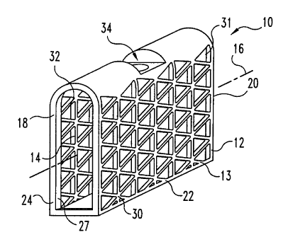
2o Figures 1-4A and 4B illustrate one embodiment of a mesh bone implant 10
according to the present invention. Bone implant 10 includes a body 12 having
an
interior chamber 11. Implant 10 also includes a first end 18 and opposite
second end 20
and a central portion 22 extending from first end 18 to second end 20. Central
portion 22
includes a first longitudinal wall 13 having a first longitudinal wall portion
30 and a
second longitudinal wall portion 32 and passageway 14 therethrough defining a
longitudinal axis 16.
First end 18 includes a support portion 24 positioned about its perimeter. In
one
form, support portion 24 includes an integrally formed support band 26
positioned
circumferentially about longitudinal axis 16. Band 26 is adapted to withstand
impaction
forces to seat impact implant 10 into a defect or a prepared cavity in the
bone structure.
In one form, band 26 is an uninterrupted band and can be provided as an
integrally
formed band having a cross section thicker than the cross section of other
wall portions,
CA 02387042 2002-04-08
WO 01/28461 PCT/LTS00/41392
7
i.e. walls 30 and 32, of body 10. Preferably, band 26 does not extend beyond
either wall
30 or wall 32 in a direction orthogonal to and away from longitudinal axis 16.
In this
form, wall portions 30 and 32 define a substantially planar surface extending
from first
end 18 to second end 20. Band 26 can taper uniformly in a direction towards
interior
chamber 11; gradually increasing in width to a maximum width proximate to
first end 18.
Extension of band 26 internally serves to provide a thickened portion to
enhance the load-
bearing capabilities of implant 10. Further, internal projections of band 26
also provide a
retaining ring about the perimeter of first end 18. Ring 27 provides
containment of
osteogenic material deposited in internal chamber 11 and facilitates greater
packing
1o density of the osteogenic material by inhibiting the escape of the packed
osteogenic
material from the implant. In other forms, band 26 can be provided as a lip or
abutment
extending from the perimeter of first end 18 toward the interior chamber 11
proximate to
first end 18.
Band 26 includes an exterior bearing surface 42. Preferably, first surface 42
defines
a substantially planar surface positioned substantially to lie in a plane
generally
perpendicular to longitudinal axis 16. Further, first surface 42 is adapted to
engage an
adjacent facing surface of a bone defect or bone cavity. In one form, the
first surface is a
roughened or knurled surface to secure implant 10 to the adjacent bone
surfaces. First
end 18 also includes an opening 21 into interior chamber 11. In the preferred
form of the
2o illustrated embodiment in Figs. 1-4, interior perimeter of band 26 defines
opening 21.
Second end 20 is opposite central portion 22 from first end 18. Second end 20
includes a second support portion 25. Second support portion 25 can be
provided as is
substantially described for first support portion 24 and can include a second
support band
27. Further, second end 20 also includes an opening into interior cavity 11 as
described
for first end 18.
In one embodiment, first end 18 and second end 20 are separated by a distance,
d 1.
Distance dl is selected so that implant 10 is matingly received within a
cavity or other
defect in a bone structure. When dl is properly selected, first end 18 and
second end 20
each can bear against respective facing bone tissue of a cavity or other
defect and provide
3o support and strength to the bone structure. As an example of implants
having varying dl
distances, an implant similar in configuration to implant 10 is illustrated in
Figure 4B.
Implant 10' is selected to have a shorter longitudinal length, dl, implant 10.
CA 02387042 2002-04-08
WO 01/28461 PCT/US00/41392
It is also intended to include within the scope of this invention a series of
implants,
each having a configuration as described for implant 10, but differing in
length dl.
Central portion 22 extends from first end 18 to second end 20 and includes a
longitudinal wall 13. Longitudinal wall 13 includes a plurality of openings 31
providing
communication with the interior chamber 11. In one form, the plurality of
openings 31
define a grid pattern or grid work on first wall 30. Each of the plurality of
openings 31
can be formed in a variety of configurations, including triangular, square,
rectangular, and
polyhedron. In a preferred form, the intersecting bars define a pattern of
equilateral
triangles or isosceles triangles. In another form, the gridwork or grid
pattern is formed by
1o a plurality of intersecting elongate bars. In a preferred form, the
plurality of intersecting
elongate bars include a first group of elongate bars have a longitudinal bar
axis arranged
perpendicular to longitudinal axis 16 and a second group of elongate bars
having a
longitudinal bar axis arranged non-perpendicular relative to longitudinal axis
16. A
plurality of joints are formed by the intersections of the elongate bars, each
joint defining
a corner of an opening into interior chamber 11.
The elongate bars can define a repeating pattern of triangles on wall sections
30 and
32, preferably isosceles triangles; more preferably, equilateral triangles.
When equilateral
triangles are used, the wall portions can have a maximum amount of open areas,
while
still retaining the requisite strength to support adjacent bone structures.
The trim open
area is intended to mean the sum of the area of the plurality of open portions
31 in walls
portions 30 and 32, respectively.
Preferably, the ratio of open area to the total surface area defined by either
wall
portion 30 (or wall portions 32) is greater than about 1:2; more preferably
greater than
about 3:4. That is, at least 50°Io of the exterior surface area of
either wall portion 30 or 32
is open area.
Longitudinal wall 13 can include a first wall section 30 and an opposing
second
wall section 32. First wall 30 extends from first end 18 to second end 20 and
defines a
plane that is substantially parallel with longitudinal axis 16. Second wall
32, similar to
first wall 30, extends from first end 18 to second end 20 and defines a plane
that is also
3o substantially parallel with longitudinal axis 16. Thus in one form, first
wall portion 30
and second wall portion 32 are positioned to lie substantial parallel to each
other.
CA 02387042 2002-04-08
WO 01/28461 PCT/US00/41392
9
Longitudinal wall 13 also includes a tool insertion end 28. Tool insertion end
28 is
positioned substantially orthogonal to first wall portion 30 and extends in a
direction
substantially parallel to longitudinal axis 16. Tool insertion end 28 includes
the tool-
engaging portion 34. Tool-engaging portion 34 can be provided in a variety of
features
adapted to engage an insertion tool for insertion of implant 10 into a
prepared bone tissue.
For example, tool-engaging portion 34 can include a variety of indents and
openings,
which may or may not be threaded, to engage corresponding configured features
on an
insertion manipulation accessory (not shown) to facilitate implantation of
implant 10 into
bone tissue. In a preferred embodiment of Figures 1-4, tool-engaging portion
34 includes
1o a longitudinally extending threaded bore 35 and a driving indent 36.
Tool insertion end 28 defines an exterior surface 37. In one form, surface 35
is
curved in a direction transverse to longitudinal axis 16 from wall portion 30
to wall
portion 32. In another form, the exterior surface defines an arcuate surface
in a direction
along axis 16 and extending from the first end.
Referring now to Figure 5-7, there is depicted another embodiment of a mesh
bone
implant according to the present invention for supporting bone structures. In
the
preferred form of the illustrated embodiment, mesh implant 110 includes a
cylindrical
body 112 having a mesh wall 113 defining an interior chamber 111 therein. Body
112
includes a passageway 114 therethrough defining a longitudinal axis 116.
Preferably,
cylindrical wall 113 extends circumferentally about longitudinal axis 116. In
the
illustrated embodiment, cylindrical wall 113 is formed in the shape of a
cylinder.
However, it is understood that the mesh wall 113 can define a variety of
shapes, including
shapes having at least one flat surface.
Body 112 includes a first end 118 and an opposite second end 120. First end
118
and second end 120 each include a support portion 124 and 125, respectively.
In one
form, support portions 124 and 125 each include a support band, 126 and 127
respectively, positioned circumferentially about longitudinal axis 116.
Support bands 126
and 127 can be provided as an uninterrupted band about the perimeter of first
end 118 and
second end 120, respectively. Support band 126 includes an exterior surface
142 that is
3o provided as a substantially smooth surface and defining a plane generally
transverse to
longitudinal axis 116. Similarly, support band 127 includes an exterior
surface 144 that is
provided as a substantially smooth surface and defining a plane generally
transverse to
CA 02387042 2002-04-08
WO 01/28461 PCT/US00/41392
longitudinal axis 116. The substantially smooth planar surfaces 142 and 144 of
support
band 126 and 127, respectively, facilitate implantation of implant 110 into
bone
structures. These surfaces provide particular advantages when implant 110 is
inserted
into a prepared cavity in a bone structure and engage the walls of the cavity
to provide
5 additional support to the bone structure.
Support portions 124 and 125 are provided to withstand the requisite impulsion
force to seat implant 110 into a bone defect or a prepared cavity. The support
portions
124, 125 can be formed from wall section having a thicker cross section then
other wall
sections of body 112. Thus, the support bands 124 and 125 can be provided in a
form as
to described above for support portions 24 and 25.
First end 118 and second end 120 are separated by a distance, d2. Distance d2
is
selected so that implant 110 is matingly received within a prepared cavity or
other defect
in a bone structure. When d2 is properly selected, first end 118 and second
end 120 each
can bear against respective facing bone tissue of a cavity, bone defect or
opposing faces
of adjacent bone structures and provide additional strength to the bone
structure(s).
First end 118 and second end 120 each include an opening, 121 into the
interior
chamber 111. Opening 121 provides communication with passageway 114 through
body
112. In the preferred form of the illustrated embodiment in Figs. 5-7, the
interior
perimeter of bands 126 and 127 each define an opening 121.
2o Mesh implant 110, similar to mesh implant 10, includes a central portion
122
extending from first end 118 to second end 120. In one aspect, cylindrical
mesh wall 113
defines central portion 122. Cylindrical mesh wall 113 also includes a
plurality of
openings 131. Openings 131 can be provided in a variety of patterns, including
triangular
(equilateral or isosceles), square, rectangular, and polyhedron, thereby
forming a mesh
wall. Preferably, outer peripheral wall 130 includes a uniform grid of a
plurality of
openings 131. In another form, cylindrical mesh wall 113 can be formed by a
plurality of
intersecting elongate bars. The plurality of intersecting elongate bars
include a first group
of elongate bars have a longitudinal bar axis arranged perpendicular to
longitudinal axis
116 and a second group of elongate bars having a longitudinal bar axis
arranged non-
perpendicular relative to longitudinal axis 116. A plurality of joints are
formed by the
intersections of the elongate bars of the first and second groups, each joint
forming an
apex that defines a corner of one of the openings of the plurality of openings
131 into
CA 02387042 2002-04-08
WO 01/28461 PCT/US00/41392
11
interior chamber 111. In another form, cylindrical wall 113 is defined by a
plurality of
intersecting elongate bars including a first group of bars defining a plane
perpendicular to
longitudinal axis 116. A second group of bars having an elongate axis arranged
non-
perpendicular to longitudinal axis 116 intersects the bars in the first group
of bars. Again,
a plurality of apexes are formed by the intersection of the first group of
bars and the
second group of bars. The apexes form one of the corners of the openings 131
in
cylindrical wall 113. Cylindrical wall 113 can be provided substantially as
described for
wall 13.
Cylindrical wall 113 includes a tool engagement portion 134. Tool engagement
1o portion 134 can be provided as described for tool engagement portion 34,
and can include
a threaded bore 135 and a driving indent 136.
Another form of the invention is illustrated in Figures 8-10. Mesh implant 210
is
depicted generally as a rectangular prism body 212 having a central portion
222 and an
interior chamber 211 formed therein. Body 212 includes a passageway 214
therethrough
defining a longitudinal axis 216. Body 212 includes a first transverse wall
240, an
opposite second transverse wall 246, and a central portion 222 extending from
first end
118 to second end 220.
First end 218 includes an opening 221 extending into interior chamber 211.
Similarly, second end 220 includes a second opening extending into interior
chamber 211.
2o First end 218 also includes a support portion 224 extending about the
perimeter of first
end 218. Similarly, second end 220 includes support portion 225 extending
about its
perimeter. Support portions 224 and 225 each include a support band 226 and
227,
respectively, positioned generally circumferentially about longitudinal axis
216. Bands
226 and 227 are adapted to withstand forces needed to impact implant 210 into
a prepared
cavity in a bone structure or between adjacent bone structures. In one form,
bands 226
and 227 can be provided as integrally formed bands having a cross section
thicker than
the cross section of other wall portions, particularly mesh walls 230 and 232,
of body 210.
In other forms, band 226 (or 227) can be provided as an abutment or a lip
extending from
the perimeter of first end 218 (or second end 220) toward the interior chamber
211
3o substantially as has been described for bands 26, 27, 126 and 127.
In a preferred form of the illustrated embodiment of implant 210, first end
218 and
second end 220 are provided as arcuate surfaces 252 and 254, respectively,
along a
CA 02387042 2002-04-08
WO 01/28461 PCT/US00/41392
12
transverse axis 256 positioned to be substantially orthogonal to longitudinal
axis 216.
Arcuate surfaces 252 and 254 each have a maximum height positioned between
first
transverse wall 240 and second transverse wall 246. In use, at least a portion
of arcuate
surfaces 252 and 254 can extend into bone tissue, such as cancellous tissue
underlying the
endplates of vertebral bodies. Arcuate surfaces 252 and 254 inhibit expulsion
of the
implant from the bone cavity by providing an implant that has a maximum height
that is
greater than height of a surgically prepared bone cavity, for example, in an
intervertebral
space between adjacent vertebrae.
Central portion 222 also includes first longitudinal wall 230 and second
to longitudinal wall 232. At least one, and preferably both, of longitudinal
mesh walls 230
and 233 are positioned to define a plane that is generally parallel to
longitudinal axis 216.
Further, first wall 230 and second wall 232 are provided with a plurality of
openings 231
into interior chamber 211. Preferably, first wall 230 and second wall 232 are
provided
with a pattern of substantially uniform apertures forming a mesh. The
apertures can be
provided in a variety of configurations, including circular, square,
rectangular,
polyhedron, and the like. A plurality of openings 231, similar to the openings
11
described for implant 10, can be formed into walls 230 and 232. In a preferred
form, the
apertures are provided in a form of an equilateral or isosceles triangle.
Further, first wall
230 and second wall 232 can be defined by a plurality of intersecting elongate
bars as
2o described for cylindrical wall 113 for implant 110 and wall 13 of implant
10.
In one form, implant 210 can be inserted in a defect or a prepared cavity
between
two bone structures to provide support and strengthen the adjacent bone
structures.
Therefore, body 212 can include a first transverse wall 240 extending between
first end
218 and second end 220 and positioned generally orthogonal to longitudinal
wall 230, and
an opposing transverse wall 246 also extending between first end 218 and
second end 220
and positioned generally orthogonal to longitudinal wall 230.
Transverse wall 240 can include a first bearing surface 242, an opposite
second
bearing surface 244, and a transverse face 247 therebetween. Preferably, first
bearing
surface 242 and second bearing surface 244 include substantially planar
surfaces 243 and
245, respectively, adapted to engage adjacent surfaces of the prepared bone
cavity or
bone defect. When inserted into the prepared cavity or bone defect, at least
one of first
bearing surface 242 or second bearing surface 244 bear against the adjacent
bone tissue.
CA 02387042 2002-04-08
WO 01/28461 PCT/US00/41392
13
In one embodiment, first bearing surface 242 and second bearing surface 244
are
separated by a distance d3 selected to engage first bearing surface 242 and
second bearing
surface 244 with corresponding opposing adjacent bone structures in the
prepared cavity
or bone defect. Further, in a preferred aspect, first bearing surface 242 and
second
bearing surface 244 are substantially planar surfaces extending generally
parallel to
transverse axis 256.
First transverse wall 240 includes a tool-engaging portion 234. Tool-engaging
portion 234 can be configured as described for tool-engaging portion 34 of
implant 10,
including a threaded bore 235 and driving indent 236.
1o In the preferred embodiments, first and/or second bearing surfaces 242 and
244
include anti-expulsion features 249, for example, ridges, teeth, and other
projections,
adapted to inhibit the expulsion of implant 210 from the prepared cavity or
bone defect.
In the preferred form, the anti-expulsion features are adapted to minimize the
force
needed to insert implant 210 into the prepared space or bone defect, yet
inhibit expulsion
of implant 210. Examples of such preferred forms include: at least one ridge
transverse
to longitudinal axis 216, a plurality of ridges, teeth, or spikes. In a
preferred form, the
anti-expulsion features are adapted to minimize the force needed to insert
implant 210
into prepared intervertebral space, yet inhibit expulsion of implant 210.
Examples of
such preferred forms include ratchet-shaped ridges or teeth that have an apex
pointing
2o toward the first terminal end. When thus configured, the ratchet-shaped
ridges or teeth
chisel deeper into the cortical bone tissue in response to an expulsive force.
Body 212 also includes a second transverse wall 246 opposite the first bearing
wall
240. Second transverse wall 246 can include a third bearing surface 248, an
opposing
fourth bearing surface 250, and a face extending therebetween. Third and
fourth bearing
surfaces 248 and 250, respectively, are separated by distance d4. In one
preferred
embodiment, distance d4 is selectively greater than distance d3 to conform to
the desired
prepared cavity in the bone structure, for example, in the intervertebral
space between
adjacent vertebrae. While third and fourth bearing surfaces 248 and 250 are
shown as
curved surfaces, it is understood that these bearing surfaces can be provided
in a variety
of shapes, including convex or ogival, in either the horizontal or vertical
plane, or both, or
substantially planar as depicted with the first and second bearing surfaces
242 and 244,
CA 02387042 2002-04-08
WO 01/28461 PCT/US00/41392
14
respectively. Further, third and fourth bearing surfaces 248 and 250 can
include anti-
expulsion features as described for the first and second bearing surfaces 242
and 246.
Further, transverse wall 246 can include a tool-engaging portion as described
for
transverse wall 240, including a threaded bore and a driving indent.
Reference will now be made to use of mesh implants 10, 110, and 210 to support
adjacent weak bony structures. Typically, mesh implants 10, 110, and 210 can
be
inserted into a bone structure after preparation of a suitable bone cavity.
For example,
implants can be inserted into the cavity resulting from harvesting an
autograft from the
iliac crest. Often, harvesting autografts leads to significant post-operative
pain and
to lengthy recovery time. Use of the implants disclosed in this invention
facilitates
reconstruction of the cavity and accommodates a quicker recovery time, often
with less
pain to the patient.
Referring now to Figures 11-14, a selected portion of the iliac crest 260 is
removed
using a surgical cutting device, such as, for example, a chisel 262, or a bone
saw. After
the selected region 264 of the iliac crest has been cut, the cut bone
autograft 266 is
removed from the residual bone structure 260' of the patient as depicted in
Figure 12. An
implant as described in the present invention is selected for cavity 268 and
to matingly
engage in the adjacent bone structures 270 and 272, respectively. The selected
implant
274 is releasably attached to an implant holder 280, preferably of a known
variety.
2o Preferably, implant holder 280 includes an implant insertion portion that
is configured to
matingly engage in tool-receiving portion 34, 134, and 234 of the selected
implants. In
preferred embodiments, the insertion portion includes a threaded shaft 284 to
readily
engage in a threaded bore in the implant. The implant insertion portion can
also include a
driving blade (not shown) to engage in a driving indent on the implant. In
other
embodiments, implant tool 280 can include a handle 288, which may or may not
include
an impaction tool, such as a slap hammer, to impact the implant into the
prepared bone
cavity or bone defect.
Preferably, implants 10, 110, and 210 are made of a single, integral piece.
The
implants may be prepared from physiologically acceptable material having a
requisite
3o strength to withstand the compressive force exerted on the spinal column
during normal
activity. Examples of such acceptable material include: titanium, composites
(carbon
fiber or glass fiber composites), ceramics, bone, stainless steel, and
surgical steel.
CA 02387042 2002-04-08
WO 01/28461 PCT/US00/41392
Preferably, implants 10, 110, and 210 are prepared of metal such as titanium
or surgical
steel.
In the preferred manufacturing procedure, implants according to the present
invention are made by an extrusion of a tube or hollow construct. The tube or
hollow
5 construct may or may not be substantially cylindrical. Preferably, the
extruded tube may
include end walls with increased thickness compared to sidewalk. After
extrusion of the
tube, the desired surface features, such as the support bands, anti-expulsion
portions, tool-
engaging portions, and the mesh configuration, may be defined or cut into the
implant
using a laser techniques well known in the art or any other suitable method.
It will be
l0 understood that mesh implants created from extruded tube may be formed
faster and with
less waste than conventional milling of implants from solid blocks. The
extruded implant
preferably has already formed therein the cavity for receipt of the bone
growth material or
osteogenic material. After extrusion and laser cutting of the desired surface
features, the
implant can be machined to prepare implants having the desired size for uses
in a variety
15 of ages of patients and bone structures.
The present invention contemplates modifications in the porous bone implant as
would occur to those skilled in the art. It is also contemplated that
processes embodied in
the present invention can be altered, rearranged, substituted, deleted,
duplicated,
combined, or added to other processes as would occur to those skilled in the
art without
2o departing from the spirit of the present invention. In addition, the
various stages, steps,
procedures, techniques, phases, and operations within these processes may be
altered,
rearranged, substituted, deleted, duplicated, or combined as would occur to
those skilled
in the art. Further, any theory of operation, proof, or finding stated herein
is meant to
further enhance understanding of the present invention and is not intended to
make the
scope of the present invention dependent upon such theory, proof, or fording.
While the invention has been illustrated and described in detail in the
drawings and
foregoing description, the same is considered to be illustrative and not
restrictive in
character, it is understood that only the preferred embodiments have been
shown and
described and that all changes and modifications that come within the spirit
of the
3o invention are desired to be protected.