Note: Descriptions are shown in the official language in which they were submitted.
CA 02395338 2002-06-14
WO 01/49363 PCTIUSOO/35525
-1-
SPLITTABLE MEDICAL VALVE
Description
Technical Field
This invention relates to medical devices, in particular to hemostatic valves
for intravascular devices.
Background of the Invention
Percutaneous placement of intravascular catheters, pacemaker leads, etc.
involves blood loss, that while easily controllable, especially during venous
access,
can become significant during long procedures. For example, procedures such as
placement of leads in the coronary sinus for biventricular pacing, can last 4
hours,
during which time the blood loss of up to 500-600 cc can represent a risk to
the
patient. Additionally, the open conduit into the body can become a source of
infection to the patient. To help reduce these potential risks, self-sealing
hemostatic
valves have been developed for use with introducer sheaths. These valves
provide
a seal against flashback of blood from the proximal end of the sheath,
including
when a second device is being manipulated within the introducer.
Medical devices with large proximal fittings, such as pacemaker leads and
PICC lines, cannot be readily used through standard hemostasis valves and
introducers because of the need to remove the introducer while leaving the
other
device in place. To address this need, splittable sheaths and hemostasis
valves
were developed so that the introducer and valve can be removed while the inner
device remains in the patient. Combinational devices exist, such as the SAFE-
SHEATHTM Splittable Valved Sheath System (Pressure Products, Inc., Rancho
Palos
Verdes, CA), which is comprised of a splittable valve attached to the end of a
scored
introducer sheath. The valve housing containing the valve membrane is split
along
scores lines which are aligned with score lines that continue down the length
of the
integral introducer. Thus, the valve and introducer are split together. One
disadvantage of this combinational system is the lack of flexibility in how
the device
is used. For example, to place a coronary sinus pacemaker lead, a physician
will
WO 01/49363 CA 02395338 2002-06-14 pCT/US00/35525
-2-
often wish to advance the long introducer sheath into the coronary vessel,
then
partially withdraw the sheath, perhaps 10 cm, prior to introducing the pacing
lead.
The large integral valve at the proximal end of the sheath cannot enter the
patient;
therefore, the physician must have an undesirably long section of introducer
exiting
the patient, where ideally, he or she would like to peel the introducer back
closer to
the entry site. In addition, the scored introducer portion of the SAFE-
SHEATHTM lacks
the structural integrity to negotiate tortuous bends of the coronary vessels.
Because
the valve and introducer are designed only to be used together, the system
cannot
be adapted to work with different sheaths and other intravascular devices that
may
offer important clinical advantages in certain procedures.
What is needed is a simple system that offers greater flexibility to fully
manipulate and adjust the splittable sheath prior to splitting away the valve.
It
would also be desirable to have a splittable valve that can be used with
different
splittable sheaths that did not require integral attachment or alignment of
split lines.
Further considerations include having a splittable hemostatic valve of simple
construction that is easy to use, inexpensive to manufacture, and can provide
superior sealing characteristics, even in the presence of high backflow
pressures
such as are seen in arterial applications.
Summary of the Invention
?0 The foregoing problems are solved and a technical advance is achieved in
a splittable hemostatic valve that includes an interfacing region sized and
configured
to permit the valve to be coupled to a separate splittable introducer sheath
or other
tubular medical device to permit passage of a catheter or device therethrough
with
minimal blood flashback. In a first embodiment, the hemostatic valve can be
placed
?5 over a splittable introducer sheath, such as a PEEL-AWAY Introducer Sheath
(COOK
Incorporated, Bloomington, IN) while typically, a dilator is initially co-
introduced,
followed by the device being placed, such as a pacemaker lead or intravenous
catheter having a large proximal hub or fitting. The hemostatic valve can then
be
split and removed from the introducer, which is also split apart, leaving the
30 indwelling device undisturbed. Advantageously, the replaceable aspect of
the valve
WO 01/49363 CA 02395338 2002-06-14 PCT/USOO/35525
-:~-
allows the physician the ability to partially withdraw the introducer and peel
it back
down, as is often done when placing certain intravascular devices, and then
place
the hemostatic valve back over the new proximal end that is formed. This
provides
a significant clinical benefit over existing splittable introducers that
include an
integral valve at the proximal end that is split along with introducer,
thereby not
allowing for replacement at a more distal location. In another embodiment, the
interfacing region can be configured to be placed at least partially within
the
passageway of the introducer sheath, instead of over the sheath's outer
surface.
The hemostatic valve comprises a valve body which is typically made of
silicone or another elastic material that allows the valve to be fitted over
or into the
introducer sheath while offering some sealing characteristics. The hemostatic
valve
includes one or more sealing elements located within the valve passageway. In
some embodiments of the invention, one or more of the sealing elements are
formed
to be integral with the valve body. They can be positioned at the proximal end
or
within the body of the valve and may include slits or apertures to allow
passage of
a medical device. Other embodiments include a valve insert disk made of
silicone
foam that is separately formed and affixed within the hemostatic valve
passageway.
In various other aspects of the present invention, the proximal end of the
hemostatic valve may be configured to receive and lock a dilator hub such that
the
dilator and introducer can be maintained in the proper longitudinal alignment
with
each other during the procedure. In addition, the distal end of the valve can
be
configured to accept a series of specific-sized introducers by including a
multiple
steps of different diameters (e.g., 3.5 to 6.0 Fr). In another aspect, the
valve can
include a side port to allow access to the passageway for procedures such as
an I.V.
drip, system flushing, air evacuation, or the infusing of medicaments or
contrast
media.
The hemostatic valve includes at least one line of fissure through which
the valve is opened to allow external access to the passageway. In one
embodiment, the silicone valve body is formed with opposing scores or grooves
formed nearly all the way through the inside or outside of the valve wall such
that
WO 01/49363 CA 02395338 2002-06-14 PCTIUSOO/35525
-4-
the two valve halves can be readily pulled apart when the two integral tabs
are
pulled outward to initiate the split. Typically, the sealing elements are
correspondingly scored or split to facilitate a complete separation of the
valve
assembly.
In another aspect of the invention, the valve is constrained by a splittable
outer sheath, such as one made of molecularly oriented, anisotropic PTFE used
to
make the PEEL-AWAY Introducer Sheath. The embodiment also includes a means
to grasp and tear the sheath away to open the valve, which may be restrained
as
two separated halves that fall apart, or scored or so affixed as to be torn
apart by
the separating action of the sheath.
In another aspect of the invention, the distal portion of the hemostatic
valve assembly includes a splittable distal extension of the valve body that
is
adapted to fit over or couple with a particular medical device. Many
intravascular
introducers and other devices, unlike the Cook PEEL-AWAY Introducer, have a
large
proximal fitting. In one embodiment, a distal portion is adapted to accept and
seal
about the proximal fitting of a standard introducer sheath. The distal portion
could
include a series of seals that are designed to fit over a multiplicity of
fittings, making
it a 'universal' splittable hemostatic valve.
In yet another aspect of the invention, a sealant filler material is provided
within the passageway of the hemostatic valve, preferably within one or more
cavities formed between the self-sealing membranes. While the self-sealing
membranes provide an adequate barrier against fluid backflow when used in the
venous system where pressures typically average around 0.2 psi, arterial
pressures
represent over a ten fold increase over that of the venous side, making
sealing much
more difficult. This sealant filler material, which provides an additional
blood barrier,
can comprise virtually any biocompatible material that can provide a seal
around a
device being passed through the valve. Possible materials include a viscous
liquid
such as glycerine; a gel; a foam or sponge; densely-packed solid particles
such as
minute beads or fibrous material; and strips of material such as collagen.
These
materials can be affixed to or incorporated into the valve body or introduced
into the
WO 01/49363 CA 02395338 2002-06-14 PCT/US00/35525
-5-
existing cavity, such as via a side port or injected through the valve body
wall.
Membranes can be used to longitudinally divide the cavity into two halves that
are
filled with a substance that allows the subcavities to be resiliently
depressed. The
resulting counter force against the residing device provides a seal with the
membranes allowing the contents of the subcavities to remain contained when
the
valve is separated.
In still yet another aspect of the invention, a biasing means is included to
provide additional force against the leaflets of the distal seal, such as a
duck-bill
valve, to provide improved sealing properties. In one embodiment, the biasing
means
comprising two biasing elements of a material such as silicone which are added
to
the valve after fabrication. The biasing elements are added by applying force
to the
valve on opposite sides such that the force is in line with a valve slit,
thereby
causing it to open slightly. The silicone or other material is then added
adjacent to
the valve leaflets at points perpendicular to the valve slit and allowed to
cure. The
force is released, returning the valve to its original shape with the cured
biasing
elements now functioning to continuously urge the leaflets closed. In other
embodiments, the biasing means comprises an 0-ring or sleeve that is included
within the valve after the valve with slit is formed to provide a biasing
force to urge
the leaflets into the closed position.
In still yet another aspect of the invention, the valve assembly can include
a plurality of valves whose passageways are joined distally into a common
passageway. In an embodiment having two proximal seals with two passageways
each representing bifurcations of the single common passageway, there are two
oppositely placed lines of fissure that allow the valve assembly to be
separated into
two halves. In an embodiment having three proximal seals and three passageways
that feed into a single common passageway, there are three lines of fissure
that
allow the valve assembly to be separated into three pieces to allow introduced
devices to remain in place. Additional valves and entry passageways are also
contemplated.
WO 01/49363 CA 02395338 2002-06-14 PCT/US00/35525
-6-
Brief Description of the Drawing_
FIG. 1 depicts a partially-sectioned pictorial view of an embodiment of the
splittable hemostatic valve assembly having a outer sheath;
FIG. 2 depicts a pictorial view of an alternative embodiment of the present
invention having an outer sheath;
FIG. 3 depicts a cross-sectional side view of an embodiment of the present
invention having a plurality of sealing elements;
FIG. 4 depicts a cross-sectional side view of the hemostatic valve
assembly of FIG. 2;
FIG. 5 depicts a top view of the embodiment of FIG. 1;
FIGs. 6-8 depicts cross-sectional views of various sealing element
embodiments of the present invention;
FIG. 9 depicts a pictorial view of an embodiment of the present invention
having a side port;
FIG. 10 depicts a top view of an embodiment of a valve body of the
present invention having a external score line;
FIG. 11 depicts a bottom view of an alternative embodiment of a valve
body of the present invention having an internal score line;
FIG. 12 depicts a side view of the embodiment of FIG. 1 being used with
a splittable introducer sheath;
FIG. 13 depicts a cross-sectional view of an embodiment of the present
invention adapted for placement over a proximal fitting;
FIG. 14 depicts a partially-sectioned side view of an embodiment of the
present invention adapted to be placed within an introducer sheath;
FIG. 15 depicts a pictorial views of a second embodiment that is adapted
for placement within a introducer sheath;
FIG. 16 depicts a partially-sectioned pictorial view of an embodiment of
the present invention adapted to be partially placed within an introducer
sheath;
FIG. 17 depicts a partially-sectioned side view of a second embodiment
that is adapted to be partially placed within an introducer sheath;
WO 01/49363 CA 02395338 2002-06-14 PCT/US00/35525
-7-
FIG. 18 depicts a pictorial view of an embodiment of the present invention
used with a helical splitting introducer sheath;
FIGs. 19-20 depict a pictorial views of embodiments of the present
invention having a grasping member or members located at the distal end of the
valve body;
FIGs. 21-22 depict cross-sectional views of a hemostatic valve having a
sealant filler material therein;
FIG. 23 depicts a cross-sectional view of a hemostatic valve having a
biasing means;
FIG. 24 depicts a cross-sectional view taken along line 24-24 of the
embodiment in FIG. 23;
FIG. 25 depicts the embodiment of FIG. 24 during the manufacturing
process;
FIG. 26 depicts a cross-sectional view of a hemostatic valve having a
second embodiment of a biasing means;
FIG. 27 depicts a top view of the biasing means of FIG. 26;
FIG. 28 depicts a pictorial view of a third embodiment of a biasing means;
FIG. 29 depicts a pictorial view of an embodiment of a splittable valve
assembly having two proximal valves with a common central passageway;
FIG. 29A depicts a cross-sectional view taken along line 29A-29A of the
embodiment of FIG. 29;
FIG. 30 depicts a pictorial view of an embodiments of a splittable valve
assembly having three proximal valves with a common central passageway;
FIG. 31 depicts a sectioned view of the embodiment of FIG. 9;
FIG. 32 depicts an exploded pictorial view of the embodiment of FIG. 9;
FIG. 33 depicts a partially sectioned view of an embodiment similar to that
of FIG. 9 being used with a dilator and introducer sheath; and
FIG. 34 depicts an embodiment of the present invention adapted to be
placed within the passageway of an introducer sheath.
WO 01/49363 CA 02395338 2002-06-14 PCT/US00/35525
-8-
Detailed Description
A better understanding of the present invention will now be had upon
reference to the following detailed description, when read in conjunction with
the
accompanying drawing, wherein like reference characters refer to like parts
throughout the several views and different embodiments of the present
invention.
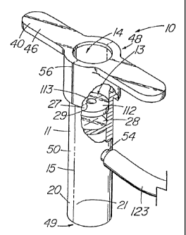
The splittable valve assembly 10 of the present invention, as embodied
in FIGs. 1-34, comprises a hemostatic valve 11 that includes a valve body 50
with
a passageway 14, at least one line of fissure 15 to permit the valve to split
and
allow external access along the length of the passageway, and at least one
sealing
element 13 configured to traverse the passageway 14, while permit the passage
of
an first medical device 57, such as a catheter, dilator, pacemaker lead, etc.,
while
substantially preventing or eliminating the leakage or 'flashback' of blood or
other
bodily fluids. The splittable valve assembly 10 is designed for use with a
second
medical device, typically a tubular medical conduit 23 such as a splittable
introducer
sheath 24. The hemostatic valve 11 of the present invention comprises an
interfacing region 120, typically located at the distal end 49 of the valve
assembly.
The interfacing region 120 is configured to permit the valve to be coupled or
attached to the tubular medical conduit 23 at some point prior to or during
the
procedure involving the tubular medical conduit and in some instances,
reattached,
particularly when the valve is removed intact and the splittable introducer
sheath is
partially peeled down to form a new proximal end. In the illustrative
embodiments
such as FIGs. 1, 2, and 9, as well as others discussed later, the interfacing
region
120 permits the splittable valve assembly 10 to be placed over the proximal
end 52
of a splittable introducer sheath 24, as depicted in FIGs. 12, 19 and 33. If
during
the course of the procedure, the physician decides to partially withdraw and
peel
back down the sheath 24, the valve can be advantageously removed, rather than
being split with the sheath 24, thereby allowing it to be placed intact back
over the
new proximal end of the splittable introducer sheath 24 and resume its
function as
a hemostatic valve 11 until such time as the first medical device 57 is
introduced to
its target location and the splittable introducer sheath 23 and hemostatic
valve 11
WO 01/49363 CA 02395338 2002-06-14 PCT/US00/35525
-9-
are split apart and discarded. It should be noted that while valve portion 11
is
referred to herein as a 'hemostatic valve,' it has possible applications in
other types
of non-vascular procedures where there is a desire to prevent leakage of
fluids and/or
reduce exposure to air-borne pathogenic organisms. For example, the splittable
valve assembly 10 of the present invention can be used in minimally invasive
neurological procedures to limit contact of the cerebral spinal fluid with
ambient air.
Another possible application would be urological procedures where the valve
could
help prevent the introduction of pathogenic organisms into the urinary tract.
A basic embodiment of the present splittable valve assembly 10 is
depicted in FIGs. 9 and 31-33. In this embodiment, the valve body 50 is insert
molded into a single piece or unit from medical grade silicone, although other
elastomeric polymers can be used, including combinations of different
compounds
for different portions of the valve. To facilitate splitting of the valve body
50 into
separate first and second halves 20,21 to expose the passageway 13 of the
hemostatic valve 1 1, opposing lines of fissure 15, located about 180 with
respect
to each other, are formed in the wall 47 of the valve body. The lines of
fissure 15
of the illustrative embodiment each comprise a score line 22 or groove formed
partially through the wall 47, leaving a small amount of material 83 (e.g.,
0.01 ") as
a bridge to join the adjacent halves 20,21. The hemostatic valve 1 1 can be
molded
as a single unit and scored to create a line of fissure 15 to facilitate
rupture of the
valve body 50 when the respective halves 20,21 are pulled outward in opposite
directions. In the embodiments of FIGs. 9-10, the score line 22 is formed into
the
outside surface 35 of the valve wall 47. To facilitate separation of the valve
body
50 along the score line 22, a starter split 56 or notch can be made at the
distal end
of the hemostatic valve 11 at the line of fissure 15. The valve body 50 is
separated
by using the integral tabs 40, thus permitting the initial separation force to
be
concentrated at the distal end 49 where the starter split 56 is located. FIG.
11
depicts yet another embodiment in which the score line 22 in formed into the
inside
surface 71 of the wall. If the hemostatic valve 11 is insert molded into the
outer
sheath 12, scoring could occur by either running the scoring tool along the
CA 02395338 2002-06-14
WO 01/49363 PCT/US00/35525
-10-
passageway 14 of the hemostatic valve 11, or configuring the die to create a
score
line 22 in the valve body 50 during the molding process such that the two
valve
halves 20,21 were bridged by a thin membrane 83 of material. A line of fissure
15
can be formed using a number of well-known techniques and assume a variety of
configurations to achieve the goal of providing a relatively predictable path
through
which the split in the valve body progresses, such that the hemostatic valve
can be
removed from around the first medical device 57.
Returning to the embodiment of FIG. 9 and 31-33, at the proximal end 48
of the hemostatic valve 11 are located two grasping elements 40 which in this
embodiment, comprise integral tabs 46 that integrally extend from valve body
50 of
the hemostatic valve 11. These grasping elements 40, which facilitate
splitting the
valve, can assume a wide variety of configurations, both integral, and
separate from
the valve body 50 with selected examples being depicted in various other
figures.
When the operator pulls the integral tabs 46 in opposite directions away from
the
valve body 50, the lines of fissure 15 split from the proximal end 48
progressing to
the distal end 49, causing the valve body 50 to separate into halves 20,21. To
initiate the split along the lines of fissure 1 5, an optional starter split
56 is included
at the proximal end 48 whereby the lines of fissure 15 completely traverse the
wall
47 for a relatively short distance (e.g., 2-7 mm) relative to the length of
the
hemostatic valve, which in the illustrative embodiment used with 3-12 Fr
intravascular introducer sheaths, measures about 30-50 mm, depending on the
size
of the companion sheath.
FIGs. 19-20 depict embodiments in which the hemostatic valve 11 is split
starting from the distal end 49, proceeding to the proximal end 48. To better
accomplish this, the grasping members 40 are located at the distal end 49 of
the
hemostatic valve assembly 10 for opening the line of fissure 15 toward the
proximal
direction, resulting in separation of the valve halves 20,21. FIG. 19 depicts
an
embodiment that is similar to that of FIG. 9 with the exception of the
reversal of the
grasping member 40 and starter split 56 orientation. As shown, the grasping
members 40 are advantageously located in proximity to the splittable
introducer
WO 01/49363 CA 02395338 2002-06-14 PCT/US00/35525
- 11 -
sheath handles 32. If each pair of grasping members/handles are pinched
together
and pulled outward from the hemostatic valve assembly 10 and splittable
introducer
sheath 24, both devices can be split together. In doing so, the hemostatic
valve 11
split initially continues upward from the starter split 56, while the
splittable
introducer sheath split initially progresses upward to the proximal end 52,
then
continues downward along a distal path. The line of fissure 15 may not extend
the
entire length of the valve body 50 if the starter split 56 or starter split
plus a partial
score line are sufficient, given the wall thickness and material, to force a
split that
continues all the way to the opposite end 48. FIG. 20 depicts a related
embodiment
that includes a single grasping member 40 and integral tab 46 that is located
at the
distal end 49 of the valve body 50 on only one half 20 of the valve. If the
device
over which the hemostatic valve 11 is placed extends a sufficient distance
into the
passageway 14 to provide adequate counter force against the opposite half 21,
a
single grasping element 40 located on the first half 20 can be used to cause a
split
that allows full separation of the valve body 50. The number and configuration
of sealing element 13 of the present invention represents a design choice
influenced
by the type of procedure involved and the instrumentation to be used with the
valve.
In the embodiments of FIGs. 31-33, the illustrative hemostatic valve 11
includes two
sealing elements 13 which comprise a proximal seal 27 and a distal seal 28.
The
distal seal 28 comprises a thin, 0.010" membrane that is integrally formed
with the
valve 50. A slit 29 is formed through the membrane to permit through passage
of
the first medical device 57, such as a dilator shaft 119, being introduced
through the
tubular medical conduit 23 for placement at the target site. In the
illustrative
embodiment, the proximal seal 27 comprises disk-shaped seal insert 1 12 made
of
silicone foam that is separately formed from the valve body 50, inserted into
the
passageway 14 and affixed with silicone adhesive or otherwise secured in
placed.
The seal insert 1 1 2 includes a small aperture 1 13 that facilitates smooth
passage of
a relatively large-diameter medical device therethrough. A transverse fissure
126 is
made partially through the seal insert 1 12 in line with the lines of fissure
15 in the
CA 02395338 2002-06-14
WO 01/49363 PCT/US00/35525
- 12-
valve body to allow the seal to split in half along with the remainder of the
hemostatic valve 11.
FIGs. 9 and 31-33 depict two related embodiments in which the proximal
seal 27 is situated within the passageway 14 such that sufficient space exists
between the proximal seal 27 and the proximal end 48 of the valve to form a
proximal receiving chamber 110 that is configured to accept a dilator hub 117.
A
locking lip 1 1 1 that is located at the proximal end of the proximal
receiving chamber
110 helps hold the dilator hub 117 therein. This permits the dilator 58 and
introducer sheath 24 to advantageously remain in a constant positional
relationship
in which the distal tapers of the two devices 58,24 match while being
manipulated
within the patient. Because the valve body 50 is typically made of a flexible
silastic
material , the dilator hub 1 17 can easily be pulled back out of the proximal
receiving
chamber 1 10 once the dilator 58 is ready to be removed from the introducer
sheath
24. In the embodiments of FIG. 31 and 33, the configuration of the proximal
1 5 receiving chamber 1 10 varies depending on the size of the dilator and the
design of
its hub. The valve embodiment of FIG. 31 is designed for a smaller dilator hub
(e.g.,
4.5-7 Fr), while the embodiment of FIG. 33, accepts a larger, longer hub used
with
a larger dilator, such as that intended for use with a 10-12 Fr introducer
sheath 24.
In valve embodiments that do not include a proximal receiving chamber
110, the proximal seal 27 is typically located at the proximal end 48 of the
valve
assembly 10 as depicted in a number of embodiments, including those in FIGs. 1-
8.
In one embodiment depicted in FIG. 5, the proximal seal 27 functions a self-
sealing
membrane 42 by virtue of one or more slits 29. In the embodiment, of FIG. 5
there
is a first slit 29 comprising a portion of the line of fissure 15 that extends
across the
self-sealing membrane. Also included are two diagonal slits 69 that along with
the
first slit 27, define a series of opposing valve leaflets 62 that seal around
a medical
device placed through the passageway 14 of the hemostatic valve 1 1. To ease
passage of a device through the self-sealing membrane 42, especially a small-
diameter device such as a biventricular pacing lead, the valve leaflets 62 can
be
CA 02395338 2002-06-14
WO 01/49363 PCT/US00/35525
-13-
coated with a lubricious material such as SLIP-COATTM or GRAFT-COATTM
(Sterilization Technical Services, Rush, NY). With regard to the illustrative
embodiment, the valve body 50 is contiguous with the sealing element 13, as
both
are formed of the same elastomeric material.
FIGs. 3 and 6-8 depict additional sealing element 13 embodiments. In
each of the illustrative examples, there is a proximal seal 27 comprising a
self-sealing
membrane 42 with at least one slit 29, and at least one distal seal 28 to
provide an
additional barrier against flashback of blood or other bodily fluid. In the
embodiment
of FIG. 3, the distal seal 28 comprises an integral ring or constriction that
provides
an second sealing element 13 in addition to the self-sealing membrane 42 that
comprises the proximal seal 27. In the embodiment of FIG. 6, there are a pair
of
distal seals 28, each comprising a disk-shaped self-sealing membrane 42 across
the
passageway 14 of the hemostatic valve 11. In the embodiment depicted in FIG.
7,
the distal seal 28 comprises a duck-bill valve 70 with a central slit 29
wherein fluid
flowing back toward the proximal end 48 of the valve helps force two halve of
the
valve 70 together and thus, assists with sealing about an device positioned in
the
passageway 14. It the embodiment of FIG. 8, the hemostatic valve 11 and
proximal
seal 27 are attachable to a series of additional seal components 43,44,45 that
interlock into a single unit. Each component comprises a distal seal 28 and
seal
supporting structure 51 which collectively, form the valve body 50 of the
expanded
splittable valve assembly 10. It is anticipated that number of components can
be
varied to achieve the desired amount of protection against flashback of blood
or
bodily fluid within the passageway 14 of the hemostatic valve 11.
FIGs. 31-34 are exemplary of two basic types of interfacing regions 120
for coupling or attaching the hemostatic valve 11 to a tubular medical conduit
23.
In the type depicted in FIGs. 31-33, which also the type found the embodiments
depicted in FIGs. 1-13 and 19-30, the interfacing region 120 is sized and
configured
such that its contact surface 124 with the tubular medical conduit 23 is
located
within the passageway 14 of the hemostatic valve 11. Coupling occurs with the
hemostatic valve 1 1 being placed over the proximal end 52 of the tubular
medical
WO 01/49363 CA 02395338 2002-06-14 PCT/US00/35525
-14-
conduit 23, as depicted in FIGs. 33, with other embodiments shown in FIG. 12
and
19. Ideally, the passageway 14 at the distal end 49 of the hemostatic valve 11
is
sized such that the proximal end 52 seals against the contact surface 124 to
greatly
reduce the possibility of leakage. Although it is within the scope of the
invention for
the valve body 50 to comprise a rigid or semi-rigid plastic or another non-
elastic
material, silicone or similar type materials provide superior sealing
characteristics, as
well as making it easier to split the valve body 50 along the lines of fissure
1 5.
In the embodiments of FIGs. 31-33, the interfacing region 1 20 is
configured to accept different-sized introducer sheaths 24 by including a
series of
steps 1 14,1 15,1 16, each corresponding to a specific sized introducer. For
example,
in the embodiment of FIGs. 31-32, the first step 114, located closest to the
distal
end 49, a diameter to readily accommodate up to a 6.0 Fr introducer sheath 24
before the proximal end 52 of sheath abuts the proximal lip of the stop 1 14
and
cannot be advanced further into the passageway 14. The second step 115,
located
proximal the first step 114, can accept up to a 4.5-5.0 Fr introducer sheath,
while
a 3.5-4.0 Fr introducer sheath can pass through the first two steps 114,115
before
abutting the third step 116. Depending on the durometer of the valve body 50
material, it is possible for the valve body 50 to yield somewhat and
accommodate
a larger-size introducer sheath 24 that for which the particular stop is
configured.
The embodiment of FIG. 33 depicts an interfacing region 120 sized to accept
either
a 10,1 1, or 12 Fr introducer sheath 24, the proximal end 52 of the latter of
the three
being shown positioned at the first step 114. The examples of FIGs. 31-33 are
merely illustrative as to the number and range of steps. It is possible to
configure
the interfacing region 120 to accept multiple sizes of introducer sheaths 24
without
having steps. One solution is to gradually taper the interfacing region 120 to
accommodate a range of different-sized introducer sheaths 24. Additionally
both
steps and tapers can be combined to accommodate a range of different
introducer
sheath diameters.
Other types of introducer sheaths 24 and tubular medical conduits 23 for
whose functionality could be improved by the present invention often include
large
CA 02395338 2002-06-14
WO 01/49363 PCT/US00/35525
-15-
proximal hubs or fittings, such as luer fittings, that the splittable valve
assembly 10
must fit over in order to provide G proper seal. FIG. 13 depicts an embodiment
in
which the distal portion 33 of the hemostatic valve 11 includes a coupling
mechanism 60 such as threads that allow the hemostatic valve assembly 10 to be
placed over an introducer sheath with a fitting such as a luer lock hub. A
valve 0-
ring 61, located within the passageway 14 toward the distal end 49, provides a
seal
13 that is located against or below the fitting when the tubular medical
conduit 23
is coupled to the hemostatic valve 11. It is also contemplated that the
coupling
mechanism 60 could be eliminated with the distal portion 33 being adapted to
slide
over and seal a standard proximal hub or fitting. This could occur by
configuring the
distal portion 33, which would include a series of seals 13 or 0-rings 61,
such that
it can resiliently stretch over large fittings and provide a tight seal for a
variety
devices. Requirements include making the passageway of a sufficient diameter
to
accommodate the fitting, constructing the valve body 50 from a sufficiently
elastic
material to provide adequate contact with the fitting, and appropriately
configuring
the seal 13 or seals that would lie distal to the fitting to prevent flashback
of blood
after the hemostatic valve assembly 10 is in place.
FIG. 34 depicts an second main type of interfacing region 120 in which
the contact surface 124 with the tubular medical conduit 23 occurs on the
outside
surface of the hemostatic valve 11 such that at that the distal end 49 is
inserted into
the passageway 121 of the tubular medical conduit 23. In the illustrative
embodiment, the passageway 121 represents a proximal receiving chamber that
has
been specially configured to mate with the distal portion 33 of an
appropriately
configured hemostatic valve 11. An optional distal lip 122 is included at the
distal
end 49 of the hemostatic valve 11 to help couple the valve within the
passageway
122. Additionally or alternatively, the proximal end 52 of the tubular medical
conduit 23 could be modified to include a locking lip similar in structure to
element
1 1 1 of the hemostatic valve 11 depicted in FIGs. 31-33. The interfacing
region 120
of the embodiment of FIG. 34 is configured such that only the distal portion
of the
valve body 50 is inserted into the proximal receiving chamber 122 of the
introducer
WO 01/49363 CA 02395338 2002-06-14 PCT/US00/35525
-16-
sheath 24; however it is also within the scope of the invention to have all or
a
substantial portion of the hemostatic valve be inserted into the passageway
121 of
the introducer sheath 24 as depicted in FIGs. 14-18.
Included in the embodiments of FIGs, 9,13, and 31-33 is a side port 54
that communicates with the central passageway 14. The side port 54 can be used
for a variety of purposes, for example, slow-drip intravenous administration
(e.g., 1-
cc/hr) to keep the vein open and prevent coagulation. A length of tubing 123,
as depicted in FIG. 33, is attached to the side port 54 which in turn, would
include
a luer lock port or similar-type fitting to connect with the I.V. line at the
end distal
10 to the patient. The side port 54 would be available to perform other
functions such
as infusion of medicaments, saline for flushing, or contrast media. It would
also
have utility for instances when air must be evacuated from the system. The
side
port 54 of FIG. 33 is depicted as a nipple over which the tubing 123 is
attached;
however, other embodiments are possible such as a luer or other fitting, or
merely
an aperture into which the tubing 123 is inserted.
In various embodiments depicted in FIGs. 1-2, 4-5, and 12-13, the
hemostatic valve assembly 10 of the illustrative embodiment further comprises
a
section of outer sheath 12 material that surrounds the hemostatic valve 11 and
offers structural reinforcement and an alternative means of splitting the
hemostatic
valve 11 open to expose the passageway 14. In the illustrative embodiment of
FIG.
1, the outer sheath 12 comprises a thin-walled tube of an molecularly-
oriented,
anisotropic material such as polytetrafluoroethylene (PTFE) whose molecular
properties permit it to be torn longitudinally along a predetermined split
line 16
whose path is determined by a cut point 55 formed in the material. The cut
point
55 comprises a V-shaped notch in the illustrative embodiment, although a short
linear cut could also work. The cut point 55 provides a starting point for the
tear
such that when the grasping members 40 are pulled apart, the tear continues
from
cut point 55 and maintains a straight path along the predetermined split line
16 that
extends from cut point 55, thereby separating the outer sheath 12
longitudinally into
two pieces. Separation of the outer sheath 12 permits the hemostatic valve 11
to
WO 01/49363 CA 02395338 2002-06-14 PCT/USOO/35525
- 17-
also separate, which allows the hemostatic valve assembly 10, when no longer
needed during the procedure, to be removed from an indwelling medical device
without having to slide the valve over the proximal end of the indwelling
device,
which may be precluded if the device has a proximal fitting larger than the
passageway 14 of the hemostatic valve 11. In the embodiment of FIG. 5, the
hemostatic valve 11 has been pre-split into two halves 20,21 and then glued
together with a layer of adhesive 41 such as silicone adhesive. Because the
outer
sheath 12 constrains the hemostatic valve 11, it should be noted that the
hemostatic valve 11 can be split into two mated valve halves 20,21 that are
not
interconnected, but rather only held together by the inward radial force of
the outer
sheath 12. For example, by taking a split 7.0 Fr O.D. hemostatic valve 11 and
pressure fitting the two valve halves 20,21 together inside a 7.0 Fr I.D.
outer sheath
12, the resiliency and surface properties of the silicone material help
provide a good
seal along the lines of fissure 15. When the sheath is removed, the first and
second
valve halves 20,21 fall away from each other. Although a material having
preferred
directional properties such as anisotropic PTFE is preferred, the present
invention
encompassed any known method of predisposing a sheath to separate along a
predetermined split line. Other methods of making a sheath splittable include
scoring
or perforating the walls of the sheath. Also included are multi-layered
sheaths where
one or more split or scores sheath layers are bonded to regular sheath to
guide the
tear through the underlying solid sheath, or subjecting the outer sheath 12
material
to chemical or energy treatment along a desired predetermined split line 16 to
create
a preweakened feature.
In reference to FIG. 1, the integral tabs 46 not only serve as grasping
members 40 for the clinician to separate the hemostatic valve 11, they also
provide
a means to secure the outer sheath 12 to the hemostatic valve 11 such that
separation of the former results in the separation of the latter. Two
longitudinally
aligned apertures 38 are made through opposite sides of the outer sheath. A
generally cylindrical die is used having recesses external to the apertures 38
such
that when the silicone is injected into the die, it flows out the apertures 38
and cures
WO 01/49363 CA 02395338 2002-06-14 pCT/US00/35525
-18-
to form a silicone bead 39 on the exterior surface 35 of the outer sheath 12.
In the
illustrative embodiment, the respective silicone beads 39 are molded so that
they
extend upward to the proximal end 48 of the hemostatic valve were they are
extended outward to conveniently form the grasping members 40. In the
embodiment of FIG. 2, the silicone bead 39 itself is not a grasping member 40,
this
function being provided by the ears 37 or extensions of the splittable PTFE
material,
and the associated handles 32 attached to the terminal ends of the ears 37.
The
embodiment of FIG. 2, shown also in cross-section in FIG. 4, basically
represents a
modified PEEL-AWAY Introducer Sheath that has been truncated and coupled to
an
internal hemostatic valve 11. The outer sheath 12 forms a double layer 36 of
material with the cut line 55 made to tear upward to the proximal end 48 of
the
assembly 10, then downward, continuing along the predetermined split line 16.
The
method of attaching the outer sheath 12 to the hemostatic valve 11 is not
considered critical and as previously noted, an attachment is may not be
necessary.
In addition to the attachment method shown in FIGs. 1-2, the valve halves
20,21
can be bonded to the sheath with adhesive or another well-known method. When
using PTFE, etching of the inner surface 17 can improve adherence of the
hemostatic valve 11 to the outer sheath 12.
The hemostatic valve assembly 10 of the present invention, as shown in
FIGs. 1-13 and 19-34, is used as a device that is separate from the splittable
introducer sheath 24, or the hemostatic valve assembly 10 can be constructed
such
that the outer sheath 12 includes an introducer extension 18 as depicted in
FIGs. 14-
18, thereby obviating the need for a separate introducer. Essentially, the
hemostatic
valves 11 of these same embodiments, if not pre-coupled to the outer sheath 12
and
?5 distal extension 18, can also be regarded as a separate components from the
sheath, such as the FIG. 1-13 and 19-34 embodiments which are adapted to be
placed into a separate tubular medical conduit 23 or introducer sheath 24. In
either
case, the interfacing region 120 extends a substantial portion (FIG. 16) or
the entire
length (FIGs. 15,17-18) of the external surface 35 of the valve. If the
hemostatic
valve 11 is not fixedly positioned within the introducer sheath24/introducer
WO 01/49363 CA 02395338 2002-06-14 PCTIUSOO/35525
-19-
extension 18 prior to use, this -,ivould allow the physician to insert the
hemostatic
valve into the introducer sheath 24 at some point into the procedure, and in
some
instances, back into the introducer sheath 24 once it has been partially
peeled back
to form a new proximal end. In the embodiment of FIG. 14, the outer sheath 12
and
introducer extension 18 comprise a single tear-apart PTFE sheath that
resembles the
COOK PEEL-AWAY Introducer Sheath with a hemostatic valve insert molded
thereinside. Optionally, the hemostatic valve 11 may be attached to the outer
sheath 12 in a manner similar to the embodiment of FIG. 2. The predetermined
split
line 16 extends the length of the outer sheath 12 and continues down the
length of
the contiguous introducer extension 18 as well. In another embodiment, the
outer
sheath configuration of FIG. 1, lacking the double layer 36 of material and
ears 37
at the proximal end 48, can be simply modified to include a introducer
extension 18
as well.
The embodiment of FIG. 15 depicts a simplified hemostatic valve 11 in
which the seal 13 and valve body 50, are essentially united into a single
cylindrical-
shaped structure that is inserted into the outer sheath 12 and introducer
extension
18 (or introducer sheath 24). In the illustrative embodiment, a single line of
fissure
15 permits the intravascular medical device, such as a pacemaker lead, to be
removed from the valve. Rather than being torn apart or falling apart from the
splitting action of the outer sheath 12, the hemostatic valve 11 is simply
slid off the
lead via the line of fissure 15 when the two pieces 25,26 of the outer sheath
1 2/introducer extension 18 are torn away. More than one hemostatic valve 11
may
be placed in the outer sheath 12 to be used in this manner. To prevent distal
migration of the hemostatic valve 11 in embodiments where the hemostatic valve
11 and, the outer sheath 12 are not securely interconnected, the outer sheath
12
portion of the hemostatic valve assembly 10 can be made to have a slightly
greater
I.D. than that of the outer sheath/introducer extension 18 or introducer
sheath 24.
FIGs. 16-17 depict alternative embodiments having a number of features,
including alternative methods of providing an secure interface between the
hemostatic valve 11 and outer sheath 12. In the embodiment of FIG. 16, a band
63,
WO 01/49363 CA 02395338 2002-06-14 PCT/US00/35525
-20-
which can be made of metal or hard plastic, is inserted into an annular recess
64 in
the outer surface 35 of the valve body 50. The band 63 includes a series of
teeth
65 that engage the inside surface 71 of the wall, preventing slippage of the
hemostatic valve 1 1'Loward the proximal end. Integral tabs 46 at the proximal
end
of the hemostatic valve 11 prevent its migration distally. Alternatively, the
teeth 65
can be directed both proximally and distally to eliminate the need for making
the
proximal end of the hemostatic valve 11 larger than the outer sheath 1
2/introducer
sheath 24. Having reverse-directed teeth 65 allows the physician to advance
the
hemostatic valve 11 into the sheath after it has been introduced into the
patient,
such as after a dilator has been removed. The band 63 can act as a means to
hold
the valve halves 20,21 together. In the illustrative embodiment the band 63
includes
a break line 66 designed to fracture when the outer sheath 12 is separated.
With
the band 63 securing the two valve halves 20,21, can remain as separate pieces
and
the line of fissure 15 need not be aligned with the break line 66 or the
predetermined
split line 16 of the outer sheath 12. The teeth 65 embedded in the valve wall
47
provide a positive fixation that allows the band to separate along the break
line 66.
Alternatively, the band 63 can be made on only partially circumscribe the
valve body
50 with the closed end being attached to the valve wall 47 of one valve half
and not
the other. Therefore, the C-shaped band 63 is pulled off the valve with the
attached
valve half, making a break line 66 unnecessary.
The embodiment of FIG. 17 depicts a hemostatic valve 11 with a annular
recess 64, wherein the annular recess is used to receive projections 72, such
as
annular ridges, that are molded into the inner surface 17 of the other sheath
12 or
introducer sheath 24. A second projection 73 distal to the position of the
hemostatic valve 11 acts as a stop, while the proximal projection 74 prevents
backward migration of the valve. The predetermined split line 16 of the outer
sheath
12 or introducer sheath 24 in FIG. 17 comprises a preweakened feature 19
extending downward to the distal end of the sheath. The preweakened feature 19
can include a groove molded into the wall 47 of the outer sheath 1
2/introducer
extension 1 8/introducer sheath 24 or the wall 47 can be scored after
extrusion. In
WO 01/49363 CA 02395338 2002-06-14 PCTIUSOO/35525
-21 -
another aspect of the embodiment of FIG. 17, the hemostatic valve assembly 10
includes both integral tabs 46 on the hemostatic valve 11 and ears 37
extending
laterally from the outer sheath 1 2/introducer sheath 24. The integral tabs 46
and
ears 37 can be made to interlock as shown so that both form the grasping
member
40, thereby creating additional force to separate the hemostatic valve
assembly 10.
In this particularly embodiment, the integral tabs 46 contain terminal knobs
67 that
snap into receptacles 68 in the outer sheath ears 37.
In another embodiment shown in FIG. 18, the predetermined split line 16
of the outer sheath 12 and introducer extension 18 or introducer sheath
comprises
a helical-shaped preweakened feature 19, such as a groove, extending generally
longitudinal along the length of the sheath. A single grasping member 40 is
used
to tear apart the outer sheath 1 2/introducer extension 18, resulting in a
single piece
of sheath material. The hemostatic valve 11 can be made to fall apart when the
outer sheath 12 or introducer sheath is separated, or it may be attached to
the outer
sheath near a line of fissure such that when splitting of the sheath is
initiated, the
valve body 50 is at least partially slit along a line of fissure 15.
To improve sealing performance, which is especially desirable for arterial
applications, FIG. 21 depicts a hemostatic valve embodiment that provides an
increase in protection against blood flashback. Lying between the proximal
seal 27
and the distal seal 28 is a valvular cavity 76 in which a sealant filler
material 77 is
placed to provide an additional blood barrier. This sealant filler material 77
can
comprises virtually any biocompatible material capable of filling the valvular
cavity
and allowing passage of an intravascular device therethrough. Possible
materials
include, but are not limited to, a viscous liquid, such as glycerine; a gel; a
foam (such
as silicone); a sponge material; densely-packed solid particles such as minute
beads
or fibrous material; and strips of material such as collagen. Collagen and
other
certain other materials are able to absorb and retain blood providing an
additional
mechanism of protection. A pathway may be preformed through the sponge or
other
solid material to ease the passage of a medical device. Materials can be used
in
combination, for example, a gel-impregnated foam or collagen sponge. Solid
WO 01/49363 CA 02395338 2002-06-14 PCT/US00/35525
-22-
materials can be affixed to, or incorporated into the valve body 50 so that
they are
carried away with the respective valve halves 20,21 during separation. In
embodiments such as FIG. 6 and FIG. 8 having more than one valvular cavity 76,
each can be filled with material and these materials can vary between the
valvular
cavities 76. The sealant filler material 77 of the illustrative embodiment can
be
placed in the placed within the mold prior to fabrication, placed within the
valvular
cavity 76 after the valve has been presplit, or injected into the valve,
including
through a side port 54 as shown in FIGs. 13 or 31-33 or through the valve wall
47
using a small or non-coring needle. If so desired, a fluidized sealant filler
material 77
could be aspirated from the valvular cavity 76 via the side port 54 prior to
splitting
the hemostatic valve 11, at which time any contents of the cavity would be
exposed
and be subject to leakage.
As shown in FIG. 22, a liquid, foam, gel, or other semi-solid or resilient
solid material can be contained within the valvular cavity 76 by the inclusion
of one
or more longitudinal membranes 79 that divide the valvular cavity 76 into two
subcavities 80,81. In the illustrative embodiment, each subcavity is enclosed
by a
longitudinal membrane 79 and completely filled with a sealant filler material
77 such
as gel or foam. When an intravascular device 57 such a dilator, pacemaker
lead,
etc., is introduced through the passageway 14 of the hemostatic valve 1 1, the
filled
subcavities 80,81 which have been laterally compressed by the introduced
device,
each exert a counteracting force upon the device and thus, provide a seal to
impede
blood flashback passing through the distal seal 28 at the distal end of the
valvular
cavity 76. During separation of the hemostatic valve 1 1, the valve halves
fall away
and the contents of the subcavities 80,81 remain intact. Referring now to
FIGs.
23-28, the hemostatic valve assembly 10 of the present invention can also
include
a biasing means 84 that urges the valve leaflets together, thereby providing
improved functionality to the sealing element 13, which in the illustrative
embodiments, include the distal seal 28 comprising a duck-bill valve 70. In
the
embodiment depicted in FIGs. 23-25, the biasing means 84 comprises a first and
a
second biasing member 85,86 that are added to the valve assembly 10 after
CA 02395338 2007-02-26
WO 01/49363 PCT/US00135525
-2.3-
fabrication of the main hemostatic valve 11 body. In the illustrative example
in
which the body of the valve 11 is made of silicone, the first and second
biasing
members 85,86 comprise additional silicone material that is applied against
each of
the opposing valve leaflets 62 and allowed to cure. One example of manufacture
is depicted in FIG. 25 wherein the steps include the application of external
force 89
to the valve 11 using a fixture (not shown) capable of maintaining the valve
in a
given position. The force 89 is applied at opposite points along the
circumference
of the valve such that it is aligned with the main slit 29 that is to be urged
closed.
This causes the slit 29 to open slightly as the valve body 11 is deformed from
its
original circular cross-sectional shape. With the hemostatic valve 11 in a
deformed
condition, the material that will form the second biasing member 86 (shown in
FIG. 24) is applied, using an injection device 88 such as a syringe, within an
intravalvular space 107 between the inner surface of the passageway 14 and a
leaflet 62 of the distal valve 28. In the illustrative embodiment, the biasing
material
is injected through an aperture 87 in the valve wall and allowed to cure while
the
pressure is maintained on the valve. The procedure is repeated for the
opposing
leaflet on opposite side of the valve 11. An alternative method of providing
biasing
material that functions as a biasing means 84 is to inject the material into
the
same intravalvular space 107 via the main passageway 14, rather than through
an
aperture 87 in the valve wall. Once curing has taken place, the external force
89
that is compressing the valve is removed, allowing the valve to return to its
previous
shape. As it does, each biasing member 85,86 urges the respective opposing
leaflets 62 together. This cantilever action provided by the biasing members
85,86
allows the valve to maintain the desired level of function under higher
backfiow
pressures than might be otherwise possible.
Another embodiment that includes a biasing means 84 is depicted in FIG.
26. The embodiment includes separate ring element 90 that is placed over the
sealing element 13 (duckbill valve 70) to urge the leaflets 62 closed. The
ring
element 90, a top view of which is shown in FIG. 27, can be comprise a rubber
0-
ring or some other material, such as metal, and is placed within the central
WO 01/49363 CA 02395338 2002-06-14 pCT/US00/35525
-24-
passageway 14 into the valvular space 107 to hold the leaflets 62 of the valve
11
together. In the illustrative embodiment, the ring element 90 has an ovoid
configuration with lines of fissure 94 that align with the those of the valve
body, as
well as aligning with the main slit 29. The ring element 90, being elastic, is
compressed into a more rounded configuration to permit the duck-bill valve 70
to
pass therethrough, whereby the ring element 90 is released to impinge on the
leaflets 62 of the sealing element 13 to urge them closed. As noted, the lines
of
fissure 94 permit the ring element 90 to be separated when the two halves
20,21
of FIG. 26 are split apart during removal of the valve assembly 10.
FIG. 28 depicts yet another embodiment of a biasing means 84
comprising a sleeve 91 that functions in a manner similar to that of the ring
element
90 of FIGs. 26-27. The sleeve 91 of the illustrative embodiment features an
optional
thickened portion 95 that projects into the aperture space 93 of the sleeve 91
and
acts to further urge the valve closed when the latter is situated therewithin.
The
embodiments of FIGs. 26-27 are merely exemplary constraining elements and
certainly do not represent the full range of possibilities that exist. Those
skilled in
medical arts would recognize that a multitude of configurations and materials
are
possible for constructing a suitable biasing means 84 that yields the desired
characteristics. Although the illustrative embodiments each include lines of
fissure
15 for splitting the valve 11 to expose the central passageway 14,
conventional,
non-splittable valves that include the disclosed biasing means 84 are to be
considered to all within the scope of the invention.
Referring now to FIGs. 29-30, the valve assembly 10 of the present
invention can include a plurality of valves 11 or proximal seals 28, each with
separate passageways that merge into a central common passageway 100 that
communicates with a common introducer sheath or medical conduit, therein
allowing
multiple devices to be used together without the disadvantage of having to
share a
common sealing element 13. For example, dual-chamber cardiac pacing requires
introduction of separate leads for placement in both the atrium and the
ventricle.
The embodiment of FIGs. 29-29A, which includes a first proximal seal 27 and a
WO 01/49363 CA 02395338 2002-06-14 PCTIUSOO/35525
- 25 -
second proximal seal 96, allows each lead to enter the introducer sheath over
which
the valve assembly 10 is situated via a dedicated sealing element 1 3, rather
than
requiring that a single sealing element 13 provide a tight seal for a pair of
leads
passing therethrough. Having dedicated proximal seals 27,96 for each device
can
also allow the clinician to better identify and track the individual leads or
devices
being placed during a procedure, especially if indicia 108 are used to
distinguish the
different proximal seals. These indicia 108 can include unique alphabetic or
numeric
identifiers, such as shown in the figure, or other standard means such as
color
coding, dots, different shapes, etc. The first and second proximal seals 27,96
communicate with a first and a second passageway 97,98, respectively, which
unite
distal to the point of bifurcation 99 to form a central common passageway 100
as
depicted in FIG. 29A In the illustrative embodiment, the main passageway 14 is
designed to receive the proximal end of a standard or splittable introducer,
however,
the valve assembly 10 can include an integral introducer (introducer extension
18)
such as in the embodiment of FIG. 14.
In reference to FIG. 29, the lines of fissure 15 are locate such that each
of the proximal seals 27,96 are split down the middle along a main slit 29
when the
two halves 20,21 of the valve assembly 10 are separated from each other by
pulling
apart the integral tabs 46. When the valve assembly includes a third proximal
seal
104 such as in the embodiment depicted in FIG. 30, the valve assembly 10 is
preferably configured to separate into three portions 101,102,103 with the
lines of
fissure 15 that divide the respective portions being configured to split two
adjacent
proximal seals 27,96,104 along the centrally located slit 29 of each. All
three of the
lines of fissure 15 converge at a central point 109 located between the three
proximal seals 27,96,104. As with the embodiment of FIGs. 29-29A, the three
proximal seals 27,96,104 of FIG. 30 each communicate with dedicated
passageways that join distally to form a central common passageway 100. One
can
appreciate that embodiments having more that three sealing elements 13 and
passageways are possible, each proximal seal added generally requiring an
additional
WO 01/49363 CA 02395338 2002-06-14 PCT/US00/35525
-26-
line of fissure and corresponding portion in order that the valve assembly 10
can be
split and removed from around each of the multiple devices that remains in
position.
It is thus seen that the present invention has utility in a variety of medical
procedures, and that variations and modifications of the splittable valve
assembly
of the present invention additional to the embodiments described herein are
within
the spirit of the invention and the scope of the claims.
15
25