Note: Descriptions are shown in the official language in which they were submitted.
CA 02396357 2002-05-24
WO 01/41657 PCT/US00/28375
LATERAL NEEDLE-LESS INJECTION APPARATUS AND METHOD
Related Applications
This application is related to co-pending U.S. Patent Application Serial
Number 09/457,453, filed on December 8, 1999, entitled INJECTION ARRAY
APPARATUS AND METHOD; co-pending U.S. Patent Application Serial Number
09/457,254, filed on December 8, 1999, entitled LATERAL NEEDLE INJECTION
APPARATUS AND METHOD; and co-pending U.S. Patent Application Serial
Number 09/456,456, filed on December 8, 1999, entitled NEEDLE-LESS
INJECTION APPARATUS AND METHOD.
Field of the Invention
The present invention generally relates to delivering and injecting fluid into
heart tissue. More specifically, the present invention relates to delivering
and
injecting fluid into heart tissue utilizing laterally directed injection
ports.
Background of the Invention
Injection catheters may be used to inject therapeutic or diagnostic agents
into a
variety of organs, such as the heart. In the case of injecting a therapeutic
agent into
the heart, 27 or 28 gauge needles are generally used to inject solutions
carrying genes,
proteins, or drugs directly into the myocardium. A typical volume of an agent
delivered to an injection site is about 100 microliters. A limitation to this
method of
2o delivering therapeutic agents to the heart is that the injected fluid tends
to leak from
the site of the injection after the needle is disengaged from the heart. In
fact, fluid
may continue to leak over several seconds. In the case of dynamic organs such
as the
heart, there may be more pronounced leakage with each muscle contraction.
Therapeutic and diagnostic agents may be delivered to a portion of the heart
as
part of a percutaneous myocardial revascularization (PMR) procedure. PMR is a
procedure which is aimed at assuring that the heart is properly oxygenated.
Assuring
that the heart muscle is adequately supplied with oxygen is critical to
sustaining the
life of a patient. To receive an adequate supply of oxygen, the heart muscle
must be
well perfused with blood. In a healthy heart, blood perfusion is accomplished
with a
3o system of blood vessels and capillaries. However, it is common for the
blood vessels
to become occluded (blocked) or stenotic (narrowed). A stenosis may be formed
by
an atheroma which is typically a harder, calcified substance which forms on
the walls
of a blood vessel.
CA 02396357 2002-05-24
WO 01/41657 PCT/US00/28375
Historically, individual stenotic lesions have been treated with a number of
medical procedures including coronary bypass surgery, angioplasty, and
atherectomy.
Coronary bypass surgery typically involves utilizing vascular tissue from
another part
of the patient's body to construct a shunt around the obstructed vessel.
Angioplasty
techniques such as percutaneous transluminal angioplasty (PTA) and
percutaneous
transluminal coronary angioplasty (PTCA) are relatively non-invasive methods
of
treating a stenotic lesion. These angioplasty techniques typically involve the
use of a
guide wire and a balloon catheter. In these procedures, a balloon catheter is
advanced
over a guide wire such that the balloon is positioned proximate a restriction
in a
1o diseased vessel. The balloon is then inflated and the restriction in the
vessel is
opened. A third technique which may be used to treat a stenotic lesion is
atherectomy. During an atherectomy procedure, the stenotic lesion is
mechanically
cut or abraded away from the blood vessel wall.
Coronary by-pass, angioplasty, and atherectomy procedures have all been
found effective in treating individual stenotic lesions in relatively large
blood vessels.
However, the heart muscle is perfused with blood through a network of small
vessels
and capillaries. In some cases, a large number of stenotic lesions may occur
in a large
number of locations throughout this network of small blood vessels and
capillaries.
The torturous path and small diameter of these blood vessels limit access to
the
2o stenotic lesions. The sheer number and small size of these stenotic lesions
make
techniques such as cardiovascular by-pass surgery, angioplasty, and
atherectomy
impractical.
When techniques which treat individual lesion are not practical, percutaneous
myocardial revascularization (PI~~IR)wnay be used to -improve the ~xygenatior~
of the
myocardial tissue. A PMR procedure generally involves the creation of holes,
craters
or channels directly into the myocardium of the heart. In a typical PMR
procedure,
these holes are created using radio frequency energy delivered by a catheter
having
one or more electrodes near its distal end. After the wound has been created,
therapeutic agents are sometimes ejected into the heart chamber from the
distal end of
a catheter.
Positive clinical results have been demonstrated in human patients receiving
PMR treatments. These results are believed to be caused in part by blood
flowing
within the heart chamber through channels in myocardial tissue formed by PMR.
Increased blood flow to the myocardium is also believed to be caused in part
by the
-2-
CA 02396357 2002-05-24
WO 01/41657 PCT/US00/28375
healing response to wound formation. Specifically, the formation of new blood
vessels is believed to occur in response to the newly created wound. This
response is
sometimes referred to as angiogenesis. After the wound has been created,
therapeutic
agents which are intended to promote angiogenesis are sometimes injected into
the
heart chamber. A limitation of this procedure is that the therapeutic agent
may be
quickly carried away by the flow of blood through the heart.
In addition to promoting increased blood flow, it is also believed that PMR
improves a patient's condition through denervation. Denervation is the
elimination of
nerves. The creation of wounds during a PMR procedure results in the
elimination of
to nerve endings which were previously sending pain signals to the brain as a
result of
hibernating tissue.
Currently available injection catheters--are not particularly suitable for
accurately delivering small volumes of therapeutic agents to heart tissue.
Improved
devices and methods are desired to address the problems associated with
retention of
the agent in the heart tissue as discussed above. This is particularly true
for agents
carrying genes, proteins, or other angiogenic drugs which may be very
expensive,
even in small doses.
Summa of the Invention
The present invention provides an improved apparatus and method for
delivering and injecting fluid into heart tissue. The present invention
addresses the
problems associated with retention of the fluid in the heart tissue by
utilizing one or
more laterally directed injection ports. The present invention may be used to
deliver
genes, proteins, or drugs directly into the myocardium for purposes of
mvocardial
revascularization. ---- --
In an exemplary embodiment, the present invention provides a catheter having
a shaft with an infusion lumen extending therethrough. The distal end of the
shaft
includes a penetrating member having one or more injection ports. The
penetrating
member penetrates the heart tissue in a first direction, and the injection
port or ports
direct fluid in a second direction different from the first direction. By
injecting the
3o fluid or fluids in a direction different than the penetration direction,
fluid leakage from
the injection site is reduced and a greater volume of tissue is treated for a
single
primary injection.
The injection ports may have a diameter of approximately 1 to 500 microns,
depending on the desired injection parameters. The second direction may be at
an
-3-
CA 02396357 2002-05-24
WO 01/41657 PCTlUS00/28375
angle of about 5 to about 90 degrees relative to the first direction, and the
first
direction is preferably orthogonal to the heart tissue at the injection site.
The catheter may include a sheath disposed about the shaft. The distal end of
the sheath may include a suction head for stabilizing the distal end of the
catheter
upon the application of suction to the sheath.
The present invention also provides a method of delivering a fluid to heart
tissue including the steps of navigating a catheter substantially as described
above in
a patient's body until the distal end of the catheter is positioned adjacent
the injection
site; actuating the penetrating member such that the penetrating member
penetrates
1o the heart tissue in a first direction; and injecting the fluid into the
heart tissue via the
injection ports at a second direction different than the first direction. This
method
reduces fluid leakage from the injection site and treats a greater volume of
tissue for a
single primary injection.
Brief Description of the Drawines
Fig. 1A is a plan view of a catheter system in accordance with an exemplary
embodiment of the present invention;
Fig. 1B is an enlarged detailed view of the distal end of the catheter
illustrated
in Fig. 1A;
Fig. 2 is a further enlarged view of the distal end of the catheter
illustrated in
2o Fig.lA;
Fig. 3 is a lateral cross-sectional view taken along line 3-3 in Fig. 2;
Fig. 4 is a lateral cross-sectional view taken along line 4-4 in Fig. 2;
Fig. 5 is a simplified longitudinal cross-sectional view of the penetrating
member;
Figs. 6A-6C illustrate a sequence of steps for using the system illustrated in
Fig 1 A; and
Figs. 7A-7C illustrate a sequence of steps for using an alternative embodiment
of the system illustrated in Fig 1A, incorporating a stabilizing suction head.
Detailed Description of the Invention
3o The following detailed description should be read with reference to the
drawings in which similar elements in different drawings are numbered the
same.
The drawings, which are not necessarily to scale, depict illustrative
embodiments and
are not intended to limit the scope of the invention.
-4-
CA 02396357 2002-05-24
WO 01/41657 PCT/US00/28375
Refer now to Figure 1A which illustrates a plan view of a catheter system 10
in accordance with an exemplary embodiment of the present invention. Catheter
system 10 includes a catheter 12 having an elongate shaft 14. A manifold 16 is
connected to the proximal end of the elongate shaft 14. The elongate shaft 14
includes a distal portion 18 which is illustrated in greater detail in Figure
1B.
A pressurized fluid source 20 is connected to the catheter 12 by way of the
manifold 16. Optionally, a vacuum source may be coupled to the side arm of the
manifold 16. 'The pressurized fluid source 20 may comprise a conventional
syringe or
an automated pressure source such as a high pressure injection system. An
example
l0 of a high pressure injection system is disclosed in U.S. Patent No.
5,520,639 to
Peterson et al. which is hereby incorporated by reference. The system may be
gas
driven, such as with-carbon dioxide, or it may be mechanically driven, with a
spring,
for example, to propel the solution. Similarly, vacuum source 22 may comprise
a
conventional syringe or other suitable vacuum means such as a vacuum bottle.
Refer now to Figure 1 B which illustrates an enlarged detailed view of the
distal portion 18 of the elongate shaft 14. The distal portion 18 of the
elongate shaft
14 includes a penetrating member 24 coaxially disposed in an elongate outer
sheath
28. The penetrating member 24 contains a plurality of injection ports 26
disposed
adjacent the distal end thereof. The injection ports 26 are in fluid
communication
2o with the pressurized fluid source 20 via penetrating member 24 and manifold
16.
With reference to Figure 2, the penetrating member 24 includes a sharpened
distal end 30 to facilitate easy penetration of tissue. The injection ports 26
extend
through the wall of the penetrating member 24. The injection ports 26 each
have an
axis that-is at -an angle- with-the -longitudinal-axis of the penetrating
member--24: --'tee
axis of each injection port 26 may be orthogonal to the axis of the
penetrating member
24 or any other desired angle. The angle of the axis of each injection port 26
determines in part the penetration angle of the fluid as discussed in greater
detail with
reference to Figures 6A - 6C.
With reference to Figure 3, a lateral cross-sectional view taken along line 3-
3
3o in Figure 2 is shown. The shaft 14 includes an annular lumen 36 defined
between the
interior of the sheath 28 and the exterior of the penetrating member 24. The
annular
lumen 36 may be used to infuse fluids for purposes of fluoroscopic
visualization
and/or aspiration. Alternatively, the annular lumen 36 may be used to
facilitate the
-5-
CA 02396357 2002-05-24
WO 01/41657 PCT/US00/28375
application of suction for stabilization purposes as will be discussed in
greater detail
with reference to Figures 7A-7C.
The elongate shaft 14 has characteristics (length, profile, flexibility,
pushability, trackability, etc.) suitable for navigation from a remote access
site to the
treatment site within the human body. For example, the elongate shaft 14 may
have
characteristics suitable for intravascular navigation to the coronary tissue
from a
remote access site in the femoral artery. Alternatively, the elongate shaft 14
may have
characteristics suitable for transthoracic navigation to the coronary tissue
from a
remote access point in the upper thorax. Those skilled in the art will
recognize that
1o the shaft 14 may have a wide variety of dimensions, materials,
constructions, etc.
depending on the particular anatomy being traversed.
- - R-efer now- to --Figure-4-which illustrates a lateral cross-sectional view
taken
along line 4-4 in Figure 2. Penetrating member 24 includes an internal lumen
38 in
fluid communication with the injection ports 26. The injection ports 26 are in
fluid
communication with the pressurized fluid source 20 via lumen 38 of penetrating
member 24 such that fluid may be readily delivered from the pressurized fluid
source
through the shaft 14 and into the heart tissue being treated. Fluid
communication
between the pressurized fluid source 20 and the injection ports 26 may be
defined by
a direct connection between the proximal end of the penetrating member 24 and
the
2o source 20 via manifold 16. Such fluid communication may also be defined in
part by
an intermediate tube connected to the proximal end of the penetrating member
24.
The penetrating member 24 may have a length slightly greater than the length
of the outer sheath 28, with a penetrating length of approximately 1 to 10 mm.
The
inside-diameter of-the-penetrating---member- 24--should- be suf~icientl~--
large to-
accommodate the desired flow rate of fluid, but sufficiently small to reduce
the
amount of fluid waste remaining in the lumen 38 after the procedure is
complete. For
example, the penetrating member 24 may have an inside diameter in the range of
1 to
250 microns and an outside diameter in the range of 10 microns to 1.25 mm. The
penetrating member 24 may be formed of stainless steel or other suitable
material
such as nickel titanium alloy. The injection ports 26 may have a diameter
ranging
from approximately 1 to 500 microns.
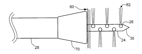
Refer now to Figures 6A-6C which illustrate operation of the catheter system
10. The heart tissue 60 (i.e., myocardium) may be accessed from the interior
of the
heart by, for example, navigating the catheter 12 through the vascular system
into a
-6-
CA 02396357 2002-05-24
WO 01/41657 PCT/US00/28375
chamber of the heart. Alternatively, the heart tissue 60 may be accessed from
the
exterior of the heart by, for example, transthoracic minimally invasive
surgery in
which the catheter 12 is navigated through the upper thoracic cavity adjacent
the
epicardium of the heart.
g Regardless of the approach, the distal portion 18 of the catheter 12 is
positioned adjacent the desired treatment site of the heart tissue 60
utilizing
conventional visualization techniques such as x-ray, fluoroscopy or endoscopic
visualization. While positioning the catheter 12, the penetrating member 24
may be
partially retracted in the outer sheath 28 such that only the distal end 30 of
the
1o penetrating member 24 is exposed, or fully retracted such that the entire
penetrating
member 24 is contained within the outer sheath 28.
With the distal portion 18 positioned adjacent the heart tissue 60 as shown in
Figure 6A, the penetrating member 24 is advanced into the heart tissue 60
until the
distal end 30 of the penetrating member 24 reaches a sufficient depth to
position the
15 injection ports 26 completely within the tissue 60 as shown in Figure 6B.
This
position may be confirmed by injecting radiopaque contrast media or colored
dye
through the inner lumen 38 of the penetrating member 24 such that the contrast
media
or dye exits the injection ports 26.
Once in position, fluid 62 may be infused from the pressurized fluid source 20
2o through the lumen 38 of the penetrating member and through the injection
ports 26
and into the heart tissue 60. After the fluid 62 has been delivered via the
injection
lumens in the injection ports 26, the penetrating member 24 may be retracted
into the
outer sheath 28. After retraction, the entire catheter 12 may be removed from
the
patient-
25 The pressure applied by the pressurized fluid source 20 to deliver the
fluid 62
into the heart tissue 60 may vary depending on the desired result. For
example, a
relatively low pressure of approximately .O1 to 1 ATM may be utilized to
deliver the
fluid 62 into the heart tissue 60 thereby minimizing trauma to the tissue
adjacent the
injection site. Alternatively, a relatively high pressure of approximately 10
to 300
3o ATM may be utilized to increase the depth penetration of the fluid 62 into
the heart
tissue 60 and/or to dispense the solution throughout the injected tissue.
The penetration depth of the fluid 62 into the tissue 60 influences fluid
retention, the volume of tissue 60 treated and the degree of trauma to the
tissue 60.
The penetration depth of the fluid 62 is dictated, in part, by the exit
velocity of the
CA 02396357 2002-05-24
WO 01/41657 PCT/tTS00/28375
fluid 62, the size of the fluid stream 62, and the properties of the tissue
60. The exit
velocity, in turn, depends on the applied pressure of the pressurized fluid
source 20,
the drag or pressure drop along the length of the lumen 38 and the ports 26,
and the
cross-sectional area or size of the ports 26. The size of the fluid stream 62
also
depends on the size of the ports 26. Thus, assuming the treatment site
dictates the
tissue 60 properties, the penetration depth may be selected by adjusting the
applied
pressure of the pressurized fluid source 20, the size and length of the lumen
38, and
the cross-sectional area of the ports 26. By adjusting these parameters, fluid
retention,
treated tissue volume and degree of trauma may be modified as required for the
1o particular clinical application.
As can be appreciated from the illustration of Figure 6C, by injecting the
fluid
62 in a direction different from the direction of penetration of the
penetrating member
24, the fluid 62 will be retained within the heart tissue 60. Retention of the
fluid 62 in
the heart tissue 60 is primarily accomplished by forming the injection ports
at an
angle relative to the direction of penetration of the penetrating member 24,
i.e., the
longitudinal axis of the penetrating member 24. In addition to providing
better
retention of the fluid 62 within the heart tissue 60, this arrangement also
allows for a
greater volume of heart tissue 60 to be treated with a single primary
penetration.
In an embodiment of the present invention, a low volume (several microliters
2o but less than 100 microliters by a single injection) of solution is
delivered to the heart
such that it may absorb the delivered solution within the time frame of the
injection.
In contrast to higher volume injections, the heart is more capable of
absorbing these
low volumes. The effect of the low volume injection is to minimize expulsion
by the
tissue: Iri order to deliver the entire-dose-of-virus it may be desirable or
necessar3r--to-
concentrate the injection (i.e., deliver the same number of viral particles or
micrograms of protein, typically delivered in 100.1, in a volume of I Owl) or
keep the
concentration of virus the same as that typically used, but increase the
number of
injections from 10 (typical) to 20, 30, or more.
Each injectate may also be delivered in a prolonged manner such that the heart
3o can absorb the solution as it is being injected (rate of delivery <_ rate
of tissue
absorption). For instance, the injection can be delivered at a defined flow
rate using a
syringe pump. The time of injection will depend on the volume to be delivered.
For
example, low volumes (a few microliters) may be delivered in under a minute
while
higher volumes (10 to 1001 or more) may be delivered over several minutes. In
this
_g_
CA 02396357 2002-05-24
WO 01/41657 PCT/US00/28375
instance, it may be beneficial to include a method which gently attaches the
injection
catheter to the wall of the heart, for instance suction or vacuum.
Thus, to accomplish this result, the injection ports 26 may be formed at an
angle to the longitudinal axis of the penetrating member 24. Preferably, the
axes of
the injection ports 26 are generally lateral to the longitudinal axis of the
penetrating
member 24. However, the axes of the injection ports 26 may be formed at an
angle of
about 5 to about 90 degrees relative to the axis of the penetrating member 24
to
accomplish essentially the same result. Also preferably, the penetrating
member 24
penetrates the heart tissue 60 in a direction generally orthogonal to the
surface of the
to heart tissue 60 adjacent the injection site.
Refer now to Figures 7A-7C which illustrate operation of an alternative
embodiment of the catheter system 10. In this particular embodiment, the
distal
portion of the catheter 12 incorporates a suction head 70 connected to the
distal head
of the outer sheath 28. The suction head 70 comprises a flexible tubular
member
having a generally conical shape. The suction head 70 has an interior which is
in
fluid communication with the inner lumen 36 of the outer sheath 28. As
mentioned
previously, the inner lumen 36 of the outer sheath 28 is in fluid
communication with
the vacuum source 22. By actuating the vacuum source 22, suction is applied to
the
suction head via the inner lumen 36 of the outer sheath 28.
2o The suction head is positioned adjacent the heart tissue 60 as illustrated
in
Figure 7A. The suction head 70 grasps the surface of the heart tissue 60
thereby
stabilizing the distal portion 18 of the catheter 12. This is particularly
beneficial when
treating tissue in a dynamic setting such as when the heart is beating. Absent
a
stabilizing -means such- as suction head- 70~ it maybe difficult to maintain-
the -distal
portion 18 in a relatively fixed position if the treatment site is not
stationary. Those
skilled in the art will recognize that other stabilizing means may be utilized
such as
removable screw anchors, miniature forceps, etc.
After suction is applied to the suction head 70 thereby stabilizing the distal
portion 18 of the catheter 12, the penetrating member 24 is advanced into the
heart
3o tissue 60 as illustrated in Figure 7B. Once the injection ports 26 of the
penetrating
member 24 are completely embedded within the heart tissue 60, fluid 62 may be
delivered into the heart tissue 60 via the injection ports 26 as discussed
previously.
After the fluid 62 has been delivered to the heart tissue 60, the penetrating
member 24 may be retracted into the outer sheath 28. After retracting the
penetrating
-9-
CA 02396357 2002-05-24
WO 01/41657 PCT/tTS00/28375
member 24, the suction applied by the suction head 70 is terminated to release
the
distal portion 18 of the catheter from the heart tissue 60. The entire
catheter system
12 may then be removed from the patient.
From the foregoing, it is apparent that the present invention provides a
device
and method for delivering and injecting fluid into heart tissue to improve
delivery
efficiency. This is accomplished by utilizing injection ports which direct
fluid in a
direction different from the direction of penetration of the penetrating
member. Thus,
fluid leakage from the injection site is reduced and the fluid is distributed
over a
greater volume of tissue.
Although treatment of the heart is used as an example herein, the medical
devices of the present invention are useful for treating any mammalian tissue
or
organ. Non-limiting examples include tumors; organs including but not limited
to the
heart, lung, brain, liver, kidney, bladder, urethra and ureters, eye,
intestines, stomach,
pancreas, ovary, prostate; skeletal muscle; smooth muscle; breast, cartilage
and bone.
The terms "therapeutic agents" and "drugs" are used interchangeably herein
and include pharmaceutically active compounds, cells, nucleic acids with and
without
carrier vectors such as lipids, compacting agents (such as histones), virus,
polymers,
proteins, and the like, with or without targeting sequences.
Specific examples of therapeutic agents used in conjunction with the present
2o invention include, for example, proteins, oligonucleotides, ribozymes, anti-
sense
genes, DNA compacting agents, gene/vector systems (i.e., anything that allows
for the
uptake and expression of nucleic acids), nucleic acids (including, for
example,
recombinant nucleic acids; naked DNA, cDNA, RNA; genomic DNA, cDNA or RNA
in a ion=infectious-vector or in a-viral vector which may have attached--
peptide
targeting sequences; antisense nucleic acid (RNA or DNA); and DNA chimeras
which
include gene sequences and encoding for ferry proteins such as membrane
translocating sequences ("MTS") and herpes simplex virus-1 ("VP22")), and
viral,
liposomes and cationic polymers that are selected from a number of types
depending
on the desired application. Other pharmaceutically active materials include
anti-
3o thrombogenic agents such as heparin, heparin derivatives, urokinase, and
PPACK
(dextrophenylalanine proline arginine chloromethylketone); antioxidants such
as
probucol and retinoic acid; angiogenic and anti-angiogenic agents; agents
blocking
smooth muscle cell proliferation such as rapamycin, angiopeptin, and
monoclonal
antibodies capable of blocking smooth muscle cell proliferation; anti-
inflammatory
-10-
CA 02396357 2002-05-24
WO 01/41657 PCT/US00/28375
agents such as dexamethasone, prednisolone, corticosterone, budesonide,
estrogen,
sulfasalazine, acetyl salicylic acid, and mesalamine; calcium entry blockers
such as
verapamil, diltiazem and nifedipine; antineoplastic / antiproliferative / anti-
mitotic
agents such as paclitaxel; 5-fluorouracil, methotrexate, doxorubicin,
daunorubicin,
cyclosporine, cisplatin, vinblastine, vincristine, epothilones, endostatin,
angiostatin
and thymidine kinase inhibitors; antimicrobials such as triclosan,
cephalosporins,
aminoglycosides, and nitorfurantoin; anesthetic agents such as lidocaine,
bupivacaine,
and ropivacaine; nitric oxide (NO) donors such as lisidomine, molsidomine, L-
arginine, NO-protein adducts, NO-carbohydrate adducts, polymeric or oligomeric
NO
1o adducts; anti-coagulants such as D-Phe-Pro-Arg chloromethyl ketone, an RGD
peptide-containing compound, heparin, antithrombin compounds, platelet
receptor
antagonists, anti-thrombin antibodies, anti-platelet -receptor antibodies,
enoxaparin,
hirudin, Warafin sodium, Dicumarol, aspirin, prostaglandin inhibitors,
platelet
inhibitors and tick antiplatelet factors; vascular cell growth promotors such
as growth
factors, growth factor receptor antagonists, transcriptional activators, and
translational
promotors; vascular cell growth inhibitors such as growth factor inhibitors,
growth
factor receptor antagonists, transcriptional repressors, translational
repressors,
replication inhibitors, inhibitory antibodies, antibodies directed against
growth
factors, bifunctional molecules consisting of a growth factor and a cytotoxin,
2o bifunctional molecules consisting of an antibody and a cytotoxin;
cholesterol-
lowering agents; vasodilating agents; agents which interfere with endogeneus
vascoactive mechanisms; survival genes which protect against cell death, such
as anti-
apoptotic Bcl-2 family factors and Akt kinase; and combinations thereof.
~Xariiples - of polynucleotide -sequences -useful in practice of-the-
invention
include DNA or RNA sequences having a therapeutic effect after being taken up
by a
cell. Examples of therapeutic polynucleotides include anti-sense DNA and RNA;
DNA coding for an anti-sense RNA; or DNA coding for tRNA or rRNA to replace
defective or deficient endogenous molecules. The polynucleotides of the
invention
can also code for therapeutic proteins or polypeptides. A polypeptide is
understood to
be any translation product of a polynucleotide regardless of size, and whether
glycosylated or not. Therapeutic proteins and polypeptides include as a
primary
example, those proteins or polypeptides that can compensate for defective or
deficient
species in an animal, or those that act through toxic effects to limit or
remove harmful
cells from the body. In addition, the polypeptides or proteins useful in the
present
-11-
CA 02396357 2002-05-24
WO 01/41657 PCT/US00/28375
invention include, without limitation, angiogenic factors and other molecules
competent to induce angiogenesis, including acidic and basic fibroblast growth
factors, vascular endothelial growth factor, hif 1, epidermal growth factor,
transforming growth factor a and (3, platelet-derived endothelial growth
factor,
S platelet-derived growth factor, tumor necrosis factor a, hepatocyte growth
factor and
insulin like growth factor; growth factors; cell cycle inhibitors including
CDK
inhibitors; anti-restenosis agents, including p15, p16, p18, p19, p21, p27,
p53, p57,
Rb, nFkB and E2F decoys, thymidine kinase ("TK") and combinations thereof and
other agents useful for interfering with cell proliferation, including agents
for treating
1o malignancies; and combinations thereof. Still other useful factors, which
can be
provided as polypeptides or as DNA encoding these polypeptides, include
monocyte
chemoattractant protein ("MCP-1 "), and the family of bone morphogenic
proteins
("BMP's"). The known proteins include BMP-2, BMP-3, BMP-4, BMP-5, BMP-6
(vgr-1), BMP-7 (oP-1), BMP-s, BMP-9, BMP-lo, BMP-11, BMP-12, BMP-13,
15 BMP-14, BMP-15, and BMP-16. Currently preferred BMP's are any of BMP-2,
BMP-3, BMP-4, BMP-5, BMP-6 and BMP-7. These dimeric proteins can be
provided as homodimers, heterodimers, or combinations thereof, alone or
together
with other molecules. Alternatively or, in addition, molecules capable of
inducing an
upstream or downstream effect of a BMP can be provided. Such molecules include
2o any of the "hedgehog" proteins, or the DNA's encoding them.
The present invention is also useful in delivering cells as the therapeutic
agent.
Cells can be of human origin (autologous or allogeneic) or from an animal
source
(xenogeneic), genetically engineered if desired to deliver proteins of
interest at a
delivery or transplant site. The delivery Iriedia is formulated as-needed to
maintain
25 cell function and viability.
Those skilled in the art will recognize that the present invention may be
manifested in a variety of forms other than the specific embodiments described
and
contemplated herein. Accordingly, departures in form and detail may be made
without departing from the scope and spirit of the present invention as
described in
30 the appended claims.
-12-