Note: Descriptions are shown in the official language in which they were submitted.
CA 02398203 2002-07-24
WO 01/57510 PCT/USO1/02465
ELECTROCHEMICAL METHODS AND DEVICES FOR USE IN THE
DETERMINATION OF HEMATOCRIT CORRECTED ANALYTE
CONCENTRATIONS
INTRODUCTION
Field of the Invention
The field of this invention is analyte determination, particularly
electrochemical
analyte determination and more particularly the electrochemical determination
of blood
analytes.
Background
Analyte detection in physiological fluids, e.g. blood or blood derived
products, is of
ever increasing importance to today's society. Analyte detection assays find
use in a variety
of applications, including clinical laboratory testing, home testing, etc.,
where the results of
such testing play a prominent role in diagnosis and management in a variety of
disease
conditions. Analytes of interest include glucose for diabetes management,
cholesterol, and
the like. In response to this growing importance of analyte detection, a
variety of analyte
detection protocols and devices for both clinical and home use have been
developed.
One type of method that is employed for analyte detection is an
electrochemical
method. In such methods, an aqueous liquid sample is placed into a reaction
zone in an
electrochemical cell comprising two electrodes, i.e. a reference and working
electrode,
where the electrodes have an impedance which renders them suitable for
amperometric
measurement. The component to be analyzed is allowed to react directly with an
electrode,
or directly or indirectly with a redox reagent to form an oxidisable (or
reducible) substance
in an amount corresponding to the concentration of the component to be
analyzed, i.e.
analyte. The quantity of the oxidisable (or reducible) substance present is
then estimated
electrochemically and related to the amount of analyte present in the initial
sample.
Where the physiological sample being assayed is whole blood or a derivative
thereof,
the hematocrit of the sample can be a source of analytical error in the
ultimate analyte
concentration measurement. For example, in electrochemical measurement
protocols where
3o the analyte concentration is derived from observed time-current transients,
hematocrit can
slow the equilibration chemistry in the electrochemical cell and/or slow the
enzyme kinetics
by increasing the sample viscosity in the cell, thereby attenuating the time
current response
and causing analytical error.
CA 02398203 2002-07-24
WO 01/57510 PCT/USO1/02465
As such, there is great interest in the development of methods of at least
minimizing
the hematocrit originated analytical error. In certain protocols, blood
filtering membranes are
employed to remove red blood cells and thereby minimize the hematocrit effect.
These
particular protocols are unsatisfactory in that increased sample volumes and
testing times are
required. Other protocols focus on the determination of the capillary fill
time. However,
these protocols add complexity to both the strips and devices that are used to
read them. In
yet other embodiments, hematocrit is separately determined using two
additional electrodes,
which also results in more complex and expensive strips/devices.
As such, there is continued interest in the identification of new methods for
1o electrochemically measuring the concentration of an analyte in a
physiological sample,
where the method minimizes the analytical error which originates with the
hematocrit of the
sample.
Relevant Literature
Patent documents of interest include: 5,942,102 and WO 97/18465.
SUMMARY OF THE INVENTION
Methods and devices for determining the concentration of an analyte in a
physiological sample are provided. In the subject methods, the physiological
sample is
introduced into an electrochemical cell having a working and reference
electrode. A first
2o electric potential is applied to the cell and the resultant cell current
over a first period of time
is measured to determine a first time-current transient. A second electric
potential of
opposite polarity is then applied to the cell and a second time-current
transient is determined.
The preliminary concentration of the analyte (Co)is then calculated from the
first and/or
second time-current transients. This preliminary analyte concentration, less a
background
value, is then multiplied by a hematocrit correction factor to obtain the
analyte concentration
in the sample, where the hematocrit correction factor is a fiznction of the
preliminary analyte
concentration and the ratio of 2 current values (y) within the time-current
transient of the
electrochemical cell. The subject methods and devices are suited for use in
the determination
of a wide variety of analytes in a wide variety of samples, and are
particularly suited for the
3o determination of analytes in whole blood or derivatives thereof, where an
analyte of
particular interest is glucose.
CA 02398203 2002-07-24
WO 01/57510 PCT/USO1/02465
BRIEF DESCRIPTION OF THE FIGURES
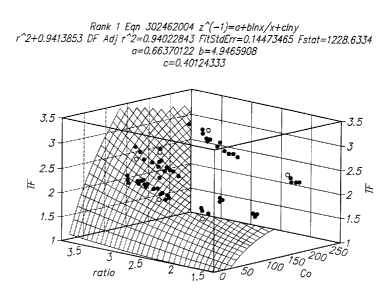
Fig. 1 provides a three-dimensional graph of Co, y and a.(Co, y) derived from
experimental data using a wide range of glucose and hematocrit values.
DESCRIPTION OF THE SPECIFIC EMBODIMENTS
Methods and devices for determining the concentration of an analyte in a
physiological sample are provided. In the subject methods, the physiological
sample is
introduced into an electrochemical cell having a working and reference
electrode. A first
electric potential is applied to the cell and the resultant cell current over
a first period of time
1o is measured to determine a first time-current transient. A second electric
potential of
opposite polarity is then applied to the cell and a second a time-current
transient is
determined. The preliminary concentration of the analyte is then calculated
from the first
and/or second time-current transient. This preliminary analyte concentration,
less a
background value, is then multiplied by a hematocrit correction factor to
obtain the analyte
concentration in the sample, where the hematocrit correction factor is a
fiznction of the
preliminary analyte concentration and the variable y of the electrochemical
cell. The subject
methods and devices are suited for use in the determination of a wide variety
of analytes in a
wide variety of samples, and are particularly suited for use in the
determination of analytes
in whole blood or derivatives thereof, where an analyte of particular interest
is glucose. In
2o fizrther describing the subject invention, the subject methods will be
described first followed
by a review of a representative device for use in practicing the subject
methods.
Before the subject invention is described fi~rther, it is to be understood
that the
invention is not limited to the particular embodiments of the invention
described below, as
variations of the particular embodiments may be made and still fall within the
scope of the
appended claims. It is also to be understood that the terminology employed is
for the purpose
of describing particular embodiments, and is not intended to be limiting.
Instead, the scope
of the present invention will be established by the appended claims.
3o In this specification and the appended claims, singular references include
the plural,
unless the context clearly dictates otherwise. Unless defined otherwise, all
technical and
scientific terms used herein have the same meaning as commonly understood to
one of
ordinary skill in the art to which this invention belongs.
CA 02398203 2002-07-24
WO 01/57510 PCT/USO1/02465
METHODS
As summarized above, the subject invention provides a method for determining a
hematocrit corrected analyte concentration value in a physiological sample. By
hematocrit
corrected analyte concentration is meant that the analyte concentration value
determined
using the subject methods has been modulated or changed to remove
substantially all
contribution of hematocrit to the value. In other words, the concentration
value that is
determined using the subject methods has been modified so that any
contribution to the
value from the hematocrit of the sample that would be present in the value but
for the
1o practicing of the subject methods is removed. As such, the hematocrit
signal is deconvoluted
from the analyte signal in the subject methods, and only the analyte signal is
employed in
arriving at the final hematocrit corrected analyte concentration.
The first step in the subject methods is to introduce a quantity of the
physiological
sample of interest into an electrochemical cell that includes spaced apart
working and
reference electrodes and a redox reagent system. The physiological sample may
vary, but in
many embodiments is generally whole blood or a derivative or fraction thereof,
where whole
blood is of particular interest in many embodiments. The amount of
physiological sample,
e.g. blood, that is introduced into the reaction area of the test strip
varies, but generally
ranges from about 0.1 to 10 p.L, usually from about 0.9 to 1.6 p,L. The sample
is introduced
2o into the reaction area using any convenient protocol, where the sample may
be injected into
the reaction area, allowed to wick into the reaction area, and the like, as
may be convenient.
While the subject methods may be used, in principle, with any type of
electrochemical cell having spaced apart working and reference electrodes and
a redox
reagent system, in many embodiments the subject methods employ an
electrochemical test
strip. The electrochemical test strips employed in these embodiments of the
subject invention
are made up of two opposing metal electrodes separated by a thin spacer layer,
where these
components define a reaction area or zone in which is located a redox reagent
system.
In certain embodiments of these electrochemical test strips, the working and
reference electrodes are generally configured in the form of elongated
rectangular strips.
3o Typically, the length of the electrodes ranges from about 1.9 to 4.5 cm,
usually from about
2.0 to 2.8 cm. The width of the electrodes ranges from about 0.38 to 0.76 cm,
usually from
about 0.51 to 0.67 cm. The reference electrodes typically have a thickness
ranging from
about 10 to 100 nm and usually from about 10 to 20 nm. In certain embodiments,
the length
4
CA 02398203 2002-07-24
WO 01/57510 PCT/USO1/02465
of one of the electrodes is shorter than the length of the other electrode,
typically about 0.32
cm. The shorter electrode may be the working or reference electrode.
The working and reference electrodes are further characterized in that at
least the
surface of the electrodes that faces the reaction area in the strip is a
metal, where metals of
interest include palladium, gold, platinum, silver, iridium, carbon, doped tin
oxide, stainless
steel and the like. In many embodiments, the metal is gold or palladium. While
in principle
the entire electrode may be made of the metal, each of the electrodes is
generally made up of
an inert support material on the surface of which is present a thin layer of
the metal
component of the electrode. In these more common embodiments, the thickness of
the inert
1o backing material typically ranges from about 51 to 356 Vim, usually from
about 102 to
153 ~m while the thickness of the metal layer typically ranges from about 10
to 100 nm and
usually from about 10 to 40 nm, e.g. a sputtered metal layer. Any convenient
inert backing
material may be employed in the subject electrodes, where typically the
material is a rigid
material that is capable of providing structural support to the electrode and,
in turn, the
electrochemical test strip as a whole. Suitable materials that may be employed
as the backing
substrate include plastics, e.g. PET, PETG, polyimide, polycarbonate,
polystyrene, silicon,
ceramic, glass, and the like.
A feature of the electrochemical test strips used in these embodiments of the
subject
methods is that the working and reference electrodes as described above face
each other and
2o are separated by only a short distance, such that the distance between the
working and
reference electrode in the reaction zone or area of the electrochemical test
strip is extremely
small. This minimal spacing of the working and reference electrodes in the
subject test strips
is a result of the presence of a thin spacer layer positioned or sandwiched
between the
working and reference electrodes. The thickness of this spacer layer generally
should be less
than or equal to 500 Vim, and usually ranges from about 102 to 153 Vim. The
spacer layer is
cut so as to provide a reaction zone or area with at least an inlet port into
the reaction zone,
and generally an outlet port out of the reaction zone as well. The spacer
layer may have a
circular reaction area cut with side inlet and outlet vents or ports, or other
configurations,
e.g. square, triangular, rectangular, irregular shaped reaction areas, etc.
The spacer layer may
3o be fabricated from any convenient material, where representative suitable
materials include
PET, PETG, polyimide, polycarbonate, and the like, where the surfaces of the
spacer layer
may be treated so as to be adhesive with respect to their respective
electrodes and thereby
CA 02398203 2002-07-24
WO 01/57510 PCT/USO1/02465
maintain the structure of the electrochemical test strip. Of particular
interest is the use of a
die-cut double-sided adhesive strip as the spacer layer.
The electrochemical test strips used in these embodiments of the subject
invention
include a reaction zone or area that is defined by the working electrode, the
reference
electrode and the spacer layer, where these elements are described above.
Specifically, the
working and reference electrodes define the top and bottom of the reaction
area, while the
spacer layer defines the walls of the reaction area. The volume of the
reaction area is at least
about 0.1 pL, usually at least about 1 ~L and more usually at least about 1.5
~L, where the
volume may be as large as 10 p.L or larger. As mentioned above, the reaction
area generally
1o includes at least an inlet port, and in many embodiments also includes an
outlet port. The
cross-sectional area of the inlet and outlet ports may vary as long as it is
sui~ciently large to
provide an effective entrance or exit of fluid from the reaction area, but
generally ranges
from about 9 x 10~ to 5 x 10⁻³ cm2, usually from about 1.3 x 10⁻³ to 2.5 x 10-
3 cm2.
Present in the reaction area is a redox reagent system, which reagent system
provides
for the species that is measured by the electrode and therefore is used to
derive the
concentration of analyte in a physiological sample. The redox reagent system
present in the
reaction area typically includes at least an enzymes) and a mediator. In many
embodiments,
the enzyme members) of the redox reagent system is an enzyme or plurality of
enzymes that
work in concert to oxidize the analyte of interest. In other words, the enzyme
component of
the redox reagent system is made up of a single analyte oxidizing enzyme or a
collection of
two or more enzymes that work in concert to oxidize the analyte of interest.
Enzymes of
interest include oxidases, dehydrogenases, lipases, kinases, diphorases,
quinoproteins, and
the like.
The specific enzyme present in the reaction area depends on the particular
analyte for
which the electrochemical test strip is designed to detect, where
representative enzymes
include: glucose oxidase, glucose dehydrogenase, cholesterol esterase,
cholesterol oxidase,
lipoprotein lipase, glycerol kinase, glycerol-3-phosphate oxidase, lactate
oxidase, lactate
dehydrogenase, pyruvate oxidase, alcohol oxidase, bilirubin oxidase, uricase,
and the like. In
many preferred embodiments where the analyte of interest is glucose, the
enzyme
3o component of the redox reagent system is a glucose oxidizing enzyme, e.g. a
glucose oxidase
or glucose dehydrogenase.
The second component of the redox reagent system is a mediator component,
which
is made up of one or more mediator agents. A variety of different mediator
agents are known
6
CA 02398203 2002-07-24
WO 01/57510 PCT/US01/02465
in the an and include: ferricyanide, phenazine ethosulphate, phenazine
methosulfate,
pheylenediamine, 1-methoxy-phenazine methosulfate, 2,6-dimethyl-1,4-
benzoquinone, 2,5-
dichloro-1,4-benzoquinone, ferrocene derivatives, osmium bipyridyl complexes,
ruthenium
complexes, and the like. In those embodiments where glucose in the analyte of
interest and
glucose oxidase or glucose dehydrogenase are the enzyme components, mediators
of
particular interest are ferricyanide, and the like.
Other reagents that may be present in the reaction area include buffering
agents, e.g.
citraconate, citrate, malic, malefic, phosphate, "Good" buffers and the like.
Yet other agents
that may be present include: divalent canons such as calcium chloride, and
magnesium
to chloride; pyrroloquinoline quinone; types of surfactants such as Triton,
Macol, Tetronic,
Silwet, Zonyl, and Pluronic; stabilizing agents such as albumin, sucrose,
trehalose, mannitol,
and lactose.
The redox reagent system is generally present in dry form. The amounts of the
various components may vary, where the amount of enzyme component typically
ranges
from about 1 to 100 mg/mL, usually from about 5 to 80mg/mL; and the amount of
mediator
component typically ranges from about 5 to 1000 mM, usually from about 90 to
900 mM.
Following sample introduction, first and second time-current transients are
obtained.
The first and second time-current transients are obtained by applying a
constant electric
potential to the cell and observing the change in current over a period of
time in the cell. In
other words, first and second pulses are applied to the cell and the resultant
time-current
transients are observed. As such, the first time-current transient is obtained
by applying a
constant electric potential or first pulse to the cell, e.g. between the
working and the
reference electrodes, and observing the change in current over time between
the electrodes,
i.e. change in cell current, to obtain the first time-current transient. The
magnitude of the first
applied electric potential generally ranges from about 0 to -0.6 V, usually
from about -0.2
to -0.4 V. The length of time over which the current between the electrodes is
observed to
obtain the first time-current transient typically ranges from about 3 to 20
seconds, usually
from about 4 to 10 seconds.
The second time current is obtained by applying a second constant electric
potential
or second pulse, typically of opposite polarity from the first constant
electric potential, to the
electrodes and observing the change in current between the electrodes for a
second period of
time. The magnitude of this second constant electric potential typically
ranges from about 0
to +0.6 V, usually from about +0.2 to +0.4 V, where in many embodiments the
magnitude
7
CA 02398203 2002-07-24
WO 01/57510 PCT/USO1/02465
of the second electric potential is the same as the magnitude of the first
electric potential.
The second time period typically ranges from about 1 to 10 seconds, usually
from about 2 to
4 seconds. By observing the change in current between the electrodes over this
second
period of time, a second time-current transient for the cell is determined.
The overall time period required to obtain the requisite first and second time-
current
transients, as described above, is relatively short in certain embodiments. In
such
embodiments, the total amount of time required to obtain the first and second
time-current
transients is less than about 30 seconds, usually less than about 20 seconds
and more usually
less than about 14 seconds.
to The next step in the subject methods is to use the observed first and
second time-
current transients, obtained as described above, to determine: (a) the
variable y of the
electrochemical cell used in the subject methods; and (b) a preliminary
analyte concentration
for the analyte of interest in the sample.
The variable ~r employed in the subject methods is defined to describe the
deviation
of the electrochemical cell from ideality. By way of background, it should be
noted that
y should approach unity under ideal conditions, i.e. reagent equilibration and
glucose
reaction are complete before the end of the first pulse. Any of these
conditions not being
complete will cause the ratio to deviate fron non-unity values. The numerator
of y is defined
as the steady-state current observed following application of the second
electric potential to
2o the cell, i.e. predicted value at t=oc of the second time-current
transient. The denominator is
defined as the average current over a short time period near the end of the
first period of
time, i.e. near the end of the application of the first electric potential or
first pulse. The short
period of time from which the average current is determined typically ranges
from .2 to 2
seconds, usually from about .2 to 1.5 seconds and more usually from about .2
to 1.25
seconds, where in many embodiments the short period of time is about .3
second. The
average current is determined at a time near the end of the first time period,
typically within
about 0.1 to 1 second,. In certain embodiments, the variable y is described by
the formula:
155~1PP
where:
iss is the steady-state current of the second applied electric potential; and
iPP is the average current over a short period of time near the end of first
time period,
i.e. near the end of the time during which the first electric potential is
applied to the cell. For
example, where the first time period is 10 seconds long, the average current
may be the
s
CA 02398203 2002-07-24
WO 01/57510 PCT/USO1/02465
average current from 8.5 to 9.5 seconds of the 10 second long period, which is
a 1.0 second
time period 0.5 seconds from the end of the first time period As mentioned
above, the
first and second time-current transients are also employed to derive a
preliminary analyte
concentration value for the sample being assayed. In many embodiments, the
preliminary
analyte concentration is determined by using the following equations:
i(t) = iss { 1 + 4 exp(-4~t2Dt/L2) }
iss = 2 FADCo/L
where
iss is the steady-state current following application of the second electric
potential;
1o i is the measured current which is a function of time
D is the diffusion coefficient of the cell, where this coefficient may be
determined
from Fick's first law, i.e. J(x,t)=-Ddc~~,t~/dX
L is the spacer thickness;
t is the time for the application of the 2"d electric potential where t=0 for
the
beginning of the pulse
Co is the preliminary concentration of the analyte;
F is faraday's constant, i.e. 9.6485x10⁴C/mol; and
A is the area of the working electrode.
2o Using the above equations and steps, the observed first and second time-
current
transients are used to determine the variable y of the electrochemical cell
employed in the
subject method and the preliminary concentration value of the analyte of
interest in the
assayed physiological sample.
From the determined variable y and preliminary analyte concentration value, a
hematocrit correction factor is determined, which hematocrit correction factor
is used to
obtain a hematocrit corrected analyte concentration value from the initial or
preliminary
analyte concentration value described above. The hematocrit correction factor
is a factor
with which the preliminary analvte concentration (typically less a background
value) may be
multiplied in order to obtain a hematocrit corrected analyte concentration
value, i.e. a
3o concentration value from which the hematocrit component has been removed.
The
hematocrit correction factor is a function of both the preliminary analyte
concentration value
and the variable 'y of the electrochemical cell.
9
CA 02398203 2002-07-24
WO 01/57510 PCT/USO1/02465
Any hematocrit correction factor that can be multiplied by the preliminary
concentration value (usually less a background value, as described in greater
detail below)
may be employed in the subject methods. One class of hematocrit correction
factors that find
use in the subject methods are those that are derived from a three dimensional
graph of Co, y
and oc(Co, ~r) obtained from experimental data using a wide range of analyte
and hematocrit
values. The hematocrit correction factor (a(Co, Y)) is determined using the
formula:
a(Co, ~/) =actual concentration/( Co -Background Value)
(For example, where the analyte is glucose, a(Co, y) in many embodiments
equals the
glucose concentration as determined using the Yellow Springs Instrument
glucose analyzer
to model 23A (as described in U.S. Patent No. 5, 968,760 the disclosure of
which is herein
incorporated by reference) divided by the Co less a background value, e.g. 22
mg/dL). This
class of hematocrit correction factors are typically equations which fit a
smooth surface
function that minimizes the error between the predicted and actual data. See
e.g. the
experimental section, infra. One representative hematocrit correction factor
that finds use in
the subject methods is:
1/((0.6637) + ((4.9466*ln(Ca))/ Co) + (-0.4012*ln(y)))
In determining the hematocrit corrected concentration of analyte according to
the
subject invention, the preliminary analyte concentration (Co) as determined
above, less a
background signal value, is multiplied by the hematocrit correction factor.
The background
value that is subtracted from the preliminary concentration value depends on
the analyte
being measured. For glucose, this value typically ranges from about 0 to 40
mg/dL, usually
from about 8 to 25 mg/dL, where in many embodiments the background value is
about 22
mg/dL or is 22 mg/dL.
Generally, the following formula is employed to determine the hematocrit
corrected
analyte concentration according to the subject invention:
hematocrit corrected concentration = hematocrit correction factor x[ Co - (3]
where
(3 is the background value; and
Co is the preliminary analyte concentration.
The above described methods yield a hematocrit corrected analyte concentration
value, i.e. a concentration value in which the hematocrit component has been
deconvoluted
CA 02398203 2002-07-24
WO 01/57510 PCT/USO1/02465
and removed. As such, the above described methods provide for an accurate
value of the
concentration of the analyte in the sample being assayed.
The above computational steps of the subject method may be accomplished
manually
or through the use of an automated computing means, where in many embodiments
the use
of an automated computing means, such as is described in connection with the
subject
devices discussed below, is of interest.
DEVICES
Also provided by the subject invention are meters for use in practicing the
subject
to invention. The subject meters are typically meters for amperometrically
measuring the
hematocrit corrected concentration of an analyte in a physiological sample.
The subject
meters typically include: (a) a means for applying a first electric potential
to an
electrochemical cell into which the sample has been introduced and measuring
cell current as
a function of time to obtain a first time-current transient; (b) a means for
applying a second
15 electric potential to the electrochemical cell and measuring cell current
as a function of time
to obtain a second time-current transient; (c) a means for determining a
preliminary analyte
concentration value and a variable y from said first and second time-currents;
and (d) a
means for removing the hematocrit component from the preliminary concentration
value to
derive the hematocrit corrected analyte concentration in said sample. Means
(a) and (b) may
2o be any suitable means, where representative means are described in WO
97/18465 and U.S.
Patent No. 5,942,102; the disclosures of which are herein incorporated by
reference. Means
(c) and (d) are typically computing means present in the meter which are
capable of using
the measured first and second time current transients to ultimately obtain the
hematocrit
corrected analyte concentration. As such, means (c) is typically a means that
is capable of
25 determining the preliminary concentration of the analyte of interest and
the variable y from
the first and second time-current transients using the equations described
above. Likewise,
means (d) is typically a means that is capable of determining the hematocrit
corrected
analyte concentration using the equations described above, where this means
typically
comprises the hematocrit correction factor.
The following examples are offered by way of illustration and not by way of
limitation.
I1
CA 02398203 2002-07-24
WO 01/57510 PCT/USO1/02465
EXPERIMENTAL
I. Electrochemical Test Strip Preparation
An electrochemical test strip consisting of two metallized electrodes oriented
in a
sandwich configuration was prepared as follows. The top layer of the test
strip was a gold
sputtered Mylar strip. The middle layer was a double-sided adhesive with a
punched hole
that defined the reaction zone or area. The punched hole was a circle with two
juxtaposed
rectangular inlet and outlet channels. The bottom layer of the test strip was
sputtered
palladium on Mylar. A film of ferricyanide and glucose dehydrogenase PQQ was
deposited
on the palladium sputtered surface.
II. Generation of Experimental Data
First and second time current transients for a number of different samples
varying by
glucose concentration and hematocrit were obtained as follows. Sample was
applied to the
strip which actuated an applied potential of -.03 V for a period of 10 seconds
which was then
followed by a second pulse of +0.3 V for a period of 3 to 10 seconds (where
these electrode
potentials are with respect to the gold electrode).
III. Derivation of Hematocrit Correction Factor for Glucose
For a wide range of glucose and hematocrit values measured as described above,
Co,
2o the variable y and a,(Co, ~r) were derived.
Co was derived using the equations:
i(t) = iss { 1 + 4 exp(-4~ZDt/LZ) ~
iSS = 2 FADCo/L
where
iss is the steady-state current following application of the second electric
potential;
i is the measured current which is a function of time
D is the diffusion coefficient of the cell, where this coefficient may be
determined
from Fick's first law, i.e. J(x,t)=-D dC(~t)/ax
L is the spacer thickness;
3o t is the time for the application of the 2"d electric potential where t=0
for the
beginning of the pulse;
Co is the preliminary concentration of the analyte;
F is faraday's constant, i. e. 9.6485 x 10⁴C/mol; and
12
CA 02398203 2002-07-24
WO 01/57510 PCT/USO1/02465
A is the area of the electrode surface.
The variable y was derived using the equation:
lss / 1PP
where:
iss is the steady-state current of the second applied electric potential or
second pulse;
and
iPP is the average current from 8.5 to 9.5 seconds of the 10 s long period
during which
the first pulse was applied.
to
oc(Co, y) was determined using the equation:
oc(Co, y) =YSI concentration/( C° -22 mg/dL)
where YSI is the glucose concentration as determined using the Yellow Springs
Instrument
glucose analyzer model 23A (as described in U.S. Patent No. 5, 968,760 the
disclosure of
which is herein incorporated by reference).
A three-dimensional graph of Co, y and oc(Co, y) as determined above for a
wide
range of glucose and hematocrit values was prepared and is shown in Fig. 1. A
simple
equation fit was then performed on the graph to define the surface. The
residual of the fitted
data was monitored to ascertain the quality of the model equation. The
empirical equation
2o was found to be:
Hematocrit Correction Factor = 1/((0.6637) + ((4.9466*ln(C°))/ Co) + (-
0.4012*ln(y)))
The above correction factor was found to be valid for those situations where
the y >0.7 and
Ca >40 mg/dL.
IV. Comparison of Hematocrit Corrected Values to YSI determined Values.
A prediction data set was generated by testing several glucose strips with a
wide
range of glucose and hematocrit levels. From this data a hematocrit correction
equation was
derived using a model which fits the terms Co, y, and oc(C°, y) . It
was found that using the
hematocrit correction equation on the prediction data set causes the majority
of data points to
3o fall within +/- 15%. It was also found that the bias of the glucose results
to 42% hematocrit,
indicating that the hematocrit effect on this data set is minimal. In order to
confirm this
algorithm, another batch of glucose sesnsors was tested with a different blood
donor. It was
13
CA 02398203 2002-07-24
WO 01/57510 PCT/USO1/02465
found that the algorithm still corrects for the hematocrit effect in a manner
analogous to the
earlier findings.
The above results and discussion demonstrate that subject invention provides a
simple and powerful tool to obtain analyte concentration values in which
hematocrit derived
error is substantially if not entirely eliminated. As the subject methods rely
solely on the
measurement of time-current transients, they may be practiced with relatively
simple
electrochemical devices. Furthermore, only small sample volumes need be
employed and
relatively quick assay times are provided. As such, the subject invention
represents a
to significant contribution to the art.
All publications and patents cited in this specification are herein
incorporated by
reference as if each individual publication or patent were specifically and
individually
indicated to be incorporated by reference. The citation of any publication is
for its disclosure
15 prior to the filing date and should not be construed as an admission that
the present invention
is not entitled to antedate such publication by virtue of prior invention.
Although the foregoing invention has been described in some detail by way of
illustration and example for purposes of clarity of understanding, it is
readily apparent to
2o those of ordinary skill in the art in light of the teachings of this
invention that certain changes
and modifications may be made thereto without departing from the spirit or
scope of the
appended claims.
1~