Note: Descriptions are shown in the official language in which they were submitted.
CA 02398278 2002-07-25
WO 01/54580 PCT/CA01/00090
VISIBLE-NEAR INFRARED SPECTROSCOPY IN BURN INJURY ASSESSMENT
FIELD OF THE INVENTION
The present invention relates generally to the field of medical devices.
BACKGROUND OF THE INVENTION
Cutaneous burns are one of the most destructive insults to the skin,
causing damage, scarring and even death of the tissue. It has been reported
that burns
alone account for over 2 million medical procedures every year in the United
States. Of
these, 150,000 refer to individuals who are hospitalized and as many as 10,000
die
(Bronzino, 1995, The Biomedical Engineering Handbook (CRC Press: Florida)).
Despite
the large number of annual burn cases, the accurate assessment of burn
severity remains
a problem for the burn specialist. The ability to distinguish between burns
that will heal on
their own versus those that will require surgical intervention is particularly
challenging.
Generally, the depth of a burn injury determines and is inversely related to
the ability of the
skin to restore and regenerate itself. Burns involve damage to the dermis in
varying
amounts, reducing the dermal blood supply and altering the skin hemodynamics.
Highly
destructive burns have only a marginal residual blood supply to the dermis
that may result in
ischemia and ultimately necrosis of the dermis, as the re-epithelialization of
the tissue
depends on the viable dermis below the burned tissue. Thermal injuries are
clinically
classified according to the depth of the injury as superficial (epidermal),
partial thickness
(epidermal and varying levels of dermal) and full thickness (epidermal and
dermal).
Superficial burns are mild burns whereby the tissue is capable of regenerating
the
epidermis. Partial thickness injuries destroy a portion of the dermal layer,
although
sufficient dermis usually remains for re-epithalization to occur with adequate
vasculature.
Deep partial and full thickness injuries involve destruction of the dermal
layer and what
little if any remains of the dermis is insufficient to regenerate due to a
reduced dermal
blood supply. Currently, the diagnosis is usually done by visual inspection
and is based on
the surface appearance of the wound..
As a research tool, biopsies followed by histological examination remain the
gold standard for gauging burn depth (Chvapil et al, 1984, Plast Reconstr Surg
73: 438-
441). However, the major drawback of this technique is that it provides a
static picture of
the injury reflecting the extend of tissue damage at the time the biopsy was
taken. Since
burn injuries are dynamic and change over the early postburn period, a single
biopsy
taken at the initial assessment of the injury may not accurately predict
outcome. For this
CA 02398278 2002-07-25
WO 01/54580 PCT/CAO1/00090
2
reason, biopsies are not generally relied upon in the clinical assessment of
burn injuries.
Fluorescent dyes, such as indocyanine green, have also been used to
assess the severity of burns. This invasive method, which is used specifically
to monitor
tissue perfusion, requires that a fluorescent dye be injected into the
systemic circulation of
a patient (Gatti et al, 1983, J. Trauma 23: 202-206). Following the injection
of dye, vessels
that are intact and have a functional blood supply will fluoresce when
illuminated by the
appropriate wavelength of light. The presence or absence of dye fluorescence
therefore
acts as an indicator of tissue perfusion. While this method has demonstrated
success in
distinguishing superficial from full thickness burns (i.e. presence or absence
of
fluorescence), it cannot easily differentiate those, burn types that are
between the two
extremes. Furthermore, the extended washout times of the dye limit the
frequency with
which it can be used to assess a dynamic injury. As a result, indocyanine
green has not
yet met with clinical acceptance even though it has been available for burn
diagnosis for
over a decade. Other techniques, including thermography (Mason et al, 1981,
Burns 7:
197-202), laser Doppler (Park et al, 1998, Plast Reconstr Surg 101: 1516-
1,523),
ultrasound (Brink et al, 1986, Invest Radio! 21: 645-651) and light
reflectance (Afromowitz
et al, 1987, IEEE Trans Biomed Eng BME34: 114-127) have also been developed to
assess burn injuries.
US Patent 5,701,902 describes the use of fluorescence excitation and
simultaneous IR spectroscopy to characterize burns. Specifically, in this
method, the
fluorescence of elastin, collagen, NADH and FAD are analyzed, and the total
amount of
hemoglobin and relative amounts of oxygenated hemoglobin and reduced
hemoglobin as
well as the water reflectance are also determined. The data is then compared
to data from
similar skin types in a database which is in turn used to characterize the
burn. As can be
seen, this process is invasive as it requires the injection of fluorescent
dyes and also relies
on the use of a database for characterizing the burn injury.
US Patent 4,170,987 teaches a medicinal skin diagnosis method which
uses a rotating mirror and three detectors (IR, red and green) onto which the
same pixels
of the patient's skin sampled in the line scan are simultaneously imaged. From
the
respective three associated stored digital values per pixel, ratio numbers are
then formed
which can be displayed on a color monitor as a false-color image or can be
printed.
Canadian Patent Application 2,287,687 teaches a device for generating
data for the diagnosis of the degree of injury to a patient's skin tissue
wherein a halogen
lamp is used to illuminate a skin portion. The remitted light from the skin
surface is
CA 02398278 2010-10-15
-3-
recorded by a multispectral camera and the spectral images are analyzed
pixelwise
using suitable software. Classification of the skin injury is carried out by
specific ratio
formations and comparison values of degrees of injury to known skin tissue
patterns.
As discussed above, the most widely used diagnostic method for diagnosing
burn injuries remains visual evaluation by an experienced physician. The prior
art
methods described above either provide a static picture of a burn injury or
rely on
databases for assistance in diagnosing burn injuries. Clearly, the need exists
for a
reliable, non-subjective, and easy to handle technique to evaluate burn
injuries in the
early post-burn period that provides diagnostic as well as prognostic
information on
the severity of the injury.
SUMMARY OF THE INVENTION
According to a first aspect of the invention, there is provided a method of
characterizing a burn comprising: emitting a beam of near infrared light into
a burnt
tissue portion to a first depth; collecting and analyzing reflected light from
the beam,
thereby producing a first depth spectrum; emitting a beam of near infrared
light into a
burnt tissue portion to a second depth; collecting and analyzing reflected
light from
the beam, thereby producing a second depth spectrum; comparing the first depth
spectrum and the second depth spectrum; and categorizing the burn based on
said
comparison.
According to a second aspect of the invention, there is provided a method of
characterizing a bum comprising: at a first time point: (a) emitting a beam of
near
infrared light from into a burnt tissue portion at a first tissue depth; (b)
collecting and
analyzing reflected light from the beam, thereby producing an early first
depth
spectrum; (c) emitting a beam of near infrared light from into a burnt tissue
portion at
a second tissue depth; (d) collecting and analyzing reflected light from the
beam,
thereby producing an early second depth spectrum; at a second time point: (e)
emitting a beam of near infrared light from into a burnt tissue portion at
said first
tissue depth; (f) collecting and analyzing reflected light from the beam,
thereby
producing a late first depth spectrum; (g) emitting a beam of near infrared
light from
into a burnt tissue portion at said second tissue depth; (h) collecting and
analyzing
reflected light from the beam, thereby producing a late second depth spectrum;
and
characterizing said burnt tissue portion based on spectral changes over time
at said
first and second tissue depths.
CA 02398278 2011-05-16
-3a-
According to another aspect of the invention, there is provided a method of
characterizing a burn comprising: emitting a beam of light at a wavelength
between
500 to 1100 nm from a source into a burnt tissue portion; collecting and
analyzing
reflected light from the beam with a detector at a first separation distance
from the
source, thereby producing a first depth spectrum; emitting a beam of light at
a
wavelength between 500 to 1100 nm from the source into a burnt tissue portion;
collecting and analyzing reflected light from the beam with the detector at a
second
separation distance from the source, thereby producing a second depth
spectrum;
investigating depth dependent circulatory alterations by comparing
oxyhemoglobin
and deoxyhemoglobin balance within the burnt tissue potion from the first
depth
spectrum and the second depth spectrum; and categorizing the burn based on
said
comparison.
According to another aspect of the invention, there is provided a method of
characterizing a burn comprising: at a first time point: (a) emitting a beam
of light at a
wavelength between 500 to 1100 nm from a source into a burnt tissue portion;
(b)
collecting and analyzing reflected light from the beam with a detector at a
first
separation distance from the source, thereby producing an early first depth
spectrum;
(c) emitting a beam of light at a wavelength between 500 to 1100 nm from the
source
into a burnt tissue portion; (d) collecting and analyzing reflected light from
the beam
with the detector at a second separation distance from the source, thereby
producing
an early second depth spectrum; at a second time point: (e) emitting a beam of
light
at a wavelength between 500 to 1100 nm from the source into a burnt tissue
portion;
(f) collecting and analyzing reflected light from the beam with the detector
at the first
separation distance from the source, thereby producing a late first depth
spectrum;
(g) emitting a beam of light at a wavelength between 500 to 1100 nm from the
source
into a burnt tissue portion; (h) collecting and analyzing reflected light from
the beam
with the detector at the second separation distance from the source, thereby
producing a late second depth spectrum; (i) investigating depth dependent
circulatory alterations over time by comparing oxyhemoglobin and
deoxyhemoglobin
balance within the burnt tissue potion from the early first depth spectrum to
oxyhemoglobin and deoxyhemoglobin balance from the early second depth spectrum
and comparing oxyhemoglobin and deoxyhemoglobin balance within the burnt
tissue
potion from the late first depth spectrum to oxyhemoglobin and deoxyhemoglobin
balance from the late second depth spectrum; and (j) determining if burn depth
has
become worse over time based on said investigation.
CA 02398278 2010-10-15
- 3b -
BRIEF DESCRIPTION OF THE DRAWINGS
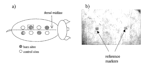
FIGURE 1 shows the layout of the burn and control sites on the dorsal
surface of the animal. (a) pictorial and (b) photographic representation.
CA 02398278 2009-08-21
-4-
FIGURE 2 is a descriptive diagram of the three-way decomposition of matrix X
into
factors A, B, and C.
FIGURE 3 is a series of spectra in which a multiplicative scatter correction
method
was applied to the raw visible-near infrared spectra of thermally injured
porcine skin. Representative
results (n=1) for superficial (A), intermediate (B), deep partial (C), and
full thickness (D) burn sites are
displayed.
FIGURE 4 is a series of spectra of thermally injured porcine skin analyzed
using the
PARAFAC method. Representative results (n=1) are displayed with respect to
wavelength (A), burn
type (B), and time (C).
FIGURE 5 is a series of spectral data reconstructed using the wavelength,
thermal
insult, and time loading factors that were determined by the PARAFAC analysis.
The reconstructed
data for one set of superficial (A), intermediate (B), deep partial (C), and
full thickness (D) burns is
shown.
FIGURE 6 shows the pre-bum (left panels) and post-burn (right panels)
injuries. The
upper panel is a visual (photographic) representation of deep partial
thickness (a), superficial (b), full
thickness (c) and intermediate partial thickness (d) burns. The lower panels
are the corresponding near
infrared oxygen saturation images.
FIGURE 7 shows burn hemodynamics as a function of source-collector (SC)
separation during the early post-burn period. The top panels denote the oxygen
saturation changes and
the lower panels the blood volume. The four source-collector separations,
denoted SC1 through SC4,
correspond to probe separation distances of 1.5, 3, 4.5 and 6 mm.
FIGURE 8 is a schematic diagram of the light path through skin (left) and the
penetration depth (right) at several source-detector positions.
FIGURE 9 shows the computed a)StO2 and b) tHb for the superficial,
intermediate
partial, deep partial and full thickness bums at the various source-detector
distances over early post
bum period.
DESCRIPTION OF THE PREFERRED EMBODIMENTS
Unless defined otherwise, all technical and scientific terms used herein have
the same
meaning as commonly understood by one of ordinary skill in the art to which
the invention belongs.
Although any methods and materials similar or equivalent to those described
herein can be used in the
practice or testing of the present invention, the preferred methods and
materials are now described.
CA 02398278 2002-07-25
WO 01/54580 PCT/CA01/00090
As used herein, superficial burns refer to mild burns having only epidermal
damage, wherein the tissue is capable of regenerating the epidermis.
As used herein, partial thickness burns refer to burns having epidermal and
varying levels of dermal damage, wherein a portion of the dermal layer is
destroyed but
sufficient dermis may remain such that effective re-epithalization will occur
with adequate
vasculature.
As used herein, deep partial thickness burns refer to burns having dermal
destruction to the extent that an insufficient dermal layer may exist such
that regeneration
of the dermis is not possible.
As used herein, full thickness burns refer to burns having dermal
destruction to the extent that the dermal blood supply is so reduced that
regeneration of
the dermis is not possible.
As used herein, oxygen saturation refers to the relative amount of
oxygenated hemoglobin to total amount of hemoglobin.
Described herein is a method of diagnosing and characterizing burn injuries
wherein near infrared spectra are taken of a burnt tissue portion at two or
more depths.
The spectra may then be compared and analyzed using an algorithm that uses
multivariate statistical analysis methods as known in the art to determine
regions of high
significance or diagnostic regions of the spectra. In some embodiments, these
diagnostic
regions correspond to spectral regions related to or that can be used to
determine oxygen
saturation, hemodynamics and hydration characteristics of the damaged tissue
portions,
as discussed below. This information allows for classification of the burn
injury as
described below, and allows for the appropriate steps to be taken by the
clinician.
In other embodiments, spectra are taken at different depths over two or
more time periods so that trends or changes in the spectra can be determined.
This
information can then be used to classify or categorize the burn.
In yet other embodiments, spectra are taken at two or more depths and the
spectra are compared at wavelengths corresponding to oxygen saturation,
hemodynamics
and hydration, as discussed below.
That is, in the described method, near infrared spectroscopy is used to
noninvasively distinguish between surface and subsurface molecular changes
that are
caused by the burn injury. Specifically, the remaining residual cutaneous
blood supply at
the dermis following an injury is directly related to the extent or depth of
tissue damage.
Increased depth of thermal injury means there is a greater portion of the
vessels that are
CA 02398278 2002-07-25
WO 01/54580 PCT/CAO1/00090
6
damaged and as a result the transport of blood to the tissue is impaired.
Therefore,
knowledge of the skin hemodynamics following an injury can define for
clinicians the
extent and depth of damage.
Furthermore, the oxygenated and deoxygenated forms of hemoglobin have
different extinction coefficients in the near infrared region. The data is
used to calculate
the oxygen saturation of the tissue, as a measure of the relative amount of
oxygenated
hemoglobin to the total amount of hemoglobin, thereby providing a quantifiable
measure of
the oxygen transport in tissue. Severe burns resulting from prolonged contact
with a heat
source are characterized by more heat conduction in deeper tissue resulting in
tissue
ischemia and vascular damage. In tissue, the total hemoglobin present can be
used to
represent a measure of tissue perfusion or blood volume. Burns result in
complex
responses related to hypo- and hypervolemia, ischemia and hypoxia. These
responses
are discernible with near infrared spectroscopy and provide meaningful insight
into the
local cutaneous microcirculatory changes associated with burn injuries.
As discussed above, the extent and depth of burn injury dictates treatment.
Therefore, the main clinical question lies in grading the thermal injury and
assessing the
extent of the viable tissue underneath the wound. The near infrared wavelength
range is
ideally suited for the noninvasive evaluation of tissue chromophores deep
within tissue.
This suitability stems from the optical properties of tissue in the near
infrared region of the
spectrum. Optical properties of tissue can be described by two basic
processes:
absorption by the tissue chromophores and scattering by tissue constituents.
When light
impinges on tissue, a portion is absorbed by chromophores dispersed throughout
the
cellular and intercellular space. The absorption of light by tissue
chromophores is weak
compared to the extent of scattering in the near infrared region. Tissue
scattering
represents the major contribution to the attenuation of near infrared light.
Scattering can.
occur due to refractive index variations in tissue or by elastic scattering
such as the
interaction with collagen in tissue. A single photon entering a tissue sample
will experience
many scattering events as it propagates through the medium. The interaction of
light with
multiple scatters results in an alteration from its original direction. In
tissue these multiple
scattering events are highly forward directed, i.e. light propagates in a
forward direction.
Therefore, the majority of the light penetrates deep within tissue before
being scattered
out of the tissue. The measured reflectance not only provides information on
the near
surface absorption but also absorptions deep inside the tissue.
In an optical geometry such as reflectance that involves placing a detector
CA 02398278 2002-07-25
WO 01/54580 PCT/CAO1/00090
7
some finite lateral distance away from the incident light, a small fraction of
the scattered
light can be measured. Tissue constituents have attenuated this light from the
original
source by the absorption of tissue chromophores as well as from tissue
scattering
components. The measured attenuation is also related to the separation
distance between
the light source and the detector. The larger the source-detector separation
the larger the
attenuation since the light has undergone additional scattering to emerge out
of the tissue.
Therefore, the penetration depth of the observed light is dependent on the
separation
distance between the source and detector. The further the detector is placed
from the
source, the greater the depth probed into the tissue as demonstrated in Figure
8. In order
for light to reach a detector a set distance away from the source, the light
must traverse a
path through the media, denoted by the shaded region. As the source-detector
separation
increases, the path increases and the depth sampled into the medium also
increases.
These paths have been described by a number of authors using both Monte Carlo
simulations (Long et al, 1996, Special Publications of the Royal Society of
Chemistry 194:
176-184; Flock et al, 1989, IEEE Trans Biomed Eng 36: 1162-1168) and time or
frequency
resolved spectroscopic techniques (Hemelt et at, 1997, Biotechnol Prog 13: 640-
649;
Hemelt et at, 1999, Biotechnol Prog 15: 622-630; Cui et al, 1991, Opt Left 16:
1632-1634).
Essentially, by acquiring spectra at various source-detector separation, one
can obtain
spectroscopic information at different depths into the tissue.
The investigation of the epidermis as well as the deep dermis is ideal for
burn depth assessment. This technique can provide depth dependent hemodynamics
that
are of vital importance in the assessment of tissue viability and burn depth.
Near infrared
spectroscopy can be applied as a noninvasive method to investigate the depth
dependent
circulatory alterations that arise from thermal damage to the skin. Herein, an
acute porcine
model to demonstrate the potential of near infrared spectroscopy to
distinguish burns of
varying severity. Thermal injuries disrupt the blood flow and oxygen delivery
to the
damaged tissue. The severe alteration of the microvascular integrity due to a
thermal
insult results in dramatic hemodynamic changes such as tissue ischemia and
impaired
tissue perfusion. These factors lead to a relative change and distribution of
the levels of
oxyhemoglobin and deoxyhemoglobin in tissue that can be measured by near
infrared
spectroscopy and used to assess the degree of thermal damage or tissue
viability.
Furthermore, near infrared reflectance spectroscopy and imaging provides
a non-invasive means of assessing the balance between oxygen delivery and
oxygen
utilization in tissue. The principal benefit of using near infrared
spectroscopy and imaging
CA 02398278 2002-07-25
WO 01/54580 PCT/CAO1/00090
8
is that regional variations in tissue hemodynamics can be discerned
objectively. The main
advantage of using near infrared light is the extended tissue sampling depth
achieved.
Near infrared light between 700-1100 nm can penetrate deep within tissue
providing vital
burn injury related information. Wound healing involves a number of different
processes
that must be carried out to accomplish repair. However, many of these
processes are
dependent on the oxygen delivery to the damaged tissue. Hemoglobin provides an
endogenous marker of oxygenation. The oxygenated and deoxygenated states of
hemoglobin have different extinction coefficients in the near infrared region.
Therefore,
contained within the near infrared absorption spectrum is the relative
concentration of both
the oxy- and dexoy- hemoglobin. A measure of the combined amounts of oxy- and
deoxy-
hemoglobin, or total hemoglobin, is related to tissue blood volume which can
be used as
an indicator of tissue perfusion while the ratio of oxygenated to total
hemoglobin
represents the oxygen saturation of tissue (Stranc et al, 1998, Br J Plast
Surg 51: 210-
217; Thorniley et al, 1997, Adv Exp Med Biol 411: 481-493; Sowa et al, 1997,
Appl Spec
51: 143-152).
EXAMPLE I - ANIMAL MODEL
Following a 10 day acclimatization period, adult Yorkshire cross swine
weighing between 40 and 50 kg were premedicated with an intramuscular
injection of
midazolam (0.3 mg/kg), atropine (0.02 mg/kg), and ketamine (20 mg/kg).
Anesthesia was
then induced by mask and the pigs were intubated and mechanically ventilated.
Isoflurane
(1.5 - 2.5 %), was delivered through the ventilator (via 40-60% oxygen mixed
with medical
air at 3.0 L/min) to maintain anesthesia for the duration of the experiment.
Systemic
oxygen saturation, heart rate, and blood pressure were monitored throughout
the
experiment. Core body temperature was maintained at 39.0 C 0.5 C. Blood
samples for
blood gas and electrolyte analyses were acquired prior to thermal injury and
every hour
thereafter.
Following anesthesia, both sides of the dorsal midline were shaved and
eight sites, each 3 cm in diameter, were marked on the back of the animal
using a custom-
made template. Four of the sites were located on the right side of the dorsal
midline while
the other four were located on the left side. Preburn spectra were then
acquired at each
site. Burn injuries were then created on four of the eight sites by applying a
heated brass
rod (100 C) to the skin with a constant pressure (2000 g). By altering the
length of time
the brass rod was applied to the skin, a variety of burn injuries were
created. Superficial
burns were created in 3 seconds, intermediate partial thickness in 12 seconds,
deep
CA 02398278 2002-07-25
WO 01/54580 PCT/CAO1/00090
9
partial thickness in 20 seconds and full thickness burns in 75 seconds. The
remaining four
uninjured sites were used as controls. For the sham-controls, the brass rod
was warmed
to 39 C (the body temperature of the pigs) and held on the animals' skin at
previously
defined locations with constant pressure (2000 g) for 3, 12, 20 and 75 seconds
to match
the times used for the burn injuries. Immediately following removal of the
brass rod,
control and postburn measurements were acquired. Measurement sequences were
then
acquired hourly until 5 sets of measurements were completed.
The animals were anesthetized for the entire experiment and closely
monitored throughout. At the end of the experiment, the animals were
immediately
euthanized without any recovery of consciousness. All procedures were
performed in
accordance with the Canadian Council on Animal Care. The protocol was approved
by the
Animal Care Committee at the Institute for Biodiagnostics (Winnipeg, MB).
The general burn layout, depicted in Figure 1, shows the relative positions
of the burn and control sites.
EXAMPLE II - VISIBLE-NEAR INFRARED SPECTROSCOPY
Visible-near infrared spectra were collected with an NIRSystems 6500
(Foss, Silver Springs, MA) spectrometer using a custom bifurcated fiber optic
bundle
(Fiberguide Industries, Stirling, NJ). The multifiber probe consisted of five
optical fibers,
one to illuminate the tissue and four to collected the remitted light. The
illumination and
collection fibers were 2m in length with a core diameter of 600 and 200 m,
respectively.
The fiber order at the head of the probe were placed in a co-linear
arrangement beginning
with the illumination and subsequent collection fibers spaced 1.5 mm from each
other.
Therefore, the distance of the four collection fiber from the illumination
source were 1.5, 3,
4.5, and 6 mm. The illumination optical fiber was coupled to a 100 W quartz
tungsten
halogen white light source (Oriel, Strattford, CT). The four collection
optical fibers were
placed at the entrance of an imaging spectragraph (Sciencetech Inc., London,
On,
Canada) covering the 500 to 1100 nm range. A back thinned illuminated 1024
x128 pixel
area image CCD detector (Hamamatsu, Bridgewater, NJ) cooled to -10 C was used
as
the detection element in the spectragraph. Each image, containing the spectrum
from all
of the four input fibers, consisted of five co-added images, which were parsed
and binned
into four separate raw reflectance spectrum. A 99% Spectralon reflectance
standard
(LabSphere Inc., North Sutton, NH) was used as a reference to convert raw data
into
reflectance spectra. Each reflectance spectrum consisted of two 32 co-added
scans
collected between 400-2500 nm at 10 nm resolution.
CA 02398278 2002-07-25
WO 01/54580 PCT/CAO1/00090
Near infrared imaging
Near infrared reflectance images of 256 x 256 pixels were collected
between 650 - 1050 nm at 10 nm increments using a Photometrics Series 200 CCD
camera (Photometrics, Tucson, AZ) fitted with a Nikon Macro AF60 lens and a 7
nm
bandpass (FWHH) Lyot type liquid crystal tunable filter (LCTF) (Cambridge
Research
Instruments, Cambridge, MA). Each image was acquired with a 200-msec exposure
time.
The white-side of a Kodak Gray Card (Rochester, NY), was used as a reference.
Near infrared depth spectroscopy
Near infrared spectra were collected with an imaging spectragraph using a
multifiber optic bundle (Fiberguide Industries, Stirling, NJ). The multifiber
probe
consisted of five optical fibers, one to illuminate the tissue and four to
collect the re-
emitted light. The distance of the four collection fibers from the
illumination source were
1.5, 3, 4.5, and 6 mm. The illumination optical fiber was coupled to a 100 W
quartz
tungsten halogen white light source model 77501(Oriel, Strattford, CT). The
four collection
optical fibers were placed at the entrance of the imaging spectrograph
(Sciencetech Inc.,
London, On, Canada) covering the 500 to 1100 nm range. A back thinned
illuminated
1024 x128 pixel area image CCD detector model C7041 (Hamamatsu, Bridgewater,
NJ)
cooled to -10 C was used as the detection element in the spectrograph. Each
image,
containing the spectrum from all of the four input fibers, which were parsed
and binned
into four separate raw reflectance spectra. A 99% Spectralon reflectance
standard
(LabSphere Inc., North Sutton, NH) was used as a reference to convert raw data
into
reflectance spectra. The measured attenuation is related to the separation
distance
between the light source and the four detection or collection fibers. The
penetration depth
of the observed light is dependent on the separation distance between the
source and the
collector. The further the collector is placed from the source, the greater
the depth probed
into the tissue.
EXAMPLE III - DATA PREPROCESSING
The multiplicative scatter correction (MSC) method was used to reduce the
multiplicative and additive scatter effects that occur between a series of
spectra. For every
animal, an average spectrum was calculated for the time series of spectra that
were taken
from each burn type. Individual spectra of the series were assumed to deviate
in both an
additive (a) and multiplicative (m) fashion from the average spectrum.
xik=a+mxk
CA 02398278 2002-07-25
WO 01/54580 PCT/CAO1/00090
11
(1 a)
The scatter contribution in the individual spectra is given by ilk. The
multiplicative and additive coefficients were determined by linear least
squares estimates
of the mean spectra over the different wavelengths for each burn. Corrected
spectra were
obtained by subtracting the additive and dividing by the multiplicative terms
of the least
squares fit.
Xik, new = (Xik - a)/m
(2a)
The MSC method was used to preprocess all data prior to implementing a
parallel factor analysis (PARAFAC).
EXAMPLE III - MULTIVARIATE DATA PROCESSING
A time series of near infrared spectra were collected from selected sites on
the dorsa of 5 animals. The first time point in the series was acquired prior
to thermal
injury. Subsequent points in the time series were acquired at fixed time
intervals after the
injury. The experimental design results in a rich set of data consisting of
many variables
(reflectance response over the 400-2500 nm wavelength range) observed on
several
occasions. (longitudinal measurements) and measured at several sites or groups
(cross-
sectional data) over a sample population of 5 animals. Parallel factor
analysis is used to
try to isolate and recover the spatial, spectral and temporal changes in
reflectance of the
skin due to the varying degree of thermal insult.
Parallel factor analysis is used to explore the time series of spectral data
by
identifying the "pure" molecular species that contribute to the spectra and
determining how
these "pure" species are affected by thermal insults of varying magnitude. The
temporal
variation of these species following the insult was also explored using the
same method.
Identifying the components in an unknown sample is one of the oldest problems
in
chemistry. The identification of the species that contribute to the response
in a highly
convolved series of spectra is particularly challenging when little or no
information is
available on the nature and relative concentrations of the constituents that
make up the
system. In such situations, exploratory factor analysis techniques can be
applied.
Two-way factor analysis approaches attempt to provide an interpretable
model of the data by imposing physically meaningful constraints in the
decomposition of
the data. Generally, this is accomplished by rotating (performing a linear
transformation) a
truncated set of the latent components (often starting from the principal
components) of
the data subject to a set of physically meaningful constraints. The resulting
factors are
CA 02398278 2002-07-25
WO 01/54580 PCT/CA01/00090
12
intended to retrieve the individual underlying "pure" components of the
measured
multicomponent system. Such approaches, which are usually referred to as self-
modelling
curve resolution methods, often suffer from rotational ambiguity when applied
to
spectroscopic data. In these instances the number of constraints are too few
to provide a
unique rotation or solution and therefore a number of possible factor models
can exist.
Parallel factor analysis is an N-way decomposition model originating from
Psychometrics
(Harshman and Lundy, 1984, in Research Methods for Multimode Data Analysis,
Law et al
eds (New York: Praeger); Burdick, 1995, Chem Intel) Lab Sys 28: 229-237) which
is aimed
at overcoming the rotational problem encountered by two-way methods. In the
experimental design, dorsal sites with varying degrees of thermal injury were
monitored
spectroscopically over time. The data has an intrinsic three-way structure
consisting of the
three fixed variables: degree of thermal injury, observation time and the
wavelengths at
which the reflectance of the skin was monitored. PARAFAC can exploit this
structure while
imposing further constraints in order to resolve the problem of rotational
ambiguity. In
spectroscopic applications, a nonnegativity constraint is usually invoked.
This constraint
ensures that the derived "pure" factors have a positive or zero contribution
at each time
point, thereby avoiding negative constituent concentrations. The nonnegativity
constraint
also ensures that there are no spectral regions of negative optical density
for the derived
"pure" components. These constraints provide rotational solutions that lead to
physically
meaningful factors and potentially, to the pure components that make up the
system.
For clarity, a brief description of the mathematical nomenclature used to
describe the variables will be presented. In tensor notation, a scalar (Oth
order tensor) is
indicated by lower-case italics, a vector (1St order tensor) by a bold lower-
case letter, two-
dimensional matrices (2d order tensor) by bold capital letters, and underlined
bold capitals
for three-dimensional matrices (3rd order tensor). The capital letters I, J,
K, L and M are
used to indicate the dimensionality of the various tensors. Therefore, the
ijth element of a
matrix X is designated as x;j and the ijkth element of a matrix Y as y;jk.
Although PARAFAC is an N-way decomposition method, its application to
burn injury evaluation is three-way involving time, burn, and spectral
absorbance. A triad
of loading matrices represents the PARAFAC model of the three-way data set.
M
xUk = aimb 'mckm +ei .k
m=1 .1 J
(2)
CA 02398278 2002-07-25
WO 01/54580 PCT/CA01/00090
13
where aim, b;,,,, and Ckm are the loading elements of the decomposed data set
consisting of
M factors and E the data residual matrix. M represents the number of
underlying factors or
components in the data. In the two-way factor analysis there is rotational
ambiguity that
leads to a continuum of factors satisfying the factor model. However, the
three-way
decomposition described in both equation 2 and Figure 2 represent the "pure"
components
contained within the data with respect to the a, b, and c factors. In the case
of spectral
data, the "pure" spectrum of each of the components contained within the data
will be
expressed as one of the matrices containing M spectra. Two-way decomposition
methods
produce one score and one loading matrix. The loadings are orthogonal
projections of the
latent variables in the data with scores representing the scalar weightings of
each of the
variables. The three-way PARAFAC decomposition produces one score and two
loading
matrices. Another common notation for both equation 2 and Figure 2 was the
Kronecker
tensor product to describe the model (Burdick, 1995).
X= ZamObm(&-m
m-I
(3)
The decomposition produces three matrices related to 1) the spectroscopic
changes 2) the variation with degree of thermal injuries and 3) the time
course of the
changes. Multi-way data analysis provides a means to investigate and compare
the
spectroscopic time-courses of the various burns. Therefore, the spectral
variations in the
(temporal) response to the burn can be effectively isolated from the static
independent
components.
The factors in the PARAFAC analysis are determined using an iterative
alternating least squares method. The convergence criterion used in the
PARAFAC
analysis to terminate the iterative procedure utilizes the relative difference
in the fit
between two consecutive iterations. The interactive procedure terminates when
this
difference is below a value of 10"6. A constraint of nonnegativity was also
placed on the
wavelength factors of the PARAFAC analysis to ensure that the extracted
wavelength
factors or "pure" components have no region of negative optical density. This
method
requires an` initial guess or starting value to determine the underlying
factors or solution.
CA 02398278 2002-07-25
WO 01/54580 PCT/CAO1/00090
14
,To avoid a solution whereby a local minimum is reached, a random set of
starting values
are used for each run. The PARAFAC analysis is run twice using a random set of
starting
values for each run. A consistent set of solutions for each run helps ensure
that the global
solution is obtained. If essentially the same factors are obtained in each
run, there is little
probability that a local minimum was reached and the solution is unique. The
PARAFAC
analysis was run twice and the results compared to ensure that the global
minimum was
reached.
Burn injury assessment
As discussed above, thermal injuries disrupt the blood flow and oxygen
delivery to the damaged tissue. The severe alteration of the microvascular
integrity due to
a thermal insult results in dramatic hemodynamic changes such as tissue
ischemia and
impaired tissue perfusion. These factors lead to a relative change and
distribution of the
levels of oxy- and deoxy-hemoglobin in tissue. These changes can be measured
by near
infrared spectroscopy and used to assess the degree of thermal damage or
tissue viability.
The relative oxygen saturation (SO2), a measure of the relative amount of
oxygenated
hemoglobin to the total amount of hemoglobin present (defined as
StO2=[HbO2]/([HbO2]+[Hb])), provides a quantifiable measure of the oxygen
transport in
tissue. The combined measure of oxy- and deoxy- hemoglobin, or total
hemoglobin ([tHb]),
is related to tissue blood volume which can be used as an indicator of tissue
perfusion.
The oxygenated and deoxygenated forms of hemoglobin have different extinction
coefficients across the near infrared region. Using two or more of the
extinction
coefficients for oxy- and deoxy-hemoglobin, the StO2 and [tHb] for tissue can
be
determined from a near infrared spectrum of tissue. Hemoglobin concentrations
per unit
photon pathlength were determined by fitting the absorption coefficients of
the oxy- and
deoxy-hemoglobin to the observed reflectance attenuation expressed in optical
density
units over the spectral range of 740 - 840 nm. The underlying water absorption
bands at
730 and 830 nm were subtracted from the spectrum prior to fitting the
reflectance
attenuation. Oxygen saturation and/or total hemoglobin can be determined from
the near
infrared images and depth dependent spectroscopic measurements. Each method
provides a particular description of the hemodynamic changes occurring with
the injury.
Spectroscopic images
Photographs of the pre- and post-burn injuries are reproduced in the upper
panels of Figure 6. A visual inspection of the wound clearly identifies the
superficial burn
(Figure 6, site b) from the more extensive and severe burns. Assessment of the
partial and
CA 02398278 2002-07-25
WO 01/54580 PCT/CAO1/00090
full thickness injuries is difficult and subjective with visual observation in
the early post
burn period. Tissue oxygen saturation images of the dorsal region of the pig
provides a
visual survey of tissue oxygenation. The lower panels of Figure 6 show tissue
oxygen
saturation images of both the control and burn injures before and 4 h after
the initial insult.
The control sites and surrounding non-involved tissue appear bright which
indicates
normal tissue oxygen saturation. However, the injured sites display a drop in
oxygenation
following the thermal insult. Sites with low tissue oxygenation appear as dark
areas on the
dorsum in the post-burn St02 images. However, the site of the superficial
injury displays a
distinctly different response compared to the more severe burn sites.
Oxygenation
increases at the superficial burn site in comparison to its pre-burn levels.
This increase
corresponds to the visible erythema or tissue reddening associated with minor
burns. Hair
also appears dark in the oxygen saturation images but is easily distinguished
from areas
of tissue with low oxygen saturation.
Depth spectroscopy
Regardless of the degree of injury, all burns show an immediate post-injury
alteration in the oxygen saturation and blood volume as displayed in Figure 7.
The figure
summarizes the hemodynamic for the various burn injuries in relation to the
source-
collector separation or sampling depth into the tissue. All burns exhibit an
instantaneous
decrease in the tissue oxygenation following the injury. Results from the
smallest source-
collector separation show no significant difference in the oxygen saturation
between burns
of different severity relative to the uninjured control tissue. These results
were expected
since the smaller source-collector separations probes primarily the epidermis,
however,
the epidermis is mainly avascular depending on the capillary beds in the
dermis for
oxygen. Thus, there is little or no hemoglobin contribution to the spectral
signature from
the smallest source-collector separation. As the source-collector separation
increases, the
tissue sampling depth is extended from the epidermis to the dermis and the
burn injuries
commence to become discernible. Despite the ability to distinguish oxygenation
at various
depths, intermediate and deep partial thickness injuries cannot be reliably
isolated on the
basis of oxygenation measurements.
Total hemoglobin, displayed in the bottom panels of Figure 7, provides an
efficient indicator of blood volume alterations following a burn injury as a
function of probe
depth. Again, the burn injuries are indiscernible when probing with the
smallest source-
collector separation since it is primarily the epidermis that is probed. As
the deeper tissue
is probed using the larger source-collector separations, the superficial and
intermediate
CA 02398278 2002-07-25
WO 01/54580 PCT/CAO1/00090
16
partial thickness burns exhibit a notable alteration from the pre-burn state.
Superficial
injuries show a sudden increase in the [tHb] resulting from the injury
illustrating. a
hypervolemic state followed by a decrease or hypovolemic and finally steady
increase
over the 4 h. Partial thickness injuries also undergo an increase in [tHb].
However, this
increase in [tHb]. peaks later (2 h following the injury) and is longer lived
than the
superficial response. The blood volume changes in the deep partial and full
thickness
injuries are remarkably different from that observed in the less severe
injuries. The deep
partial wound also demonstrates a hypervolemic peak, however hypervolemia
occurs
towards the end of the study. Small source-collector separation distances
probe the
topmost layer of the skin, however, this layer has sustained heavy damage with
only a
limited micro-circulation to supply blood to the injured site. Examining deep
wounds at
these small source-collector separations, one is primarily sampling heavily
wounded
tissue. On the other hand, large source-collector separations permit sampling
of deep
tissue, in particular, the viable tissue underneath the destroyed visible
tissue. Therefore,
large source-collector separations are ideally suited to distinguish deep
partial from full
thickness injuries, whereas, small source-collector separations are better
suited for
distinguishing between superficial and intermediate wounds. Each of the
various
hemodynamic parameters provides information on the status of the tissue
following a
thermal insult. Combining several of these parameters along with the added
information of
sampling depth, one can isolate and grade the burn injury.
EXAMPLE IV - RESULTS AND DISCUSSION
Burn injuries drastically modify both the physical and optical properties of
skin. PARAFAC is used to investigate the spectral changes that accompany a
thermal
injury. Prior to the PARAFAC analysis, a multiplicative scatter correction is
applied to the
data to compensate for additive and multiplicative differences between spectra
taken at
different times at the same burn site. Applying a multiplicative scatter
correction to the
data has the advantage of removing unwanted constants and multiplicative
effects in the
data. The multiplicative scatter correction also simplifies data comparison by
standardizing
data across replicated series of measurements. Representative time series of
multiplicatively scatter corrected spectra for each type of burn are presented
in Figure 3.
The results from a two-factor three-way PARAFAC analysis of the data for
a single animal are summarized by the computed wavelength, thermal insult and
time
loadings shown in Figure 4. The loading wavelengths of Figure 4A reveal the
two "pure"
spectral components or factors that are retrieved by the analysis. The first
wavelength
CA 02398278 2002-07-25
WO 01/54580 PCT/CAO1/00090
17
factor (dashed line) has distinctive spectral features at 555 and 760 nm
consistent with the
spectrum of deoxyhemoglobin as well as an absorption feature at 980 nm which
is
characteristic of water. Thus the first factor appears to consist of
contributions from both
deoxyhemoglobin and water. The second factor (solid line) resembles an
oxyhemoglobin
spectrum with its unique vibronic transition bands at 540 and 580 nm. The
broad
absorption band between 800 and 1000 nm, which is consistent with the broad
near
infrared charge transfer band of oxyhemoglobin, is also evident. These results
suggest the
data can be represented by two distinct spectra (factors), one resembling the
combined
deoxyhemoglobin and water contribution and the other representing the
oxyhemoglobin
contribution.
The wavelength loading factors obtained from the PARAFAC analysis
suggest that thermal burns alter oxy- and deoxy- hemoglobin concentrations.
The thermal
insult loading vectors given in Figure 4B indicate the relative changes in the
wavelength
factors (components) for the different types of burns. The first thermal
insult factor (dashed
line) displays only a moderate change between the different burn types with a
slight
upward trend between the superficial (1 ) and full thickness (3 ) burns. This
suggests the
deoxyhemoglobin and water contribution in the spectral response undergoes a
minor
change with the thermal insult. The second factor in the loading (solid line)
shows a steady
decline, suggesting that the oxyhemoglobin content of tissue decreases with
increasing
severity of the burn injury. These results indicate that the severity of a
burn injury has a
direct effect on the oxy- and deoxy- hemoglobin balance within the injured
tissue. The
superficial and full thickness burns represent the two extremes in the second
thermal
insult loading. The intermediate and deep partial thickness burns are similar,
differing only
in the extent' of damage to the dermis. These injuries have intermediate
values in the
thermal loading vector. More importantly, the loading values scale with the
magnitude of
the injury.
Burn injuries are a dynamic process whereby burn depth can become
progressively worse during the first few hours following the injury. The time
loading
factors, Figure 4C, describe the general trends of the two wavelength factors
over the time
course of the study. The first time loading factor (dashed line) which is
associated with the
deoxyhemoglobin content of tissue, exhibits a small but slow increase with
time. This
indicates that the deoxyhemoglobin - water content of tissue increases
slightly and slowly
following the burn injury. On the other hand, the second time loading factor
(solid line)
which corresponds to the oxyhemoglobin content of tissue, displays an enormous
change
CA 02398278 2002-07-25
WO 01/54580 PCT/CAO1/00090
18
over time. This change is equivalent to a large positive change in the oxygen
content of
the tissue over time. The thermal insult loadings suggest that thermal
injuries have a
profound effect on the oxygen balance not only at the onset of the injury but
also over
time. In order to assess the quality of the two-factor model and more clearly
demonstrate
the changing oxygen balance between burns of different severity, the data was
reconstructed using the factors determined from the PARAFAC analysis. Applying
equation 3 to the PARAFAC wavelength, thermal insult, and time loading
factors, the data
was reconstructed and is displayed in Figure 5.
The main trend observable with the superficial burn is the large increase in
the oxyhemoglobin contribution to the spectrum as demonstrated in Figure 5A.
This
increase in oxyhemoglobin, which results from an influx of blood to the
injured tissue,
becomes apparent immediately following the injury and continues to rise over
the 4 h
postburn monitoring period. On the other hand, intermediate and deep partial
thickness
burns (Figures 5B and 5C) undergo a greater amount of tissue damage. Although
they do
show an increase in oxyhemoglobin following the injury, the response is
invariably less
dramatic. While it only takes 10 minutes for the oxyhemoglobin content of the
tissue to
increase significantly following a superficial burn, the response of
intermediately burned
tissue is minimal. Following a deep partial thickness injury, the change in
oxyhemoglobin
is further reduced, almost nil. The cutaneous blood supply at the dermis and
its relation to
the extent or depth of tissue damage explains why there are varying degrees of
oxyhemoglobin following burn injuries. Increased depth of thermal injury means
there is a
greater portion of the vessels that are damaged and as a result are no longer
capable of
transporting oxygenated blood to the tissues. Since deep partial thickness
injuries extend
further into the dermis than intermediate ones, it is not surprising that less
oxygen is
available for tissue consumption. The even deeper, full thickness burns
obliterate both the
epidermis and dermis completely, thereby making the skin avascular. Rather
than
observing an increase in the oxygen content of the tissue, the full thickness
injuries
experience a drop in the oxyhemoglobin contribution to the spectra following
the insult.
This drop becomes evident within 10 minutes of the thermal insult and
continues to
decrease at the full thickness burn site over the 4 h monitoring period of the
study. Since
full thickness burns involve total destruction of the vascular supply to
cutaneous tissue,
skin that has suffered such an insult has no supply of oxygenated blood and
will eventually
die.
Multi-way parallel factor analysis was used to investigate and compare the
CA 02398278 2002-07-25
WO 01/54580 PCT/CAO1/00090
19
spectroscopic time courses of the response of tissue to burn injuries of
varying magnitude.
The multi-way analysis could be done in an exploratory fashion requiring
little or no prior
information on the system other than that information required to determine
the constraints
imposed by the experimental design and the nonnegativity constraint associated
with
generic spectral response of any component species present in a multi-
component
system. The two-component three-way analysis extracts two wavelength factors,
one
resembling the combined deoxyhemoglobin and water contribution and the other
representing the oxyhemoglobin contribution to the spectra. These results
indicate that the
severity of a burn injury has a direct effect on the oxy- and deoxy-
hemoglobin balance
within the injured tissue. Reduced tissue oxygenation in deep partial
thickness and full
thickness injuries may ultimately contribute to tissue necrosis following a
severe burn
injury. Parallel factor analysis reveals that the spectral changes in the
early post-burn
period can be faithfully represented by two "pure" components that summarize
the oxy-
and deoxy- hemoglobin balance within the injured tissue. Analysis of the
visible-near
infrared spectroscopic data indicated that the oxy- and deoxy- hemoglobin
balance in
injured tissue changed over time following the injury. However, within the
early post-burn
period the oxy- and deoxy- hemoglobin balance scaled with the degree or depth
of thermal
injury. Visible-near infrared spectroscopic assessment of the oxy- and deoxy-
hemoglobin
balance in injured tissue could clearly distinguish superficial and full
thickness injuries and
may also provide an indicator sensitive enough to distinguish between
intermediate and
deep partial thickness burns.
In general, the injured sites all show an immediate visual change following
the thermal injury as displayed in Figures 2b and c. Based on the visual
appearance of the
wound, the superficial wound is distinct from remaining wounds. However, the
partial and
full thickness injuries are difficult to identify using visual observation.
Physiological
monitoring (heart rate and blood pressure) as well as blood samples were taken
prior to
the burn injury, immediately following the injury and at hourly intervals for
the duration of
the protocol. Since less than 1 % of the total body surface area is injured in
this model, no
systemic response was expected. Blood gas analysis showed no indication of
systemic
response to the thermal injury. Blood gases remained within the normal range
for all
animals for the duration of the protocol. The near infrared spectral data from
both the
uninjured (control) and injured (burn) sites were processed as described in
the methods
section to obtain StO2 and tHb hemodynamic parameters. The control sites
displayed no
significant variation in any of the parameters 4 hrs following the thermal
burn insult. These
CA 02398278 2002-07-25
WO 01/54580 PCT/CAO1/00090
results are consistent with the physiological monitoring and blood gas results
that indicate
no significant systemic effects on the animal due to the thermal injury. While
no systemic
response was observed as a result of the burn injury, regionally changes were
observed in
the areas of tissue subjected to thermal damage.
Oxygen Saturation
Figure 9a describes the difference in the oxygen saturation for the various
burns injuries in relation to the response measured at a nearby control site
for the various
source-detector separations. All burns show an instantaneous decrease in the
oxygenation following a burn injury.
Results from this panel are greatly inconsistent and were expected with
regards to tissue oxygenation. This panel contains the St02 for the smallest
source-
detector separation which was designed to probe the outermost layer of the
skin, namely
the epidermis. However, the epidermis is mainly an avascular layer of tissue
which
depends on the capillary beds in the dermis for oxygen. As such, the method is
not
appropriate to determine the oxygen content in this tissue layer. The
superficial injuries
show a minor decreasing change St02 with certain level of recovery observed
towards the
end of the 4hrs period. Examining the St02 at the various source-detector
geometries, one
notices changes throughout the different fiber optic separations implying that
superficial
burns actually extent deep within the tissue as well. However, the magnitude
of the
changes would suggest that this type of injury is minor, only disrupting the
tissue.
The remaining burns are highly destructive, obliterating the epidermis and
damaging or destroying the dermis. Regardless of the severity of the injury,
all show a
distinct drop in the St02 succeeded by a steady decline over the 4hrs post
burn period. As
a whole, the superficial and full thickness burns are well distinguishable at
any of the
source-detector separations. At the appropriate source-detector separation,
the
intermediate partial thickness burn is also discernible from the deep partial
thickness
injury. Considering the extent of the damage and the volume probe by the
various source-
detector separations, partition of the various burns is possible. At the
smaller probe
separation, it becomes difficult to identify the intermediate partial, deep
partial, and full
thickness thermal injuries. At smaller source-detector separations, the depth
probed into
the tissue is not deep enough to distinguish the damage from the viable
tissue. As the
probe separation is increases, damaged and healthy tissue is probed depending
on the
depth of the thermal injury, thus permitting the separation of the various
burns. Despite
this ability to distinguish oxygenation at various depths, intermediate and
deep partial
CA 02398278 2002-07-25
WO 01/54580 PCT/CAO1/00090
21
thickness injuries cannot be reliably isolated on the basis of StO2
measurements made in
the early post-burn period of a thermal injury.
Total Hemoglobin
Total hemoglobin, as displayed in Figure 9b, provided an efficient indicator
of blood volume status following a burn injury. Superficial injuries show a
sudden increase
in the tHb resulting from the injury illustrating a hypervolemic state
followed by a decrease
or hypovolemic and finally steady increase over the 4hrs. Superficial burns
are considered
minor burns, mildly disrupting normal microcirculation in the skin.
Partial thickness injuries also undergo an increase in tHb. However, this
increase peaks later (2hrs following the injury) and is longer lived than the
superficial
response. At the shortest probe separation, it is difficult to distinguish the
various injuries
other than the superficial burn. As stated earlier, the epidermis is primarily
avascular;
however, the influx resulting from the thermal injury does extend to some
degree into the
dermis. As source-detector separation increases, the injuries become
distinguishable,
beginning with the superficial with the familiar hyper hypo increase, followed
by the
intermediate partial thickness burn. Intermediate partial thickness burns
impairs the
uppermost layer of the dermis, with the remaining basement layer intact. This
bottom layer
is capable of providing a large inrush of blood to the injured site. In some
respects,
intermediate partial thickness injuries are not much more than slightly,
extended superficial
burns. These results would suggest tHb with the intermediate partial thickness
injury
would continue to decrease analogous to the superficial but at a reduced rate
to provide
the necessary blood to remedy the injury. The deep partial wound also
demonstrates a
hypervolemic peak, however hypervolemia occurs towards the end of the study.
At the
shorter source-detector separation, it is difficult to observe the sudden
change in tHb
associated with thermal injuries. As mentioned earlier, short probe separation
distances
probe the topmost layer of the skin; however, this layer has sustained heavy
damaged
with only a limited microcirculation to supply blood to the injured site. At
these separations
one is moreover sampling heavily wounded tissue. An increased separation
permits
sampling of the viable tissue underneath the wound and it is this layer which
supports and
advances healing.
Full thickness injuries, the most destructive of the thermal injuries
exhibited
large changes over the 4hrs study period. This is consistent with the premise
that full
thickness injuries extensively damage the tissue to the extent of destroying
the
microcirculation to the epidermis and the dermis. The results suggest a very
large
CA 02398278 2002-07-25
WO 01/54580 PCT/CAO1/00090
22
difference prior and post the injury suggesting the tissue has sustained=
heavy damage
with no possibility of recovery. Each of the various hemodynamic parameters
provides
information on the status of the tissue following a thermal insult. The
combined result of
lack of tHb increase and decrease StO2 in combination correlates well with the
expected
outcome, which is that the circulation has been damaged and that whatever
blood is
present is slowly being dissipated. Eventually, since the vasculature has been
destroyed
and the site is slowly becoming deoxygenated, this injury will result in
necrosis and tissue
death.
Individually, categorizing the various thermal injuries based solely on any
one parameter is not possible. Each of the various parameters provides
information on the
status of the tissue following a thermal insult. Oxygen saturation can be used
to
differentiate a minor injury (superficial burn).from the damaging partial and
full thickness
injuries. This parameter was also explored against the different source-
detector positions.
The oxygen saturation results indicate that at the proper probe position, one
can
distinguish the partial thickness wounds from the other injuries. More
importantly, the
oxygen saturation of the various partial thickness injuries are discernible
from one another.
In general, a burn specialist is capable of classifying superficial and full
thickness burns
using exclusively the visual appearance of the wound. The difficulty lies in
distinguishing
the various levels of damage caused by partial thickness burns. Of the
visually discernible
burn injuries, the superficial is the simplest to assess. Burns have a
dramatic influence on
the relative amount of blood present after a burn injury, especially with the
more invasive
of the thermal injuries. Total hemoglobin levels reflect tissue blood volume
or the extent of
blood perfusion. The larger the destruction to the tissue, the lower the blood
or available
blood present at the injured site. As seen in Figure 9, the intermediate,
deep, and- full
thickness injuries are definitely differentiated based on tHb changes at the
injured site.
These results correlate well with what is known about these injuries and the
effects they
have on the microcirculation. Total destruction of the microcirculation
results in no blood
available to regenerate the injury as seen with the full thickness injury.
Using the
combined available information of a decrease in the StO2 and lack of an
increase of tHb,
the predicted outcome of this tissue is necrosis. Separately, each parameter
provides a
portion of the description of the thermal injury with time. When all the
measurements are
taken as a whole and examined relative to one another, the superficial,
intermediate,
deep, and full thickness injuries can be differentiated.
Tissue responds differently depending on the severity of the insult. The
CA 02398278 2002-07-25
WO 01/54580 PCT/CA01/00090
23
results suggest burns can be differentiates based on a time series over the
first 4hrs
following the injury. The two extreme cases of a thermal injury, namely the
superficial (a
mild injury) and full thickness (a severe injury) injuries, were definitely
distinguishable.
Using the proper probe arrangement, the intermediate partial thickness injury
can be
isolated from the superficial and deep partial thickness injuries based on
oxygen
saturation measurements. However, deep partials and full thickness resemble
one another
with regards to this hemodynamic parameter. The different source-detector
separations
were unable to separate these injuries as well. Total hemoglobin, a useful
parameter,
demonstrated a marked difference in response with the partial and full
thickness injuries.
The combined parameters of oxygen saturation and total hemoglobin provide a
more
appropriate description of the thermal injury. In particular, using both
hemodynamic
parameters, one can identify the different burn injuries. This study was
designed to
investigate thermal injuries immediately following the burn. Water may be
usefully
parameter a few hours after the initial thermal injury once edema has settled
in.
Near infrared spectral measurements in tissue can provide a clinically
noninvasive or minimally invasive method to distinguish between thermal
injuries of
varying severity. Thermal injuries alter the oxygen content and blood
perfusion as well as
the water content of the tissue. Physiological parameters of oxygen saturation
total
hemoglobin, and water were determined from a time series of near infrared
reflectance
spectra. These parameters were used to assess and distinguish different
thermal injuries
utilizing a porcine burn model.
While the preferred embodiments of the invention have been described
above, it will be recognized and understood that various modifications may be
made
therein, and the appended claims are intended to cover all such modifications
which may
fall within the spirit and scope of the invention.