Note: Descriptions are shown in the official language in which they were submitted.
CA 02398949 2002-07-31
WO 01/56454 PCT/USO1/03625
-1-
INDIRECT CALORIMETER FOR MEDICAL APPLICATIONS
Field of the Invention
This invention relates to the use of indirect calorimetry within health
management, in particular for use with ventilators. ,
Background of the Invention
In U.S. Pat. No. 5,989,188 Birldioelzer et al. describe the use of an indirect
calorimeter for determining the energy balance of a person. However, there is
no
description of how this would be achieved for a patient on a ventilator.
In U.S. Pat. No. 5,705,735, Acorn describes a method of determining
nutritional
requirements for a patient ttsi~ng an indirect calorimeter. However, the
described system
uses a presstu-e differential sensor to determine gas flow. The presence of a
restriction in
a flow tube can cause problems in medical applications. This system uses gas
sampling
for respiratory analysis, whereas the Applicant's invention uses analysis of
gases in the
flow path, providing an effectively instantaneous analysis of gas composition.
In U.S. Pat. No. 5,647,370 Harnoncourt describes an ultrasonic spirometer. In
this application, the transducers are at an oblique angle to the flow tube
axis. In U.S. Pat.
No. 5,645,071 Harnoncourt et al. describe a method for determining the molar
mass of a
gas mixture using an ultrasonic method. In U.S. Pat. No. 5,503,151,
Harnoncouut et al.
describe the use of ultrasonic transducers in analyzing respiratory gases. The
use of these
spirometers in a mechanical ventilator system is not described.
In U.S. Pat. No. 5,179,958, Mault describes an indirect calorimeter from which
the respiratory quotient and resting metabolic rate ca.n be determined.
However, this
device is not optimized for use with an intubated patient. The use of a carbon
dioxide
scrubber adds weight and vohune to a respiratory analyzer.
In U.S. patent 5,285,794, Lynch describes a respiratory gas monitor; however
this
device uses a gas mixing chamber and does not provide real time measurements
of flow
rates and gas component concentrations.
CA 02398949 2002-07-31
WO 01/56454 PCT/USO1/03625
-2-
Other patents describing the use of oxygen and carbon dioxide sensors for
metabolic monitoring include U.S. Pat. No. 5,072,737 to Goulding; U.S. Pat.
No.
5,069,220 to Casparie et al.; U.S. Pat. No. 5,060,65.6 to Howard; U.S. Pat.
No. 4,856,531
to Merilainen; U.S. Pat. Nos. 4,619,269 and 4,572,208 both to Cutler; and U.S.
Pat. No.
' 4,233,842 to Raemer et al.
United States Pat. Nos. 4,917,108; 5,038,792; 5,178,155; 5,179,958; and
5,836,300, all to Mault, a co-inventor of the present application, are
incorporated herein
by reference. These patents disclose systems for measuring metabolism and
related
respiratory parameters through indirect calorimetry. ~ These instruments
generally employ
flow meters which pass both the inhalations and the exhalations of a user
breathing
through the instrument and integrate the resulting instantaneous flow signals
to determine
total full flow volumes. In some embodiments, the exhaled gases generated by
the user
are passed through a carbon dioxide scrubber before passing through the flow
meter so
that the differences between the inhaled and exhaled volumes is essentially a
measurement of the oxygen consumed by the lungs. In other embodiments, the
concentration of carbon dioxide exhaled by the user is determined by passing
the exhaled
volume through a capnometer and integrating that signal with the exhaled flow
volume.
The oxygen consumption can then be calculated as the difference between the
inhaled
and exhaled oxygen volumes, corrected to standard conditions.
Recently, James R. Mault, M.D. and others invented an improved indirect
calorimeter, more fully described in U.S. application 09/630,398, the contents
of which
are incorporated herein by reference. The improved calorimeter comprises an
ultrasonic
detection apparatus combined with a fluorescence oxygen sensor. This improved
calorimeter can be adapted for use with an intubated patient, or other patient
connected in
some ma~taer to a mechanical ventilator or respirator.
The oxygen consumption of a person . is related to their resting metabolic
rate
(RMR). This can increase up to several hundred percent in certain trawna
victims, such
as bllrll patients. In addition, the nutritional requirements of a person are
also determined
by their metabolic rate. An eWanced RMR can lead to muscle wasting of a
patient, as
muscle burning proceeds in order to supply the person with the required
additional
CA 02398949 2002-07-31
WO 01/56454 PCT/USO1/03625
-3-
energy. , Hence, for optimized recovery of a patient, it would be valuable to
lalow their
nutritional requirements.
In addition, the correct ventilation of a patient requires laiowledge of
carbon
dioxide and oxygen levels in the blood. The carbon dioxide and oxygen levels
in arterial
blood can be determined using the end tidal gas component concentrations of
exhaled
breath.
Summary of the Invention
The present invention provides an improved respiratory analyzer for use in a
ventilator system, or other system to assist with breathing. An improved
ventilator system
for a patient comprises a ventilator emit providing respiratory gases, a tube
(or line or
conduit) for conveying respiratory , gases to the patient, a flow module
holder located
within the tube (such as a slot, holder, clip, or the like); a flow module
being be placed in
the holder so that respiratory gases pass through a flow path of the flow
module; and an
electronics module, comzected to the flow module and containing an electronic
circuit
having processor, designed to calculate a flow rate for respiratory gases
flowing through
the flow path. In a preferred e111bOd1llle1lt, oxygen consumption volumes and
metabolic
rates are calculated by the electronics module.
It is an object of the present invention to provide an improved system by
which
the metabolic rate of an intubated patient can be determined.
It is a further object of the present invention to provide a system for
improved
respiratory control of a patient on a ventilator.
It is a object of the present invention to provide improved respiratory
analysis for
a patient on a ventilator or other means of respiratory assistance.
Brief Description of the Drawings
Figure 1 shows a general schematic of a ventilator system;
Figure 2 shows a schematic of an improved respiratory analyzer;
Figure 3 shows a design for an improved respiratory analyzer;
CA 02398949 2002-07-31
WO 01/56454 PCT/USO1/03625
-4-
Figure 4 shows a flow module embodiment;
Figure 5 shows an electronics module embodiment;
Figure 6 shows a flow module embodiment having a coaxial flow geometry;
Figure 6B shows a coaxial flow module docked to an electronics module;
Figures 7 and 8 show further flow module embodiments having coaxial flow
geometries;
Figure 9 shows a flow module having ultrasonic transducers in an oblique
configuration;
Figure 10 shows a flow module having 'ultrasonic transducers in the flow;
Figure 11 shows a flow module adapted to receive an oxygen sensor;
Figure 12 shows a flow module adapted to receive an optical fiber for oxygen
sensing;
Figure 13 shows an oxygen sensor with a fluorescent coating in contact with
the
flow path;
Figure 14 shows a pathogen resistant liner for a flow tube;
Figures 15 and 16 illustrate a system embodiment with automatic control of
patient feeding;
Figure 17 shows a respiratory analyzer system with a helmet momted electronics
module;
Figure 18 shows a system for determination of cardiac output;
Figure 19 shows a tracheal flow module; and
Figures 20 and 21 show designs for other embodiments using an electronics
module.
Detailed Description of the Invention
Figure 1 shows a ventilator system. The system comprises a ventilator 10, an
inlet tube 12, a valve unit 14, a valve connector 16, a return tube 18, a
respiratory
analyzer 20, and a patient intubation device 22 comiecting to patient 24. In a
conventional
respirator system, the respiratory analyzer'20 may be the pneumotach described
by Acorn
CA 02398949 2002-07-31
WO 01/56454 PCT/USO1/03625
-5-
in U.S. Pat. No. 5,705,735. In embodiments of the present invention, the
improved
. indirect calorimeter described in U.S. application 09/630,398 is adapted for
use as an
improved respiratory analyzer in a ventilator systems, such as the system
shown in Figure
1. However, the present invention can be adapted for other ventilator systems
lalown in
the art.
Referring to Figure l, ventilator 10 provides a source of inhalation gas
during
inhalation of the patient, which passes through the inlet tube 12 to the valve
14. The
valve 14 allows the ilihalation gas to pass tluough to the valve connector 16,
and so
through the respiratory analyzer 20 and the intubation device 22 to the
patient 24. The
intubation device may be placed in the mouth of the patient, or into the
trachea. During
exhalation, exhaled gas passes out through the intubation device 22,
respiratory analyzer
20, and valve connector 16 to the valve 14. The valve 14 allows the exhaled
gas to pass
through to the return tube 18, and so pass back to the ventilator unit 10. The
valve unit is
typically T-shaped or Y-shaped, and in part acts to prevent exhaled gases
entering the
inlet tube 12, to minimize rebreathing of exhaled carbon dioxide.
In preferred embodiments, the respiratory analyzer 20 is located close to. the
mouth of the patient, but outside of the patient's body. In other embodiments,
described
later, components of an improved respiratory analyzer may be located inside
the body of
the patient within a respiratory tube such as the trachea.
Referring again to Figl~re 1, the inlet tube 12 forms an inlet conduit (or
inhalation
conduit) for directing il~halation gases to the patient. The connector 16 and
111tLlbat1011
device 22 form a respiratory conduit, tluough which both i1W aled and exhaled
air flow.
The flow module is preferably inserted into the respiratory conduit, so that
both i1W aled
and exhaled gases pass tluough the flow module. The return tube 18 forms a
return
conduit (or exhalation conduit) for exhaled gases. The valve 14 allows
il~haled gases to
pass from the inhalation conduit to the respiratory conduit, and allows
exhaled gases to
pass from the respiratory conduit to the exhalation conduit. Exhaled gases may
also be
vented to the atmosphere. In the configuration of Figure 1, respiratory gases
pass in both
directions through the flow module (inhaled gases and exhaled gases pass in
opposite
directions). The ventilator 10 serves as a supply of respiratory gases. For
partial
CA 02398949 2002-07-31
WO 01/56454 PCT/USO1/03625
-6-
rebreathing and cardiac output studies, the valve may be configured to allow
some gas to
pass from the exhalation conduit baclc into the respiratory conduit, as
discussed, later in
relation to cardiac output measurements.
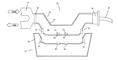
Figure 2 shows a cross-sectional view of an improved respiratory analyzer,
shown
generally at 20, and comprising two parts: a flow module 30, and an
electronics module
32. The modules 30 and 32, shown in cross-section, are adapted to be attached
to each
other for analysis of respiratory gases flowing through the flow module, and
to be
detached from each other for sterilization or disposal of the flow module. In
a preferred
embodiment, the flow module 30 is a disposable part of the respiratory
analyzer 20,
whereas, the electronics module 32 is a non-disposable part. Figure 2 shows
the flow
module 30 connected between valve 14 and intubation device (o,r respiratory
comiecior)
22 using connector 36 and collar 38. A valve comiector may be added between
the valve
14 and analyzer 20, for optimized placement of the flow module relative to the
patient.
Flow module 30 has a housing 34 which encloses as flow path 40. Comiector 36
and collar 38 provide fluid coupling between gases in the flow path 40 and
gases in the
valve 14 and intubation~device 22. Respired gases pass through the flow module
30,
which is changed if.another patient is connected to the ventilator, and may be
changed at
intervals for the same intubated patient. The flow module has first and second
ultrasonic
transducers 50 and 52, an oxygen sensor film 54 and a carbon dioxide sensor
film 56.
Ultrasonic transducers 50 and 52 are mounted on the housing 34 so as to have
ultrasonic
coupling with gases flowing along the flow path 40, to provide a flow rate
sensor. Gas
sensor films 54 and 56 have elements in fluid communication with the flow path
40, so as
to allow compositional analysis of gases flowing through the flow path 40.
Embodiments
of flow module axe described more fully below. The electronics module shown
generally
at 32 forms a reusable portion of the respiratory analyzer. The module 32
comprises a
housing 58, a first transducer interface 60. and a second transducer interface
62. The
module 32 preferably contains the electronic circuitry required to measure and
analyze
flow rates and gas component compositions, as discussed in parent application
09/630,398 and in fiu-ther detail below. The housing of module 32 is adapted
to be
removably mounted to module 30, using any convenient attachment, so that an
electrical
CA 02398949 2002-07-31
WO 01/56454 PCT/USO1/03625
connection is formed between the transducers (50 and 52) and transducer
interfaces (60
and 62). The attachment may comprise a clip, screw, magnetic strip, hook-and-
loop
attachment (Velcro). Preferably the housing of the two modules are formed so
that the
housing of module 32 snaps into a mechanical guide in the housing of 30 (or
vice versa).
Figure 2 shows the flow module to be supported between two sections of the
respiratory gas conduit using the connector 36 and collar 38. The coimector
and collar
hence form a flow module holder. The holder is in series with the respirator
line, so that
gases passing along the line pass through a flow module placed within the
holder. In
other embodiments, the flow module holder may comprise a mechanical bridge
connecting two sections of the respiratory conduit, such as the valve and the
intubation
device, having a slot or other mechanical structure into which the flow module
is inserted
so that the flow path of the flow module becomes part of the respiratory gas
conduit. A
locking mechanism may be provided to prevent the flow module from falling out
of the
holder. In other embodiments, the non-disposable electronics module is used to
form a
mechanical bridge between two sections, and the flow module is inserted so as
to make
electrical comlection with the electronics module and Iluid connection between
the flow
path and the sections of the respiratory conduit.
Figure 2 also shows an oxygen sensor module 82 and carbon dioxide sensor
module 84 mounted on the housing of the electronics module. When the
electronics
module is comlected to the flow module, the sensor modules detect gas
component
concentration levels in the flow path of the flow module using fluorescent
sensor films 54
and 56. Poets 64 and 66 allow interaction of the sensor modules and the
fluorescent
sensor films. In the preferred embodiment, the sensor modules contain a
radiation source
(such as a light emitting diode), reference photodetector, and a sensor
photodetector. The
ports are recesses having a transparent window. The radiation fr0111 the light
emitting
diode in the sensor module irradiates sensing and reference regions of the
fluorescent
film. Sensing regions produce fluorescent radiation with an intensity,
frequency, or decay
time correlated with gas component concentration. Reference regions are
insulated from
the effects of gas component concentration, for example using a gas-impervious
film. The
sensor and reference photodetectors detect radiation. from sensing and
reference regions
CA 02398949 2002-07-31
WO 01/56454 PCT/USO1/03625
_g_
of the fluorescent film, respectively. Analysis of the signals is fully
described in the
parent application, and gas sensor configurations discussed further below. In
other
embodiments, gas sensors are contained within the flow module. These may be
extracted
and sterilized for re-use when the flow module is disposed of.,
In other embodiments, comiections to transducers 50 and 52 may be brought to a
single socket, and a corresponding plug provided on the electronics module.
The modules
may then be connected by a cable. In further embodiments, the ultrasonic
transducers
may be located in the electronics module, in place of transducer interfaces 60
and 62, and
pathogen-resistant, ultrasound-transmitting windows used in the flow module in
place of
transducers 50 and 52.
Figure 3 shows a design for an improved respiratory analyzer, in the form of a
three-dimensional computer rendering. This perspective drawing shows a non-
disposable
section, incorporating the primary portions of the sensors and electronics,
indicated at 57,
having cable connector 59, supported by correction to the disposable section
58, which
is in turn supported in the respirator line between the patient and a
respirator pwnp.
Figure 4 shows further details of the flow module embodiment shown at 30
Figure 2. The housing 34 of flow module 30 encloses a flow path 40. The flow
path has
an inlet portion A, a ~ fir st lateral offset portion B, a central portion C,
a second lateral
offset portion D, and an outlet portion E. The terms inlet and outlet portion
relate to the
direction of gas flow during inhalation. For convenience, we will discuss the
flow path
with reference to iWalation (the flow direction is reversed during
exhalation).
The flow path 40 is not straight, having the flow path central portion C with
a
lateral spatial offset from a path which would directly liuc sections A and E,
due to the
presence of lateral offset (or oblique) flow sections B and D. The purpose of
this design
is to allow ultrasonic flow analysis of gases flowing along the central
portion of the flow
path C, using ultrasonic pulses communicated between transducers 50 and 52. In
this
configuration, the path', of the ultrasonic pulses is directly along the flow
path section C.
This is an improvement over the configurations described by Harnoncom-t in
which
ultrasonic pulse propagation is in a direction oblique to the gas flow
direction, for
example as shown in U.S. Pat. Nos. 5,503,151 and 5,645,071. If the pulse
direction and
CA 02398949 2002-07-31
WO 01/56454 PCT/USO1/03625
-9-
gas flow directions are not parallel, angular corrections need to be applied
to the
ultrasonic data, and the sensitivity of the ultrasonic technique is reduced.
Gas sensor f lms 54 and 56 are disposed on the side of the flow path 40 so as
to
allow gas component concentrations to be determined. Preferably, the
concentrations are
determined at a point near midway between the ultrasonic transducers, to allow
more
accurate integration of component gas volumes from flow rates and gas
concentration
values. However, gas sensor films may be located elsewhere in the flow path if
convenient. The sensor films will be discussed in more detail later.
The ultrasonic transducers are used to determine flow rates, and by
integration
with gas component concentration measurements, flow volumes are determined, as
described in U.S. Application 09/630,398. 'fhe molar mass of inhaled and
exhaled gases
can be determined using ultrasonic pulse transit time measurements, as
described more
fully in International Pat. App. No. WO 00/7498 to Mault. Hence, gas
concentration
sensors can be omitted in some embodiments, for the purpose of lowering costs.
Recently, low cost ~ micro-machined ultrasonic transducers became available
frOlll
suppliers such as Sensant, of San Leandro, CA, as described in International
Pat. App.
Nos. WO 00/11730 and WO 00/72631, herein incorporated by reference. Low-cost
transducers are preferably used in the disposable flow module 30. Amplified or
processed
transducer signals may be transmitted to the electronics module.
The electronics module shown generally at 32, best shown in Figure 5, forms a
reusable portion of the respiratory analyzer. The module 32 comprises a
housing 58, a
circuit board 72 disposed within the housing, a first transducer interface 60
and
transducer cormector 74 so as to allow communication with transducer 50 of the
flow
module, a second transducer interface 62 and connector 76 so as to allow
communication
with the transducer 52. Electronic circuitry is provided to determine gas flow
from .
ultrasonic pulse transit data. The module 32 preferably contains the
electronic circuitry
required to measure and analyze flow rates and gas component compositions,
including a
processor 78, ASIC 80, and other circuitry adapted to process the signals from
the
sensors, such as a timer, memory, and display, for example as described in
U.S.
application 09/630,398. The electronics module also contains sensor film
analyzers 82
CA 02398949 2002-07-31
WO 01/56454 PCT/USO1/03625
-10-
and 84, and sensor analysis circuitry 86. The electronics module 32 processes
the signals
from the sensors and provides data on flow volumes and gas component
concentrations.
Data can be transmitted to another device using interface unit 88 and cable
90.
In other embodiments, the electronics module 32 can be a stand-alone unit, a
unit
which clips onto the flow tube, or may be integrated into the electronic
circuitry of the
ventilator 10, or integrated into other medical equipment in proximity to the
patient.
Circuitry to allow ultrasonic analysis of gas flow rates, suitable for
inclusion in the
electronics module, is described in U.S. App. No. 5,214,966 to Delsiilg,
incorporated
herein by reference. In other embodiments, a respiratory analyzer adapted to
measure
flow rates and carbon dioxide concentration of exhaled air may be located at
any
convenient point along the return tube (element 18 in Figure 1), or inside the
ventilator
itself.
The electronics module receives data from ultrasonic flow sensors, gas
analysis
sensors, and any other ~ sensors which may be included in the flow module,
such as a
humidity sensor, pressure sensor, and a temperature sensor. Micromachined
ultrasonic
transducers may be designed containing micromachined temperature, pressure,
and
humidity sensing elements. The processing of collected data is preferably as
described in
parent application 09/630,398.
Hence, when the flow module and electronics module are attached together, the
electronics module receives signals from the ultrasonic transducers and gas
sensors. The
electronic circuitry needed to analyze these signals has been discussed in the
parent
application. The electronic circuit determines flow rates from the transit
time of
ultrasonic pulses along the flow path, between the two ultrasonic transducers.
Gas
concentrations are determined using the ratio of sensing to reference level
fluorescence
from the fluorescent films. Flow rates and gas concentrations are determined
effectively
on an instantaneous basis, i.e. on a time scale such as milliseconds which is
much faster
than that of breathing. In this context, real time measurements of flow and
gas component
concentrations are those made on an effectively instantaneous time scale. A
processor
within the electronics module then integrates the flow values with gas
component
concentration value so, as to determined volumes of gases itW aled or exhaled.
Breath
CA 02398949 2002-07-31
WO 01/56454 PCT/USO1/03625
-11-
direction, beginning, and end are determined as described in the parent
application. More
generally, the term instantaneous, in regard to flow or concentration sensing,
refers to a
time period much less (such as one tenth or less) than the time period over
which a flow
volume is to be calculated. Fluorescence gas component concentration sensors
and
ultrasonic flow sensors, are effectively instantaneous with regard~to
respiratory analysis
applications. Preferably, an in-line flow meter is used, which provides
measures flow
rates directly within the flow path, such as a pair of ultrasonic transducers.
The combination of flow module and electronics module fOrlIlS the respiratory
analyzer, and in the preferred embodiment the respiratory analyzer has the
functionality
of an indirect calorimeter, as described fully in the parent application. The
combination
of flow rate and oxygen concentration measurements in inhaled and exhaled
breaths
allows oxygen consumption to be measured, and hence metabolic rate to be
determined.
The respiratory quotient may be assumed, determined directly using a carbon
dioxide
sensor, determined using the methods of the parent application, or estimated
from the
nutritional balance of the food that the patient is receiving. The metabolic
rate determined
by the indirect calorimeter can be displayed on a display, such as a liquid
crystal display,
on the housing of the flow module; or transmitted to another electronic
device, such as
the ventilator or feeding device, for display. In other embodiments, a
portable computing
device adapted to receive data from the flow module, for example a PDA
(personal
digital assistant) provided with a data logging card, may be used as the
electronics
module.
In other embodiments, sensors and transducers within the flow module are
connected to an interface module, such as a plug, socket, Bluetooth wireless
transmitter,
IR transmitter, or the like, which enables data to be transmitted to the
electronics module.
In. other embodiments, some or all of the sensor and transducer drive and
detection circuitry are contained within the flow module. The electronics
module
preferably contains a processor for combining, correcting, and analyzing data,
and an
ASIC for analysis of ultrasound data: The flow module preferably contains a
power
supply, such as a battery or electrical power input, and power to sensors in
the flow
module is preferably supplied through the connection to the electronics
module. The gas
CA 02398949 2002-07-31
WO 01/56454 PCT/USO1/03625
-12-
component sensors may contain analog to digital conversion circuitry, so as to
provide a
digital signal or concentration dependent frequency signal to the electronics
module. In
further embodiments, the flow module communicates with the electronics module
using a
wireless liuc. The flow module then will contain a power supply of its own,
and circuitry
sufficient to 'transmit data signals from the transducers and sensors to the
electronics
module.
In ventilator applications, it is preferable to locate the processing
electronics away
from the face of the person. This reduces the weight of the flow tube pressing
on the
person, and removes soLUCes of heat away from the sensors, improving sensor
accuracy.
To achieve this, the flow module and electronics module may be connected by a
cable
connection, or a wireless communication link such as the Bluetooth protocol
can be used.
Further embodiments of the flow module are now described below.
The coaxial flow geometry of preferred embodiments of the indirect calorimeter
(Gas Exchange Monitor, or GEM) described in U.S. Application 09/630,398 can be
adapted for use in ventilator systems. The flow resistance should be low
enough so as not
to present problems for patients with respiratory problems, and so the
diameter of the
flow path may be increased in relation to that described in U.S. Application
09/630,398.
However, increasing the diameter of the flow path can reduce accuracy of the
flow
measurements and lead to increased dead space. From studies with the Gas
Exchange
Monitor (GEM) described in U.S. Application 09/630,398, the coaxial geometry
is
lenown to give accurate results.
Figure 6A shows another preferred embodiment of the flow module (shown in
cross-section) having a coaxial flow geometry. The coaxial flow tube module
shown
generally at 120 has a housing 122, enclosing a flow path formed by first
chamber 124,
central flow path 126, and second chamber 128. A flow tube 130, generally
circular in a
preferred embodiment, surrounds the central flow path 126. The chambers 124
and 128
have toroidal portions surrounding the flow tube 130. Ultrasonic transducers
140 and 142
are molmted so as to communicate ultrasonic pulses along the flow path 126
formed by
the flow tube 130. Chambers 124 and 128 are separated by partition 132.
CA 02398949 2002-07-31
WO 01/56454 PCT/USO1/03625
-13-
In the case of inhaled air, inspired air enters chamber 124 from the valve 14,
and
passes into the chamber portion surrounding the flow tube 130. Inhaled air
then enters
central flow path 126, for example as indicated by arrow A. Air then passes
through the
central flow path 126, and then enters second chamber 128, for example as
shown by
arrow B. The ultrasonic transducers 140 and 142 are used to measure the flow
rate along
the central flow path 126. ~An oxygen sensor 134 measures the concentration of
oxygen
in the gas flowing through the main flow path. The oxygen sensor may be also
located at
other positions, such as in first chamber 124, or in second chamber 128.
Wires may connect the oxygen sensor and ultrasonic transducers to an interface
connector, into which an electronics module, cable (for example leading to an
electronics
module), or wireless transmitter (for example communicating with an
electronics
module) may be plugged.
Figure 6B shows an end-view of a detachable electronics module attached to the
housing 122 of the flow module. The electronics module has housing 146, with
an
extended portion 148 having an ultrasonic transducer interface 150, which
forms a
correction to ultrasonic transducer 142 of the flow module. At the other end
of the flow
module and electronics module, a similar connection is made between a
transducer
interface of the detachable electronics module and the ultrasonic transducer
140.
Figure 7 shows an embodiment of the flow module that is a slight modification
from the design shown in Figure 6. In this embodiment, the direction of the
central flow
path is substantially perpendicular to the flow direction of iWaled air path
through
connector 36 and collar 38.
The flow module embodiment, shown generally at 170, has a housing 172,
enclosing a flow path formed by first chamber 174, central flow path 176, and
second
chamber 178. The central flow path is formed by flow tube 180, which is
preferably
generally circular. Ultrasonic transducers 182 and 184 are disposed so as to
communicate
via ultrasonic pulses propagating along central flow path 176. An oxygen
sensor is
provided in the second chamber 178 so as to measure instantaneous oxygen
concentration
in the gas flow through the flow module. A chamber separator 188 separates the
first and
second chambers 174 and 178.
CA 02398949 2002-07-31
WO 01/56454 PCT/USO1/03625
-14-
Figure 8 shows a further embodiment of the flow module that is another
modification of the design shown in Figure 6. The flow module, show~l in cross-
section at
200, has housing 202 enclosing a ventilator-side chamber 204, a central flow
path 206
formed by flow tube 210, and a patient-side chamber 208. Ultrasonic
transducers 212 and
214 are located so as to measure'the transmission times of ultrasonic pulses
along the
flow path 206. Transducer 212 is mounted on transducer support 216, and
transducer 214
is mounted on transducer support 218. The transducer supports are supported,
relative to
the housing 202, by one or more struts 220, drawn as. dotted lines as they may
IlOt be in
the plane of the cross section. An oxygen sensor 222 is preferably located on
the inside
surface of the flow tube 210. Chamber separator 224 separates the two chambers
204 and
208, and supports flow tube 210 generally centrally within housing 200. An
interface
module 226 mounted on the housing 200, in the form of an electrical socket,
allows
connection to an electronics module.
Electrical correction to the ultrasonic transducers is made thlOllgh 011e Or
lllole
struts. Electrical or optical access to the oxygen sensor 222 is made tluough
the chamber
separator 224. The struts 220 do not substantially impede gas flow through the
device,
and do not divide up the chambers 204 and 208. The struts 220 are shown
perpendicular
to the long axis of the housing 200, but they can have any reasonable angle
and point of
attachment .to the housing. In other embodiments, the oxygen sensor may be
located on
the inside surface of the housing 202 for easier electrical or optical access.
Other possible embodiments of the flow module will now be described.
Figure 9 shows a flow module, shown generally at 240, having housing 242
surrounding a central flow path 244. Recesses 246 and 248 are formed within
the inside
surface of the housing, coupling ultrasonic transducers 250 and 252 with the
central flow
path. An oxygen sensor 254 is mounted so as to be exposed to a gas flow along
the
central flow path. The ultrasonic transducers and oxygen sensor are connected
to an
interface unit 256, using wires such as 258. The oxygen sensor may also be
comlected
optically to the interface unit.
The ultrasonic transducers communicate along a path oblique to the flow path
204. The use of ultrasound to measure flow rates and volumes in such a
configuration is
CA 02398949 2002-07-31
WO 01/56454 PCT/USO1/03625
-15-
described by Harnoncourt in U.S. Pat Nos. 5,647,370, 5,645,071, 5,503,151, and
5,419,326, the contents of which are incorporated herein in their entirety by
reference.
The use of bacteria=resistant membranes with such transducers is described by
Wallen et
al. in U.S. Pat. No. 6,058,786, incorporated herein by reference.
In the preferred embodiment, the electronics module plugs into the flow module
using the interface unit 256. In other embodiments, a cable can be connected
to the flow
module 240 so.as to provide power to the transducers, and to allow data
transfer between
the flow module and the electronics module, which can be a free-standing unit.
In other
embodiments, the electronics module can be in wireless commmication with the
flow
module. In this case, the flow module preferably contains an independent power
source,
such as a battery.
Figure 10 shows a flow module, shown generally at 270, having a housing 272
surrounding flow path 274. The housing is generally cylindrical. Ultrasonic
transducers
276 and 278 are disposed directly within the flow path 274, and are each
supported by
one or more struts such as 280 and 282, designed to minimize flow path
impedance. An
oxygen sensor 284 is located on the inner surface of housing. Wires such as
286 connect
the transducers and sensor to an interface unit 288, to which an electronics
module or
communications module can be connected.
Micromachined ultrasonic transducers may be sufficiently inexpensive to be
used
in a disposable flow module. A plurality of transducers may be supported at
various
positions in the flow path, so as to measure flow profiles and so determine
more accurate
'a
flow volumes. In this case, the transducers are preferably small so as not to
significantly
disturb the flow prof 1e. The flow distribution across the flow tube cross
section can be
modeled using conventional techniques, as a function of measured flow rate,
and the
model results used to improve the accuracy of the flow rate data. The
interface unit 288
forms an electrical interface between the transducers; sensor and external
devices. A
cable can connect to an electronics module.
In preferred embodiments, the flow module contains one or more a gas sensors,
so
as to determine concentration of gases passing tluough the flow path of the
flow module.
For metabolic rate measurements, one or more gas sensors sensitive to oxygen
or carbon
CA 02398949 2002-07-31
WO 01/56454 PCT/USO1/03625
-16-
dioxide are preferably used. However, oxygen consumption (or carbon dioxide
production) can be determined from ultrasound measurements alone, as discussed
in
International Pat. App. No. WO 00/7498 to Mault, the contents of which are
included
herein by reference.
In preferred embodiments, the, gas sensor used is (or axe) fluorescence
sensors,
such as described by Colvin and others in U.S. Pat. Nos. 5,917,605, 5,910,661,
5,894,351, and 5,517,313, the contents of which are herein incorporated by
reference, and
World Pat. Appl. Nos. W098/52024, W098/52023, W099/46600, and WO00/13003, the
contents of which are incorporated herein by reference. However, other sensors
ca~i be
used. For example, a laser based sensor can be used, for example as described
in U.S.
Pat. Nos. 5,570,697 and 6,091,504, incorporated herein by reference. Other
sensor
technologies may be used, including Raman scattering based sensors, IR
absorption or
emission based sensors, zirconia detectors such as described in U.S. Pat. No.
4,995,256,
photoacoustic sensors, and micromachined sensors.
Figure 11 shows a partial cross-section of a flow module adapted to receive an
external oxygen sensor. The flow module 300 has a generally cylindrical body
302
having an indentation 304 in the top surface, (as shown) adapted to receive an
oxygen
sensor 306. The oxygen sensor is separated from the flow path 310 by oxygen
permeable
membrane 308. The oxygen sensor responds effectively instantaneously to oxygen
concentration changes in the flow path 310. In this context, instantaneous
refers to time
scale much shorter,than that of a breath, such as on a millisecond scale. A
cable 312 is
used to convey data from the sensor to an electronic processing module.
Preferably, the
oxygen sensor can be removed from the flow module, allowing disposal or
sterilization of
the flow module.
Figure 12 shows a portion of a flow module 320 having a housing 322 and an
oxygen sensitive fluorescent coating 324 on the inner surface of housing 322.
A
transparent membrane 326 separates the fluorescent element 324 from a.n
external
radiation source and detector. In this example, an optical fiber 330 used to
convey
excitation radiation to the fluorescent coating, and fluorescent radiation
from the coating
returns along the fiber. An electronics module 332 contains an excitation
radiation source
CA 02398949 2002-07-31
WO 01/56454 PCT/USO1/03625
-17-
and fluorescent radiation detector. The electronics module can be' detachably
mounted to
the housing of the flow module, or may be at some distance away using a longer
fiber.
The housing of the flov~ module may be transparent, which would allow optical
coupling
to fluorescent films, and viewing of any colorimetric detectors of diagnostic
respiratory
components. In this example, removal of the fiber allows disposal of the flow
module.
The fluorescent chemistry may also be included in a film at the end of the
fiber, or within
the end' of the fiber. In this case the fiber would fit through a hole in the
housing 322 so
as to expose the oxygen-sensitive chemistry to the flow path 328, and the
fiber would be
removed and disposed of between patients.
Figure 13 illustrates an oxygen sensor analysis module 340, having a housing
342,
electronics circuit 344, an excitation radiation source 346, a sensing charnel
optical
detector 348, a reference channel optical detector 350, and external
connection 352. The
oxygen sensor analysis module is shown placed into an indentation 354 in the
housing
356 (shown in part) of a flow module 358. The inner surface of the housing 356
has
sensing chamzel fluorescent film 360 and reference channel fluorescent film
362 disposed
on its inner surface. The surface of the sensing channel f hn 360 is exposed
to respiratory
gases passing through the flow path 366, whereas the reference charnel film
362 is
protected against the influence of oxygen by oxygen-impermeable film 364. A
transparent waveguide film 368 allows optical coupling between the oxygen
sensor
analysis module and the fluorescent films. Preferably, the oxygen sensors in
the flow
module contain electronic circuitry so as to provide a signal, correlated with
oxygen
content in the gas flow. Determination of oxygen concentration, including the
application
of calibration factors, is preferably. achieved using the electronics module.
The oxygen
detector may comprise an analog to digital conveuter, so as to provide a
digital signal to
the electronics module.
The oxygen sensor malysis module can be separated from the fluorescent
elements disposed on the inner surface of the tube body. The object of this
configuration
is to allow reuse of the electronic part of the oxygen sensor, while allowing
the
fluorescent element to be disposed along with the flow tube. The module may be
~0~ removed before sterilization or disposal of the flow module. This has the
advantage of
CA 02398949 2002-07-31
WO 01/56454 PCT/USO1/03625
_18_
allowing a lower cost disposable element, which is an important aspect of
reducing the
cost of the inventive ventilator system.
The excitation radiation source 346 is preferably a blue light emitting diode,
and
optical detectors (photodetectors) 348 and 350 are located so as to receive
fluorescent
radiation from oxygen sensitive fluorescent element 360 and oxygen insensitive
fluorescent element 362, respectively. The cable 352 allows com2ection to an
external
electronics module, possibly using the interface module of previous
embodiments. The
electronic circuitry necessary for analysis of fluorescence oxygen sensor
signals is
described in parent application 09/630,398.
In other embodiments, an oxygen sensor may be a unitary device, comprising the
analysis module, transparent film, and fluorescent films, which reversibly
pushes into a
hole through the wall of the flow tube. The fluorescent films may be
periodically
replaced if they degrade over time.
The oxygen sensor is preferably combined with an ultrasonic flow module for
measurement of oxygen consumption by the patient. Other flow determination
methods
may be used, such as flow meters based on the cooling rate of an element, and
flow
meters based on pressure drops across an obstruction (such as described by
Rodder in
U.S. Pat. No. 5,313,955, incorporated by reference).
In other, embodiments, a carbon dioxide detector may be present, in addition
to or
instead of the oxygen detector. The carbon dioxide sensor preferably uses a
chemical
with a fast response charge in carbon dioxide concentration. Carbon dioxide
sensor
technologies include fluorescent films, IR detection, Raman detection, and
other
spectroscopic techniques. Micro-mechanical sensors may be used, in which the
frequency of an oscillation is modified by surface absorbed carbon dioxide.
There are advantages to including gas sensors sensitive to other gases. For
example, nitric oxide is sometimes administered to a patient to improve
breathing. The
use of nitric oxide sensors, coupled with flow measurements, allows the volume
of nitric
oxide gas administered to the patient to be determined. Exhaled gas contains
gas
components which may be usefully detected as. diagnostic of the patient
condition. For
example, exhaled nitric oxide can indicate airway inflammation. In this case,
a qualitative
CA 02398949 2002-07-31
WO 01/56454 PCT/USO1/03625
-19-
nitric oxide indication, for example colorimetric, may be used. The housing of
the flow
module may be transparent, or contain a window, so as to allow a colorimetric
sensor to
be observed. It is also advantageous to detect exhalation components
indicative of
abnormal metabolism. For example, lcetones (such as acetone), aldehydes (such
as
acetaldehyde), and acid components (such as acetoacetic acid and 3-
hydroxybutyric acid)
can be indicative of low or unavailable blood sugar. In this case,
administration of
feeding or insulin may be urgently required. Ammonia in exhaled breath can be
indicative of liver failure, as discussed in U.S. Pat. No. 5425374 to Ueda,
incorporated
herein by reference. The flow module of the present invention can be combined
with
other devices, such as those using sampling methods, to analyze respiratory
components.
These and other gas detecting methods are further described in co-pending U.S.
provisional application 60/228,388 (filed 8/28/2000), incorporated herein by
reference.
Figure 14 shows a flow module in cross-section at 400, having a generally
cylindrical housing 402. The flow module is similar to that shown in Figure 9,
having
ultrasonic transducers 404 and 406 transmitting and receiving ultrasonic
pulses along a
path oblique to the main flow path 408. The inside surface of the housing 402,
which
forms the flow path 408, is lined with pathogen-resistant liner 410. The liner
material
preferably does not significantly attenuate ultrasound radiation, and the
cross-sectional
shape of liner is matched that. of the flow path. The liner 410 is permeable
to molecular
gases so that an oxygen sensor 412 and a carbon dioxide sensor 414 are
responsive to
compositional changes in respiratory gases passing tluough the flow path. The
indentations 416 and 418 are air filled, but in other embodiments may be
filled with gel
so as to increase ultrasonic coupling between the transducers and the flow
path. The
sensors and transducers are connected to an interface module 420 using wires
such as
422. The interface module is preferably a .socket to which an electronics
module, cable,
or wireless communications module is connected. The pathogen resistant liner
element
410 protects the ultrasonic transducers and gas sensors from contamination due
to the
flow of gas through the tube. The liner can be removed and replaced between
patients.
CA 02398949 2002-07-31
WO 01/56454 PCT/USO1/03625
-20-
In other embodiments, the liner 410 may be provided with a fluorescent gas
sensing element, which may be used in conjunction with an oxygen sensor
analysis
module and electronic module in a manner similar to those methods described
above.
In addition to the functionality described in U.S. application 09/630,398,
other
respiratory parameters may be calculated, such as peals flow, tidal volume,
respiratory
frequency,° FEV1, and the like. These parameters are useful iii
monitoring respiratory
performance, and diagnosing problems. Further discussion is given in co-
pending U.S.
provisional application Serial No. 60/236,829 (filed 9/2912000), which is
incorporated by
reference. Flow parameters which may be determined have been listed by Acorn
in U.S.
I0 Pat. No. 5,705,735 (column 7, line 24 through column 8, line 4), which is
incorporated
herein by reference. Respiratory parameters may be detemnined using methods
described
by Daniels et al. in U.S. Pat. No. 6,099,481, incorporated herein by
reference. Respiratory
parameters, respiratory quotient, resting metabolic rate (or resting energy
expenditure),
flow-volume curves, and other tabular or graphical data may be shown on a
display on
the housing of the electronics module. If the functionality of the electronics
module is
incorporated into other medical equipment, such as a ventilator, intravenous
feeding unit
control, EKG monitor, oximeter, or the like, then the display of that device
can be used.
The respiratory analyzer sensors may also provide additional information such
as
pulmonary function, lung mechanics, work of breathing, FRG, and nitric oxide
(iWaled
and/or exhaled). This information can be communicated via cable or other means
to the
mechanical ventilator, whereby ventilator settings can be optimally adjusted
to suit the
conditions of the lung. Alternatively, the calorimeter could communicate with
an enteral
or parenteral infusion pump to adjust the nutrition support according to the
measured
nutritional needs as determined by the calorimeter. This is discussed in more
detail
below.
Integration of flow and gas concentration data gives the gas volumes inhaled
and
exhaled. Subtraction of exhaled oxygen volume from inhaled oxygen volume gives
the
volume of oxygen consmnption, VOz. USlng a carbon dioxide detector VCOZ can
also be
determined. This parameter may also be determined without using the carbon
dioxide
detector, for example by assuming a respiratory quotient.
CA 02398949 2002-07-31
WO 01/56454 PCT/USO1/03625
-21 -
If the diet of a person is known, for example using an electronic diet log, or
by
administrating a controlled food composition to a patient, the respiratory
quotient may be
calculated from the nutrients that the person is expected to be consuming at
the time of
measurement. A model of a person's physiology can be developed so as to allow
calculation of respiratory quotient based on the time and nature of meals
eaten.
The metabolic rate of the person can be found using the Weir equation, as
described in U.S. Pat. No. 5,705,735 to Acorn, and U.S. Pat. Nos. 6,135,107
and
5,836,300 to Mault, incoxporated herein by reference. Nitrogen metabolism
levels can be
determined from analysis of urine, or since this factor is relatively small,
an estimated
value may be used. The determined resting metabolic rate can be used to
control the
feeding of a patient. If a patient is consuming a nutritionally balanced food
composition,
the amount of food which needs to be administered can be determined using the
metabolic rate. If a patient is being fed intravenously, an infusion pump can
be controlled
by the electronics module. The infusion pump preferably comprises an
electronic
controller responsive to metabolic rate data provided by an indirect
calorimeter.
In another embodiment, the infusion pump has an electronic control system that
comprises the functionality of the electronics module. The control system
contains an
electronic circuit to analyze the signals from the sensors and/or transducers
of the flow
module, calculate a metabolic rate for the patient on the ventilator, and
control the rate of
the infusion pump motor as a function of metabolic rate.
Figure 15 shows a system in which sensor data from flow module 450 is analyzed
by electronics module 452, and the determined metabolic rate used to control
an
intravenous feeding system 454. The electronics module also receives data from
m
oximeter, which determines the oxygen content of the patient's blood. This may
be
placed on a finger of the patient and wired to the electronics module. Blood
oxygenation
and carbon dioxide content can be determined from end tidal oxygen and carbon
dioxide
content for exhaled gas. Data is sent to the ventilator to control the oxygen
supply to the
patient.
Figure 16 shows a possible intravenous feeding system which can be used with
the present invention. A nutritional pump 470 draws a nutritional fluid
(liquid food) from
CA 02398949 2002-07-31
WO 01/56454 PCT/USO1/03625
-22-
food supply device 472 along food supply pipe 474. Based on data received from
the
electronics module (452 in Figure 15) concerning the metabolic rate of the
patient, the
nutritional pump (or infusion pump) 470 supplies nutritional fluid to the
patient along
feeding tube 478 and through feeding needle 480. The needle may be inserted
under the
skin of the patient into a vein. The pump 470 preferably has a preferred range
of
operation and caru~ot be operated outside this range without medical
intervention. A
display 482 is provided on the housing of the pump to show the rate of
intravenous
administration of nutrition, and possibly the metabolic rate of the person as
determined
by the improved respiratory analyzer. An alarm 480 sounds and flashes if the
patient's
metabolic rate is outside of a predetermined range.
The metabolism of a patient, or other person, walking around or otherwise
mobile, can be monitored using a helmet based calorimeter system. Figure 17
shows a
person breathing through face ,mask 502. A flow module 500 is in fluid
communication
with the face mask. Oxygen is provided by cylinder 504, mounted on trolley
506.
Oxygen pressure is controlled by regulator 508, and oxygen then passes along
tube 510 to
flow module 500. The flow module is linlced by cable 514 to an electronics
module
contained within helmet 512. In another preferred embodiment the flow tube is
in
wireless communication with the electronics module, so that cable 514 is not
required.
An electronics module may also clip on to a helmet. Exhaled air is vented to
the
atmosphere, preferably after passing through the flow module. A valve system
can be
used to ,control the flow of in iWaled and exhaled gases, so as to direct
exhaled gases to
the atmosphere. The mouthpiece can be supported by the helmet, or by elastic
straps
around the head of the person.
The helmet based system can be used with other ventilator systems, or with a
gas
cylinder carried in a backpaclc, e.g. for high altitude breathing assistance
for healthy
individuals. The helmet based system can also be used to determine the
metabolic rate of
a person breathing air. Advantages include the removal of heat-generating
electronics
from the sensors and transducers, and reduction of the weight of the flow
module
supported by the mask or a mouthpiece.
CA 02398949 2002-07-31
WO 01/56454 PCT/USO1/03625
- 23 -
The improved respiratory analyzer of the present' invention can be used to
determine cardiac output. Figure 18 shows a modified ventilator system having
a
reservoir for exhaled gas. The modified system comprises a ventilator 550, a
inlet tube
552, a valve unit 554, a flow module 556, a patient intubation device 558, an
exhaled gas
reservoir 560, a return tube 562, and an electronics module 564, which is
comlected to the
flow module by cable 566 and to the ventilator by cable 568. In normal
breathing, the
valve 554 allows only gases supplied by the ventilator to the patient. During
exhalation,
exhaled gas passes out through the valve 554 into the reservoir 560 and
retLlrll tube SG2.
The cardiac output of the patient can be determined by the method described by
Mault in
U.S. Patent No. 6,135,107, incorporated herein by reference. The valve unit
554 is
reconfigured so as to allow exhaled air stored in reservoir 560 to be re-
breathed by the
patient. The flow module comprises a flow path, a pair of ultrasonic
transducers disposed
so as to measure flow rates through the flow path, and a capnometer (carbon
dioxide
sensor). The flow module provides a signal to the electronics module,
containing data
correlated with flow rate and carbon dioxide concentration in the respired
gases. The
change in arterial carbon dioxide (carbonate)' due to the partial rebreathing
of exhaled
carbon dioxide is monitored. using the end tidal carbon dioxide level of
exhaled breath.
Flow rate data is integrated with carbon dioxide concentration data, using a
processor in
the electronics module, so as to determine total i1W aled and exhaled carbon
dioxide
volumes and the concentration of carbon dioxide at the end of an exhalation
(the end tidal
concentration). These values are converted to cardiac output using an
algoritlnn running
on the electronics module based on the method of U.S. patent 6,135,107, in
which cardiac
output is determined from change in carbon dioxide production divided by
change in end
tidal carbon dioxide concentration. Valve 554 is returned to the normal
configuration
after the end of the test, which may take approximately 30 seconds.
The gas stored in the reservoir 560 can also be analyzed for trace components,
such as nitric oxide and metabolic disorder indicators, LlSlllg teC1ll11C1ueS
SllCh aS
spectroscopy which benefit from larger gas volumes.
If a patient is intubated, the flow module is' preferably located near the
point of
intubation. If the patient is not intubated, a mouthpiece or mask is provided,
or the flow .
CA 02398949 2002-07-31
WO 01/56454 PCT/USO1/03625
-24-
module can be shaped so as to be placed iri the patient's mouth. A general
cylindrical
shape is suitable to be placed in the mouth.
The separation of the respiratory analyzer into a flow module and an
electronics
module allows placing the flow module into the patient's trachea, reducing
dead space
and increasing the accuracy of the measurements. In this embodiment, the flow
module is
preferably cylindrical, having a pair of micromachined ultrasonic transducers
disposed to
determine flow rates through the flow path. If the person's ventilatory
equivalent is
determined, then the oxygen consLUnption and metabolic . rate of the person
can be
determined from flow rates alone. Alternatively, oxygen consumption can be
determined
from flow rates and gas density determination using ultrasound transducers, as
described
in Int. Pat. App. No. WO 00/7498 to Mault, incorporated herein by reference.
Figure 19 shows a flow module 600 installed in the trachea of a patient,
having a
generally cylindrical housing 602, enclosing a flow path 604, and a pair of
ultrasonic
transducers 606 and 608 disposed to transmit and receive ultrasonic pulses
along a
direction having a direction component along the flow path. A flexible tube G
10 exits the
patient's trachea through a hole (or through the mouth), and connects to a
valve unit 612.
Gas from the ventilator arrives along inhalation conduit 614, and exhaled gas
passes
along exhalation conduit 616. A cable 618 corrects the transducers in the flow
module to
suitable drive and analysis circuitry in the electronics module 620.
Preferably, the
ultrasonic transducers are micromachined devices adapted to operate at non-
hazardous
voltages. A capnometer 622 in the exhalation conduit provides an independent
measurement of carbon dioxide exhalation volume. Element 626 is a transducer
support.
In other embodiments, ultrasonic analysis of gas flow within the body is
achieved
by electromagnetic excitation of transducers within the body using a radiation
source
outside of the body. For example, .an inductor in the flow module is used to
provide
electrical power to a transducer, and is powered by an external radiation
source. The
tracheal module can also be pushed down through the mouth of a breathing
subject, so
that the flexible tube and wire emerge from the mouth. The flow module and
electronics
module may also communicate using a wireless communications liuc.
CA 02398949 2002-07-31
WO 01/56454 PCT/USO1/03625
-25-
Figures 20 and 21 show embodiments in which an electronics module 700 may be
used in various form factors of flow module and flow path. In Figure 20,
electronics
module 700 forms an interface with a flow module 702 in fluid connection with
a
mouthpiece 704. This configuration is an alternative embodiment of the gas
exchange
S monitor (GEM). Figure 21 shows electronics module 706 forming an electrical
and
mechanical interface with flow module 708 which is then in connection with
face mask
710.
We have described embodiments of an indirect calorimeter for use with a
mechanical ventilator apparatus in which the disposable flow tube is adapted
to be
IO removably inserted in the ventilator line connecting the mouthpiece or
endotracheal tube
with the forced ventilator apparatus. The non-disposable section of the
calorimeter,
incorporating the flow meter and gas sensor apparatus and associated
electronics, may be
physically attached to the forced ventilator apparatus so as to be engaged
with the
disposable section when it is inserted into the ventilator line, or,
alternatively, may be
15 supported on the disposable section which is in turn supported by the
ventilator
apparatus. The disposable section may incorporate a fluorescent coating
forming part of a
fluorescence oxygen quench sensor.
Other embodiments of the invention will be clear to those slcilled in the art.
The
examples and embodiments given are not limiting. The invention is defined by
the
20 following claims.
We claim: