Note: Descriptions are shown in the official language in which they were submitted.
CA 02403477 2006-06-09
ANESTHESIA CONDUCTION CATHETER
Fie1d Of The Invention
[0001] This invention relates in general to catheters, and more specifically,
to anesthesia
conduction catheters, such as epidural catheters and catheters used for local
or regional anesthesia and
peripheral pain management.
BackQround Of Tbe Invention
10002] Local or regional anesthesia consists of injecting an agent about the
nerves thereby
producing a loss of sensation in a region or regions of the body. An example
of local or regional
anesthesia is an epidural block given to women to ease the pain of childbirth.
[0003] Peripheral pain management procedures are continuous peripheral nerve
blocks which
can be categorized into two types depending on the area of the body where the
block is introduced. In
upper extremity blocks, the majority of the continuous peripheral nerve blocks
performed are in the
brachial plexus, i.e., the shoulder and neck regions. Such nerve blocks in the
area of the brachial plexus
include: interscalene block, supraclavicular block and axillary block.
[0004] . In lower extremity blocks, the majority of the continuous nerve
blocks performed are in
the lumbar plexus and the celiac plexus, i.e., the hips and waist areas. Nerve
blocks perfortned in the
region of the lumbar plexus are: sciatic block, femoral block, lateral femoral
block, obturator block,
popliteal block, ankle block and lumbar sympathetic block. Nerve blocks
performed in the area of the
celiac plexus include: the celiac plexus block, which blocks the splanchnic
nerve bundle.
[0005] Continuous epidural anesthesia is a technique used to relieve acute and
chronic pain, in
which an anesthesiologist introduces a conduction catheter via an epidural
needle into the patient's
epidural space between T10 and IA. The catheter is advanced and checked for
"flashback" to indicate
whether the distal end of the catheter is properly emplaced in the patient,
the needle is removed, and
anesthesia is administered. The anesthesia blocks nerve impulses from nerves
in the T10 (thoracic
vertebra number 10) through S5 (sacral vertebra number 5) region.
[0006] The most popular epidural catheter currently in use is a closed-end,
non-reinforced
catheter with three to five side ports. The preference for the closed end
catheter with side ports is due
to lowered incidences of inadequate analgesia, i.e., improved drug dispersion,
and less need for catheter
1
CA 02403477 2002-09-23
WO 01/70322 PCT/US01/09472
manipulation. However, a significant drawback to using this type of catheter
is the lack of
reinforcement that makes the catheter prone to collapsing and/or kink
formation during use. Kink
formation can make proper placement of the catheter difficult.
[0007] To avoid the problems of catheter collapse and kinking associated with
closed-end
catheters, many anesthesiologists prefer to use a reinforced catheter.
Reinforcement may come from a
variety of sources including the incorporation into the catheter of wire,
polymers, metallic ribbons and
the like. A major drawback limiting the use of reinforced catheters is that
these catheters are open-
ended, which may result in the reinforcement, i.e., the wire or metallic
ribbon, becoming detached from
inside the catheter and presenting a puncture hazard to the patient. Open-
ended catheters were also
found in at least one study to provide a less even distribution of analgesia
than closed-end catheters
(See: Epidural catheters for obstetrics. Terminal hole or lateral eyes? Reg.
Anesth. Nov.-Dec.; 19
(6):378-85, 1994).
[0008] A number of inventions have been directed to solving this problem. For
example, U.S.
Pat. No. 3,757,768 issued to Kline provides an example of a catheter with the
helices of a reinforcing
spring embedded into the inner wall. Although this catheter can resist
collapse of the catheter wall, it
may be prone to kink formation because the spring is part of the catheter and
cannot relieve torsional
stress accumulated from inserting and advancing the catheter into a patient.
[0009] U.S. Pat. No. 3,942,632 issued to Cook provides a catheter consisting
of an inner core
wrapped by a spiraling fiber glass reinforcement surrounded by an outer core.
The layered nature of
this construction may help prevent catheter wall collapse but may not permit
the release of torsional
stress and therefore may not make the catheter resistant to kink formation.
[0010] U.S. Pat. No. 5,004,456 issued to Botterbosch et al. provides a
catheter with a relatively
soft distal portion joined to a relatively stiff portion to help prevent
catheter wall collapse. This catheter
does not provide reinforcement at the distal portion where it may be needed
and further does not make
any provision for kink resistance.
[0011] U.S. Pat. No. 5,947,940 issued to Beisel provides a catheter that is
reinforced with a
coil made of plastic ribbons. The reinforcing coil is incorporated directly
into the catheter's body by
being layered between an inner and outer core. The reinforcing coil can be
wound less tightly to allow.
fluid to flow out of side holes in the catheter wall to provide a more even
distribution of anesthesia.
Although this catheter can provide resistance to catheter wall collapse, the
invention of Beisel does not
provide for kink resistance because of its construction incorporating the
reinforcing coil directly into
the catheter. Beisel teaches at col. 4, lines 4-6, that, "(i)t is impossible
to terminate the coil short of the
side holes since the unsupported, thin polyurethane tube would then kink or
collapse and occlude."
[0012] Therefore, a need exists in the art for an anesthesia conduction
catheter that will
provide the benefits of better dispersion of analgesia obtained from the use
of closed-end, side port
catheters with the kink and collapse resistance encountered in the use of
reinforced catheters.
2
CA 02403477 2006-11-06
Summarv of the Invention
[0013] The present invention provides such a catheter that combines the
benefits of improved
drug dispersion, increased accuracy of placement, kink resistance and
reinforcement.
[0014] The present invention provides a catheter comprising, a flexible
cylindrical tube having
a proximal portion open at one end and a distal portion, the portions in fluid
communication, the distal
portion comprising a closed, rounded tip and a diffusion area having at least
one opening therein which
permits fluid flow, the proximal portion including a proximal tip, and at
least one reinforcement
member attached at the distal tip and at the proximal tip of the flexible
cylindrical tube, with the
remainder of the at least one reinforcement member movable within the flexible
cylindrical tube.
[0015] The present invention further provides a method of administering a
fluid, the method
comprising, inserting into a patient a catheter comprising a flexible
cylindrical tube having a proximal
portion open at one end and a distal portion, the portions in fluid
communication, the distal portion
comprising a closed, rounded tip and a diffusion area having at least one
opening therethrough which
permits fluid flow, the proximal portion including a proximal tip, and at
least one reinforcement
member attached at the distal tip and at the proximal tip of the flexible
cylindrical tube, with the
remainder of the at least one reinforcement member movable within the flexible
cylindrical tube;
connecting the open proximal end of the catheter to a fluid source; and
flowing the fluid from the
source through the catheter into the patient.
[0016] The present invention yet further provides a method of making a
catheter, the method
comprising, surrounding at least one reinforcement member with a flexible
cylindrical tube, the flexible
cylindrical tube having a proximal portion open at one end and a distal
portion, the portions in fluid
communication, the distal portion comprising a closed, rounded tip and a
diffusion area having at least
one opening therein which permits fluid flow, the proximal portion including a
proximal tip; and
attaching the at least one reinforcement member at the distal tip and at the
proximal tip of the flexible
cylindrical tube such that the at least one reinforcement member is movable
within the flexible
cylindrical tube.
[0017] The present invention still further provides a method of managing
peripheral pain, the
method comprising inserting into a patient a catheter comprising a flexible
cylindrical tube having a
proximal portion open at one end and a distal portion, the portions in fluid
communication, the distal
portion comprising a closed, rounded tip and a diffusion area having at least
one opening therethrough
which permits fluid flow, the proximal portion including a proximal tip, and
at least one reinforcement
member attached at the distal tip. and at the proximal tip of the flexible
cylindrical tube, with the
remainder of the at least one reinforcement member movable within the flexible
cylindrical tube;
connecting the open proximal end of the catheter to a pain management fluid
source; and flowing the
pain management fluid from the source through the catheter into the patient.
3 =
CA 02403477 2002-09-23
WO 01/70322 PCT/US01/09472
[0018] The present invention also provides a method of administering a nerve
block, the
method comprising inserting into a patient a catheter comprising a flexible
cylindrical tube having a
proximal portion open at one end and a distal portion, the portions in fluid
communication, the distal
portion comprising a closed, rounded tip and a diffusion area having at least
one opening therethrough
which permits fluid flow, the proximal portion including a proximal tip, and
at least one reinforcement
member attached at the distal tip and at the proximal tip of the flexible
cylindrical tube, with the
remainder of the at least one reinforcement member movable within the flexible
cylindrical tube;
connecting the open proximal end of the catheter to a nerve block fluid
source; and flowing the nerve
block fluid from the source through the catheter into the patient.
[0019] These and other advantages and benefits of the present invention will
be apparent from
the Detailed Description of the Invention herein below.
Brief Description of the Figures
[0020] The present invention will now be described for the purpose of
illustration and not
limitation in conjunction with the following figures wherein:
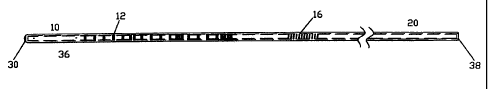
[0021] Figure 1 is a depiction of the catheter of the present invention;
[0022] Figure 2 is an enlarged view of the catheter of Figure 1 showing the
distal portion and
flashback window; and
[0023] Figure 3 is an illustration of the distal portion of the catheter of
the present invention.
Detailed Description of the Invention
[0024] The term "patient" generally refers to living humans and/or animals on
which the
catheter of the present invention may be employed, but may also include
cadavers used for
training/teaching purposes.
[0025] In the following detailed description, the terms "distal" and
"proximal" will be used.
As used herein, the term "proximal" refers to that region, portion or end of a
device or procedure
nearest the person using the device or performing the procedure, while the
term "distal" refers to that
region, portion or end of a device or procedure nearest a patient upon whom
the device is being used or
the procedure is being performed.
[0026] The catheters of the present invention are intended to be utilized in
the introduction of
fluids, particularly those fluids containing a pharmaceutically active
ingredient, such as anesthesia, into
a patient in a wide variety of local, regional and peripheral pain management
situations. Such
situations, include but are not limited to: interscalene blocks;
supraclavicular blocks; axillary blocks;
sciatic blocks; femoral blocks; lateral femoral blocks; obturator blocks;
popliteal blocks; ankle blocks;
lumbar sympathetic blocks; and celiac plexus blocks. More than one catheter of
the present invention
may be used in situations where a temporary block must be administered
followed by a longer-term
4
CA 02403477 2002-09-23
WO 01/70322 PCT/US01/09472
administration of a pharmaceutically active ingredient. Such situations may
include, but are not limited
to, caesarian section and hernia surgery.
[0027] The catheter of the present invention, comprised of closed-end distal
portion 10,
flashback window 16 and proximal portion 20, is shown in Figure 1. The
catheter of the present
invention can preferably be about 12 in. (30.5 cm) to about 36 in. (92 cm) in
length and preferably has
an inner diameter (I.D.) of between about 0.005 in. (0.127 mm) and about 0.020
in. (0.508 mm) with an
outer diameter (OD) of between 28 gauge (G) and 16G, more preferably between
24G and 18G, and
most preferably 20G. The thickness of the walls of the catheter of the present
invention preferably can
be from about 0.003 in. (0.076 mm) to about 0.011 in. (0.279 mm).
[0028] Closed-end distal portion 10 is comprised of a distal tip 30 and a
diffusion area 36.
The diffusion area 36 can be preferably about 0.5 in. (1.27 cm) up to about 20
in. (50.8 cm) in length.
The catheter may preferably have one-centimeter marks 12 plus a mark at distal
tip 30, as is commonly
practiced in the art. Such marks are typically provided on the catheter to
allow the anesthesiologist to
easily determine how far the catheter has been inserted into a patient and to
confirm the catheter's
complete removal from a patient. Proximal portion 20 includes a proximal tip
38.
[0029] Flashback window 16 can preferably be about 1.0 in. (2.54 cm) in length
and.is
provided to allow the anesthesiologist to visualize blood or cerebrospinal
fluid in the catheter upon
aspiration, called "flashback" and thereby avoid subarachnoid or intravenous
injection of anesthetic.
The coils of the reinforcement member 34 in the flashback window 16 region of
the catheter of the
present invention preferably have a spread of about 0.006 in. (0.152 mm) to
about 0.021 in. (0.53 mm)
from center to center of the coils.
[0030] Figure 2 depicts an enlarged view of closed-end distal portion 10 and
flashback
window 16 of the catheter of figure 1. The coils of the reinforcement member
34 in distal portion 10 of
the catheter of the present invention preferably have a spread of up to about
0.015 (0.381 mm) from
center to center of the coils.
[0031] The catheter of the present invention can be made from a variety of
sterilizable plastics
known to those in the art including, but not limited to, polyamides and
copolymers thereof, low density
polyethylene, high density polyethylene, polypropylene, polystyrene,
polycarbonate,
polytetrafluoroethylene, tetrafluoroethylene and fluorinated ethylene
propylene. A particularly
preferred sterilizable plastic for use in the present invention is nylon
polymer. The catheter of the
present invention may also be made of polyurethanes. Particularly preferred
polyurethanes are those
incorporating siloxane available as Elast-EonTM and described in the following
patent applications:
PCT/AU91/00270; PCT/AU91/00545; PCT/AU98/00497; PCT/AU97/00619;
PCT/AU98/00546; and
PCT/AU99/00236.
[0032] Figure 3 illustrates the closed-end distal portion 10 of the catheter
of figure 1. Distal
tip 30 is closed and preferably may be rounded, to prevent reinforcement
member 34 from becoming
dislodged from the catheter of the present invention and thereby posing a
puncture hazard to the patient.
CA 02403477 2002-09-23
WO 01/70322 PCT/US01/09472
The combination of a closed, rounded tip and the flexibility of the above-
mentioned sterilizable plastics
helps prevent venous cannulations and/or dura matter punctures.
[0033] Reinforcement member 34 provides collapse resistance for the catheter
during use.
The reinforcement member 34 is preferably not embedded in, or in any way
attached to, the catheter's
inner wall, except at the distal tip 30 and the proximal tip 38. The lack of
attachment allows the
reinforcement member 34 to retain its flexibility by being able to move
relatively freely within the
catheter's body. The flexibility in turn allows the catheter of the present
invention to better resist
kinking.
[0034] The reinforcement member 34 can be made of a variety of materials,
including but not
limited to stainless steel, titanium, nickel-titanium and plastic
monofilament. A particularly preferred
material for use in reinforcement member of the catheter of the present
invention is stainless steel, such
as #304 wire. Although the reinforcement member of the present invention is
depicted herein as a coil,
the inventor contemplates that it may take a variety of shapes, including but
not limited to strips,
ribbons, filaments, braids or mesh.
[0035] If radiopacity is desired, the reinforcement member 34 preferably can
be made of a
radiopaque substance such as steel, titanium or,nickel titanium or radiopacity
can be conferred by the
incorporation of barium, bismuth, etc. in the wall of the catheter.
Radiopacity, coupled with
fluoroscopy, can facilitate easier placement of the catheter of the present
invention as is known by those
skilled in the art.
[0036] As illustrated in Figure 3, in one embodiment of the catheter of the
present invention
the diffusion area 36 may have three openings 32 arranged about 4 mm from each
other with the distal
most opening being positioned about 5 mm from the distal tip 30. Each opening
32 may preferably be
offset from adjacent openings by about 120 circumferentially to provide for a
more even distribution
of fluid from the catheter. It will readily be apparent to those skilled in
the art that a greater number of
openings and/or openings in different arrangements can be provided in the
catheter of the present
invention.
[0037] In another embodiment, the openings 32 may be aligned in a straight
line or may be in
the form of rows. In yet another embodiment, the openings 32 of the catheter
of the present invention
may be offset from each other by any amount from 0 to 360 . The inventor
contemplates that a
catheter of the present invention may in some situations have as many as about
100 or more openings in
the diffusion area 36. The openings 32 may also be spaced from as little as
about 2 mm to as much as
about 300 mm apart. Although the catheter of the present invention can be
sized to be inserted with
16G to 24G needles as required by the intended application, it can preferably
be sized to permit its
insertion using a 16G to 21 G epidural needle.
[0038] Tests were used to determine the percentage of diffusion area through
which flow was
achieved and the flow rates of catheters of the present invention, and the
results are summarized in
Table I. The catheters of the present invention tested varied in the length of
diffusion area, i.e., the
6
CA 02403477 2002-09-23
WO 01/70322 PCT/US01/09472
length of catheter measured from the distal end, through which openings may be
drilled and therefore
through which fluid flow may occur.
[0039] Because there is no standard test for catheter flow rates, the inventor
used one test, ISO
10555-3:1996(E), to measure natural, i.e., gravity, flow rates for catheters
of the present invention and a
pump test to demonstrate achievable flow for the catheter of the present
invention using a pump.
[0040] Briefly, in the pump test, a Touhy-Borst adapter was attached to the
catheter at the
proximal end. The catheter was primed with a 3 mL syringe containing distilled
water. The outlet line
from an appropriate pump (Sorenson or Baxter'), that also contained distilled
water was attached to the
adapter and the pump was operated. The flow through the diffusion length and
the percent diffusion
was recorded over a period of 5 to 60 minutes and is reported in Table I.
TABLE I
Length of Diffusion Percentage of Diffusion Area Flow Rate Coil
Catheter Area Through Which Flow Was mL/hour Spread ?
in inches (cm) Achieved
I-A 1.0 (2.54) 100 0.5 No
I-B 1.0 (2.54) 100 5.0 No
II-A' 2.0 (5.08) 78 ' 0.5 No
II-B 2.0 (5.08) 86 5.0 No
III-A 3.5 (8.89) 10 141.0* Yes
III-B 3.5 (8.89) 50 38.0* No
IV-A 5.0 (12.7) 86 5.0 No
IV-B 5.0 (12.7) 100 125.0 No
V 7.5 (19.05) 85 5.0 No
VI 10.2 (25.91) 70 5.0 No
*Flow rate measurement made by ISO 10555-3:1996(E).
[0041] As can be seen from a review of table I, catheter I provided flow
through 100% of its
diffusion area at the very slow flow rate of 0.5 mL/hr (I-A) and at 2.0 mL/h
(I-B).
[0042] Catheter II, with a diffusion area having a length of 2.0 in. (5.08
cm), also showed
excellent performance, flowing out of 78% and 86% of the diffusion area, at
flow rates of 0.5 mL/hr (11-
A) and 5.0 mL/hr (II-B), respectively.
[0043] Catheters III-A and B were identical except for the coil being spread
in III-A compared
to catheter III-B. The data in Table I demonstrate that spreading the coil, as
in catheter III-A, resulted
in a much higher flow rate, 141 mL/hr, compared to 38 mL/hr for catheter III-
B. It should be noted that
flow occurred out of only 10% of the diffusion area in catheter III-A compared
to 50 % in catheter III-
B, resulting from the effect of the coil spread in catheter 111-A.
[0044] Using a diffusion area having a length of 5.0 in. (12.7 cm), resulted
in flow occurring
out of 86 % of the diffusion area at 5.0 mL/hr (IV-A) and out of 100% of the
diffusion area at 125
7
CA 02403477 2002-09-23
WO 01/70322 PCT/US01/09472
mL/hr (IV-B). This catheter achieved not only a large range of flow rates, but
did so with excellent
diffusion. As the length of the diffusion area was increased to 7.5 in. (19.05
cm) in catheter V and 10.2
in. (25.91 cm) in catheter VI, flow occurred out of 85 and 70 % of the
diffusion area, respectively.
[0045] The above results demonstrate that catheters of the present invention
are capable of
flow rates ranging from very low (0.5 mL/hr) to very high (125 mL/hr) with the
ability to achieve flow
out of 70% to 100% of the diffusion area. The only exceptions to this being
catheters 111-A and Ill-B
wherein diffusions of 10% and 50 % respectively were obtained. However, the
inventor contemplates
use of catheter III-A in situations where a very high flow rate is required,
but where the percentage
diffusion is not so important, such as epidural anesthesia.
[0046] Although the results summarized in Table I demonstrate that excellent
diffusion is
obtained in catheters of the present invention having a length of diffusion
area as little as 1.0 in. (2.54
cm) to as long an as 10.2 in. (25.91 cm), the inventor contemplates that the
length of the diffusion area
could be up to about 20 in. (50.8 cm). The results also demonstrate that using
catheters of the present
invention, control can be achieved over flow rate as well as the diffusion
area through which flow
occurs.
[0047] The conduction.catheter of the present invention is intended for
administration of local
anesthetic or narcotics into intraoperative sites for post-operative pain
management and for regional
anesthesia outside of the epidural space. Routes of administration may include
intraoperative,
subcutaneous and percutaneous.
[0048] The foregoing illustrations of embodiments of the present invention are
offered for the
purposes of illustration and not limitation. It will be readily apparent to
those skilled in the art that the
embodiments described herein may be modified or revised in various ways
without departing from the
spirit and scope of the invention. The scope of the invention is to be
measured by the appended claims.
8