Note: Descriptions are shown in the official language in which they were submitted.
CA 02408327 2005-11-15
1
BONE PLATE SYSTEM
The present invention is directed to a bone plating system for fracture
fixation, and in
particular to a system including a bone plate having plate holes for both
locking and
non-locking screws.
The clinical success of plate and screw systems for internal fixation of
fractures is
well-documented. However, treatment of certain fractures, such as peri-
articular
fractures, which require a fixed angular relationship between the bone plate
and
screws, remains problematic. Fixed angle devices for treatment of these
fractures
are available and include the Dynamic Condylar Screw SysterriMcommercially
available from Synthes (USA) of Paoli, PA and a wide variety of blade plates.
All of
these devices require a high level of surgical skill, suitable bone quantity
and quality,
and a fracture pattern compatible with the device.
In cases in which these requirements are not satisfied, e.g. severely
comminuted
bone or missing bone segments, conventional bone plate and screw systems must
be used. Although these conventional systems are particularly well-suited to
promoting healing of the fracture by compressing the fracture ends together
and
drawing the bone into close apposition with other fragments and the bone
plate, the
angular relationships between the plate and screws are not fixed and can
change
postoperatively. This can lead to mal-alignment and poor clinical results.
The primary mechanism for the change in angular relationship is related to
energy
storage. As previously noted, threading a bone screw into bone compresses the
bone against the plate. The compression results in high strain in the bone,
and,
consequently, energy storage. With the dynamic loading resulting from
physiological
conditions, loosening of the plate and screw and loss of the stored energy can
result.
Securing the screws to the plate provides a fixed angle relationship between
the
plate and screw and reduces the incidence of loosening. One method of securing
the screw to the plate involves the use of so-called "locking screws". A
locking screw
has threading on an outer surface of its head that mates with corresponding
threading on the surface of a plate hole to lock the screw to the plate. Bone
plates
CA 02408327 2002-11-06
WO 01/19267 PCT/CH00/00474
2
having threaded holes for accommodating locking screws are known. For example,
German Patent Application No. 43 43 117 discloses a bone plate with threaded
holes
for locking screws. As the relationship between the locking screws and the
plate is
fixed, locking screws provide a high resistance to shear or torsional forces.
However,
locking screws have a limited capability to compress bone fragments.
In summary, conventional bone screws, i.e. screws that are not secured to a
plate so
that a fixed angular relationship between the plate and screw is maintained
(hereinafter "non-locking screws") effectively compress bone fragments, but
possess
a low resistance to shear force that can lead to loosening of the screw.
Locking
screws have a high resistance to shear force that ensure stability at the bone
screw/plate hole interface, but possess a limited ability to compress bone
fragments.
Thus, a bone plating system that combines non-locking screws with locking
screws
would be ideal for certain clinical situations.
U.S. Patent No. 5,601,553 discloses a locking plate and bone screw. The plate
has
a plurality of threaded plate holes for receiving locking screws. The plate
also has
non-threaded plate holes for receiving temporary screws that keep the plate in
place
while the locking screws are inserted. After the locking screws are inserted,
the
temporary screws are removed. Thus, the long term benefits of combining non-
locking screws with locking screv~rs are not obtained. U.S. Patent No.
5,709,686
discloses a bone plate with partially threaded plate holes. The partially
threaded
holes allow either non-locking or locking screws to be used. Because the plate
holes
are only partially threaded, the locking screws used may not be able to
maintain the
fixed angular relationship between the screws and plate under physiological
loads.
Specifically, the locking screws within the plate are only partially
captivated and thus
only partially surrounded by threads. Under high stress and loading
conditions, the
locking plate hole may distort and allow the fixed angular relationship
between the
locking screw and plate to change. This can result in loss of fixation or loss
of
established intraoperative plate orientation. Additionally, because of the
plate hole
geometry, translation of the plate with the non-locking screws is limited to
one
direction only. This may be a disadvantage in reduction and manipulation of
fragments.
01-10-200 i CH000047
CA 02408327 2002-11-06
- 2 continuation -
From EP-B 0 486 762 LfN another bone plate is known which has'a plurality of
elongated non-threaded plate holes and a plurality of circular plate holes
with an
interior thread for accomodating a screw having a head with a corresponding
exterior
thread: The screws to be introduced into the elongated holes are provided with
a
threaded head having a rectangular cross-section and corresponding nuts so
that all
of the screws are to be fixed in a rigid manner to the plate.
AMENDED SHEET
CA 02408327 2002-11-06
WO 01/19267 PCT/CH00/00474
3
Thus, there exists a need for an improved bone plating system that overcomes
the
deficiencies of the prior art.
The bone plating system for fixation of bone according to the present
invention
includes a bone plate having an upper surface, a bone-contacting surface, at
least
one first hole passing through the upper and bone-contacting surfaces and
having a
thread, and at least one second hole passing through the upper and bone-
contacting
surfaces. The bone plating system also includes a first screw having a shaft
with a
thread for engaging bone and a head with a thread configured and dimensioned
to
mate with the thread of the first hole, and a second screw having a shaft with
a
thread for engaging bone and a head. The first and second screws remain seated
in
their respective holes for substantially as long as the bone plate is
implanted.
Preferably, the bone plate includes a plurality of first and second holes, and
a
corresponding plurality of first and second screws are provided.
In order to facilitate insertion, the first and second screws can be a self-
tapping
screws. These screws can also be self-drilling screws. Additionally, the first
and
second screws can be cannulated for insertion of a guide wire to guide screw
placement. The first plate hole can have a substantially conical shape with a
double-
lead thread.
In one embodiment, the bone plate has a trapezoidal shaped cross section in
regions
between the first and second plate holes for minimizing contact between bone
and
the bone-contacting surface. Additionally, at least one of the second plate
holes is
longitudinally elongated and has an edge inclined at an angle to the upper
surface
toward the bone-contacting surface for displacing the bone plate when engaged
by
the head of a second bone screw.
In an exemplary embodiment, the bone plate includes a head portion configured
and
dimensioned to conform to a metaphysis of a bone and a shaft portion
configured
and dimensioned to conform to a diaphysis of a bone. The head portion has only
first
plate holes and the shaft portion has both first and second plate holes. In
one
embodiment, the head portion has a curved surface, includes an anterior fork
substantially parallel to an anterior side of the shaft portion, and includes
a posterior
CA 02408327 2002-11-06
WO 01/19267 PCT/CH00/00474
4
fork extending out from a posterior side of the shaft portion. In another
embodiment,
the head portion flares outward from the shaft portion and is curved, tapered,
and
twisted. The head portion can also be provided with suture holes from suture
anchoring of the bone plate.
The method for fracture fixation of bone according to the present invention
comprises
the steps of reducing the fracture to bring bone fragments in close
apposition;
compressing a bone plate against the bone with at least one first fastener to
hold the
fracture reduction; and securing at least one second fastener at a fixed
angular
relationship to the bone plate. The first fasteners are inserted before the
second
fasteners and both the first and second fasteners remain in bone for
substantially as
long as the bone plate is implanted.
Brief Description of the Drawings
FIG. 1 is a side view of one embodiment of a non-locking screw according to
the
present invention;
FIG. 2 is a side view of one embodiment of a locking screw according to the
present
invention;
FIG. 3 is a perspective view of a portion of a bone plate according to the
present
invention;
FIG. 4 shows a cross-sectional view of one of the first plate holes through
line 4-4 of
FIG. 3;
FIG. 5 shows a cross-sectional view of one of the second plate holes through
line 5-5
of FIG. 3;
FIG. 6 shows another cross-sectional view of the second plate hole of FIG. 5
through
line 6-6 of FIG. 3;
FIG. 7 shows a top view of an embodiment of a bone plate according to the
present
CA 02408327 2002-11-06
WO 01/19267 PCT/CH00/00474
invention designed for use in the distal femur;
FIG. 8 shows a side view of the bone plate of FIG. 7;
FIG. 9 shows a perspective view of the bone plate of FIG. 7 implanted in a
distal
femur;
FIG. 10 shows a top view of the bone plate of FIG. 7 with various cross
sections
labeled;
FIG. 11 shows a cross-section of the bone plate of FIG. 7 through line A-A;
FIG. 12 shows a cross-section of the bone plate of FIG. 7 through line B-B;
FIG. 13 shows a cross-section of the bone plate of FIG. 7 through line C-C;
FIG. 14 shows a cross-section of the bone plate of FIG. 7 through line D-D;
FIG. 15 shows a cross-section of the bone plate of FIG. 7 through line E-E;
FIG. 16 shows a cross-section of the bone plate of FIG. 7 through line F-F;
FIG. 17 shows a cross-section of the bone plate of FIG. 7 through line G-G;
FIG. 18 shows a cross-section of the bone plate of FIG. 7 through line H-H;
FIG. 19 shows a cross-section of the bone plate of FIG. 7 through line I-I;
FIG. 20 shows a side view of an embodiment of a bone plate according to the
present invention designed for use in the proximal tibia;
F1G. 21 shows a top view of the bone plate of FIG. 20;
FIG. 22 shows a perspective view of the bone plate of FIG. 20 implanted in a
CA 02408327 2005-11-15
6
proximal tibia;
FIG. 23 shows an end view of the bone plate of FIG. 20 with various cross
sections
labeled;
F1G. 24 shows a cross-section of the bone plate of FIG. 21 through line A-A;
FIG. 25 shows a cross-section of the bone plate of FIG. 21 through line I-I;
and
FIG. 26 shows a cross-section of the bone plate of FIG. 21 through line D-D.
Description of the Preferred Embodiments
The bone plating system according to the present invention includes a bone
plate,
non-locking screws, and locking screws. FIG. 1 shows an example of a non-
locking
screw 10 that can be used with the present invention. In general and as
described in
more detail below, any surgical screw that has a non-threaded head 12 of an
appropriate size and geometry for select plate holes of the bone plate can be
used.
Non-locking screw 10 has a shaft 14 that is at least partially threaded for
attachment
to bone. The length of shaft 14 and the shaft thread configuration can be
selected for
the particular application. As is well known in the art, the threads and a tip
16 can be
made to be self-tapping andlor self-drilling to facilitate implantation. Shaft
14 can
also be cannulated with a channel 18 for receiving a guide wire to aid in
proper
placement.
FIG. 2 shows an example of a locking screw 20 that can be used with the
present
invention. In general and as described in more detail below, any surgical
screw that
has a head 22 with threads 24 can be used as long as head 22 is of an
appropriate
size and geometry for select plate holes of the bone plate and threads 24 mate
with
the threads of the plate holes. Locking screw 20 has a shaft 26 that is at
least
partially threaded for attachment to bone. The length of shaft 26 and the
shaft thread
configuration can be selected for the particular application. As is well known
in the
art, the threads and a tip 28 can be made to be self-tapping and/or self-
drilling to
facilitate implantation. Shaft 26 can be cannulated for receiving a guide
wire.
CA 02408327 2005-11-15
7
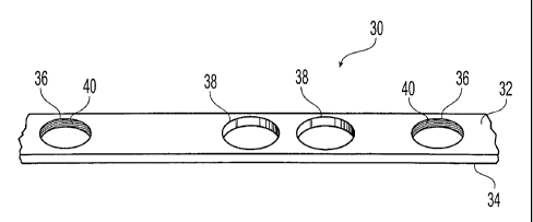
FIG. 3 shows a portion of a bone plate 30 according to the present invention.
Bone
plate 30 can be made in different shapes and sizes for use in a wide variety
of clinical
applications. Bone plate 30 includes an upper surface 32 and a bone contacting
surface 34. Bone plate 30 has a plurality of first plate holes 36 and a
plurality of
second plate holes 38. Each of first and second plate holes 36, 38 passes
through
upper.32 and bone-contacting surfaces 34. Each first plate hole 36 has a
thread 40
that mates with thread 24 on head 22 of locking screw 20 to secure locking
screw 20
to bone plate 30 at a temporally fixed angular orientation. Second plate holes
38 are
not threaded and receive non-locking screws 10 with non-threaded heads 12.
Insertion of non-locking screws 10 in second plate holes 38 draws the bone
toward
bone-contacting surface 34 to compress the bone. Thus, seating of non-locking
screws 10 in second plate holes 38 compresses the bone against bone-contacting
surface 34 and seating of locking screws 20 in first plate holes 36 secures
heads 22
to bone plate 30 for maintaining a fixed angular relationship between locking
screws
20 and bone plate 30. Simultaneous use of bone plate 30 with both non-locking
and
locking screws 10, 20 for as long as bone plate 30 is implanted provides
stability
between both the screw and bone plate and between the bone plate and bone. As
non-locking screws 10 are generally secured in cancellous bone, the threads on
shaft
14 are typically larger than the threads on shaft 26 of locking screws 20.
First plate holes 36 are preferably conical in shape. As shown in FIG. 4,
threads 40
on first plate holes 36 are also preferably double lead threads. The double
lead
conical threads enables multiple threads to engage while maintaining a low
profile.
Additionally, the double lead conical threads are less susceptible to cross-
threading
compared to other threads, e.g. cylindrical threaded arrangements.
As seen best in FIGS. 5 and 6, second plate holes 38 are preferably dynamic
compression unit (DCU) screw holes substantially similar to those disclosed in
reissued U.S. Patent No. Re. 31,628 to Allgower et al.
The DCU screw holes promote healing of the bone
by compressing the fracture ends together. Briefly, second plate holes 38 have
an
edge 42 which includes an oblique portion or ramp 44 having an inclination
such that
when ramp 44 is engaged by the underside of head 12 of non-locking screw 10,
bone
CA 02408327 2005-11-15
a
plate 30 is displaced in a direction to move ramp 44 away from non-locking
screw 10
and to cause bone plate 30 to apply a pressure to hold the fracture ends in
tight
engagement.
Bone-contacting surface 34 on bone plate 30 can be shaped to minimize contact
with
bone. Limiting contact between the bone plate and bone has a number of
biological
and mechanical advantages including reduced damage to blood supply and easier
plate removal. Providing bone plate 30 with a trapezoidal cross section (FIG.
11 ) in
the regions between first and second plate holes 36, 38 is one way to minimize
contact. Other ways are disclosed in U.S. Patent Nos. 5,151,103; 5,053,036;
5,002,544; and 4,838,252.
By combining locking screws and non-locking screws on the same bone plate, the
present invention provides a novel mixed fixation. With the non-Pocking
screws,
fracture reduction is held by friction between the bone plate and bone. This
friction is
generated by tightening the non-locking screws in bone. However, micromotion
between the non-locking screws and bone leads to bone resorption, and toss of
reduction. Additionally, insertion of the non-locking screws requires bone to
withstand the stresses of tightening of the screws. This results in high
stress in bone
surrounding the non-locking screws. Ordinarily, the high stress can cause the
non-
locking screw threads to strip (threads in bone fail in shear) and/or creep in
bone
(since bone is a viscoelastic material). Either one of these phenomenon also
results
in loss of reduction.
By adding at least one locking screw, loss of reduction is minimized or
eliminated by
the present invention. Specifically, by securing the locking screws to the
bone plate
and not the bone, the effect of the viscoelastic behavior of bone is reduced,
the
threads do not strip, and micromotion is prevented. The attachment between the
locking screws and bone plate is a high strength connection in which the
locking
screw must cut sidevrays through bone to fail.
As management of certain peri-articular fractures typically involves insertion
of.
screws at various angles with respect to the bone plate and it is highly
desirable to
CA 02408327 2002-11-06
WO 01/19267 PCT/CH00/00474
9
maintain the initial angular relationships between the individual screws and
the bone
plate, the bone plating system according to the present invention is
particularly well-
suited for these clinical applications. FIGS. 7-19 show a bone plate 50
according to
the present invention specifically designed for use in the distal femur. Bone
plate 50
would be used primarily for, but not limited to, severely comminuted fractures
including Hoffa type fractures.
Bone plate 50 has an upper surface 52 and a bone-contacting surface 54. Bone
plate 50 has a plurality of threaded plate holes 56a, 56b, 56c (collectively
referred to
as threaded plate holes 56) for receiving locking screws 20 and a plurality of
non-
threaded plate holes 58 for receiving non-locking screws 10. Each of threaded
and
non-threaded plate holes 56, 58 passes through upper 52 and bone-contacting
surfaces 54. As was the case for bone plate 30, the thread on threaded plate
holes
56 mates with threaded head 22 of locking screw 20 to secure locking screw 20
to
bone plate 50 at a temporally fixed angular orientation and insertion of non-
locking
screws 10 in non-threaded plate holes 58 draws the bone toward bone-contacting
surface 54 to compress the bone.
Bone plate 50 includes a head portion 60 configured and dimensioned to conform
to
the metaphysis of the distal femur and a shaft portion 62 configured and
dimensioned
to conform to a diaphysis of a bone. As best seen in FIG. 8, bone contacting
surface
54 of head portion 60 is a curved surface to fit the contours of the distal
femur. Head
portion 60 includes an anterior fork 64 substantially parallel to an anterior
side 66 of
shaft portion 62 and a posterior fork 68 extending laterally out from a
posterior side
70 of shaft portion 62.
The non-threaded plate holes 58 are preferably dynamic compression unit (DCU)
screw holes substantially similar to second plate holes 38. Shaft portion 62
has both
threaded plate holes 56a and non-threaded plate holes 58 so that both locking
and
non-locking screws can be used in shaft portion 62. The ability to use locking
screws
in shaft portion 62 is particularly useful when the far cortex of part of the
diaphysis is
missing or severely damaged since fixation with non-locking screws is
problematic
because of the condition of the far cortex. As best seen in FIG. 11, the
regions
between threaded and non-threaded plate holes 56a, 58 have a trapezoidal cross
CA 02408327 2002-11-06
WO 01/19267 PCT/CH00/00474
section that limits contact between bone-contacting surface 54 of shaft
portion 62
and the femur. Shaft portion 62 terminates in a tapered tail 72 (FIG. 19).
In contrast to shaft portion 62, head portion 60 contains only threaded holes
56.
Specifically, threaded plate holes 56b that surround a centrally located
threaded plate
hole 56c. Threaded plate hole 56c has a larger diameter than threaded plate
holes
56b to accommodate a locking screw with a larger diameter, e.g. threaded plate
hole
56b have a diameter of 5.0 mm and threaded plate hole 56c has a diameter of
7.3
mm. FIGS. 12-18 show the various angular orientations of the individual
threaded
holes 56b, 56c. In generally, threaded holes 56b, 56c are arranged so that the
inserted locking screws converge towards each other. It should be noted that,
if a
surgeon elects, non-locking screws can be used in any of threaded plate holes
56.
Finally, it should also be noted that bone plate 50 has several structural
differences
from the condylar buttress plate commercially available from Synthes (U.S.A.)
of
Paoli, Pennsylvania. For example, the head of the condylar buttress plate is
contoured in both the longitudinal and transverse directions while head
portion 60 of
bone plate 50 is contoured only in the longitudinal direction for a more
anatomical fit.
Additionally, tail 72 has an elevated end to get under tissue.
FIGS. 20-26 show a bone plate 80 according to the present invention
specifically
designed for use in the proximal tibia. Bone plate 80 would be primarily used
for, but
not limited to fractures of the lateral proximal tibial plateau. Bone plate 80
has an
upper surface 82 and a bone-contacting surface 84. Bone plate 80 has a
plurality of
threaded plate holes 86a, 86b and 86c (collectively referred to as threaded
plate
holes 86) for receiving locking screws 20 and a plurality of non-threaded
plate holes
88 for receiving non-locking screws 10. Each of threaded and non-threaded
plate
holes 86 and 88 pass through upper 82 and bone-contacting surfaces 84. As was
the case for bone plate 30, the threads on threaded plate holes 86 mate with
the
threaded head 22 of locking screw 20 to secure locking screw 20 to bone plate
80 at
a fixed angular orientation. Insertion of non-locking screws 10 in non-
threaded plate
holes 88 draws the bone-contacting surface 84 toward the bone to compress the
plate to the bone.
Bone plate 80 includes a head portion 90 configured and dimensioned to conform
to
CA 02408327 2002-11-06
WO 01/19267 PCT/CH00/00474
11
the metaphysis of the lateral proximal tibia and a shaft portion 92 configured
and
dimensioned to conform to a diaphysis of the lateral proximal tibia. As seen
in FIGS.
20 and 26, bone contacting surface 84 of head portion 90 is a curved, tapered,
and
twisted to fit the contours of the lateral proximal tibial plateau. Head
portion 90 also
features sutures holes for suture anchoring and for provisional fixation of
bone plate
80.
The non-threaded plate holes 88 are preferably dynamic compression unit (DCU)
screw holes substantially similar to second plate holes 38. Shaft portion 92
has both
threaded plate holes 86a and non-threaded plate holes 88 so that both locking
and
non-locking screws can be used in shaft portion 92. The ability to use locking
screws
in shaft portion 92 is particularly useful when the far cortex of part of the
diaphysis is
missing or severely damaged since fixation with non-locking screws is
problematic
because of the condition of the far cortex. As best seen in FIG. 24, the
regions
between threaded and non-threaded plate holes 86a and 88 have a rectangular
cross section that limits contact between bone-contacting surface 84 of shaft
portion
92 and the tibia. Shaft portion 92 terminates in a tapered tail 102 (FIG. 25).
In similar fashion to shaft portion 92, head portion 90 contains threaded
holes 86 and
non-threaded holes 88. Head portion 90 features threaded plate holes 86b and
86c.
Holes 86b and 86c have a diameter of 5.0 mm and are oriented as shown in FIGS.
23 and 26. In general, threaded holes 86b, 86c are arranged so that the
inserted
locking screws converge towards each other. As shown in FIG. 23, plate holes
86b
are oriented to converge at a predetermined distance from plate surface 84 to
optimize the position of locking screws 20 within the tibia plateau. As shown
in FIG
26, plate hole 86c is oriented to converge with plate hole 86b at
predetermined
distance to provide additional stability to the locked fixed-angle construct.
It should
be noted that if a surgeon elects, non-locking screws can be used in any of
threaded
plate holes 86.
While it is apparent that the illustrative embodiments of the invention herein
disclosed
fulfil the objectives stated above, it will be appreciated that numerous
modifications
and other embodiments may be devised by those skilled in the art. For example,
for
some fractures only one first plate hole and one second plate hole are needed,
CA 02408327 2002-11-06
WO 01/19267 PCT/CH00/00474
12
although at least two of each is advantageous. Furthermore, additional plate
holes
without screws can be present in the plate, if desired to allow the surgeon
further
flexibility in use. Therefore, it will be understood that the appended claims
are
intended to cover all such modifications and embodiments which come within the
scope of the present invention.