Note: Descriptions are shown in the official language in which they were submitted.
CA 02412826 2002-12-20
WO 01/97702 PCT/USO1/19454
Device for Biopsy and Treatment of Breast Tumors
Field of the Inventions
The devices and method described below relate to the
diagnosis and treatment of breast lesions, and more generally,
to the diagnosis and treatment of tumors and lesions
throughout the body.
Background of the Inventions
Biopsy is an important procedure used for the diagnosis
of patients with cancerous tumors, pre-malignant conditions,
and other diseases and disorders. Typically, in the case of
cancer, when the physician establishes by means of procedures
such as palpation, mammography or x-ray, or ultrasound imaging
that suspicious circumstances exist, a biopsy is performed.
The biopsy will help determine whether the cells are
cancerous, the type of cancer, and what treatment should be
used to treat the cancer. Biopsy may be done by an open or
percutaneous technique. Open biopsy, which is an invasive
surgical procedure using a scalpel and involving direct vision
of the target area, removes the entire mass (excisional
biopsy) or a part of the mass (incisional biopsy).
Percutaneous biopsy, on the other hand, is usually done with a
needle-like instrument through a relatively small incision,
blindly or with the aid of an imaging device, and may be
either a fine needle aspiration (FNA) or a core biopsy. In
FNA biopsy, individual cells or clusters of cells are obtained
for cytologic examination and may be prepared such as in a
Papanicolaou smear. In core biopsy, as the term suggests, a
core or fragment of tissue is obtained for histologic
examination which may be done via a frozen section or paraffin
1
CA 02412826 2002-12-20
WO 01/97702 PCT/USO1/19454
section. One important area where biopsies are performed is
the diagnosis of breast tumors.
Traditionally, the biopsy technique for breast tumors
involves placing a biopsy device multiple times into the
breast and taking several samples of tissue from a mass or
tumor which is suspected of being cancerous. Several samples
are required to be sure that some tissue from the suspect mass
has been captured, and enough tissue has been sampled to
ensure that, if disperse cancer cells exist in the suspect
mass some of those cancer cells will be captured in the
samples. Each time the device is placed the physician must
locate and direct the device with ultrasound imaging into the
correct position near the suspect mass. Some breast tumors
and lesions are very well defined, hard spherical masses which
grow within the soft, compliant breast tissue. It is
difficult to force a needle into these lesions because they
are resistant to puncture and fairly mobile. Forcing the
biopsy needle into the lesion is like trying to spear an apple
floating in water.
Vacuum assisted biopsy system proposed by Biopsys
involves sucking a breast lesion into a cannula and shearing
off the captured edge of the lesion to obtain a biopsy sample.
The device uses a vacuum to collect tissue into the side of an
open tubular device, and then uses a rotating corer to cut the
~5 tissue collected. The rotating core is slidable within the
tubular section and can be pulled back to remove the tissue
collected in the rotating core. An additional stylet inside
the rotating core can be used to push the tissue out of the
core. The device can,be rotated on its axis to remove a
sample, 360 degrees around the central placement of the
device. Typically, physicians sample six to eight cores. One
advantage of this device is that the physician does not have
to remove the device for additional biopsy samples. However,
2
CA 02412826 2002-12-20
WO 01/97702 PCT/USO1/19454
the tumor itself must be re-engaged after every coring
operation, which entails substantial effort in relocation and
confirmation that the target suspect mass has been engaged by
the side aperture. Tumors may be too tough to yield to the
suction and deform as necessary to enter the side opening of
the cannula. Doctors also currently use the device to take a
circular sequence of cores by rotating the device about its
long axis or by sideways movement of the suction head to take
a line of cores.
After biopsy and analysis, the tumor must be treated with
a separate device, as Biopsys teaches that their coring device
should not be used for resection. Indeed, the device is not
designed to perform resection with assurance that complete
resection of a suspect mass has been accomplished. Mechanical
cutting and disruption of the tissue structure and cancer cell
dispersion (that is, tearing of the tissue around the cancer
and movement of the cancer cells amongst normal tissue) will
result in unintentional delivery of cancer cells into healthy
tissue adjacent the lesion.
0 Summary
The devices and methods described below provide for
diagnosis and treatment of tumors within the breast. The
devices include structures which permit the surgeon to secure
a suspect mass or tumor within the breast for an extended
~5 period of time and for several biopsies, coring procedures, or
resections. The suspect mass or tumor is secured to a cannula
for the entire diagnostic and treatment procedure, or subsets
of the procedure such as biopsy or ablation. This allows the
placement of the cannula with a single step utilizing methods
30 such as ultrasound to guide the cannula toward the tumor.
The cannula includes a lumen adapted to be connected to a
source of vacuum, which can be used to secure a breast lesion
3
CA 02412826 2002-12-20
WO 01/97702 PCT/USO1/19454
to the cannula. A ring seal on the proximal end of the
catheter permits biopsy needles, cryoprobes or other ablation
devices to be inserted through the cannula and into the lesion
while the vacuum on the cannula is maintained. In this
manner, the needles and ablation devices may be inserted into
the lesion while the lesion in held securely in place by the
suction applied to the cannula.
Brief Description of The Drawings
Figure 1 illustrates the cannula adapted for use in
securing a breast tumor during a biopsy or ablation procedure.
Figure 2 illustrates the biopsy needle in use with the
cannula of Figure 1.
Figure 3 illustrates a multiple coring needle which may
be used with the cannula of Figure 1.
Figure 4 illustrates the placement of a cryoprobe or
other ablative device within the cannula of Figure 1.
Figure 5 illustrates a method of breast tumor ablation
for tumors located near the skin.
Figure 6 illustrates a method of breast tumor ablation
a0 for tumors located near the skin.
Figure 7 illustrates and adaptation of the cannula to
provide additional protection to the skin.
Detailed Description of the Inventions
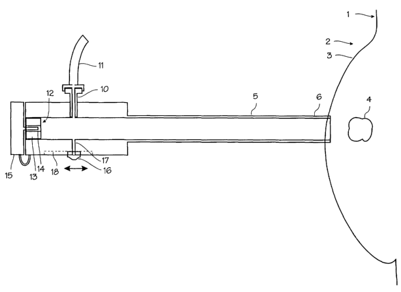
Figure 1 illustrates the biopsy and treatment device
?5 adapted for use in securing a breast tumor during the biopsy
and treatment procedure. The patient 1 and the patient's
breast 2 and skin 3 of the breast are shown schematically.
The tumor, lesion or other suspect mass 4 is located within
4
CA 02412826 2002-12-20
WO 01/97702 PCT/USO1/19454
the breast, surrounded by soft tissue and fatty tissue. The
tumor in this illustration is a well defined, hard mass
ranging in size from 3 to 40 mm in diameter, typical of a
benign palpable tumor or fibro-adenoma, although the device
and method may be used to treat fibrocystic disease and other
conditions. The device comprises a cannula 5 with a straight
cut distal edge 6 adapted for insertion through a small
incision in the skin overlying the tumor and a proximal end 7
which remains outside the breast. The proximal end of the
cannula is fitted with hub 8 which serves as a handle and a
manifold for the several connections to the cannula. This hub
may be integral with the cannula or provided as a separate
piece secured to the proximal end of the cannula. The cannula
has a lumen 9 extending through the cannula from the distal
edge to the proximal end of the cannula. On the hub, a
vacuum connection 10 in the form of Luer fitting provides a
fluid connection between the lumen of the cannula and a vacuum
tube 11. The vacuum hose may be connected to any source of
vacuum or suction. On the proximal end of the hub, a valve 12
seals the cannula proximal end against air pressure but allows
passage of the needles and probes used in the procedure. The
valve may be a self-sealing silicone plug 13 provided with a
slit 14 capable of accommodating the needles and probes by
resiliently expanding and conforming around a needle or probe
when a needle or probe is forced through the slit, and
resiliently closing to an airtight seal when the needles or
probes are removed. Thus, the valve allows for insertion of
various instruments and elongate medical devices while
maintaining the seal necessary to provide sufficient suction
to hold the tumor. A stopper or cap 15 is provided for
insertion into the slit when the valve is not occupied by a
needle or probe to positively seal the valve. A backup valve,
such as ball valve which opens to form a clear,.and straight
lumen, may be placed in line before the valve 12 in. place of
5
CA 02412826 2002-12-20
WO 01/97702 PCT/USO1/19454
the stopper. The cannula is made of an acceptable biological
material such as Teflon, carbon fiber, metal or metal
composite for maximum strength with minimal wall thickness.
The self-sealing valve is comprised of silicone or other
material of similar resilience and conformability. An
additional valve 16 may be added on the proximal handle,
controlling a port 17 communicating between the vacuum lumen
and the exterior of the cannula. The valve illustrated is
merely a thumbslide mounted in a recess 18. This valve may be
used to break the vacuum established in the vacuum lumen to
release a lesion from the distal tip of the device, or to
bleed the vacuum from the lumen to lessen the suction on a
lesion.
Figure 2 illustrates the cannula in use with a biopsy
needle 20 in place within the lumen. A biopsy needle 20 fits
within the lumen of the cannula and passes through the valve
12. The valve deforms and opens enough to allow the needle to
pass through, yet still maintains a sufficiently airtight seal
to maintain the vacuum within the cannula lumen. The needle
has a sharp distal tip 21 which can pierce the tumor 4. The
distal tip is shaped with a coring edge to collect tissue
within the lumen 22 of the needle. As depicted in Figure 2,
suction has been applied to the cannula lumen through the
vacuum hose 11 and connection 10, thus drawing the tumor to
the distal edge of the cannula and securely holding it in
place. The biopsy needle has been inserted through the self-
sealing valve and through the cannula lumen into and through
the tumor. A small core of tumor tissue 23 has been forced
into the lumen of the needle. The needle may now be removed
and the core of tumor tissue extracted and analyzed for the
presence of cancer cells. When the needle is removed, the
suction is maintained on the cannula lumen, and the tumor
remains securely engaged with the cannula distal edge. The
biopsy needle (or another) can then be inserted through the
6
CA 02412826 2002-12-20
WO 01/97702 PCT/USO1/19454
cannula and into the tumor without having to relocate and
reengage the tumor with the cannula. After all necessary
biopsies have been taken, the sample tissue may be analyzed
for the presence of cancer cells or other undesirable tissue
for which ablation is indicated.
Figure 3 illustrates a multiple coring needle 24 for use
with the system. This needle includes several coring lumens
25 opening at the distal end of the needle into coring edges
26. The coring lumens are spaced in a circle about the
circumference of the needle, and extend from the distal tip 21
of the needle proximally to the proximal end of the needle.
It may be used in place of the single biopsy coring needle as
illustrated in Figure 2. By providing suction to one or more
of the lumens, the tumor is secured to the coring needle.
Figure 4 illustrates the use of an ablative device, such
as cryoprobe, with the cannula. The cryoprobe 27 fits within
the lumen of the cannula and passes through the valve 12, and
the distal tip of the cryoprobe is forced into the tumor until
the active freezing portion of the probe resides within the
tumor. During placement of the cryoprobe, the vacuum is
maintained within the lumen so that the tumor is securely
engaged by the cannula. With the tumor secured by the vacuum,
the cryoprobe may be easily forced into the tumor. The
cryoprobe may be operated to ablate the tumor with cryogenic
freezing as required to destroy the tumor. To operate the
cryoprobe, liquid or gas cryogenic fluids (such as liquid
nitrogen, or gaseous argon in combination with a Joule-Thomson
cryostat in the probe tip) are passed through the probe,
supplied from a cryosurgical control system (not shown). The
operation of the cryoprobe creates an iceball 28 which
encompasses the lesion 4, and cools the lesion to lethal
cryogenic temperatures. Any ablation device may be used in
place of the cryoprobe, including RF ablation probes,
7
CA 02412826 2002-12-20
WO 01/97702 PCT/USO1/19454
microwave ablation probes, laser ablation probes, or focused
ultrasound energy probes. Temperature sensors 29 may be
mounted on the skin over the lesion in order to monitor skin
temperature, so that the surgeon may avoid ablating the skin.
In use, the devices described above are used in place of
traditional biopsy, coring and ablation devices. Prior to
use, the patient is prepared and the breast is appropriately
prepped and draped. The site is prepared using local
anesthesia and, optionally, intravenous sedation. The patient
is positioned on an operating table in the supine position,
with the patient on her back. (If the procedure is
accomplished under stereotactic guidance, the patient may be
prone on a stereotactic table, exposing the breast below the
table.) The breast is imaged, if not previously imaged, to
determine the location of lesions. A small incision is made
in the breast to allow the cannula to be easily inserted into
the skin. The surgeon inserts the cannula into the patient's
breast through the incision, pushes it into the breast until
the distal edge of the cannula is proximate to the boundary of
the tumor. An ultrasound scanner, MRI, stereotactic,
mammographic, infrared or other imaging device is used to
obtain an image of the breast, including the tumor and any
device inserted into the breast, and the surgeon uses the
display from the imaging device to assist in guidance of the
cannula to the tumor. With the cannula distal edge in
position near the tumor, the surgeon applies vacuum to the
cannula through the side port on the cannula. The vacuum
draws the tumor toward the cannula, and the cannula securely
engages the tumor until the suction is broken at the end of
the procedure. The surgical biopsy needle can be inserted
through the cannula and into the tumor to retrieve a sample of
tissue for analysis. Because coring can be accomplished
without removing the portion of the tumor engaged by the
cannula, or otherwise disrupting the suction between the
8
CA 02412826 2002-12-20
WO 01/97702 PCT/USO1/19454
cannula and the tumor, several biopsy samples may be taken
without having to relocate and re-engage the tumor.
Depending on the analysis of the biopsy (whether or not
the samples obtained contain cancerous cells or other
conditions), treatment of the tumor may be required. If
analysis can be accomplished intra-operatively (that is,
during a period of time in which it is feasible to keep the
patient in the operating room and maintain the tumor engaged
with the cannula), and indicates the presence of cancerous
cells or other condition for which ablation is indicated, an
ablation instrument can be inserted through the cannula and
into the tumor. If so, the surgeon inserts an ablation
instrument, such as a small caliber cryoprobe, into the tumor.
Preferably, the surgeon inserts a cryoprobe through the valve
and cannula and into the tumor, while maintaining suction on
the cannula. The surgeon initiates cooling of the cryoprobe,
and cools the tumor through one or more cycles of cooling to
cryogenic temperatures and subsequent warming and thawing. A
double freeze-thaw cycle is currently recommended. Each cycle
consists of a 6 to 15 minute freeze followed by thawing until
the internal cryoprobe temperature reaches 0°C (approximately 6
to 15 minutes). The device may also be used without regard to
biopsy results. Patients prefer to have these lesions
treated, even if they prove to be benign. In current
practice, should biopsy results indicate the presence of
cancer, the patient must return to the operating room shortly
after the biopsy, undergo preparation, anesthesia, relocation
of the lesion and ablation. Instead, the lesions may be
ablated intraoperatively with the biopsy, immediately after
biopsy and without interrupting the procedure to await the
biopsy results. Should the biopsy prove negative for the
presence of cancer, the patient will have received a
substantially cosmetic treatment. Should the biopsy prove
positive, the patient will have received a necessary
9
CA 02412826 2002-12-20
WO 01/97702 PCT/USO1/19454
therapeutic procedure. In addition to the ablative procedure,
the positive biopsy may indicate the need for additional
monitoring and treatment.
For lesions deeper than 1 cm from the skin surface, the
cryoprobe is advanced until the distal tip is located
approximately in the center of the lesion or just beyond the
lesion. For smaller lesions (<2cm diameter) the ice ball may
grow beyond the margins of the tumor, while for larger
lesions, the ice ball may remain within the confines of the
tumor. The cryoprobe tip temperatures and skin mounted
thermocouple readings are monitored throughout the ablation
procedure. If the temperature of the skin overlying the
cryoprobe measures below freezing, freezing operation of the
cryoprobes should be paused until it returns to 10°C (the
temperature at the edge of the ice ball edge is 0°C and
exposure to such a temperature for the few minutes will not
harm the skin, but caution should always be employed).
The procedure may be augmented with additional steps.
Just prior to ablation treatment, prophylactic antibiotics can
be administered at the surgeon's discretion. Just prior to
cryosurgical ablation, cryogenic enhancement agents may be
injected directed into the tumor through a hypodermic needle
inserted through. the valve and cannula and into the tumor
while it is secured by suction to the cannula. During cooling
operation of the cryoprobes, warm saline may be washed over
the skin overlying the tumor and iceball to prevent freezing
of the skin.
If the lesion being treated is close to the skin such
that cryoablation of the lesion entails a danger of
cryoablation of the overlying skin, several milliliters of a
resorbable material such as sterile saline may be injected or
inserted into the subcutaneous tissue between the skin and the
CA 02412826 2002-12-20
WO 01/97702 PCT/USO1/19454
lesion. This will create a thermally protective mass or
barrier layer between the tumor and the skin. Thermal
protection may arise from insulative effect of the thermally
protective mass or merely by the distension or separation of
the skin away from the tumor and thus away from the iceball.
As illustrated in Figure 5, where the tumor 4 is close to the
skin 3, the thermally protective mass 30 is injected between
the skin 3 and the subcutaneous fat 31 of the breast. When
the cryoprobe 27 is operated to create the iceball, the ice-
ball 32 either grows into the thermally protective mass or is
inhibited in growth in the direction of the thermally
protective mass (as illustrated by the non-spherical shape of
the iceball in this illustration). This method basically
distends the skin away from the iceball. This may also be
accomplished by dissecting the skin away from the tumor with a
balloon inserted between the skin and fat in the area
overlying the tumor. Balloon dissection can be accomplished
as illustrated in Figure 6. Here, a balloon 33 has been
inserted subcutaneously between the tumor 4 and the overlying
skin 3. The balloon is inflated with air or other sterile
gas, through inflation tube 34, creating a good layer of
insulation between the cryoprobe and the overlying skin.
Figure 7 illustrates and adaptation of the cannula to
provide additional protection to the skin. The cryoprobe 27
is inserted through a side lumen 35 provided on the cannula 5.
The breast lesion 4 is drawn by vacuum to the tip of the
cannula. The cryoprobe is advances distally out of the side
lumen until the freezing region underlies the lesion, and it
operated to create the iceball 36. The iceball extends
superficially toward the skin and to encompass the lesion, and
also extends posteriorly into the breast, where some healthy
breast tissue is ablated but the overlying skin is not. This
system and procedure also has the advantage that the lesion
itself is not punctured, limiting the potential for seeding
11
CA 02412826 2002-12-20
WO 01/97702 PCT/USO1/19454
due to the release of cancerous cells from the disruption of
the tissue of the tumor.
The cannula illustrated above is preferably 10 to 20 cm
in length and about 3 mm in diameter with an internal diameter
of 2.8 mm, and a clearance of about .25 mm between the inner
bore of the cannula and any device inserted through the
cannula during suction. The cryoprobes may be Joule-Thomson
probes, liquid cryogen probes, or probes of other designs.
Various other ablative devices may be used in place of the
cryoprobe, including laser ablation devices, RF ablation
devices, chemical ablation catheters and any other ablative
technology proposed for use to destroy tumors and lesions.
The vacuum applied is preferably in the range of 14 to 21
inches of mercury vacuum.
The devices and methods illustrated above have been
illustrated in relation to the treatment of tumors and lesions
within the breast. However, they may be used to treat tumors
and lesions throughout the body wherever the tumors which are
difficult to secure and locate are encountered, and wherever
nearby tissue must be protected from freezing. Thus the
devices and methods may be used for tumors and lesions of the
uterine tube (such as uterine fibroids), kidney, liver,
prostate or brain.
Thus, while the preferred embodiments of the devices and
methods have been described in reference to the environment in
which they were developed, they are merely illustrative of the
principles of the inventions. Other embodiments and
configurations may be devised without departing from the
spirit of the inventions and the scope of the appended claims.
12