Note: Descriptions are shown in the official language in which they were submitted.
CA 02414168 2007-08-30
1
PERCUTANEOUS VERTEBRAL FUSION SYSTEM
BACKGROUND
The himnan vertebrae and associated connective elements are subject to a
variety of
diseases and conditions wbich cause pain and disabiliiy. Among these diseases
and
conditions are spondylosis, spondylolisthesis, vertebral instability, spinal
stenosis and
degenerated, herniated, or degenerated and henniated intervertebral discs.
Additionally, tlye
vertebrae and associated connective elements are subject to injnries,
inclnding fractares and
torn ligaments and surgical manipulations, including laminectomies.
The pain and disability related to these diseases, conditions, injuries and
manipulations often results from the displacement of all or part of a vertebra
from the
remainder of the vertebral column. A variety of inethods have been developed
to restore the
displaced vertebrae or portions of displaced vertebrae to their normal
position and to fix them
within the vertebral column. For example, open reduction with screw fiaation
is one
currently used method. These methods, however, are associated with a variety
of
disadvantages, such as high cost, lengthy inpatient hospital stays and the
potential morbidity
associated with open procedures.
Therefore, there is a need for a method of repositioning and fixing displaced
vertebrae
or portions of displaced vertebrae to a position within the vertebral column
which is more
stable or which causes less morbidity. Further, there is a need for a system
for performing a
method of-repositioning and fixing displaced vertebrae or portions of
displaced vertebrae to a
position within the vertebral column which is more stable or which causes less
morbidity.
SUr1VIlVIARY
According to one embodiment of the present invention, there is provided a bone
screw comprising a proaimal portion comprising a head with a proximal end and
a portal; a
distal portion comprising threads and a tip with a distal end; and a cenfiral
lumen configured
to receive a gaidewire extending coaxially completely through the bone screw
from the
CA 02414168 2002-12-19
WO 02/00126 PCT/US00/34855
2
proximal end to the distal end. The head comprises a proximal portion
configured to mate
with the tip of a screwdriver.
According to another embodiment of the present invention, there is provided a
screwdriver comprising a proximal end comprising a handle configured to permit
grasping of
the screwdriver and to permit the application of torque to a bone screw; a
distal end
comprising a shaft having a tip configured to interface with a bone screw; and
a central
lumen configured to receive a guidewire extending coaxially completely through
the
screwdriver from the proximal end to the distal end.,
According to yet another embodiment of the present invention, there is
provided an
inflatable connection rod comprising a proximal end comprising a self-sealing
valve; a distal
end comprising a tip; and a compliant, inflatable balloon between the proximal
end and the
distal end. The balloon comprises thin, reinforcing wires.
According to another embodiment of the present invention, there is provided a
directing sheath comprising a proximal portion with a proximal end; a distal
portion with a
distal end; a central portion between the proximal portion and the distal
portion comprising at
least two openings; and a lumen extending through the directing sheath from
the proximal
end to distal end. The directing sheath is preferably scored along its
longitudinal axis to
allow the directing sheath to be split into two separate halves by peeling the
directing sheath
apart at either its proximal end or its distal end or both along the scoring.
In a preferred embodiment of the present invention, there is provided a method
of
repositioning or fixing one or more unstable, separated or displaced vertebrae
or one or more
portions of one or more vertebrae in a patient's vertebral column. The method
comprises:
a) identifying a patient who is a suitable candidate for undergoing the
method;
b) making a stab incision in the patient's skin overlying the patient's
vertebral column
at or near the level of the vertebrae or portion of vertebrae to be
repositioned or fixed;
c) creating a first tract from the incision to the posterior periosteal
surface of the
vertebrae;
d) incising the periosteum and extending the first tract into the cortex of
the vertebrae;
e) inserting a first guidewire into the first tract;
f) advancing a bone screw comprising a portal and a tip, and a screwdriver
over the
first guidewire;
g) applying torque to the bone screw using the screwdriver, thereby fixing
part of the
CA 02414168 2002-12-19
WO 02/00126 PCT/US00/34855
3
tip of the bone screw into the vertebrae while the portal of the bone screw is
exterior and
dorsal to the vertebrae and the portal is open parallel to the long axis of
the vertebral column;
h) removing the screwdriver and the first guidewire;
i) repeating c) through h) for at least one vertebrae which is neither
unstable,
separated or displaced and which is adjacent the vertebrae or portion of the
vertebrae that is
being repositioned or fixed, or repeating c) through h) for the cranial-ward
portion of the
sacrum of the patient;
j) inserting an inflatable connection rod comprising a proximal end, a distal
end and
an inflatable balloon between the proximal end and the distal end, between the
portals of the
bone screws; and
k) inflating the inflatable balloon thereby creating a rigid structure between
the
inflated inflatable connection rod and the bone screws;
thereby repositioning or fixing the one or more unstable, separated or
displaced
vertebrae or the one or more portions of one or more vertebrae unilaterally.
Identifying a patient who is a suitable candidate comprises identifying a
patient who
has one or more unstable vertebrae, one or more portions of a vertebrae at
least partly
separated from the remainder of the vertebrae with potential or complete
separation, or who
has one or more vertebrae or a portion of one or more vertebrae displaced from
its normal
position relative to the vertebral column, or who has one or more portions of
a vertebrae at
least partly separated from the remainder of the vertebrae and displaced from
its normal
position relative to the vertebral columm; and where the patient has either
pain, loss of
function or real or potential instability which is likely due to the
separation or displacement,
or separation and displacement.
The method can comprise enlarging the first tract from the incision to the
posterior
periosteal surface using a high-pressure fascial dilator balloon after
creating the first tract.
Further, inserting an inflatable connection rod can comprise:
i) percutaneously inserting a hollow needle and advancing the hollow needle to
the
portal of one of the bone screws;
ii) introducing a second guidewire through the lumen of the hollow needle and
into
the portal of one of the bone screws; and
iii) passing the second guidewire through all of the portals in the bone
screws, thereby
creating a second tract.
CA 02414168 2002-12-19
WO 02/00126 PCT/US00/34855
4
The method can also comprise:
i) dilating the second tract created by the second guidewire using a high
pressure
balloon;
ii) passing an introducer sheath over the guidewire along the entire guidewire
second
tract;
iii) removing the guidewire; and
iv) advancing the inflatable connection rod through the introducer sheath
until the
inflatable connection rod advances between the bone screw portals.
The method can also comprise using a guidewire directing device to direct the
advancing second guidewire through at least one bone screw portal, or can
comprise using a
guidewire capture device to pull the second guidewire through the patient's
skin. Further,
inflating the inflatable balloon can comprise inflating the balloon with a
rapid setting, liquid
polymer.
In a particularly preferred embodiment, the method further comprises repeating
c)
through h) for one additional vertebrae, where the one additional vertebrae is
either unstable,
separated or displaced, or where one or more portions of the one additional
vertebrae is
unstable, separated or displaced. In another particularly preferred
embodiment, the method
further comprises repeating b) through k) on the opposite side of the spinous
processes of the
patient's vertebrae column, thereby repositioning or fixing the one or more
unstable,
separated or displaced vertebrae or the one or more portions of one or more
vertebrae
bilaterally.
In a preferred embodiment, the method further comprises using a directing
sheath to
position the bone screws.
According to another embodiment of the present invention, there is provided a
method of repositioning or fixing a first vertebrae that is unstable,
separated or displaced or
that has one or more unstable, separated or displaced portions. The method
comprises:
a) fixing one or more than one bone screw in the first vertebrae and one or
more than
one bone screw in a second vertebrae;
b) inserting an inflatable balloon between the portal of the bone screw in the
first
vertebrae and the portal of the bone screw in the second vertebrae; and
c) inflating the inflatable balloon thereby creating a rigid structure between
the
balloon and the bone screws;
CA 02414168 2007-08-30
thereby repositioning or fixing the first vertebrae or-porkion of the Srst
vertebrae.
In a preferred embodiment, the mothod further comprises advancing each bone
screw
over a guidewire before a). In another preferred embod_, a) comprises applying
torque
to each bone screw using a screwdriver advanced over a guidewire. In anothet
preferred
5 embodiment, c) comprises inflating the balloon with a rapid setting, liquid
polymer.
In a particularly prefened embodiment, the method further comprlses repeatiyqg
a)
through c) on the opposite side of the spinous processes of the patient's
verbebrae colmmm,
thereby bilaterally repositioning or fixing the one or more unstable,
separated or displaced
vertebrae or the one or more portions of one or more vertebrae. In a preferred
embodiment,
the method furtber comprises using a directing sheath to position the bone
screws before a).
According to anotler ~~~odiment of the present invention, there is provided a
kit for
repositioning or Ixi.ng a First vertebrae that is unstable, separated or
displaced or that has one
or more unstable, separated or displaced portions. The kit comprises one or
more devices
selected from the group consisting of a bone screw according to the present
invention, a
screwdriver according to the present invention, an inflaxable connection rod
according to the
present invention, andd a directing sheath according to the present invention.
25
CA 02414168 2008-05-14
5a
According to another embodiment of the present invention,
there is provided an inflatable connection rod comprising: a
proximal end comprising a self-sealing valve; a distal end
comprising a tip; and a compliant, inflatable balloon between
the proximal end and the distal end. The balloon may comprise
thin, reinforcing wires.
According to another embodiment of the present invention,
there is provided a kit for repositioning or fixing a first
vertebrae that is unstable, separated or displaced or that has
one or more unstable, separated or displaced portions, the kit
comprising an inflatable connection rod described herein
According to another embodiment of the present invention,
there is provided use of an inflatable connection rod for
repositioning or fixing a first vertebrae that is unstable,
separated or displaced or has one or more unstable, separated or
displaced portions, wherein the inflatable connection rod
comprises a proximal end, a distal end and an inflatable balloon
between the proximal end and the distal end, and the inflatable
connection rod is for insertion between portals of a plurality
of bone screws when said plurality of bone screws are fixed in
said first vertebrae and a second vertebrae or a cranial ward
portion of the sacrum, whereby inflation of the inflatable
balloon creates a rigid structure between the inflatable
connection rod and the bone screws, repositioning or fixing the
first vertebrae or portion of the first vertebrae. The
inflatable balloon may be suitable for inflating with a rapid
setting, liquid polymer. The liquid polymer may be a light
activated polymer.
According to another embodiment of the present invention,
there is provided use of an inflatable connection rod and a
plurality of bone screws for repositioning or fixing a first
vertebrae that is unstable, separated or displaced or has one or
more unstable, separated or displaced portions, wherein the
inflatable connection rod comprises a proximal end, a distal end
CA 02414168 2008-05-14
5b
and an inflatable balloon between the proximal end and distal
end and the bone screws each comprise a portal, and the
inflatable connection rod is for insertion between the portals
of bone screws when fixed in said first vertebrae and a second
vertebrae or cranial ward portion of the sacrum, whereby
inflation of the balloon creates a rigid structure between the
inflatable connection rod and the bone screws, repositioning or
fixing the first vertebrae or portion of the first vertebrae.
The inflatable balloon may be suitable for inflating with a
rapid setting, liquid polymer. The liquid polymer may be a
light activated polymer.
According to another embodiment of the present invention,
there is provided a kit as described above further comprising a
bone screw comprising: a proximal portion comprising a head with
a proximal end and a portal; a distal portion comprising threads
and a tip with a distal end; and a central lumen configured to
received a guidewire extending coaxially completely through the
bone screw from the proximal end to the distal end; where the
head comprises a proximal portion configured to mate with the
tip of a screwdriver. The proximal portion of the bone screw
may comprise a slot. The proximal portion of the bone screw may
be configured to mate with a Phillips head screwdriver. The
proximal portion of the bone screw may comprise a raised
platform having a plurality of substantially straight sides.
The portal of the bone screw may have has a minimum diameter of
between about 4 mm and about 8 mm in a proximal to distal plane
or a minimum diameter of about 6 mm of a proximal to distal
plane. The distal portion of the bone screw may comprise at
least one perforation completely laterally through the distal
portion.
According to another embodiment of the present invention,
there is provided a surgical system, comprising: a first bone
screw comprising a proximal portion and a distal portion, the
proximal portion including a first head having a first portal; a
CA 02414168 2008-05-14
5c
second bone screw comprising a proximal portion and a distal
portion, the proximal portion including a second head having a
second portal; an inflatable connection rod for extending
through the first and second portals, the inflatable connection
rod comprising: a proximal end comprising a self-sealing valve;
a distal end comprising a tip; and a compliant, inflatable
balloon between the proximal end and the distal end, the balloon
comprising reinforcing wires; and a hardenable media for
inflating the inflatable balloon such that at least a first
portion of the inflatable connection rod extends radially beyond
the first portal and at least a second portion of the inflatable
connection rod extends radially beyond the second portal to
rigidly secure the inflatable connection rod to the first and
second bone screws.
CA 02414168 2002-12-19
WO 02/00126 PCT/US00/34855
6
Figure 8 shows an elevated perspective view of an inflatable connection rod
elevated
according to the present invention along the proximal to distal axis;
Figure 9 shows a top perspective view of a directing sheath according to the
present
invention along the proximal to distal axis;
Figure 10 through Figure 20 show partial cutaway, perspective, midline
sagittal views
of a portion of a vertebral column undergoing the method of the present
invention;
Figure 21 shows a posterior perspective view of a portion of a vertebral
column which
has had some vertebrae repositioned and fixed bilaterally according to the
method of the
present invention; and
Figure 22 through Figure 24 show a posterior perspective view of a portion of
a
vertebral column undergoing the method of the present invention using a
directing sheath
according to the present invention.
DESCRIPTION
In one embodiment of the present invention, there is provided a method of
repositioning or fixing one or more unstable, separated or displaced vertebrae
or one or more
portions of one or more vertebrae such that the one or more unstable,
separated or displaced
vertebrae or portions are more stable or are associated with less morbidity.
In another
preferred embodiment, there is provided a system for performing a method of
repositioning
or fixing one or more unstable, separated or displaced vertebrae or one or
more portions of
one or more vertebrae such that the one or more unstable, separated or
displaced vertebrae or
portions are associated with less morbidity.
The method of the present invention can be used to reposition or fix one or
more
unstable, separated or displaced vertebrae or one or more portions of one or
more vertebrae
in the cervical, thoracic or lumbar regions of the vertebral column.
Additionally, the method
can be used to reposition or fix one or more unstable, separated or displaced
vertebrae or one
or more portions of one or more vertebrae in the lumbar region, using the
cranial-ward
portion of the sacrum and the "vertebrae" against which the lumbar vertebrae
or portion is
anchored.
As used in this disclosure, "morbidity" comprises pain, loss of function,
instability
and increased tendency to degenerate, as well as other aspects of morbidity,
as will be
understood by those with skill in the art with reference to this disclosure.
As used in this disclosure, the term "fixed" with respect to a vertebra
comprises
CA 02414168 2002-12-19
WO 02/00126 PCT/US00/34855
7
stabilizing the vertebra.
As used in this disclosure, the phrase "repositioned or fixed" and its
grammatical
permutations means repositioned, or fixed or both repositioned and fixed.
The system of the present invention comprises several devices, some of which
will
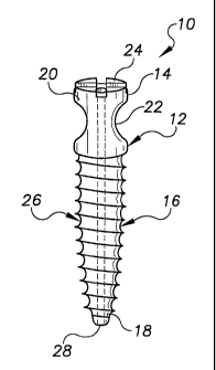
now be disclosed in detail. Referring now to Figure 1 and Figure 2, there are
shown two
elevated perspective views of a bone screw according to the present invention
along the
proximal to distal axis, where Figure 2 shows the bone screw in Figure 1
rotated ninety
degrees around its proximal to distal axis. Referring now to Figure 3, there
is shown a
cutaway, elevated perspective view of the bone screw shown in Figure 2 along
the proximal
to distal axis. In one embodiment, the bone screw is made of a biocompatible
material such
as titanium or stainless steel. In one embodiment, the bone screw has a
proximal length to
distal length of between about 40 mm and about 60 mm. In a particularly
preferred
embodiment, the bone screw has a proximal length to distal length of about 50
mm.
As can be seen, the bone screw 10 comprises a proximal portion 12 with a
proximal
end 14 and a distal portion 16 with a distal end 18. The proximal portion 12
comprises a
head 20 and a portal 22. In a preferred embodiment, the head 20 comprises a
proximal
portion 24 configured to mate with the tip of a screwdriver (not shown). In a
particularly
preferred embodiment, the top 24 portion comprises a slot. In another
particularly preferred
embodiment, as shown, the proximal portion 24 is configured to mate with a
Phillips head
screwdriver. Other indentation configurations are also suitable, as will be
understood by
those with skill in the art with reference to this disclosure. For example, as
shown in Figure
4, the proximal portion 24 can comprise a raised platform 25 having a
plurality of
substantially straight sides, such as a hexagonal platform, configured to mate
with a
corresponding depression in the distal end of a screwdriver.
The portal 22 of the bone screw extends through the head 20 and is preferably
between about 4 mm and about 8 mm in minimum diameter in the proximal to
distal plane
and is preferably either oval or round in shape when viewed perpendicular to
the proximal to
distal plane. In. a particularly preferred embodiment, the portal 22 is about
6 mm in
minimum diameter in the proximal to distal plane.
The distal portion 16 of the bone screw.10 comprises threads 26 and a sharp
tip 28.
Additionally, the bone screw 10 comprises a central lumen 30 extending
coaxially completely
through the bone screw 10 from the proximal end 14 to the distal end 18 and
configured to
CA 02414168 2002-12-19
WO 02/00126 PCT/US00/34855
8
receive a guidewire used in the present method. Preferably, but not
essentially, the bone
screw comprises one or more than one perforation 32. The one or more than one
perforation
can extend into the central lumen 30, or can extend completely laterally
through the distal
portion 16. Additionally, the one or more than one perforation 32 can be
aligned axially, as
shown, or can be staggered axially, not shown. The one or more than one
perforation 32
permits bone to grow into the bone screw 10 and help stabilize the bone screw
10 within the
bone. Additionally, bone matrix material such as a hydroxyapatite preparation
can be
injected into the central lumen 30 and through the one or more than one
perforation 32 to
promote bone ingrowth.
The system of the present invention further comprises a screwdriver configured
to
apply torque to the bone screw. Referring now to Figure 5 and to Figure 6,
there are shown
elevated perspective views of two embodiments of a screwdriver 40 according to
the present
invention along the proximal to distal axis. As can be seen, the screwdriver
comprises a
proximal portion 42 comprising a proximal end 44 and a distal portion 46
comprising a distal
end 48. The proximal portion 42 comprises handles 50 configured to permit
grasping of the
screwdriver and to permit the application of torque to a bone screw. Various
configurations
of the proximal end are possible, as will be understood by those with skill in
the art with
reference to this disclosure. Preferably, the handles 50 should be able to
rotate around their
axis independently of each other.
The distal portion 46 of the screwdriver 40 comprises a shaft 52 having a tip
54
configured to interface with the proximal portion of a bone screw according to
the present
invention. Therefore, the configuration of the distal end 48 will depend upon
the
configuration of the head of the bone screws being used in conjunction with
the screwdriver
40. The screwdriver 40 further comprises a central lumen 55 extending
coaxially completely
through the screwdriver 40 from the proximal end 44 to the distal end 48 and
configured to
receive a guidewire used in the present method.
The system of the present invention can optionally comprise a guidewire
directing
device. Referring now to Figure 7, there is shown an elevated perspective view
of a
guidewire directing device 60 according to the present inventioia along the
proximal to distal
axis. As can be seen, the guidewire directing device 60 comprises a proximal
portion 62
with a proximal end 64 and a distal portion 66 with a distal end 68. The
proximal portion 62
comprises a handle 70. Preferably, the handle 70 is configured to assist in
grasping and
CA 02414168 2002-12-19
WO 02/00126 PCT/US00/34855
9
manipulating the handle 70. The distal portion 66 comprises a shaft 72 having
a fork-tipped
end 68. The guidewire directing device 60 is used to percutaneously alter the
direction of an
advancing guidewire by engaging the guidewire in the fork-tipped end 68,
rotating the handle
70 and advancing and withdrawing the handle 70 along the proximal to distal
axis, thereby
altering the direction of the advancing guidewire.
The system of the present invention further comprises an inflatable connection
rod.
Referring now to Figure 8, there is shown an elevated perspective view of an
inflatable
connection rod according to the present invention along the proximal to distal
axis in the
uninflated state. The rod 80 comprises a proximal end 82, a distal end 84 and
a compliant,
inflatable balloon 86 between the proximal end 82 and the distal end 84. The
proximal end
82 comprises a self-sealing valve 88. The distal end 84 comprises a tip 90,
preferably
comprising a biocompatible metal. The balloon comprises any suitable material,
but
preferably comprises a biocompatible-braided polymer, such as for example a
material
selected from the group consisting of nylon, polyethylene and polyurethane.
Further
preferably, the balloon 86 comprises thin, reinforcing metallic wires 92
running the entire
proximal to distal length of the lumen of the balloon 86, but separate from
the balloon wall.
The wires 92 increase the tensile strength of the balloon 86 when inflated, as
will be
understood by those with skill in the art with reference to this disclosure.
The wires 92
preferably comprise titanium or nitinol, but can comprise another suitable
material as will be
understood by those with skill in the art with reference to this disclosure.
The system of the present invention can optionally comprise a directing sheath
that
assists in aligning a structure such as a guidewire or inflatable connection
rod to pass through
the portals in the bone screws according to the present invention. Referring
now to Figure 9,
there is shown a top perspective view of a directing sheath according to the
present invention
along the proximal to distal axis. As can be seen, the directing sheath 100
comprises a
proximal portion 102 with a proximal end (not shown), a distal portion 104
with a distal end
(not shown), and a central portion 106 between the proximal portion 102 and
the distal
portion 106. The central portion 106 comprises at least two openings 108 sized
substantially
the same as the portal on a bone screw according to the present invention, or
slightly larger.
The directing sheath 100 has a lumen 110 extending through its entire length
from the
proximal end to the distal end. The lumen 110 is of sufficient internal
diameter to allow a
structure such as a guidewire or inflatable connection rod to pass through the
directing sheath
CA 02414168 2002-12-19
WO 02/00126 PCT/US00/34855
between the proximal end and distal end. The directing sheath 100 is scored
112 along its
longitudinal axis, on either one line or preferably on two opposing lines, to
allow the
directing sheath 100 to be split into two separate halves by peeling the
directing sheath 100
apart at either its proximal end or its distal end or both along the scoring
112. The scoring
5 112 can be partially or completely through the sheath wall as will be
understood by those
with skill in the art with reference to this disclosure.
The directing sheath 100 preferably comprises a biocompatible polymer, though
other
mate'rials are suitable, as will be understood by those with skill in the art
with reference to
this disclosure. The directing sheath 100 further preferably comprises a
radiopaque filament
10 114 passing around each opening in the central portion, and more preferably
running the
entire longitudinal length of the directing sheath from the proximal end to
the distal end.
This filament 114 aids in localizing the directing sheath 100 once it has been
percutaneously
placed.
The method of the present invention involves percutaneously inserting one or
more
fusion devices into two or more than two adjacent vertebrae, either
unilaterally or, preferably
bilaterally, where a portion or all of at least one of the vertebrae is
unstable, separated or
displaced. The fusion devices reposition or fix the displaced vertebra or
portion of the
displaced vertebra to a position within the vertebral column which is more
stable or which
causes less morbidity.
Referring now to Figure 10 through Figure 19, there are shown a series of
drawings
depicting various stages of the method of repositioning and fixing a displaced
vertebra or
portion of a displaced, vertebra, unilaterally, according to the present
invention. Figures 9-18
show partial cutaway, perspective, midline sagittal views of a portion of a
vertebral column
undergoing the method of the present invention.
The method will now be disclosed and depicted with reference to only two
vertebrae,
one which is either unstable, separated or displaced and one of which is
neither unstable,
separated nor displaced. However, the method can also be applied to three or
more vertebrae
simultaneously, as will be understood by those with skill in the art with
reference to this
disclosure. Additionally, the method can be used to stabilize the L5
vertebrae, using the
cranial-ward portion of the sacrum as the "vertebrae" with which L5 is
anchored. Further,
though the method is disclosed and depicted as applied on the left side of the
vertebral
column,.the method can also be applied on the right side of the vertebral
column or,
CA 02414168 2002-12-19
WO 02/00126 PCT/US00/34855
11
preferably, can be applied on both sides of the vertebral column
simultaneously, as will be
understood by those with skill in the art with reference to this disclosure.
First, the present method comprises identifying a patient who is a suitable
candidate
for undergoing the method. A suitable candidate has one or more unstable
vertebrae, one or
more portions of one or more vertebrae at least partly separated from the
remainder of the
vertebrae with potential or complete separation, or has one or more vertebrae
or a portion of
one or more vertebrae displaced from its normal position relative to the
vertebral column, or
has one or more portions of one or more vertebrae at least partly separated
from the
remainder of the vertebrae and displaced from its normal position relative to
the vertebral
column. Further, the suitable candidate will preferably have either pain, loss
of function or
real or potential instability which is likely due to the separation or
displacement, or
separation and displacement. If only a portion of the vertebra is unstable,
separated or
displaced, the portion of the vertebra that is unstable, separated or
displaced will generally
include at least part of the vertebral body and adjoining pedicle. However,
other unstable,
separated or displaced portions of a vertebra can be repositioned or fixed
using the present
method, as will be understood by those with skill in the art with reference to
this disclosure.
For example, a suitable patient can have a disease or condition such as
spondylosis,
spondylolisthesis, vertebral instability, spinal stenosis and degenerated,
herniated, or
degenerated and herniated intervertebral discs, though actual indications
require the expertise
of one of skill in the art as will be understood by those with skill in the
art with reference to
this disclosure.
Next, the present method comprises making a stab incision in the patient's
skin
overlying the patient's vertebral column at or near the level of the vertebrae
or portion of
vertebrae to be repositioned or fixed. In a preferred embodiment, the incision
is made at or
near the level of the pedicle of the vertebrae or portion of vertebrae to be
repositioned or
fixed. The pedicle level is located preferably by identifying the pedicle
shadow using
fluoroscopy. In a preferred embodiment, the stab incision is made using a #11
scalpel blade.
Then, as shown in Figure 10, an 11-gauge bone biopsy needle or its equivalent
is
placed through the stab incision to create a tract to the posterior periosteal
surface of the
vertebrae 200 which is to be stabilized, repositioned or fixed. Next, the
biopsy needle 202 is
used to make a small incision in the periosteum and into the cortex of the
vertebrae.
Then, as shown in Figure 11, a rigid, needle-tipped guidewire 204 having a
needle
CA 02414168 2002-12-19
WO 02/00126 PCT/US00/34855
12
diameter of 13 or 15-gauge is inserted through the biopsy needle 202 into the
tract, through
the periosteal incision and into the cortex of the bone, and the guidewire 204
is advanced into
the anterior aspect of the vertebral body 200 or into another suitable portion
of the vertebrae
200, as will be understood by those with skill in the art with reference to
this disclosure.
Insertion of the guidewire 204 is preferably accomplished using fluoroscopy.
This process
creates a continuous tract from the skin surface into the anterior vertebral
body or suitable
portion of the vertebrae 200.
The biopsy needle 202 is then removed and the tract from the skin surface to
the
nicked periosteal surface is enlarged by using a high-pressure fascial dilator
balloon (not
shown) over the needle-tipped guidewire. Then, the balloon is removed and a
working
sheath 206 is introduced into the dilated tract. Alternately, a metallic
sheath with a central
dilator is advanced over the guidewire from the skin surface to the periosteal
surface.
Next, as shown in Figure 12, a bone screw 208 according to the present
invention is
introduced into the working sheath 206 over the guidewire 204 by introducing
the central
lumen of the bone screw 208 over the proximal end of the guidewire 204. A
screwdriver
210 according to the present invention is similarly introduced over the
guidewire 204. The
bone screw 208 and distal portion of the screwdriver 210 are then advanced
distally through
the sheath 206 and the tract to the periosteal surface of the vertebra1200
until the proximal
portion of the bone screw 208 is engaged by the tip of the screwdriver 210.
Torque is
applied to the bone screw 208 using the screwdriver 210 and the bone screw 208
is advanced
until the distal portion of the bone screw 208 enters the anterior vertebral
body or other
suitable portion of the vertebra 200, while the portal of the bone screw 208
is exterior and
dorsal to the vertebra 200 and the portal is open parallel to the long axis of
the vertebral
colunin. Then, as shown in Figure 13, the guidewire 204, sheath 206 and
screwdriver 210
are removed after satisfactory placement of the bone screw 208 has been
obtained and
confirmed by fluoroscopy. Additionally, bone matrix material such as a
hydroxyapatite
preparation can be injected into the central lumen of the bone screw and
through the one or
more than one perforation, if present, to promote bone ingrowth.
The stages disclosed above are repeated for at least one additional vertebra
212 until
each vertebra that is to be repositioned or fixed has a bone screw 208
applied, and
additionally for at least one vertebra which is neither unstable, separated
nor displaced and
which lies adjacent the cranial-most or caudal-most vertebra that is being
repositioned or
CA 02414168 2002-12-19
WO 02/00126 PCT/US00/34855
13
fixed. The bone screw 208 placed into the vertebra 212 which is neither
unstable, separated
nor displaced is used as the anchor to reposition or fix each vertebra 200
which is unstable,
separated or displaced as follows. As will be understood by those with skill
in the art with
reference to this disclosure, the bone screws can be placed into the vertebrae
in a different
order to that described above.
After a bone screw is positioned in each vertebra, the portals are connected
using an
inflatable connection rod according to the present invention where the rod is
inserted between
the portals of the bone screws and inflated to create a rigid structure with
the bone screws,
thereby repositioning and fixing the one or more than one previously unstable,
separated or
displaced vertebra, or one or more previously unstable, separated or displaced
portions of
one or more vertebrae with the vertebra that is neither unstable, separated
nor displaced.
Connection of the bone screws with the inflatable rod is accomplished as
follows.
Referring now to Figure 14 and Figure 15, a hollow needle 214, such as a 16
gauge
or 18 gauge needle, is inserted percutaneously and fluoroscopically advanced
to the portal of
one of the bone screws 208. While the hollow needle is shown engaging the bone
screw 208
in the cranial-ward vertebrae 212, the hollow needle can engage the bone screw
208 in the
caudal-ward vertebrae 200 first, as will be understood by those with skill in
the art with
reference to this disclosure. Figure 15 is a detailed view of Figure 14.
Then, as shown in Figure 16, a needle-tipped, semi-rigid guidewire 216 is
introduced
through the lumen of the hollow needle 214 and into the portal of the bone
screw 208 in the
cranial-ward vertebrae 212. The hollow needle 214 preferably has a Tuohy
needle tip which
causes the guidewire 216 to exit the hollow needle 214 perpendicular to the
distal-proximal
axis of the bone screw 208, thereby orienting the guidewire 216 perpendicular
to the aligned
portals in each bone screw 208 and parallel to the long axis of the vertebral
column.
Alternately, the hollow needle 214 can have an angled-tip modified Ross needle
or other
suitable structure as will be understood by those with skill in the art with
reference to this
disclosure.
In a preferred embodiment, as fiirther shown in Figure 16, a guidewire 218
directing
device according to the present invention is inserted percutaneously between
the portals of
each bone screw 208 and the fork-tipped end is used to direct the advancing
guidewire 216
through the second bone screw portal, and to reorient the guidewire 216 after
the guidewire
216 has passed through the portal on the bone screw 208 of the caudal-ward
vertebrae 212.
CA 02414168 2002-12-19
WO 02/00126 PCT/US00/34855
14
In another preferred embodiment, as further shown in Figure 16, a guidewire
capture
device 219, such as a snare or grasping forceps, is inserted percutaneously,
caudal to the
portal of the bone screw in the caudal-ward vertebrae. The capture device 219
engages the
guidewire after it passes through the portal of the bone screw in the caudal-
ward vertebrae
and allows the distal end of the guidewire to be pulled through the skin
posteriorly to obtain
control of both the proximal and distal ends of the guidewire.
In another preferred embodiment, the needle-tipped, semi-rigid guidewire 216
comprises an outer helical, flat wire sheath and an inner retractable sharp
tip stylet. Once the
needle-tipped, semi-rigid guidewire is placed, the stylet can be removed to
allow for easier
capture by the capture device with less trauma to the surrounding tissue.
Then, as shown in Figure 17, the entire guidewire tract is dilated using a
high
pressure balloon and a flexible introducer sheath 220 is passed over the
guidewire 216 along
the entire guidewire tract exiting the caudal-ward stab incision. The
guidewire 216 is
removed after the introducer sheath 220 is placed.
Next, as shown in Figure 18, an uninflated, inflatable connection rod 222
according
to the present invention which is attached to a proximal pushing catheter 224
is advanced
through the introducer sheath 220 until the inflatable connection rod 222
advances between
the two portals and the proximal end of the inflatable connection rod 222 lies
cranial to the
portal on the bone screw 208 in the cranial-ward vertebrae 212 while the
distal end of the
inflatable connection rod 222 lies caudal to the portal on the bone screw 208
in the caudal-
ward vertebrae 200. The sheath 220 is removed and the placement is confirmed
by
fluoroscopy.
Then, as shown in Figure 19, the balloon of the inflatable connection rod 222
is
inflated with a rapid setting, liquid polymer, or its equivalent, and the
polymer is allowed to
set fixing each bone screw 208 in relation to each other and repositioning and
fixing the
vertebra 200 or portion of the vertebra that was unstable, separated or
displaced. In a
preferred embodiment, the liquid polymer is polymethylmethacrylate. The rapid
setting,
liquid polymer can comprise a light activated polymer and the method can
comprise applying
light to promote setting of the polymer. The inflated balloon of the
inflatable connection rod
222 expands radially beyond the diameter of the portals of each bone screw 208
which helps
fix the bone screws 208 in relation to each other.
Finally, as shown in Figure 20, the pushing catheter 224 is detached from the
CA 02414168 2002-12-19
WO 02/00126 PCT/US00/34855
inflatable connection rod 222 by pulling on the pushing catheter 224 while
holding the
inflatable connection rod 222 to disengage the inflatable connection rod 222
from the pushing
catheter 224 and the pushing catheter 224 is removed. The inflatable
connection rod 222
comprises a self-sealing valve which prevents the polymer from leaking once
the pushing
5 catheter is detached. The vertebra is then fixed unilaterally. The method
can be repeated on
the opposite side of the spinous processes of the patient's vertebrae column,
thereby
repositioning or fixing the one or more unstable, separated or displaced
vertebrae or the one
or more portions of one or more vertebrae bilaterally. The stab incisions are
closed or sealed
as necessary and routine postoperative care administered.
10 Referring now to Figure 21, there is shown a posterior perspective view of
a portion
of a vertebral column which has had some vertebrae repositioned and fixed
bilaterally
according to a preferred embodiment of the method of the present invention.
When bilateral
fixation is accomplished, it is preferred to place all bone screws before
connecting the portals
with inflatable connection rods.
15 In another embodiment of the present method, a directing sheath 226
according to the
present invention is advanced over a guidewire until the openings in the
directing sheath 226
overlie the position in each vertebra which will receive a bone screw 208. The
bone screws
208 are then placed as disclosed in this disclosure, but through the openings
in the directing
sheath 226, which aligns the lumen in the directing sheath with the portals of
the bone screw
208. Then (not shown), a guidewire is then inserted into the lumen of the
directing sheath at
the proximal end of the directing sheath and advanced until the guidewire
passes through
each portal of the bone screws and exits the body through the lumen of the
directing sheath at
the distal end. The directing sheath is then removed by peeling the sheath
apart along the
scored lines and pulling the two halves out from the body. The guidewire that
was in the
lumen of the directing sheath remains in place to guide the placement of the
uninflated,
inflatable connection rod. Alternately, the uninflated, inflatable connection
rod can be
inserted directly into the lumen of the directing sheath at the proximal end
and advanced until
the uninflated, inflatable connection rod is properly positioned between the
portals of the
bone screws. Referring now to Figure 22 through Figure 24, there are shown
posterior
perspective views of a portion of a vertebral column undergoing the method of
the present
invention using a directing sheath according to the present invention, showing
the bone
screws placed through the openings of the directing sheath. As can be seen in
Figure 22, the
CA 02414168 2002-12-19
WO 02/00126 PCT/US00/34855
16
directing sheath 300 is positioned adjacent the vertebral column 302 according
to the present
invention. Next as can be seen in Figure 23, guidewires 304 are used to place
bone screws
306 through openings 308 in the directing sheath 300. Finally as can be seen
in Figure 24,
the directing sheath 300 is removed by the directing sheath 300 into two
separate halves.
In a preferred embodiment, there is provided a kit for performing the method
of the
present invention. The kit comprises a plurality of bone screws according to
the present
invention. The kit can also comprise other components of the system of the
present
invention, such as a guidewire directing device, an inflatable connection rod
and a directing
sheath. In another preferred embodiment, the kit also comprises a screwdriver
according to
the present invention.
Although the present invention has been discussed in considerable detail with
reference to certain preferred embodiments, other embodiments are possible.
Therefore, the
scope of the appended claims should not be limited to the description of
preferred
embodiments contained in this disclosure.