Note: Descriptions are shown in the official language in which they were submitted.
CA 02416286 2003-O1-14
WO 02/07812 PCT/USO1/22972
SYRINGE PLUNGER LOCKING MECHANISM
FIELD OF THE INVENTION
The present invention relates to angiography and more specifically, injectors
used to
inject a medical fluid such as radiographic material into living organisms.
BACKGROUND OF THE INVENTION
One of the major systems in the human body is the circulatory system. The
major
components of the circulatory system are the heart, blood vessels, and the
blood, all of which
are vital to the transportation of materials between the external environment
and the different
cells and tissues of the human body.
The blood vessels are the network of passageways through which the blood
travels in
the human body. Specifically, arteries carry the oxygenated blood away from
the left
ventricle of the heart. These arteries are aligned in progressively decreasing
diameter and
pressure capability from the aorta, which carries the blood immediately out of
the heart to
other major arteries, to smaller arteries, to arterioles, and finally to tiny
capillaries, which feed
the cells and tissues of the human body. Similarly, veins carry the oxygen-
depleted blood
back to the right atrium of the heart using a progressively increasing
diameter network of
venules and veins.
If the heart chambers, valves, arteries, veins or other capillaries connected
thereto are
either abnormal (such as from a birth defect), restricted (such as from
atherosclerotic plaque
buildup), or deteriorating (such as from aneurysm formation), then a physician
may need to
examine the heart and connected network of vessels. The physician may also
need to correct
any problems encountered during the examination with a catheter or similar
medical
instrument.
Angiography is a procedure used in the detection and treatment of
abnormalities or
restrictions in blood vessels. During angiography, a radiographic image of a
vascular
structure is obtained by injecting radiographic contrast material through a
catheter into a vein
or artery. The vascular structures fluidly connected with the vein or artery
in which the
injection occurred are filled with contrast material. X-rays are passed
through the region of
the body in which the contrast, material was injected. The X-rays are absorbed
by the
contrast material, causing a radiographic outline or image of the blood vessel
containing the
contrast material. The x-ray images of the blood vessels filled with contrast
material are
usually recorded onto film or videotape and are displayed on a fluoroscope
monitor.
Angiography gives the doctor an image of the vascular structures in question.
This
image may be used solely for diagnostic purposes, or the image may be used
during a
CA 02416286 2003-O1-14
WO 02/07812 PCT/USO1/22972
procedure such as angioplasty where a balloon is inserted into the vascular
system and
inflated to open a stenosis caused by atherosclerotic plaque buildup.
Currently, during angiography, after a physician places a catheter into a vein
or artery
(by direct insertion into the vessel or through a skin puncture site), the
angiographic catheter
is connected to either a manual or an automatic contrast injection mechanism.
A simple manual contrast injection mechanism typically has a syringe and a
catheter
connection. The syringe includes a chamber with a plunger therein.
Radiographic contrast
material is suctioned into the chamber. Any air is removed by actuating the
plunger while the
catheter connection is facing upward so that any air, which floats on the
radiographic contrast
material, is ejected from the chamber into the air. The catheter connection is
then attached to
a catheter that is positioned in a vein or artery in the patient.
The plunger is manually actuated to eject the radiographic contrast material
from the
chamber, through the catheter, and into a vein or artery. The user of the
manual contrast
injection mechanism may adjust the rate and volume of injection by altering
the manual
actuation force applied to the plunger.
Often, more than one type of fluid injection is desired, such as a saline
flush followed
by the radiographic contrast material. One of the most common manual injection
mechanisms
used today includes a valve mechanism which controls which of the fluids will
flow into the
valuing mechanism and out to the catheter within the patient. The valve
mechanism contains
a plurality of manual valves that the user operates manually to open and close
that particular
fluid channel. When the user suctions or injects contrast fluid into the
chamber, the fluid is
pulled from the valve mechanism via the open valves. By changing the valve
positions,
another fluid may be injected.
These manual injection mechanisms are typically hand actuated. This allows
user
control over the quantity and pressure of the injection. However, all of the
manual systems
are only capable of injecting the radiographic contrast material at maximum
pressure that can
be applied by the human hand (i.e., 150 p.s.i). Also, the quantity of
radiographic contrast
material is typically limited to a maximum of about l2cc. Finally, there are
no safety limits
on these manual contrast injection mechanisms which act to restrict or stop
injections that are
outside of reasonable parameters (such as rate or pressure) and no active
sensors to detect air
bubbles or other hazards.
Currently used motorized injection devices consist of a syringe connected to a
linear
actuator. The linear actuator is connected to a motor, which is controlled
electronically. The
operator enters into the electronic control a fixed volume of contrast
material to be injected at
2
CA 02416286 2003-O1-14
WO 02/07812 PCT/USO1/22972
a fixed rate of injection. The fixed rate of injection consists of a specified
initial rate of flow
increase and a final rate of injection until the entire volume of contrast
material is injected.
There is no interactive control between the operator and machine, except to
start or stop the
injection. Any change in flow rate must occur by stopping the machine and
resetting the
parameters.
The lack of ability to vary the rate of injection during the injection results
in
suboptimal quality of angiographic studies. This is because the optimal flow
rate of injections
varies considerably between patients. In the cardiovascular system, the rate
and volume of
contrast injection is dependent on the size of and blood flow rate within the
chamber or blood
vessel being injected. In many or most cases, these parameters are not known
precisely.
Moreover, the optimal rate of injection can change rapidly, as the patient's
condition changes
in response to drugs, illness, or normal physiology. Consequently, the initial
injection of
contrast material may be insufficient in flow rate to outline the structure on
x-ray imaging,
necessitating another injection. Conversely, an excessive flow rate might
injure the chamber
or blood vessel being injected, cause the catheter to be displaced (from the
jet of contrast
material exiting the catheter tip), or lead to toxic effects from contrast
overdose (such as
abnormal heart rhythm).
Furthermore, the linear actuator is usually connected to the plunger by a
"snap fit"
arrangement wherein automatic engagement and disengagement of the plunger with
the linear
actuator is desirable to prevent contamination of the pumping chamber of the
syringe and to
simplify the operation of the injection system. In some situations, it is
desirable to damage or
destroy the connection portion of the plunger to prevent reuse of the syringe.
As a result,
particulates may remain in the connection area and cause problems during
subsequent
interconnections.
Another problem often encountered when providing a syringe plunger arrangement
which automatically engages and disengages with the linear actuator is that
the force
necessary for engagement is too high, while the force necessary for
disengagement is too low.
With such an arrangement, it may be difficult to maintain the plunger in a
fixed position
relative to the pumping chamber because the plunger may be driven forward
during the
engagement procedure, and it may be difficult to maintain the plunger in an
engaged position
with the linear actuator when the linear actuator is retracted.
At present, the operator can choose between two systems for injecting contrast
material: a manual injection system which allows for a variable, operator
interactive flow rate
3
CA 02416286 2003-O1-14
WO 02/07812 PCT/USO1/22972
of limited flow rate and a preprogrammed motorized system without operator
interactive
feedback (other than the operator can startlstop the procedure).
SUMMARY OF THE INVENTION
The present invention provides a number of devices and methods fox releasably
connecting a syringe plunger to a drive member of an angiographic injector of
a type having a
syringe holder. A syringe includes a syringe body having a distal end and a
proximal end,
and the syringe body defines a pumping chamber. The syringe plunger is located
in the
pumping chamber and is adapted for reciprocal motion. An actuating shaft is
coupled to the
syringe plunger and is movable through the syringe body to control movement.
In one embodiment of the present invention, a syringe plunger includes a
capture
member projecting outwardly in a proximal direction, while an actuating shaft
includes a
circumferential groove. A latch is disposed on an inner surface of the capture
member, and a
finger extends radially outwardly from the capture member. During the
connecting
procedure, the actuating shaft is driven forward to contact the syringe
plunger, and the capture
member flexes radially outwardly and engages with the circumferential groove
to form a
releasable connection. A release actuator is disposed proximal to the syringe
body. The
release actuator may be a plate defining an aperture for allowing manipulation
of the syringe
plunger. During the disconnecting procedure, the syringe plunger is retracted
such that the
finger abuts against an inner surface of the plate and causes the capture
member to flex
radially outwardly. The latch disengages with the circumferential groove and
the syringe is
disconnected from the actuating shaft. In the exemplary embodiment, the
capture members
are designed to permanently deform during the disconnecting procedure to
ensure that the
syringe is disposed of after use with one patient and not accidentally re-used
on a new,
different patient.
In another embodiment of the present invention, a syringe plunger includes a
circumferential groove, and an actuating shaft includes a pivot member for
capturing the
syringe plunger. The pivot member is disposed at a distal portion of an
actuating shaft. A
distal portion of the pivot member includes a latch projecting outwardly from
an inner surface
thereof, and a proximal portion of the pivot member includes a ramped lug
projecting
outwardly from an outer surface thereof. A release actuator may be a plate
located proximal
to the syringe body defining an aperture for allowing manipulation of the
syringe plunger.
During the connecting procedure, the actuating shaft is driven forward to
contact the syringe
plunger, the distal portion of the pivot member projects radially outwardly,
and the latch
engages with the circumferential groove to form a releasable connection.
During the
4
CA 02416286 2003-O1-14
WO 02/07812 PCT/USO1/22972
disconnecting procedure, the syringe plunger is retracted such that the ramped
lug slidingly
engages with a sidewall of the aperture. The proximal end of the pivot member
is driven
radially inwardly and the distal end of the pivot member is projected radially
outwardly such
that the latch disengages with the circumferential groove.
In another embodiment, a syringe plunger is magnetically connected to an
actuating
shaft. The syringe plunger includes a first insert comprising a ferrous metal,
permanent
magnet, or electromagnet, while the actuating shaft includes a second insert
comprising a
ferrous metal, permanent magnet, or electromagnet. Any combination of ferrous
metal,
permanent magnet or electromagnet may be used when configuring the first and
second insert
as long as a permanent magnet or electromagnet is included in one of the
inserts. During the
connecting procedure, the actuating shaft is driven forward to contact the
syringe plunger, and
the syringe plunger remains magnetically attached to the actuating shaft. One
of the benefits
of utilizing a magnetic syringe plunger arrangement is that a "zero"
engagement force is
required. As described in the previous embodiment, a release actuator is
disposed proximal to
the syringe body. The release actuator may be a plate located proximal to the
syringe body,
and the plate defines an aperture for allowing manipulation of the .syringe
plunger. The
syringe plunger further includes a base having an outer diameter larger than
the aperture of
the plate. During the disconnecting procedure, the syringe plunger is
retracted until the base
abuts against an inner surface of the plate. Upon further retraction, the
plunger disengages
from the actuating shaft.
In another embodiment, a syringe plunger is attached to an actuator having a
split
collet actuator head. The syringe plunger includes a plunger support member
having a
receiving aperture formed therein, and at least one retaining member located
thereon. The
split collet actuator head comprises an alignment shaft positioned between a
first collet
member and a second collet member. The individual collet members have flange
portions
formed thereon. Biasing members are positioned between the first and second
collet members
and bias the collet members outwardly. During the connecting procedure, the
actuating shaft
is driven forward to contact the syringe plunger, wherein the first and second
collet members
are forced inwardly. Thereafter, the at least one retaining member engages the
individual
flange members formed on the collet members and the biasing members force the
first and
second collet members outwardly. Thereafter, the syringe plunger is coupled to
the actuating
shaft.
' To detach the syringe plunger from the actuator the actuator is retracted to
a position
proximal the rear plate of the syringe holder assembly. During the retraction
procedure, the
5
CA 02416286 2003-O1-14
WO 02/07812 PCT/USO1/22972
detachment members located on the first and second collet members are caused
to engage the
rear plate of the syringe holder assembly. As a result, the first and second
collet members are
forced inwardly and the at least one retaining member disengages the flange
portion. Upon
further retraction, the plunger disengages from the actuating shaft.
S Other objects, features, and advantages of the present invention will become
apparent
from a consideration of the following detailed description and from the
accompanying
descriptions.
BRIEF DESCRIPTION OF THE DRAWINGS
FIG. 1 is a perspective view illustrating a preferred embodiment of the
angiographic
injector system in accordance with the present invention.
FIGS. 2A-ZG are diagrams illustrating operations of the system of FIG. 1.
FIG. 3 is an electrical block diagram of the control system of the injector
system of
FIG. 1.
FIG. 4 illustrates front panel controls and displays of a preferred embodiment
of the
injector system in accordance with the present invention.
FIGS. 5A and SB are side and partial top perspective views of the remote
control of
the system of FIG. 1.
FIG. 6 is a perspective view of a foot operated remote control.
FIGS. 7A-7D illustrate the operation of the inlet check valve and manifold
during
contrast fill, air purge, and patient inject operations.
FIGS. 8A-8C illustrate operation of the inlet check valve in greater detail.
FIG. 9 shows a conventional syringe body adapted for dual port.
FIG. 10 is a perspective view of an adapter insert used in the dual port
syringe of
FIG. 9.
FIGS. 11A-11B are top and side views of the adapter insert of FIG. 10.
FIG. 12 is a perspective view of one embodiment of a syringe usable in the
angiographic injector system, according to the present invention.
FIG. 13 is a bottom plan view of the syringe depicted in FIG. 12.
FIG. 14 is a top plan view of the syringe depicted in FIG. 12.
FIG. 15 is a side elevational view of the syringe depicted in FIG. 12.
FIG. 16 is a front side elevational view of the syringe depicted in FIG. 12.
FIG. 17 is a rear side elevational view of the syringe depicted in FIG. 12,
and without
the plunger therein.
6
CA 02416286 2003-O1-14
WO 02/07812 PCT/USO1/22972
FIG. 18 is a perspective view of one embodiment of a syringe holder
arrangement,
according to the present invention.
FIG. 19 is a perspective view of the syringe holder arrangement depicted in
FIG. 18,
and holding a syringe and a bottle of fluid.
FIG. 20 is an exploded, perspective view of a subassembly of the syringe
holder
arrangement depicted in FIG. 18.
FIG. 21 is a rear side elevational view of the syringe depicted in FIG. 12,
and
analogous to FIG. 17, but with the plunger therein.
FIG. 22 is a schematic, side elevational view of an air column detector and
tubing, in
accordance with the present invention.
FIG. 23 is a side plan view of a drive piston and an actuator head for the
injector
systems of FIGS. 1 and 18 in accordance with the present invention, wherein
the drive piston
is coupled to the actuator head by flexible capture members.
FIG. 24A is a perspective view of a syringe plunger for the drive piston and
actuator
head of FIG. 23.
FIG. 24B is a cross sectional side view of the syringe plunger shown in FIG.
24A.
FIG. 24C is a plan bottom view of the syringe plunger shown in FIG. 24A.
FIG. 25 is a plan side view of another embodiment of a syringe plunger
arrangement
for the injector systems of FIGS. 1 and 18 in accordance with the present
invention, wherein a
syringe plunger is magnetically coupled to an actuator.
FIG. 26 is a plan side view of another embodiment of a syringe plunger
arrangement
for the injector systems of FIGS. 1 and 18 in accordance with the present
invention, wherein a
syringe plunger is coupled to an actuator by pivoting members.
FIGS. 27A-27G illustrate another embodiment of a syringe plunger arrangement
for
the injector systems in accordance with the present invention, wherein a
syringe plunger is
coupled to an actuator by a tongue and groove system.
FIG. 28 is a plan side view of another embodiment of a syringe plunger
arrangement
for the injector systems of FIGS. 1 and 18 in accordance with the present
invention, wherein a
syringe plunger is coupled to an actuator by pivoting members.
FIGS. 29A-29B illustrate another embodiment of a syringe plunger arrangement
for
the injector systems of FIGS. 1 and 18 in accordance with the present
invention, wherein a
retainer ring maintains a syringe plunger in a fixed position during
engagement.
7
CA 02416286 2003-O1-14
WO 02/07812 PCT/USO1/22972
FIG. 30 illustrates a still further embodiment of a syringe plunger
arrangement for the
injector systems of FIGS. 1 and 18 in accordance with the present invention,
wherein a
syringe plunger is coupled to an actuator by a "Christmas tree" type fastener.
FIG. 31A-31M illustrate yet another embodiment of a syringe plunger
arrangement for
injector systems in accordance with the present invention, wherein the syringe
plunger is
coupled to an actuator having a split collet design.
DETAILED DESCRIPTION OF THE PREFERRED EMBODIMENTS
As used herein to describe the angiographic injection system of the present
invention,
the terms "axial" or "axially" refer generally to an axis A around which the
system is formed.
The terms "proximal" or "rearward" refer generally to an axial direction
toward the end of
injector housing opposite the main console. The terms "distal" or "forward"
refer generally to
an axial direction towards a syringe tip. The term "radial" refers generally
to a direction
normal to axis A.
EXEMPLARY ANGIOGRAPHIC INJECTOR SYSTEM
FIG. 1 shows angiographic injector system 10 for injecting radiographic
contrast
material into a blood vessel under interactive physician control. System 10
includes main
console 12, hand held remote control 14, syringe holder 16, syringe body 18,
syringe plunger
20, radiographic material reservoir (bottle) 22, one-way valve 24, manifold
26, high pressure
tube 28, catheter 30, patient medication port 32, three-way stop-cock 34, T-
connector 36,
pressure transducer 38, stop-cock 40, tubing 42, peristaltic pump 44, saline
check valve 46,
waste check valve 48, saline bag 50, waste bag 52, and bag support rack 54.
Console 12 houses the electrical controls for system 10, together with the
motors
which drive piston 20 and peristaltic pump 44. On the front surface of console
12, user
interface 54 provides control switches 56 and display 58 through which the
user may enter
control settings and monitor the operational state of system 10.
Remote control 14 is connected to console 12 by cable 60 (although in other
embodiments remote control 14 may be connected by a wireless connection such
as a RF,
infrared optic, or ultrasonic link). Remote control 14 is, in the embodiment
shown in FIG. 1,
a hand-held control which includes reset and saline push button switches 62
and 64,
respectively, and flow rate control lever or trigger 66. By squeezing trigger
66, the user can
provide a command signal to console 12 to provide a continuously variable
injection rate.
Syringe holder 16 projects from the left hand side of console 12. Syringe
holder 16 is
preferably a clear material, and includes a half cylindrical back shell 68, a
half cylindrical
front door 70 (which is shown in open position in FIG. 1), and reservoir
holder 72.
8
CA 02416286 2003-O1-14
WO 02/07812 PCT/USO1/22972
Syringe 18 is a transparent or translucent plastic cylinder having its open
end 74
connected to console 12. Closed end 76 of syringe 18 contains two ports: upper
port 78 and
lower port 80.
Plunger 20 is movable within syringe body 18. Plunger 20 is connected to, and
driven
by a motor located within console 12.
Radiographic contrast material reservoir 22 is connected through one-way check
valve
24 to upper port 78. Radiographic contrast material is drawn from reservoir 22
through check
valve 24 and upper port 78 into the pumping chamber defined by syringe body 18
and plunger
20. Check valve 24 is preferably a weighted one-way valve which permits air to
flow from
syringe body 18 back into reservoir 22, but will not permit radiographic
contrast material to
flow from syringe body 18 to reservoir 22. This permits automatic purging of
air from the
system, as will be described in more detail later.
Lower port 80 of syringe body 18 is connected to manifold 26. Manifold 26
includes
a spring biased spool valve which normally connects transducer/saline port 82
and patient port
84. When radiographic contrast material is to be injected, the pressure of the
radiographic
material causes the spool valve to change states so that lower port 80 is
connected to patient
port 84.
High pressure tube 28 is a flexible tube which connects patient port 84 to
catheter 30.
Three-way stop-cock 34 is located at the distal end of tube 28. Rotatable luer
lock connector
86 is connected to stop-cock 34 and mates with luer connector 88 at the
proximal end of
catheter 30. Stopcock 34 either blocks flow between tube 28 and catheter 30,
permits flow, or
connects medication port 32 to catheter 30.
In addition to injecting radiographic material into a patient through catheter
30, system
10 also permits other related functions to be performed. A device for
delivering the patient
medication (not shown in FIG. 1) may be connected to medication port 32 when
medication is
to be delivered through catheter 30 to the patient.
When catheter 30 is in place in the patient, and an injection of radiographic
contrast
material is not taking place, pressure transducer 38 monitors the blood
pressure through the
column of fluid which extends from catheter 30, tube 28, patient port 84,
manifold 26,
transducer/saline port 82, tubing 90, T-connector 36, and tubing 92.
Transducer 38 has an
associated stop-cock 40 which allows transducer 38 to be exposed to
atmospheric pressure
during calibration and also allows for removal/expulsion of trapped air so the
dome chamber
of transducer 38 can be flushed with saline.
9
CA 02416286 2003-O1-14
WO 02/07812 PCT/USO1/22972
Peristaltic pump 44 supplies saline solution from bag 50 through saline check
valve
46, tubing 42, T-connector 36 and tubing 90 to saline port 82. When
peristaltic pump 44 is
operating to supply saline solution, the saline solution is supplied through
manifold 26 to
patient port 84 and then through tube 28 to catheter 30.
Peristaltic pump 44 also operates in an opposite direction to draw fluid from
catheter
30 and through tube 28, manifold 26, tubing 90, T-connector 36 and tubing 42
to waste check
valve 48 and then into waste collection bag 52.
In one embodiment of the present invention, syringe body 18, manifold 26, tube
28,
catheter 30, T-connector 36, tubing 42, check valves 46 and 48, bags 50 and
52, and tubing 90
and 92 are all disposable items. They must be installed in system 10 each time
an
angiography procedure is to be performed with a new patient. Once system 10 is
set up with
all the disposable items installed, door 70 is closed, and syringe body 18
filled with contrast
material and purged of air, the user (typically a physician) enters into
system 10 the safety
parameters that will apply to the injection of radiographic contrast material.
These safety
parameters typically include the maximum amount of radiographic contrast
material to be
injected during any one injection, the maximum flow rate of the injection, the
maximum
pressure developed within syringe body 18, and the maximum rise time or
acceleration of the
injection. To actuate an injection of contrast material, the user operates
remote control 14 by
squeezing trigger 66. Within the preset safety parameters, system 10 causes
the flow rate of
the injection to increase as the force or distance of travel of trigger 66 is
increased.
Typically, the user will meter the amount and rate of contrast material
injected based
upon continuous observation of the contrast outflow into the structure being
injected using
fluoroscopy or other imaging methods. System 10 allows the user to tailor the
contrast
injections to the needs of the patient, thereby maximizing the quality of the
procedure,
increasing the safety, and reducing the amount of contrast material required
to perform the
fluoroscopic examination.
FIGS. 2A-2G are diagrams illustrating fluid flow paths during seven different
operations of system 10. Those operations are contrast fill (FIG. 2A), air
purge (FIG. 2B),
patient inject (FIG. 2C), patient pressure (FIG. 2D), saline flush (FIG. 2E),
aspirate waste
(FIG. 2F), and medicate patient (FIG. 2G).
The contrast fill operation illustrated in FIG. 2A involves the filling of
syringe body
18 with radiographic contrast material from reservoir (contrast media supply)
22. The
contrast fill operation is performed during initial set up of system 10, and
may be repeated
CA 02416286 2003-O1-14
WO 02/07812 PCT/USO1/22972
during operation of system 10 whenever syringe body 18 is running low on
radiographic
contrast material.
During initial set up of system 10, plunger 20 is initially driven to a
forward position
approximately 20% from closed end 76 of syringe body 18. In other words,
plunger 20 is
driven forward to a position which corresponds to approximately
80°f° of the length of the
syringe. This will expel to the atmosphere a portion of the air which is
located within syringe
body 18.
Plunger 20 is then retracted, which creates a vacuum within syringe body 18
which
draws contrast material from reservoir 22 through check valve 24 into syringe
body 18
through upper port 78. If it is desirable to transfer additional contrast
material into syringe
body 18, a "sipping" procedure may be implemented where the process of driving
plunger 20
approximately 20% from closed end 76 and retracting plunger 20 is repeated.
The Contrast Fill operation typically will result in some air being drawn into
or
remaining within syringe body 18. It is important, of course, to prevent air
from being
injected into the patient through catheter 30. That is the purpose of the Air
Purge operation
shown in FIG. 2B. Also, the location of two ports at different elevations
allows for a greater
amount of safety in preventing air bubbles in the injection.
During the Air Purge operation, plunger 20 travels forward to expel trapped
air within
syringe body 18. The air, being lighter than the contrast material, gathers
near the top of
syringe body 18. As plunger 20 moves forward, the air is expelled from syringe
body 18
through upper port 78 and one-way valve 24. In the embodiment illustrated in
FIG. 2B, one-
way valve 24 is a weighted one-way valve which allows flow of radiographic
contrast
material from reservoir 22 to upper port 78, but will not allow radiographic
contrast material
to flow in the opposite direction from upper port 78 to reservoir 22. Valve 24
will, however,
allow air to flow from port 78 to reservoir 22. As soon as radiographic
contrast material
begins flowing out of syringe body 18 through upper port 78 to valve 24, valve
24 closes to
prevent any further flow toward reservoir 22.
Valve 24 can also, in alternative embodiments, can be a solenoid actuated or
motor
driven valve operated under control of the electric circuitry within console
12. In either case,
valve 24 is capable to withstanding the relatively high pressures to which it
will be subjected
during the inject operation. Preferably, valve 24 is capable of withstanding
static fluid
pressures up to about 1200 p.s.i.
FIG. 2C illustrates the Patient Inject operation. Plunger 20 travels forward
under the
interactive control of the user, who is controlling trigger 66 of remote
control 14. The
11
CA 02416286 2003-O1-14
WO 02/07812 PCT/USO1/22972
movement of plunger 20 creates hydraulic pressure to force contrast material
out of syringe
body 18 through lower port 80 and through manifold 26 and high pressure tube
28 into
catheter 30. As shown in FIG. 2C, syringe lower port 80 and patient port 84
are connected for
fluid flow during the patient inject operation.
Manifold 26 contains a valve which controls the routing of fluid connections
between
patient port 84 and either syringe bottom port 80 or transducer/saline port
82. In one
embodiment of the invention, manifold 26 includes a spool valve which is
spring biased so
that patient port 84 is normally connected to transducer/saline port 82 (as
illustrated in
FIGS. 2A and 2B). When the pressure at syringe bottom port 80 builds with the
movement of
plunger 20 forward, the bias force against the spool valve is overcome so that
syringe bottom
port 80 is connected to patient port 84, and transducer/saline port 82 is
disconnected the valve
within manifold 26 protects pressure transducer 38 from being exposed to the
high pressure
generated by the patient inject operation.
The spool valve opens automatically during the patient inject operation in
response to
increase pressure exerted on it from the syringe lower port 80. The spool
valve closes and
returns to its original position allowing for connection of patient port 84 to
transducer 38
when a slight vacuum is applied by retraction of plunger 20 at the end of each
Patient Inject
operation.
In an alternative embodiment, the valve within manifold 26 is an
electromechanical or
motor driven valve which is actuated at appropriate times to connect either
syringe lower port
80 or transducer/saline port 82 to patient port 84. The actuator mechanism is
controlled by
console 12. Once again in this alternative embodiment, the valve protects
pressure transducer
38 from being exposed to high pressure.
FIG. 2D illustrates the Patient Pressure operation. System 10 allows for
reading of the
patient's blood pressure, which is monitored through catheter 30. Patient
blood pressure can
be monitored through the use of pressure transducer 38 at any time except
during the patient
inject, saline flush, and waste aspirate operations. The pressure reading
being produced by
pressure transducer 38 may be normalized by manually opening stop-cock 40 and
closing
stop-cock 34 to expose pressure transducer 38 to atmospheric pressure.
During the Saline Flush operation illustrated in. FIG. 2E, saline solution is
used to
flush all of the internal lines, pressure transducer chamber 38, tube 28, and
catheter 30. As
shown in FIG. 2E, peristaltic pump 44 is operating in a direction which causes
saline solution
to be drawn from bag 50 through check valve 46 and through tubing 42 to saline
port 82.
12
CA 02416286 2003-O1-14
WO 02/07812 PCT/USO1/22972
Manifold 26 connects saline port 82 to patient port 84 so that saline solution
is pumped out of
patient port 84 and through tube 28 and catheter 30.
During the Aspirate Waste operation, patient port 84 is again connected to
saline port
82. During this operation, peristaltic pump 44 is operating in the opposite
direction from its
rotation during the saline flush operation. As a result, patient fluids are
aspirated from patient
port 84 to saline port 82 and then through tubing 42 and check valve 48 into
waste collection
bag 52. Peristaltic pump 44 acts as a valve pinching/occluding tubing 42 and
preventing back
flow to/from saline and waste containers 50 and 52 in conjunction with check
valves 46 and
48.
With catheter 30 in place within the patient, it may be desirable to supply
patient
medication. System 10 allows for that option by providing patient medication
port 32. As
shown in FIG. 2G, when stop-cock 34 is open, a medication source connected to
port 32 will
be connected to patient port 84, and thereby to catheter 30. During the
medicate patient
operation, peristaltic pump 44 and plunger 20 are not moving.
FIG. 3 is an electrical block diagram of the control system which controls the
operation of angiographic injector system 10. The electrical control system
includes digital
computer 100, which receives input signals from remote control 14 and front
panel controls
56 through interface 102, and provides signals to display 58 to display
operation data, alerts,
status information and operator prompts.
Computer 100 controls the motion of plunger 20 through a motor drive circuit
which
includes motor 104, motor amplifier 106, tachometer 108, potentiometer 110, a
rectifier 112,
pressure sensing load cell 114, and A/D converter 160.
Motor amplifier 106 provides a drive signal to motor 104 in response to
Control
Voltage, Fwd/Rev, andBrake signals from computer 100 and a speed feedback
signal from
tachometer 108 through rectifier 112. The outputs of tachometer 108 and
potentiometer 110
are supplied to computer 100 through A/D converter 116 as Speed Monitor and
Position
Monitor signals. These allow computer 100 to check motor speed, motor
direction, and
position (volume is a calculated value).
Pressure sensor 114 senses motor current or plunger force in order to measure
the
pressure being applied to the radiographic contrast material within syringe
body 18. This
Pressure Monitor Signal is supplied through A/D converter 116 and interface
102 to
computer 100.
Peristaltic pump 44 is driven under the control of computer 100 through pump
motor
120, motor driver 122 and optical encoder 124. Computer 100 provides Saline
(Forward) and
13
CA 02416286 2003-O1-14
WO 02/07812 PCT/USO1/22972
Waste (Reverse) drive signals to motor driver 122 to operate pump motor 120 in
a forward
direction for saline flush and a reverse direction for waste aspiration.
Optical encoder 124
provides the Speed Direction Monitor signal to interface 102 which indicates
both the speed
and the direction of rotation of pump motor 120.
S FIG. 3 illustrates an embodiment of the control system in which valve motor
130 is
used to actuate valves such as one-way valve 24 and the valve within manifold
26. In this
embodiment, computer 100 controls valve motor 130 through motor driver 132,
and monitors
position through a Position Monitor feedback signal from potentiometer 134. In
this
particular embodiment, valve motor 130 is a stepper motor.
Computer 100 monitors temperature of the contrast material based upon a Temp
Monitor signal from temperature sensor 140. Temperature sensor 140 is
preferably positioned
near syringe body 18. If the temperature being sensed by temperature sensor
140 is too high,
computer 100 will disable operation motor 104 to discontinue patient
injection. If the
temperature is too low, computer 100 provides a /Temp Enable drive signal to
heater drive
150, which energizes heater 152. In one preferred embodiment, heater 152 is a
resistive film
heater which is positioned within syringe holder 116 adjacent to syringe body
18.
Computer 100 also receives feedback signals from contrast bottle sensor 160,
forward
limit sensor 162, reverse limit sensor 164, syringe missing sensor 166,
chamber open sensor
168, no contrast bubble detector 170, and air in line bubble detector 172.
Contrast bottle sensor 160 is a miniature switch located within reservoir
holder 72.
The state of the Contrast Bottle Present signal from sensor 160 indicates
whether a reservoir
22 is in position within holder 72. If reservoir 22 is not present, computer
100 will disable the
fill operation.
Forward limit and reverse limit sensors 162 sense the end limit positions of
plunger
20. When plunger 20 reaches its forward limit position, no further forward
movement of
plunger 20 is permitted. Similarly, when reverse limit sensor 164 indicates
that plunger 20
has reached its reverse limit position, no further reverse movements are
permitted.
Syringe missing sensor 166 is a miniature switch or infrared emitter/detector
which
indicates when syringe body 18 is not in position within syringe holder 16. If
syringe body 18
is not in position, all movement functions are disabled except that plunger 20
can move to its
reverse limit position (i.e., return to zero).
Chamber open sensor 168 is a miniature switch or infrared emitter/detector
which
senses when door 70 of syringe holder 16 is open. When the signal from sensor
168 indicates
that door 70 is open, all movement functions are disabled. Only when door 70
is closed and
14
CA 02416286 2003-O1-14
WO 02/07812 PCT/USO1/22972
locked may any movement be allowed. When door 70 is indicated as closed and
sensor 166
indicates the syringe body 18 is in position, other normal functions of the
system 10 can
proceed.
Bubble detector 170 is positioned between reservoir 22 and top port 78, and is
preferably an infrared emitter/detector which senses air bubbles. If an air
bubble is sensed in
the flow path between reservoir 22 and top port 78 during a fill operation,
the fill operation is
disabled until a new reservoir is connected.
Bubble detector 172 is positioned to sense air bubbles in high pressure line
28. It is
preferably an infrared emitter/detector type of bubble detector. Any air
bubble which is
sensed in high pressure line 28 results in the disabling of all fluid push out
functions, whether
the fluid is saline solution from peristaltic pump 44 or contrast material
from syringe body 18.
The control system of FIG. 3 also includes the capability to provide a control
signal to
x-ray equipment through relay 180 which is controlled by computer 100. In
addition,
computer 100 receives data from blood pressure transducer 38 and from an
electrocardiograph
(ECG) system which is separate from injector system 10. The Pressure and ECG
signals are
received through signal conditioners and A/D converter 190, and are
transferred to computer
100. The ECG signal is used by computer 100 in one preferred embodiment, to
synchronize
operation of motor 104 (and thus the Patient Inject operation) with heart
beats.
Blood flow to the heart occurs predominantly in diastole (when the heart is
between
contractions). Continuous injection of contrast material results in spillage
of the contrast
material into the aorta during systole (during contraction). By injecting
primarily during
diastole, contrast dosage can be reduced without impairing the completeness of
the contrast
injection into the coronary artery.
In a preferred embodiment, the injection of radiographic contrast material is
synchronized to the coronary artery blood flow. The time periods of systole
and diastole are
determined using an electrocardiographic (ECG) electrical signal, arterial
blood pressure
waveform analysis, or other timing based on the heart rate. By controlling
speed of motor
104, speed and therefore movement of plunger 20, the injection of contrast
material is
interrupted during the period of systole, which reduces or stops contrast
injection during this
time. In combination with remote control 14, the operator can vary the rate of
contrast
injection into the coronary artery while computer 100 automatically pulses the
contrast
injection to the cardiac cycle.
The inertial forces of the moving contrast material and expansion of the
containers and
tubing holding the contrast material and transmitting it to the patient can
cause a phase lag
CA 02416286 2003-O1-14
WO 02/07812 PCT/USO1/22972
between movement of plunger 20 within syringe body 18 and movement of contrast
material
out of catheter 30 into the patient. To adjust to the phase lag between the
plunger 20
movement and contrast expulsion into the patient, a variable time offset can
be entered
through control panel 54 such that the timing of the cardiac cycle can be
offset by a selected
time. Since the magnitude of the phase lag may be dependent on the frequency
of the heart
rate, an algorithm within computer 100 continuously and automatically adjusts
the magnitude
of the time offset, based on the instantaneous heart rate during the injection
of contrast
material.
FIG. 4 shows one embodiment of control panel 54 which illustrates the front
panel
control switches 56 and display 58 of one embodiment of the present invention.
Front panel
control switches 56 include Set Up/Fill/End switch 200, Purge switch 202,
Aspirate switch
204, Saline switch 206, Enable OK switch 208, Injection Volume Limit switches
210a and
210b, Injection Flow Rate Limit switches 212a and 212b, Injection Pressure
Limit switches
214a and 214b, Rise Time switches 216a and 216b, OK switch 218, Injection
Range Toggle
switch 220, Large Injection OK switch 222, and Stop switch 224.
Set Up/FillBnd switch 200 is a momentary, push button switch. When it is first
activated, the user will be notified to place syringe 18 in syringe holder 16.
When syringe 18
has been placed in syringe holder 16 (which is indicated to computer 100 by
sensor 166), the
user will be instructed to close and lock the chamber (i.e., to close door
7()). Plunger 20 is
moved to a forward position approximately 20% from closed end 76 of syringe
body 18.
Display 58 then indicates to the operator that contrast reservoir 22 should be
connected. Once
contrast reservoir 22 has been put in place, the operator is requested to
depress OK switch
218, at which time plunger 20 will retract at a set rate (preferably
corresponding to a flow rate
of 10 ml per second) to the maximum syringe volume. If the real speed (as
indicated by
feedback to computer 100 from A/D converter 116) is greater than the set
speed, system 10
will stop.
Once plunger 20 is at its rearward most position, motor 104 is actuated to
move
plunger 20 forward to purge all air bubbles. Pressure sensor 114 provides an
indication of
when one-way valve 24 is closed and pressure is beginning to build up within
syringe body
18. Once the purge is completed, the total volume injected and the number of
injections
counter is reset.
The actuation of switch 200 also allows for full retraction and disengagement
of
plunger 20 from syringe body 18,
16
CA 02416286 2003-O1-14
WO 02/07812 PCT/USO1/22972
Purge switch 202 is a protected momentary push button switch. When activated,
Purge switch 202 causes plunger 20 to move forward to expel air through top
port 78. The
forward movement of plunger 20 is limited and stopped when a predetermined
pressure
within syringe 18 is reached. This is sensed by pressure sensor 114. The purge
operation
which is initiated by Purge switch 202 will expel air within syringe 20. The
user may also use
Purge switch 202 to purge fluid through patient port 84 by depressing and
holding Purge
switch 202 continuously on.
Aspirate switch 204 is a momentary push button switch which causes computer
100 to
activate pump motor 120 of peristaltic pump 44. Pump motor 120 is operated to
aspirate
catheter 30 at a set speed, with the aspirated fluid being collected in waste
bag 52. All other
motion functions are disengaged during aspiration. If the real speed of motor
120 is greater
than a set speed, computer 100 will stop motor 120.
Saline switch 206 is an alternate action switch. Pump motor 120 is activated
in
response to Saline switch 206 being pushed on, and saline solution from bag 50
is introduced
into manifold 26 and catheter 30 at a set speed. If Saline switch 206 is not
pushed a second
time to stop the flow of saline solution within 10 seconds, computer 100
automatically stops
pump motor 120. If a time-out is reached, Saline switch 206 must be reset to
its original state
prior to initiating any further actions.
Enable OK switch 208 is a momentary push button switch. After the system has
detected a disabling function at the end of an injection other than a limit,
Enable OK switch
208 must be activated prior to activating OK switch 218 and initiating any
further function.
Injection Volume Limit keys 210a and 210b are pushed to either increase or
decrease
the maximum injection volume that the system will inject during any one
injection. Key ZlOa
causes an increase in the maximum volume value, and key 210b causes a
decrease. Once the
maximum injection volume limit has been set, if the measured volume reaches
the set value,
computer 100 will stop motor 104 and will not restart until OK switch 218 has
been
depressed. If a large injection (i.e., greater than 10 ml) has been selected,
OK switch 218 and
Large Injection OK switch 220 must both be reset prior to initiating the large
injection.
Injection Flow Rate Limit keys 212a and 212b allow the physician to select the
maximum flow rate that the system can reach during any one injection. If the
measured rate
(which is determined by the feedback signals from tachometer 108 and
potentiometer 111)
reaches the set value, computer 100 will control motor 104 to limit the flow
rate to the set
value.
17
CA 02416286 2003-O1-14
WO 02/07812 PCT/USO1/22972
Injection Pressure Limit keys 214a and 214b allow the physician to select the
maximum pressure that the system can reach during any one injection. If the
measured
pressure, as determined by pressure sensor 114, reaches the set value,
computer 100 will
control motor 104 to limit the pressure to the injection pressure limit. The
injection rate will
also be limited as a result.
Rise Time keys 216a and 216b allow the physician to select the rise time that
the
system will allow while changing flow rate during any one injection. Computer
100 controls
motor 104 to limit the rise time to the set value.
In alternative embodiments, keys 210a-210b, 212a-212b, 214a-214b, and 216a-
216b
can be replaced by other devices for selecting numerical values. These include
selector dials,
numerical keypads, and touch screens.
OK switch 218 is a momentary push button switch which resets functions and
hardware sensors. In response to OK switch 218 being activated, computer 100
controls
display 58 to ask the operator to acknowledge that the correct function has
been selected.
Activation of OK switch 218 causes the status to be set to Ready.
Injection Range switch 220 is a toggle switch. Depending on whether switch 220
is in
the "small" or "large" position, it selects either a high or a low injection
volume range for the
next injection.
Large Injection OIC switch 222 is a momentary push button switch. When the
large
injection range has been selected by injection range switch 220, the Large
Injection OK
button 222 must be activated to enable OK switch 218. OK switch 218 must be
activated
prior to each injection. On large volume injections, the user is required to
verify the volume
selected by activating first Large Injection OK switch 222 and then OK switch
218.
Stop switch 224 is a momentary push button switch. When stop switch 224 is
pushed,
it disables all functions. Display 58 remains active.
Display panel 58 includes Set-Up display 250, Status display 252, Alerts
display 254,
Limits display 256, total number of injections display 260, total volume
injection display 262,
flow rate display 264, injection volume display 266, injection volume limit
display 268,
injection rate limit display 270, pressure limit display 272, rise time
minimum display 274,
large injection display 276, and real time clock display 278.
Set-Up display 250 contains a series of messages which are displayed as the
operator
goes through the set up procedure. The display of messages in set up display
250 are initiated
by the actuation of set up switch 200 as described previously.
18
CA 02416286 2003-O1-14
WO 02/07812 PCT/USO1/22972
Status display 252 provides a flashing indication of one of several different
operating
conditions. In the embodiment shown in FIG. 4, these status conditions which
can be
displayed include "Ready", "Set-Up", "Injecting", "Filling" "Flushing", and
"Aspirating".
Alerts display 254 and Limits display 256 notify the operator of conditions in
which
S system 10 has encountered a critical control parameter and will disable
operation, or has
reached an upper or lower limit and will continue to function in a limited
fashion, or has
reached an upper or lower limit and will continue to operate.
Total number of injections display 260 displays the total number of injections
(cumulative) given for the current patient case. The cumulative total volume
injected during
the current patient case is displayed by total volume display 262.
Displays 264 and 266 provide information on the current or last injection.
Display
264 shows digital value of the real time flow rate to the patient during
injection. Once the
injection is completed, the value displayed on display 264 represents the peak
flow rate
reached during that injection. Display 266 shows the digital value of the
volume injected
during the most recent injection.
Display 268 displays the digital value of the maximum injection volume
selected by
operation of switches 210a and 210b. Similarly, display 270 shows the digital
value of the
maximum flow rate that the system will allow, as selected by switches 212a and
212b.
Display 272 shows the digital value of the maximum pressure that the system
will
allow to be developed in syringe 18. The pressure limit is selected by
switches 214a and
214b.
Display 274 displays the minimum rise time that the system will allow while
changing
flow rate. The minimum rise time is selected through switches 216a and 216b.
Large injection display 276 provides a clear indication when the large
injection scale
has been selected by the operator.
Real-time clock display 278 shows the current time in hours, minutes, and
seconds.
FIGS. 5A and SB show remote control 14 which includes main housing 300, which
is
designed to conform to the user's hand. Trigger 66 is movable with respect to
housing 300,
and the position of trigger 66 generates a command signal which is a function
of trigger
position. In one embodiment, trigger 66 is linked to a potentiometer within
housing 300. The
command signal controls the injunction flow rate or speed. The flow rate is
directly
proportional to trigger position.
Reset switch 62 is a momentary push button switch whose function is identical
to that
of OK switch 218. Alternatively, Reset switch 62 may also be labeled "OK".
19
CA 02416286 2003-O1-14
WO 02/07812 PCT/USO1/22972
Saline switch 64 on remote control 14 is an alternate action push button
switch which
is pushed to turn on and pushed again to turn off. The function of Saline
switch 62 is the
same as that of Saline switch 206 on front panel 54.
As illustrated in another embodiment of the present invention, an alternative
remote
control 14' in the form of a foot pedal is used instead of the hand held
remote control 14
illustrated in FIG. 1 and in FIGS. 5A and SB. Foot pedal remote control 14'
includes foot
operated speed pedal or trigger 66' for providing a command signal, as well as
Reset or OK
switch 62' and Saline switch 64'. Covers 310 and 312 protect switches 62' and
64' so that
they can only be actuated by hand and not accidentally by foot. Foot pedal
remote control 14'
is connected to console 12 by cable 60', but could alternatively be connected
by a wireless
link.
FIGS. 7A-7D and FIGS. SA-8C illustrate the construction and operation of one
way
valve 24 and manifold 26 during Contrast Fill, Air Purge and Patient Injection
operation.
FIGS. 7A and 8A illustrate one way or check valve 24, manifold 26, syringe
body 18,
and plunger 20 during a Contrast Fill operation. Inlet check valve of one way
valve 24
includes weighted ball 350 which is positioned at its lower seated position
within valve
chamber 352 in FIGS. 7A and 7B. Contrast material is being drawn into syringe
body 18 by
the rearward movement of plunger 20. The contrast material flows through
passages 354
around ball 350 and into upper port 78.
Manifold 26 contains spring loaded spool valve 360, which includes spool body
362,
shaft 364, O-rings 366, 368 and 370, bias spring 372, and retainer 374. As
shown in FIG. 7A,
during the Contrast Fill operation, bias spring 372 urges spool body 362 to
its right-most
position toward syringe body 18. In this position, spool body 362 blocks lower
port 80 of
syringe body 18 while connecting transducer saline port 82 to patient port 84
through
diagonal passage 376. O-rings 366 and 368 on the one hand, and O-ring 370 on
the other
hand, are positioned on the opposite sides of diagonal passage 376 to provide
a fluid seal.
FIGS. 7B and 8B illustrate the Air Purge operation. Syringe body 18 has been
filled
with contrast fluid, but also contains trapped air. Plunger 20 is driven
forward to force the air
out of syringe body 18 through upper port 78 and through check valve 24. The
force of the
air may cause a slight lifting of ball 350 in check valve 20. Ball 350,
however, is sufficiently
heavy that the air being forced out of syringe body 18 and back toward
reservoir 22 cannot lift
ball 350 into its uppermost seated position where it would block the flow of
air out of syringe
body 18.
CA 02416286 2003-O1-14
WO 02/07812 PCT/USO1/22972
During the Air Purge operation, spool valve 360 is in the same position as in
FIG. 7A.
Diagonal passage 376 connects transducer saline port 82 with patient port 84.
As a result,
pressure monitoring by pressure transducer 38 can be performed during the Air
Purge (as well
as the Contrast Fill) operation.
FIGS. 7C and 8C illustrate the state of manifold 26 and check valve 24 at the
end of
the Air Purge operation and at the beginning of a Patient Inject operation.
In FIG. 7C, all air has been expelled from syringe body 18. Ball 350 floats on
the
radiographic contrast material, so that when all air has been removed and the
radiographic
contrast material begins to flow out of syringe body 18 and through upper port
78 to valve
chamber 352, ball 350 is moved upwards to its upper seated position. Ball 350
blocks any
continued upward flow of radiographic contrast material, as is illustrated in
FIGS. 7C and 8C.
In the state which is illustrated in FIG. 7C, the pressure within syringe body
18, and
specifically the pressure in lower port 80 has not yet reached a level at
which the bias force of
spring 372 has been overcome. As a result, spool body 362 has not yet moved to
the left and
diagonal passage 376 continues to connect transducer saline port 82 with
patient port 84.
FIG. 7D illustrates the patient inject operation. Plunger 20 is moving
forward, and
inlet check valve 24 is closed. The pressure at lower port 80 has become
sufficiently high to
overcome the bias force of spring 372. Spool body 362 has been driven to the
left so that
lower port 80 is connected to patient port 84. At the same time spool body 362
blocks
transducer/saline port 82.
By virtue of the operation of spool valve 360, the high pressure generated by
movement of plunger 20 and syringe body 18 is directly connected to patient
port 84, while
saline port 82 and pressure transducer 38 are protected from the high
pressure. The pressure
to actuate may be variable and determined after manufacture by increasing or
decreasing the
syringe preload.
FIGS. 9-11B illustrate another embodiment of the dual port syringe in the
present
invention. In this embodiment, conventional syringe body 400 is modified to
provide dual
port functionality. The modification is accomplished by adapter insert 402 and
T-connector
404.
Syringe body 400 has a cylindrical side wall 410, frustoconical end wall 412,
and
tubular end port 414. Adapter insert 402, which is shown in more detail in
FIGS. 10 and 11 is
inserted into syringe body 400 so that it mates with end wall 412 and tube
414. T-connector
404 connects to the end of tube 414, and provides upper port 420 and lower
port 422.
21
CA 02416286 2003-O1-14
WO 02/07812 PCT/USO1/22972
Adapter insert 402 has a frustoconical flange 430 and a generally cylindrical
shaft 432.
Flange 430 mates against the inner surface of end wall 412 of syringe body
400. Shaft 432
extends through tube 414 and through T-connector 404, so that end surface 434
of shaft 432 is
generally located at the distal end of T-connector 404. Upper port groove 436
extends along
the upper surface of shaft 432 and the inclined upper surface of flange 430.
Upper port
groove 436 stops just short of end 434.
Lower port groove 438 extends the entire length of shaft 432, along its lower
surface,
and then extends downward on the inclined lower surface flange 430.
When adapter insert 402 is positioned within syringe body 400 as shown in FIG.
9, it
forms a close press fit with both syringe body 400 and T-connector 404. Upper
port groove
436 provides an upper port passage which extends from port 420 to the interior
of syringe
body 400. As shown in FIG. 9, upper port groove 436 opens into the interior of
syringe body
400 at the uppermost portion of the interior.
Lower port groove 438 extends from the distal end of T-connector 404 to the
lowermost position in the interior of syringe body 400.
The embodiment of the present invention shown in FIGS. 9-11B provides an
inexpensive adaptation of a conventional syringe body so that it can exhibit
the advantages of
dual port capability.
In conclusion, the angiographic injector system of the present invention
provides
interactive control of the delivery of radiographic contrast material to a
catheter through a
user-actuated proportional control. This allows the user to. adjust the flow
rate of contrast
material interactively as needed and as the patient's condition changes.
Although the present invention has been described with reference to preferred
embodiments, workers skilled in the art will recognize that changes may be
made in form and
detail without departing from the spirit and scope of the invention. For
example, syringe
holder 16 may take other forms, such as an end loaded cylinder. Similarly,
manifold 26 can
take other configurations and can incorporate, for example, a part of ports 78
and 80.
ANOTHER EXEMPLARY SYRINGE
FIGS. 12-17 depict one preferred syringe S00 usable in the angiographic system
described above. Syringe 500 includes a syringe body 502 having a wall
defining first and
second opposite ends 504, 506. The first end 504 corresponds to a distal end
of syringe 500,
and the second end 506 corresponds to a proximal end of syringe 500. The wall
of body 502
is cylindrical in the illustrated embodiment and includes a central axis 508
extending
longitudinally therethrough.
22
CA 02416286 2003-O1-14
WO 02/07812 PCT/USO1/22972
Syringe body 502 defines a pumping chamber 510 in an interior thereof. A
plunger
512 is located in the pumping chamber 510 and is constructed and arranged for
reciprocal
motion between a position adjacent to first end 504 and second end 506. That
is, when
syringe 500 is mounted in a system analogous to the angiographic system
described herein
above, an actuator from the system energizes plunger 512 and causes it to move
between the
second end 506 and the first end 504. The plunger 512 includes a plunger
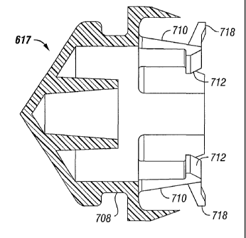
support member
617 and a cover 618. Plunger support member 617 preferably comprises a rigid,
hard
material, for example, an ABS plastic, to interface between an actuator and
the plunger 512.
Member 617 attaches to the actuator by, preferably, a snap fit.
Syringe 500 includes an end wall S 14 located at the first end 504 of the
syringe body
502. End wall 514 is located generally normal to the central, longitudinal
axis 508 of syringe
500. The end wall 514 includes a flat face 516. The flat face 516 is
particularly adapted for
mating engagement with a syringe holder, to be described further below, in an
angiographic
system as described above. Flat face 516 is advantageous in the preferred
arrangement. In
the angiographic system as described herein, significant thrust loads must be
borne in order to
suitably inject the contrast material into the cardiovascular system of the
patient. Flat face
516 allows the thrust load from the injections to be distributed in a
manageable fashion. An
angled face, in contrast, would create a wedge action, which would
unnecessarily stress the
syringe and create an unnecessary side load in the syringe holder. The
inventors have
recognized that a spherical or cone face would require a large door in the
syringe holder to
support the thrust and would also require some elaborate mechanism to properly
position the
door against the syringe. Flat face 516 on syringe 500, however, allows the
thrust load to be
managed by a thin flat door, to be described in more detail below, and is able
to bear the
thrust load from the angiographic injections.
Syringe 500 defines at least one port for providing fluid flow communication
with
pumping chamber 510. In the particular embodiment illustrated, syringe 500
includes two
ports providing fluid flow communication with the pumping chamber 510.
Specifically, an
inlet port 518, Fig. 14, allows the pumping chamber S 10 in syringe 500 to be
filled with
contrast material, and purged or air through inlet port 518, allowing for an
infinite capacity
syringe. By "infinite capacity" it is meant that syringe 500 continues to take
in contrast media
from a bottle of contrast media, wherein the bottles are replaced when empty.
A housing 520
circumscribes inlet port 518 and allows inlet port S 18 to be connected with
an appropriate
bottle 602 of contrast fluid. When syringe 500 is oriented in a syringe holder
in an
angiographic system as described above, syringe 500 defines a top portion and
a bottom
23
CA 02416286 2003-O1-14
WO 02/07812 PCT/USO1/22972
portion. Fig. 15 illustrates the orientation of syringe 500 as it would be
mounted in an
angiographic system of the preferred embodiment. When in such an orientation,
the inlet port
518 is located in the top portion 522 of syringe 500.
In preferred embodiments, the syringe 500 is mounted in an angiographic system
such
that the syringe 500 angles somewhat from the horizontal. By angling the
syringe 500 from
the horizontal, air is allowed to gather around the inlet port 518 in order to
be expelled
through the inlet port 518 during an air purge operation. Angles within the
range of about 5
30°, and preferably about 10-15° from the horizontal are
preferable.
Inlet housing 520 houses a valve assembly analogous to check valve 24,
described and
illustrated above. Check valve 24 is competent to fluid, and incompetent to
air. That is,
check valve 24 permits air to be expelled or purged from the syringe 500, but
does not allow
fluid to flow out of the pumping chamber 510 and back into the bottle 602 of
contrast fluid
when pressure movement is applied on the syringe side of the check valve 24.
Syringe 500 also includes an outlet port 524, FIG. 16, in fluid flow
communication
with pumping chamber 510. Outlet port 524 permits fluid flow from pumping
chamber 510
to downstream fluid passageways, and ultimately into the patient's
cardiovascular system.
Outlet port 524 is surrounded, or circumscribed, by outlet port housing 526
extending, or
projecting, from end wall 514. The outlet port housing 526 is adapted, i.e.,
constructed and
arranged, to receive an outlet tube. Outlet port 524 and outlet housing 526
are analogous to
lower port 80, described in detail above.
When syringe 500 is oriented in the preferred angiographic system of the
present
invention, the outlet port 524 is located adjacent to the bottom portion 523
of syringe 500.
The syringe end wall 514 includes an interior portion 528, FIG. 17, and an
exterior
portion 530, FIG. 14. It is the exterior portion 530 which defines the flat
face 516 of syringe
500. The exterior portion 530 includes a plurality of ribs 532. In the
embodiment illustrated,
there are seven ribs 532 extending transversely across the end wall 514. Ribs
532 help to
provide a reinforcing function. Ribs 532 also provide an attractive,
ornamental appearance to
syringe 500.
Ribs 532 each have end portions 534 terminating in a plane transverse to
longitudinal
axis 508 of syringe body 502. The end portions 534 define the flat face 516.
The interior portion 528 defines a cone-shaped surface 536, Fig. 17. This cone-
shaped
surface 536 is illustrated in Fig. 17 by the shading therein. Cone-shaped
surface 536 helps to
direct the liquid in pumping chamber 510 to an appropriate fluid port.
24
CA 02416286 2003-O1-14
WO 02/07812 PCT/USO1/22972
Preferred dimensions for syringe 500 are described herein below. Syringe body
502
has a diameter of about 1.3 inches. The length of syringe body 502 between
first end 504 and
second end 506 is about 6-7 inches. The inside of syringe body 502 is tapered
so that second
end 506 has an inside diameter greater than the inside diameter of interior
portion 528 of the
end wall 514. This taper is about 0.1° from horizontal for the majority
of its length. The
angle of tapering increases to about 1° at a point about 1 inch from
the second end 506 of
syringe 500. The interior portion 528 defining the cone-shaped surface 536
slopes at an angle
of about 27° from vertical, and the vertex of the cone is rounded at a
radius of about 0.25
inches. Each of ribs 532 is about 0.1 inches thick. The ribs 532 are spaced
about 0.12 inches
apart. The outlet port housing 526 has an outer diameter of about 0.3 inches,
and an inner
diameter of about 0.2 inches. The longitudinal axis of the outlet port housing
526 is parallel
to and about 0.5 inches lower than the central longitudinal axis 508 of
syringe body 502. The
outlet port housing 526 is arranged relative to the syringe body 502, such
that the outer
diameter of the outlet port housing 526 intersects at a tangent point of the
diameter of syringe
body 502. The inlet port housing 520 has an outer diameter of about 0.4 inches
and an inside
diameter of about 0.2 inches. The longitudinal axis of the inlet port housing
520 is tilted
about 10° from vertical toward the end wall 514. The inlet port 518 has
a diameter of about
0.1 inches. The inlet port housing 520 is about 0.5 inches long measured from
where the inlet
housing 520 meets the syringe body 502 in the top portion 522 of the syringe
500.
ANOTHER EXEMPLARY SYRINGE HOLDER ARRANGEMENT
In reference now to FIGS. 18-20, a syringe holder arrangement is illustrated
generally
at 540 which may be used with syringe 500 depicted in FIGS. 12-17. It is noted
that syringe
holder arrangement 540 is compatible with angiographic injector system 10
illustrated in FIG.
l, wherein syringe holder arrangement 540 interfaces with main console 12,
hand held remote
control 14, tubing 42, peristaltic pump 44, saline check valve 46, waste check
valve 48, saline
bag 50, waste bag 52, and bag support rack 54. The operation of the injector
system utilizing
syringe holder arrangement 540 is essentially the same as the operations
depicted and
described in FIGS. 2A-2G, FIGS. 7A-7D, and FIGS. 8A-8C. Of course, some of the
operations may differ due to the structural differences of the syringe holder
arrangement
(syringe holder 16, syringe body 18, syringe plunger 20, radiographic material
reservoir 22,
etc.) shown in FIG. 1 and the syringe holder arrangement 540 and syringe 500
shown in
FIGS. 12 and 18.
In general, the syringe holder arrangement 540 includes a mounting chamber
body
542, a door member 544, a rear plate 546, and a pressure containment sleeve
548. Preferred
CA 02416286 2003-O1-14
WO 02/07812 PCT/USO1/22972
assemblies further include a bottle holder assembly 550, an air column
detector 552, and a
manifold holder 554.
Mounting chamber body 542 is for holding syringe 500 in place during an
angiographic operation. Mounting chamber body 542 is constructed and arranged
to be
durable enough to sustain large pressure loads from the fluid push through
syringe 500.
Mounting chamber body 542 has an arcuate configuration for receipt of sleeve
548. It
includes a loading end 556 for receipt of syringe 500, and an actuating end
558 for receiving
the actuator to reciprocate plunger 512 between its respective proximal and
distal positions
within syringe 500. The loading end 556 also corresponds to the front of
mounting chamber
body 542, and actuating end 558 corresponds to the back or rear of mounting
chamber body
542.
Preferably, mounting chamber body 542 comprises a series of layers in order to
provide a convenient and preferred structure for holding syringe 500. In
particular, the
outermost layer is an electroilluminescent layer. The electroilluminescent
layer permits
illumination of mounting chamber body 542 and the associated tubing. That is,
the
electroluminescent layer illuminates the fluid pathway of the contrast
material as it is being
conveyed from syringe 500 to downstream components and ultimately into the
patient's
cardiovascular system.
Adjacent to the electroilluminescent layer is a membrane heating element. This
layer
maintains heat of the contrast fluid in order to sustain a desired viscosity
in the contrast fluid
for conveying into the patient's cardiovascular system.
The next layer of mounting chamber body 542 and adjacent to the membrane
heating
element layer is a layer of foam. The foam layer keeps contact resistance with
the syringe
500 high and thermal resistance low. It functions to take up tolerances and
helps to snugly
hold syringe 500 in place in syringe holder arrangement 540.
The last layer of mounting chamber body 542 is an aluminum extrusion. It
provides
for a rigid shape and for convenient manufacturing. A layer of adhesive
attaches the foam
layer to the aluminum extrusion.
As illustrated in FIG. 20, mounting chamber body 542 includes a pair of back
flanges
559, 560 defining a groove 561 therebetween. Groove 561 provides for storage
and
containment of wires to syringe holder arrangement 540. A plate 562 slides in
groove 561
and is securably attached thereto to provide for neat and convenient storage.
Still referring to FIG. 18, door member 544 is provided to allow for selective
opening
and closing of loading end 556 of body 542. That is, door member 544 is
movable relative to
26
CA 02416286 2003-O1-14
WO 02/07812 PCT/USO1/22972
mounting chamber body 542 between positions allowing access to mounting
chamber body
542, and into the interior of sleeve 548, and a position which blocks, or
closes access to, the
interior of sleeve 548. In the position where it closes access, door 544
provides for a stop
surface to support and resist the load applied through the syringe 500 when
the plunger is
depressed.
In the particular embodiment illustrated, door member 544 is pivotable
relative to
mounting chamber body 542. This allows for quick and convenient loading and
unloading of
syringe 500 into holding arrangement 540. When door 544 is in its closed
position, FIG. 19,
it locks syringe 500 into place in holder arrangement 540.
In reference again to FIG. 18 and FIG. 20, door member 544 is a structure with
a pair
of flat, planar, opposite surfaces 563, 564. Preferably, it is a fabricated
stainless steel plate
with a thickness of about 0.4 - 1 inches. Flat surface 564, FIG. 20, is
constructed and
arranged for a sliding, abutting engagement with flat end wall 514 of syringe
500 and pressure
sleeve 548. It also slides relative to and abuts against the end surface of
sleeve 548. Because
of the geometry of flat face 516 of end wall 514 of syringe 500, the thrust
load exerted by the
angiographic system 10 through syringe 500 can be managed by flat door member
544.
In reference again to FIG. 18, door member 544 defines a channel, groove, or
slot 565.
Slot 565 is an open, through-hole penetrating door member 544 and extending to
the edge of
door member 544. Slot 565 provides for slidable communication with outlet port
housing 526
of syringe 500. That is, when syringe 500 is oriented properly for loading in
holding
arrangement 540, after syringe 500 is resting within sleeve 548, as door
member 544 is
pivoted to the closed position, FIG. 19, outlet port housing 526 slides within
groove 565.
Groove 565 permits outlet port housing 526 to extend and penetrate through
door member
544 to permit liquid from syringe 500 to be conveyed to downstream components.
Still referring to FIG. 18, door member 544 includes a handle 566. Handle 566
extends from a side edge of door member 544 and allows for a user to
conveniently pivot door
member 544 between its closed position and its open positions. Door member 544
pivots
about its lowest point preventing door member 544 from acting as a guillotine
when acted
upon by gravity. That is, the arrangement of door member 544 relative to its
pivot point
prevents injury to fingers.
In accordance with the invention, a door open sensor is provided. The door
open
sensor tells the user or operator if door member 544 is in an open position.
That is, it
functions as a safety feature such that angiographic system 10 will not be
operated if door
member 544 is not in a securely closed position. In the particular embodiment
illustrated, the
27
CA 02416286 2003-O1-14
WO 02/07812 PCT/USO1/22972
door open sensor includes a magnet S67 in door member 544, and a Hall effect
sensor in the
mounting chamber body 542. When door member 544 is pivoted to its closed
position, FIG.
19, magnet 567 is in contact with mounting chamber body 542. The Hall effect
sensor senses
the presence of magnet 567 and provides an indication to the operator that
door member S44
S is closed. When the Hall effect sensor does not sense the presence of magnet
567, it provides
a signal to the operator that the door member 544 is not in the closed
position, but in an open
position. One suitable sensor is Hall effect sensor 55449A, available from
Microswitch (a
division of Honeywell).
Still referring to FIG. 18, pressure containment sleeve 548 is provided in the
holding
arrangement 540 to hold syringe S00 snugly between door member S44 and rear
plate 546.
Sleeve S48 helps to contain the pressure exerted through the syringe 500, and
allows for large
pressure forces through the syringe 500. The close fit between rear plate 546
and door
member S44 holds the syringe S00 so that no forward/rearward movement is
allowed.
In the particular embodiment illustrated, sleeve 548 is constructed and
arranged to fit,
or slide in the mounting chamber body 542. In the preferred embodiment, sleeve
548 is
cylindrical, or tubular in shape with first and second open ends 568, S69
(FIG. 20). Sleeve
548 is preferably constructed from a strong, durable, basically transparent
material in order to
sustain large pressure loads and allow for visibility of the syringe
therethrough. One preferred
material includes polycarbonate.
In reference to FIG. 19, first end 568 of sleeve 548 is open and allows outlet
port
housing S26 to project, or extend therefrom and through slot 565 in door
member 544.
Second end 569, FIG. 20, permits an actuator from angiographic system 10 to
penetrate sleeve
S48 and access syringe plunger support member 617.
Referring again to FIGS. 18 and 19, it can be seen that door member 544 slides
2S relative to first end S68 of sleeve 548, as door member S44 is moved
between its closed
position and open positions.
Sleeve 548 defines an open groove, or channel 570 extending from first end
568.
Channel 570 accommodates a sensor 571. Sensor 571 is oriented relative to the
valve
assembly in inlet housing S20 in order to detect the state of the check valve.
That is, sensor
571 detects whether the ball in the check valve is seated in its lowermost
position or whether
it has been moved out of its lowermost position. In the particular arrangement
illustrated,
sensor 571 is an emitter/detector interruptable infrared photodetector device.
When the ball
interrupts the infrared beam, a signal is sent indicating that the ball is
seated in its lowermost
position or seat. When the ball is moved out of its lowermost position or
seat, the infrared
28
CA 02416286 2003-O1-14
WO 02/07812 PCT/USO1/22972
beam is not interrupted, and a signal is generated which indicates that the
ball is out of its
lower seat.
A connector 690 and wire 692 energize sensor 571. That is, connector 690
connects
the electrical components and wires within groove 691 to sensor 571.
As can be seen in FIGS. 18 and 20, sensor 571 is generally U-shaped. The U-
shape,
in addition to enabling detecting of the ball in the check valve, also allows
the inlet port
housing 520 to be accommodated within sleeve 548 in holding arrangement 540.
As
illustrated in FIG. 19, when syringe 500 is loaded into holding arrangement
540, it is slid
through sleeve 548, and sensor 571 permits inlet port housing 520 to rest
within the U-shape
of the sensor 571 and extend radially from sleeve 548. In this way, fluid
communication is
permitted from the source of contrast fluid and into syringe 500, even after
syringe 500 is
loaded within the holding arrangement 540. One type of sensor 571 useable is
an infrared
diode (part number SE-1450-004L) and photo-transistor pair (part number SD-
1440-004L),
both available from Microswitch (a division of Honeywell).
In the preferred embodiment, sleeve 548 is conveniently removable from
mounting
chamber body 542. In this manner, it may be cleaned and disinfected separate
from chamber
body 542. In the particular embodiment illustrated in FIG. 20, sleeve 548 is
slidable relative
to mounting chamber body 542 and can be lockably secured thereto through the
cooperation
of a locking pin 572 and a locking assembly 574 in the rear plate 546. Locking
pin 572
extends from second end 569 of sleeve 548. Locking assembly 574 is a spring
loaded locking
member that engages and holds pin 572.
Again, in reference to FIG. 18, rear plate 546 is secured to mounting chamber
body
542 and is in covering relation to second end 569 of sleeve 548. Rear plate
546 supports
actuating end 558 of mounting chamber body 542.
In the particular embodiment illustrated, rear plate 546 has a rectangular
configuration.
Preferably, it is an aluminum fabricated plate, with a thickness of about 0.6
inches.
In reference now to FIG. 20, rear plate 546 defines an aperture 576 in a
central portion
therethrough. Aperture 576 allows access to the interior of sleeve 548. That
is, aperture 576
permits the angiographic actuator to penetrate and move syringe plunger 512
between its
respective proximal ends and distal ends of syringe 500.
In reference now to FIGS. 18 and 19, bottle holder assembly 550 is provided to
hold a
bottle 602 of contrast fluid in order to quickly and conveniently provide a
constant source of
contrast media to syringe S00 when it is loaded in holding assembly 540.
29
CA 02416286 2003-O1-14
WO 02/07812 PCT/USO1/22972
In the illustrated embodiment, bottle holder assembly 550 is secured to
mounting
chamber body 542. Bottle holder assembly 550 includes a column 578 and a neck
portion
580.
Neck portion 580 is pivotable with respect to mounting chamber body 542 in the
direction of arrow 581. The pivotable nature of neck portion 580 aids the ease
of connecting
the tubing from the bottle 602 of contrast media to inlet housing 520 of
syringe 500.
Neck 580 includes universal detail 584 within grooves 586. Universal detail
584 is
preferably a spring loaded configured member which allows bottle holder S50 to
accommodate and hold bottles of various sizes.
In accordance with the present invention, an indicator arrangement is provided
to
provide information whether a bottle is in bottle holder assembly 550. In the
preferred
embodiment, a switch is provided in universal detail 584. When a bottle 602 of
contrast is
within neck 580, bottle 602 presses against the spring in universal detail
584, which actuates
the switch. When the switch is actuated, it provides a visual signal to the
system operator that
a bottle is in fact in the bottle holder assembly 550. If the switch is not
actuated, a signal is
provided to the user that there is no bottle in holder assembly 550. One
suitable switch is a
microswitch MMGGDILOO, available from C&K.
In accordance with the present invention, a sensor is provided to indicate if
the fluid
level in bottle 602 of contrast is either below a certain level or empty.
Preferably, the sensor
includes a sensor provided within groove 586 in neck 580. The sensor detects
when the fluid
level in the bottle 602 has dropped below the level of the sensor in neck 580.
Preferably, the
sensor is a reflective, infrared device. One type of sensor useable is
infrared sensor
HOA1405-2, available from Microswitch (a division of Honeywell).
In reference again to FIGS. 18 and 19, air column detector 552 is provided to
detect
the presence of air in fluid line 588 (FIG. 19). Air column detector 552 is
analogous to air
bubble detector 172, described above. It uses ultrasonic means to detect the
presence of air in
line 588. One suitable ultrasonic means is available from Introtek of New
York.
Air column detector 552 defines a groove 590, FIG. 18, which provides a
friction fit
with fluid line 588. That is, the tubing snaps into groove 590 where it is
securely held therein.
Holders 627, 628 swing down over fluid line 588 to secure it in place (FIG.
19). A flange 592
provides for attachment of the air column detector 552 to mounting chamber
body 552.
In reference now to FIG. 22, air column detector 552 is shown engaging fluid
line 588
which has been wrapped around itself to form a loop 651. Although no
particular theory with
respect to this arrangement is asserted hereto, it is believed that by forming
a loop 651 in fluid
CA 02416286 2003-O1-14
WO 02/07812 PCT/USO1/22972
line 588, any air bubbles present within fluid line 588 will be at a top side
of the tube due to
buoyancy resulting from gravitational forces and centrifugal forces due to the
fluid flow.
Gravitational forces push the bubble to the top side of tube 588. Centrifugal
forces push the
bubble to the inside of the bend radius of loop 651. By the section being at
the bottom
quadrant, both ofthese forces will be in the same direction pushing the bubble
to the inside of
the bend of the loop 651 and the top of tube 588, independent of bend radius
or fluid velocity.
Thus, the bubble is forced to the top side of tube 588. In certain
arrangements, this tends to
enhance the detection of any air bubbles by air column detector 552.
Again in reference to FIGS. 18 and 19, manifold holder 554 is provided to
secure and
hold a manifold, analogous to manifold 26, described above. A clamp structure
597 holds the
manifold securely in place. Manifold holder 554 is mounted on a flange 594,
which is
secured to mounting chamber body 542. Manifold holder 554 is mounted on flange
594 in a
slot 596, FIG. 20, to permit manifold holder 554 to slide back and forth
within groove 596.
This permits manifold holder 554 to accommodate different lengths of tubing
598, FIG. 19,
from outlet port housing 526 of syringe 500.
Manifold holder 554 is configured and shaped to permit the manifold to snap in
only
one orientation. In this way, it can be assured that the manifold is always
oriented in the same
position relative to manifold holder 554. Because of this, a sensor 599 can
detect the position
of the valve within the manifold. Sensor 599 is positioned in an integral part
with manifold
554. Sensor 599 preferably is an inductive type device. One type of sensor
useable in the
embodiment shown is an inductive sensor (part number IFRM 12P1701~L.)
available from
Baumer.
Attention is again directed to FIG. 20. In FIG. 20, a pair of volume
indicators 606,
607 are illustrated. Volume indicators 606, 607 are oriented relative to
mounting chamber
body 542, such that when syringe S00 is situated within mounting chamber body
542, volume
indicators 606, 607 provide a visual cue and indication for the level of fluid
within syringe
body 502. As shown in FIG. 20, volume indicators 606, 607 each include a
plurality of marks
608. As the fluid level within syringe body 502 changes, the user is able to
visually detect
where the level is by comparing it against the marks 608.
In accordance with the present invention, a method for mounting or loading a
syringe
is provided. The method includes a step of positioning a syringe through a
front aperture in a
syringe holder arrangement. This includes sliding a syringe, such as syringe
500 in through
the front end of a syringe holder arrangement 540. Using the components
illustrated in the
drawings, syringe 500 is oriented to line up with the open end of the first
end of sleeve 548.
31
CA 02416286 2003-O1-14
WO 02/07812 PCT/USO1/22972
That is, second end 506 of syringe 500 is aligned with the front of sleeve
548, and the inlet
port housing 570 is aligned with slot 570. The rear, or second end 506, of
syringe 500 (that
is, the plunger receiving end) is first slid through the open end defined by
first end 568 of
sleeve 548. This is followed by the fluid-dispersement end of the sleeve,
i.e., first end 504
defining flat face 516. Syringe 500 is slid into the interior of sleeve 548.
Next, the door is closed. This blocks further access to the interior of sleeve
548. This
also provides for a stop surface, engagement surface, or abutting surface for
syringe 500 in
order to absorb and sustain pressure load through syringe 500. Specifically,
door member 564
is pivoted from one of its open positions, FIG. 18, to its closed position,
FIG. 19. The user
grasps handle 566 and pivots the door to close the opening. As door member 544
is being
pivoted, flat surface 564 of the door is slid relative to flat face 516 of
syringe 500, and relative
to first end portion 568 of sleeve 548. As door member 544 is moved into its
closed position,
outlet tube housing 526 communicates with and slides through groove 565.
To unload syringe 500 from syringe holder arrangement 540, the above process
is
basically done in reverse. Door member 544 is pivoted from its closed
position, FIG. 19, into
one of its open positions, such as that illustrated in FIG. 18. Syringe 500 is
then removed
from holder assembly 540. Specifically, syringe 500 is slid from the interior
of sleeve 548.
The front end of syringe 500, that is, the end with flat face 516, is slid.
out first, followed by
the rear end, or second end 506.
In accordance with the present invention, the angiographic system described
herein is
constructed and arranged to ensure that syringe 500 is not re-used. That is,
the angiographic
system of the present invention includes features to ensure that syringe 500
is disposed of
after use with one patient and not accidentally re-used on a new, different
patient. As
embodied herein, syringe 500 includes structure on its plunger support member
617 to ensure
single use. As illustrated in FIG. 21, plunger support member 617 defines a
plurality of
projections or tabs 610. Tabs 610 project or extend radially inwardly toward
the center or
apex of plunger 512. Tabs 610 are constructed of a flexible, deformable
material, but also
frangible or breakable, such that Whell the actuator engages plunger support
member 617, tabs
610 are bent inwardly to accommodate the actuator. However, when syringe 500
is removed
from the actuator, tabs 610 are broken, and plunger support member 617 is
destroyed. This
prevents syringe 500 from being re-used.
After a period of use, it may be desirable to remove pressure containment
sleeve 548
for cleaning. To do this, rear plate 546 is shifted to disengage and release
locking pin 572.
While locking pin 572 is disengaged from locking assembly 574, sleeve 548 may
be grasped
32
CA 02416286 2003-O1-14
WO 02/07812 PCT/USO1/22972
at a first end 568 and slid out from its snug engagement with mounting chamber
body 542.
At this point, sleeve 548 may be cleaned.
To reinsert the sleeve 548, sleeve 548 is slid back into secure, snug
engagement with
the mounting chamber 542. Locking assembly 574 is shifted to permit locking
engagement
with locking pin 572.
EXEMPLARY SYRINGE PLUNGER ARRANGEMENTS
Referring back to FIG. 13, syringe plunger is usable in the angiographic
systems
described above. As discussed previously, plunger 512 includes plunger support
member 617
and cover 618. Preferably, cover 618 is injection-fluid-compatible (that is,
chemically
compatible and biochemically compatible with the injection fluid). Any contact
with the
injection fluid is preferably made with cover 618 and, therefore, plunger
support member 917
need not be fabricated from a long-term chemically and biochemically
compatible material.
Plunger support member 617 may be fabricated from a relatively rigid and
strong polymer
such as ABS, styrene, acetal, nylon, polycarbonate, and the like or from a
metal such as
stainless steel, aluminum, and the like. When plunger support member 617 is
fabricated from
a metal, a coating may be provided to prevent the injection fluid from
reacting with plunger
support member 617. Cover 618 may comprise, for example, an elastomeric
material such as
a thermoplastic elastomer, synthetic materials, or rubber.. Such elastomeric
materials for
cover 618 generally have hardness in the range of approximately SO-60 Shore A,
low
compression set characteristic at elevated temperatures, high chemical
resistance and isotropic
characteristics, no plasticizers, and low levels of organic and metallic
extractables.
Referring back to FIG. 13, exemplary cover 618 includes a forward portion
having a
cone-shaped surface. A side portion of cover 618 forms a cylindrical sealing
engagement
with the inner sidewall of pumping chamber 510 and includes a plurality of
circumferential
seal ribs 619 which protrude from the outer perimeter of the cover 618 to more
effectively
create a seal between cover 618 and the inner sidewall of syringe body 502 for
containment of
the injection fluid.
Referring to FIGS. 23 and 24A-24C, an actuator 700 cooperates with plunger 512
to
impart reciprocal motion thereto. Actuator 700 comprises a drive piston 702
and an actuator
head 704. A distal end of the drive piston 702 is connected to actuator head
704 while a
proximal end of drive piston 702 is coupled to the motors housed within
console 12. Actuator
head 704 includes a circumferential flange 706.
As best illustrated in FIGS. 24A-24C, plunger support member 617 has a main
body
708 which is cone-shaped so that the proximal diameter is greater than the
distal diameter.
33
CA 02416286 2003-O1-14
WO 02/07812 PCT/USO1/22972
The reduction in diameter allows the distal portion of main body 708 to pass
through aperture
576 of rear plate 546. Plunger support member 617 includes three capture
members 710
cantileverly protruding in a proximal direction from main body 708, and
capture members
710 include latches 712 which capture and retain flange 706 of actuator head
704. Capture
members 710 are configured to flex radially outwardly when contacted by flange
706 and
subsequently "snap back" to capture flange 706. As actuator head 704 moves
forward to
contact plunger 512, beveled surfaces 714 engage flange 706 and are forced
radially
outwardly until flange 706 passes beyond and over inner shoulders 716 of
latches 712. This
design enables actuator head 704 to engage plunger 512 easily at any axial
position within
syringe body 502. For the present embodiment, approximately 7-10 lbs of axial
force is
required for engagement, which is less than the initial stiction force of
cover 618 such that
plunger S 12 remains stationary relative to syringe body 502 during the
engagement procedure.
If the initial stiction force is inadequate, outlet port 524 and inlet port
520 of syringe 500 may
be occluded. The actuator may be extended rapidly to increase pressure within
the syringe
body 502. As the pressure increases, further forward movement of plunger 512
is restricted
and the actuator engages with plunger 512 at a force lower than "cracking"
pressure of the
occlusions (e.g. 60 psi).
After retention of actuator head 704 by capture members 710, plunger 512
remains
engaged with actuator 700 at relatively high rearwardly directed forces (up to
approximately
100 lbs), while plunger 512 is capable transmitting forward thrusts greater
than 1500 lbs to
the syringe contents during an injection.
Referring back to FIGS. 24A-24C, fingers 718 extend radially outwardly from an
outer surface of capture members 710. Ends of fingers 718 extend outwardly at
a distance
approximately equivalent to an outer circumference of plunger support member
617. During
the disengagement procedure, actuator 700 is retracted and the distal portion
of main body
708 passes through aperture 576 of rear plate 546. Rearward movement of
plunger 512
substantially terminates when fingers 718 abut against an inner surface 720 of
rear plate 546
(see FIG. 18), wherein capture members 710 flex radially outwardly and
disengage with
flange 706 of actuator head 704. In the exemplary embodiment, capture members
710 are
designed to permanently deform during disengagement to ensure that syringe 500
is disposed
of after use with one patient and not accidentally re-used on a new, different
patient. By
providing capture members 710 which are deformable instead of frangible or
breakable,
problems encountered with particulates remaining and causing problems during
subsequent
interconnections are avoided. Thus, plunger 512 may be connected and
disconnected with
34
CA 02416286 2003-O1-14
WO 02/07812 PCT/USO1/22972
actuator 700 within syringe body 502 or at a remote location without the need
for outside
toggling or actuation. Furthermore, connection may be made at any location
within syringe
500 and any time, but disconnection may be performed at only one predetermined
location
(plunger 512 at its fully retracted position). Of course, the capture members
may be
configured to break away if desired to prevent re-use of the syringe. For
example, capture
members may be formed of a relatively rigid material which will break away
from the main
body when the fingers abut against the inner surface of the rear plate. In an
alternate
embodiment of the present invention, the advancement of the actuator 700 may
be used to
couple the plunger 512 to the actuator head 704. For example, the actuator 700
may be
advanced such that the actuator head 704 engages the plunger 512. Thereafter,
the plunger
512 and actuator head 704 are advanced through the syringe body 502 and
forcibly engages
the forward plate of the main body, thereby seating the plunger 512 on the
actuator head 704.
FIG. 25 illustrates another embodiment of a syringe plunger arrangement in
accordance with the present invention. Plunger 750 includes cover 618 and a
plunger support
member 752. Plunger support member 752 is magnetically attached to an actuator
754.
Plunger support member 752 includes a first insert 756 comprising a ferrous
metal, permanent
magnet, or electromagnet, while actuator 754 includes a second insert 758
comprising a
ferrous metal, permanent magnet, or electromagnet. Any combination of a
ferrous metal,
permanent magnet, and electromagnet may be used when configuring first 756 and
second
insert 758 as long as a permanent magnet or an electromagnet is included in
one of the inserts
756, 758. For example, the syringe plunger arrangement may be configured so
that first 756
includes a ferrous metal and second insert 758 includes an electromagnet, or
first insert 756
includes a ferrous metal and second insert 758 includes a permanent magnet, or
first insert
756 includes a permanent magnet and second insert 758 includes a permanent
magnet, or first
insert 756 includes a permanent magnet and second insert 758 includes an
electromagnet, etc.
Referring back to FIG. 25, plunger support member 752 includes a base 760
having an
outer diameter larger than aperture 576 of rear plate 546 (see FIG. 18). As
actuator 754 is
retracted through aperture 576, plunger 750 is driven rearwardly until base
760 abuts against
inner surface 720 of rear plate 546. Upon further retraction of actuator 754,
plunger 750 is
disengaged from actuator 754. Some of the advantages of utilizing a magnetic
syringe
plunger arrangement is that a "zero" engagement force is required to engage
plunger 750 with
actuator, a desired disengagement force can be easily obtained by selecting
appropriate inserts
756, 758, reconnection and re-use of syringe 500 can be easily performed
because plunger
CA 02416286 2003-O1-14
WO 02/07812 PCT/USO1/22972
support member 752 is not damaged during the disconnection procedure, and
particulates
from frangible or breakable parts are not present to cause problems during
reconnection.
It should be noted that construction of the syringe plunger arrangement shown
in FIG.
25 is not limited to the above description. For example, the plunger support
member may
include deformable fingers similar to the embodiment described in FIGS. 24A-
24C to prevent
re-use of syringe 500. Furthermore, at least one of the first and second
insert may be
provided with a switchable electromagnet such that the plunger may be
disengaged from the
actuator at any position relative to the syringe body with a substantially
"zero" disengagement
force.
FIG. 26 illustrates another embodiment of a syringe plunger arrangement in
accordance with the present invention. A plunger 800 includes a plunger
support member 802
and cover 618, wherein plunger support member 802 is removably connected to an
actuator
804. A main body 806 of plunger support 802 includes a circumferential groove
808. A first
capture member 810 is pivotally coupled to a distal portion of actuator 804,
and an opposing
second capture member 812 is pivotally coupled to the distal portion of
actuator 804. Distal
ends of first 810 and second capture member 812 include a latch 814 projecting
outwardly
from an inner surface thereof, and proximal ends of first 810 and second
capture member 812
include a ramped lug 816 projecting outwardly from an outer surface thereof.
The distal ends
of capture members 810, 812 are biased in a radially outward direction by a
spring 818.
As actuator 804 moves forward to contact plunger 800, beveled surfaces 820
engage
main body 806 of plunger support member 802 and are forced radially outwardly
until latches
814 engage circumferential groove 808 and move radially inwardly. After
retention of
actuator 804 by capture members 810, 812, plunger 800 remains engaged with
actuator 804.
The syringe plunger arrangement exhibits a relatively low attachment force
(less than 20 lbs)
and relatively high retention force (greater than 100 lbs).
During the disengagement procedure, actuator 804 is retracted and the distal
portion
passes through aperture 576, ramped lugs 816 slidingly engage with sidewall
822 of aperture
576, capture members 810, 812 pivot such that the proximal ends are driven
radially inwardly
and the distal ends are driven radially outwardly, and latches 814 disengage
from
circumferential groove 808.
FIGS. 27A-27G illustrate another embodiment of a syringe plunger arrangement
in
accordance with the present invention. More specifically, the present
embodiment is
particularly useful with injection systems capable of being radially loaded,
such as the system
disclosed in the applicant's co-pending U.S. Patent Application Serial No.
09/577,906 filed on
36
CA 02416286 2003-O1-14
WO 02/07812 PCT/USO1/22972
May 24, 2000 entitled "A Pressure Sleeve Assembly". Those skilled in the art,
however, will
appreciate the present embodiment may be used with a plurality of radially
loaded injection
systems. FIGS. 27A-27B show a pressure sleeve assembly 830, which includes a
longitudinal
base member 832 having a receptacle area 834, a cylinder 836 for housing a
syringe (not
shown), a pivotal arm structure 838, a rear plate 840, and a forward plate
842. The cylinder
836, which is capable of receiving an injection syringe (not shown), is
mounted on the pivotal
or "hinged" arm structure 838, which, in turn, is movable between an open
position, as shown
in FIG. 27A, where the cylinder 836 is disposed away from the receptacle area
834, and a
closed position, as shown in FIG. 27B, where the cylinder 836 is disposed
within the
receptacle area 834. The rear plate 840 disposes an actuator orifice 844,
which the actuator
854 traverses during linear movement. The front plate 842 includes a curved
slot 846, which
receives and guides a fluid exit port 848 formed on the syringe (not shown) as
the cylinder
836 is rotated into the closed position. The actuator 854 further comprises a
circumferential
groove 856, as shown in FIG. 27F, which protrudes slightly into the receptacle
area 834 of the
pressure sleeve assembly 830. The portion of the cylinder 836 located proximal
to the rear
plate 840 disposes a plunger engagement slot 850, which prevents the
protruding actuator 854
from impeding the rotation of the cylinder 836.
FIGS. 27C-27G show the plunger 850 of the present embodiment in greater
detail. As
shown in FIGS. 27C-27E, the plunger 850 includes a plunger support member 852
defining a
U-shaped tongue 858, and a cover 618 positionable on the plunger support
member 852. As
shown in FIG. 27F, the rotation of the cylinder 836 from an open position to a
closed position
causes the U-shaped tongue 858 to slidably engage the circumferential groove
856 formed on
the actuator 854, thereby resulting in the coupling of the plunger 850 to the
actuator 854.
Conversely, movement of the cylinder 836 from a closed position to an open
position causes
the U-shaped tongue 858 formed on the plunger 850 to slidably disengage the
circumferential
groove 856 formed on the actuator 854, thereby resulting in the detachment of
the plunger
850 to the actuator 854. FIG. 27G shows the circumferential groove 856 formed
on the
actuator 854 protruding from the actuator orifice 844 formed in the rear plate
840, thereby
permitting the plunger 850 to be slidably coupled to the actuator 854.
In an alternate embodiment, the plunger 850 may be manually connected to
actuator
854 by sliding a U-shaped tongue 858 of plunger support member 852 onto
circumferential
groove 856, wherein U-shaped tongue 858 remains engaged with circumferential
groove 856
when plunger 850 is reciprocated within syringe body 502. Plunger 850 is
disconnected from
37
CA 02416286 2003-O1-14
WO 02/07812 PCT/USO1/22972
actuator 854 by manually sliding U-shaped tongue 858 away from circumferential
groove
856.
FIG. 28 illustrates another embodiment of a syringe plunger arrangement in
accordance with the present invention. A plunger 900 includes a plunger
support member 902
and cover 618. Plunger support member 902 includes a proximal base 904
defining an
aperture 906. A first capture member 908 and a second capture member 910 are
pivotally
coupled to a distal portion of an actuator 912 at a common point 914. Distal
ends of first 908
and second capture member 910 each include a ramped lug 916 extending
outwardly from an
outer surface thereof, and proximal ends of first 908 and second capture
members 910 each
include a latch 918 extending outwardly from an inner surface thereof. The
distal ends of
capture members 908, 910 are biased in a radially outward direction by a
spring 920.
As actuator 912 moves forward to contact plunger 900, beveled surfaces 922
engage
aperture 906 of plunger support member 902 and are forced radially inwardly
until latches
918 engage an inner surface 924 of base 904. After retention of actuator 912
by capture
members 908, 910, plunger 900 remains engaged with actuator 912. The syringe
plunger
arrangement exhibits a relatively low attachment force (less than 20 lbs) and
relatively high
retention force (greater than 100 lbs). During the disengagement procedure,
actuator 912 is
retracted and the distal portion passes through aperture 576, ramped lugs 916
slidingly engage
with sidewall 822 of aperture 576, capture members 908, 910 pivot such that
both the
proximal and distal ends are driven radially inwardly, and latches 918
disengage from inner
surface 924 of base 904.
FIG. 29 illustrates another embodiment of a syringe plunger arrangement in
accordance with the present invention. A plunger 950 includes a plunger
support member 952
and a cover 618. A retaining ring 954 is disposed at the proximal end of the
syringe body
502. Retaining ring 954 is configured to remain fixedly attached to syringe
body 502,
wherein flange 956 abuts against the proximal end of syringe body 502. Plunger
950 is
releasably connected to retaining ring 954 by ribs 958. Plunger 950 further
includes
capture/engaging members which engage with the actuator. These
capture/engaging members
may be configured similarly to the embodiments described above. Plunger 950
and retaining
ring 954 are configured such that the retaining force of the ribs 958 is
greater than the
engagement force of the capture members. As actuator 960 is extended and
contacts plunger
950, plunger 950 remains stationary within syringe body 502 by retaining ring
954. After
plunger 950 engages with the actuator, retaining ring 954 releases plunger
950, and plunger
950 is allowed to be driven forward and rearward.
38
CA 02416286 2003-O1-14
WO 02/07812 PCT/USO1/22972
FIG. 30 illustrates a still further embodiment of a syringe plunger
arrangement in
accordance with the present invention. A plunger 1000 includes a plunger
support member
1002 and cover 618. Plunger support member 1002 includes a "Christmas tree"
type fastener
1004. Fastener 1004 includes a plurality of tongue members 1006 extending
radially
outward. An actuator 1008 moves forward to contact plunger 1000. An aperture
1010 is
defined within a distal end of actuator 1008. When actuator 1008 is extended
to contact
plunger 1000, fastener 1004 engages with aperture 1010. Tongue members 1006
are angled
so that the engagement force is less than the retaining force.
FIGS. 31A-31M illustrate yet another embodiment of a syringe plunger
arrangement
capable of detachably mounting to an actuator 1034 in accordance with the
present invention.
FIG. 31A shows the plunger 1020 of the present embodiment, which includes a
plunger
support member 1022 and a cover 1024. As shown in FIG. 31B, the plunger
support member
1022 comprises a receiving aperture 1026 formed by a base portion 1028. As
shown in FIGS.
31C and 31D, the at least one retaining member 1030 is positioned on said base
portion 1028
and projects inwardly to the receiving aperture 1026. The at least one
retaining member 1030
is capable of capturing and retaining a flange 1032 formed on the actuator
head 1038. The at
least one retaining member 1030 is configured to flex outwardly when contacted
by an
engagement member 1062A and 1062B, and subsequently "snap back" to capture
flange
1032. As actuator 1034 moves forward to contact plunger 1020, the at least one
retaining
member 1030 is forced outwardly by the engagement members 1062A and 1062B
until the
flange 1032 is engaged by the at least one retaining member 1030. The at least
one retaining
member 1030 may be beveled or angled to effectuate the coupling of the plunger
1020 to the
actuator 1034. This design enables actuator 1034 to engage plunger 1020 easily
at any axial
position within syringe body (not shown).
FIGS. 31E-31G show various views of the actuator 1034 in accordance with the
present invention. As shown in FIG. 31E, the actuator 1034 comprises an
actuator body 1036
connected to actuator head 1038. The actuator head 1038 comprises an alignment
shaft 1040
co-axially aligned along the longitudinal axis of the actuator body 1034,
which is positioned
between a first collet member 1042 and a second collet member 1044. An
alignment device
1046 may be attached to or integrally formed on the alignment shaft 1040.
As shown in FIGS. 3IF-31G, the first collet member 1042 comprises a planar
surface
1048 and an arcuate second surface 1060. The planar surface 1048 further
comprises an
alignment shaft receiving channel 1050a and further disposes a pair of biasing
lumens, 1052a
and 1052b, respectively. An alignment pin orifice 1054a may be formed within
the alignment
39
CA 02416286 2003-O1-14
WO 02/07812 PCT/USO1/22972
shaft receiving channel 1050a of the first collet member 1042. The arcuate
second surface
1060a comprises an engagement member 1062x, a flange portion 1064a, and a
detachment
member 1066x.
The second collet member 1044 is substantially identical to the first collet
member
1042, in that the second collet member 1044 comprises a planar surface 1068
and an arcuate
second surface 1070. The planar surface 1068 further comprises an alignment
shaft receiving
channel 1050b and further disposes a pair of biasing lumens, 1072a and 1072b,
respectively.
An alignment pin orifice 1054b may be formed within the alignment shaft
receiving channel
1050b of the second collet member 1044. The arcuate second surface 1060b forms
or
positions an engagement member 1062b, a flange portion 1064b, and a detachment
member
1066b.
FIGS. 31H-31I show various views of the actuator 1034 in accordance with the
present invention. FIG. 31H shows a bottom view of the actuator head 1034.
Likewise, FIG.
31I shows a top view of the actuator head 1034. As shown in FIGS. 31H-31I,
biasing
members 1076a and 1076b are used to slidably couple the first and second
collet members
1042 and 1044, respectively. In addition, the biasing members 1076a and 1076b
bias the first
and second collet members 1042 and 1044 outwardly. As a result of the outward
force
applied by the biasing members 1076a and 1076b, the flange 1032, which is
formed by flange
portions 1064a and 1064b, is capable of forming at least a first diameter D
when no
constrictive force is applied to the actuator head 1038, and at least a second
smaller diameter
D' when a constrictive force is applied to the first and second collet members
1042 and 1044.
An alignment pin 1078 may be used to retain the alignment shaft 1040 within
the
actuator head 1038 and to slidably couple and align the actuator head 1038.
The alignment
pin 1078 is positioned within an alignment pin orifice 1080 formed in the
alignment shaft
1040, and is in communication with the alignment pin orif ces 1052a and 1052b
formed on
the first and second collet members 1042 and 1044.
The alignment device 1046, which may be attached to or integrally formed on
the
alignment shaft 1040, assures the first and second collet portions 1042 and
1044 are
equidistant from the alignment shaft 1040. In addition, the alignment device
1046 may be
used to define a minimum smaller diameter D' of the flange 1032.
FIGS. 31J-31K show the process of the plunger 1020 being coupled to the
actuator
head 1038. As shown in FIG. 31J, the actuator head 1038 is coaxially aligned
with the
receiving aperture 1026 formed on the base portion 1028 of the plunger support
member
1022. Thereafter, the actuator 1034 is advanced, thereby causing the
engagement members
CA 02416286 2003-O1-14
WO 02/07812 PCT/USO1/22972
1062a and 1062b formed on the actuator head 1038 to engage the at least one
retaining
member 1030 positioned on base portion 1028 of the plunger support member
1022. In
response, the at least one retaining 1030 flexes outwardly while radially
applying a
constrictive force to the actuator head 1038. This application of constrictive
force results in
the first and second collet members 1042 and 1044 sliding inwardly and
decreasing diameter
of the flange 1032 to diameter D'.
As the movement of the actuator 1034 continues, the engagement members 1062a
and
1062b advance into the receiving aperture 1026 formed on the base portion 1028
of the
plunger support member 1022. Thereafter, the at least one retaining member
1030 engages
the flange 1032. As shown in FIG. 31I~, the biasing members 1076a and 1076b
cause the first
and second collet portions 1042 and 1044 to slide outwardly and effectuate an
increase in the
diameter of the flange 1032 from diameter D'. At this point, the plunger 1020
is coupled to
the actuator 1034.
FIGS. 31L-31M show the process of the plunger 1020 being detached to the
actuator
head 1038. Following the injection of material, the plunger 1020 and actuator
1034 are
retracted. During retraction, the actuator 1034 approaches the aperture 576
(see FIG. 18)
formed on the rear plate 546 of the syringe holder arrangement 540. The
diameter of the
aperture 576 is slightly smaller than the diameter of the actuator head 1038,
thereby causing
the detachment members 1066a and 1066b formed on the first and second collet
members
1042 and 1044 engage the rear plate 546 of the syringe holder arrangement 540.
The
retraction of the actuator and the angled wall forming the detachment members
1066a and
1066b cooperatively apply a constrictive force to the actuator head 1038,
resulting in the first
and second collet members sliding inwardly and decreasing diameter of the
flange 1032 to
diameter D'. Thereafter, the at least one retaining member 1030 disengages the
flange 1032
and the actuator 1034 detaches from the plunger 1020.
Additional embodiments of a syringe plunger arrangement can be found on
Exhibit A,
and further operation of the embodiments illustrated in Figures 1 and 18 can
be found on
Exhibit B, attached hereto and specifically made a part thereof.
Having thus described exemplary embodiments of the present invention, it is
noted
that the disclosure herein are exemplary only and that various other
alterations, adaptations
and modifications may be made within the scope of the present invention.
Accordingly, the
present invention is not limited to the specific embodiments as illustrated
herein. For
example, the various syringe plunger arrangements may be configured with
additional or
fewer capture members. For example, the actuator described in FIGS. 26 may
comprise an
41
CA 02416286 2003-O1-14
WO 02/07812 PCT/USO1/22972
additional two capture members such that four capture members are provided to
secure the
syringe plunger with the actuator.
42