Note: Descriptions are shown in the official language in which they were submitted.
CA 02418105 2003-02-03
WO 02/12893 PCT/USO1/24264
MICROARRAYS OF FUNCTIONAL BIOMOLECULES,
AND USES THEREFOR
Related Application
This application is based on and claims priority of U.S. Provisional Patent
Application
No. 60/222,763, filed on August 3, 2000, the disclosure of which is hereby
incorporated by
reference.
Field of the Invention
The present invention relates to the field of diagnostic and analytical
chemistry, and
particularly to devices for screening complex chemical or biological samples
to identify, isolate
or quantify components within a sample based upon their ability to bind to
specific binding
elements. The invention is particularly related to the production and use of
arrays, preferably
IO microarrays, of binding elements which are of biological significance or
which bind to ligands of
biological signif cance.
Background of the Invention
To construct high-density arrays of functional biomolecules for efficient
screening of
complex chemical or biological samples or large numbers of compounds, the
binding elements
15 need to be immobilized onto a solid support. A variety of methods are known
in the art for
attaching biological molecules to solid supports. See generally, Acuity
Techniques, Ey~zyme
Purification: Past B, Meth. Enz. 34 (ed. W. B. Jakoby and M. Wilchelc, Acad.
Press, N.Y. 1974)
and Immobilized Biochemicals and Amity Chromatography, Adv. Exp. Med. Biol. 42
(ed. R.
Dunlap, Plenum Press, N.Y. 1974). Arenkov et al., for example, have described
a way to
20 immobilize proteins while preserving their function by using
microfabricated polyacrylamide gel
pads to capture proteins, and then accelerating diffusion through the matrix
by
microelectrophoresis (Arenkov et al. (2000), Anal Biochem 278(2):123-31). The
patent literature
also describes a number of different methods for attaching biological
molecules to solid supports.
For example, U.S. Pat. No. 4,282,287 describes a method for modifying a
polymer surface
25 through the successive application of multiple layers of biotin, avidin,
and extenders. U.S. Pat.
No. 4,562,157 describes a technique for attaching biochemical ligands to
surfaces by attachment
CA 02418105 2003-02-03
WO 02/12893 PCT/USO1/24264
-2-
to a photochemically reactive arylazide. h -radiation of the azide creates a
reactive nitrene that
reacts irreversibly with macromolecules in solution, resulting in the
formation of a covalent
bond. The high reactivity of the nitrene intermediate, however, results in
both low coupling
efficiencies and many potentially unwanted products due to nonspecific
reactions. U.S. Pat. No.
4,681,870 describes a method for introducing free amino or carboxyl groups
onto a silica matrix,
in which the groups may subsequently be covalently linlced to a protein in the
presence of a
carbodiimide. In addition, U.S. Pat. No. 4,762,881 describes a method for
attaching a
polypeptide chain to a solid substrate by incorporating a light-sensitive
unnatural amino acid
group into the polypeptide chain and exposing the product to low-energy
ultraviolet light.
to There remains, however, a need for more efficient and easy-to-male array
systems that
identifies, isolates and/or quantifies components within complex samples, as
well as to screen
large numbers of compounds based upon their ability to bind to a variety of
different binding
partners.
Summary of the Invention
The present invention provides microarray assay systems where binding elements
of
interest are immobilized on a substrate and are able to interact with and bind
to sample analytes.
The microarrays are useful for screening large libraries of natural or
synthetic compounds to
identify natural binding partners for the binding elements, as well as to
identify non-natural
binding partners which may be of diagnostic or therapeutic interest. The
invention is particularly
2o useful in providing microarrays of antibodies or antibody fragments such as
scFv, which have
previously not been successfully incorporated into high-density arrays while
maintaining their
specific binding activity. The invention also provides methods for using such
microarrays,
methods for selecting epitopes for the antibodies or antibody fragments useful
in such arrays, and
methods for analyzing the data obtained from assays conducted on the
microarrays.
Preferably, the immobilized binding~elerrierits axe arranged in an array on a
solid support,
such as a silicon-based chip or glass slide. The surface of the support is
chosen to possess, or are
chemically derivatized to possess, at least one reactive chemical group that
can be used for
further attachment chemistry. There may be optional flexible molecular
linlcers interposed
between the support and the binding elements. Examples of such linkers include
bovine serum
3o albumin (BSA) molecules, maleimide and vinyl sulfone groups.
CA 02418105 2003-02-03
WO 02/12893 PCT/USO1/24264
_ 3. _.,
In certain embodiments of the invention, a binding element is irmnobilized on
a support
in ways that separate the binding element's region responsible for binding to
its cognate ligand
and the region where it is linlced to the support. In a preferred embodiment,
the two regions are
two separate termini, and the binding element is engineered to form covalent
bond, through one
of the termini, to a linker molecule on the support. Such covalent bond may be
formed through a
Schiff base linkage, a linkage generated by a Michael addition, or a tluoether
linkage. In a
particularly preferred embodiment, an antibody fragment is engineered to
comprise a reduced
cysteine at its carboxyl terminus.
In preferred embodiments, the microarrays,.comprise an array of immobilized
yet
functional binding elements at a density of at least '1.000 spots per cm2. In
some embodiments, to
prevent dehydration, the invention provides for adding a humectant such as
glycerol to the layer
of immobilized binding elements. In other embodiments, the invention provides
for the addition
of a blocking agent solution such as BSA to the substrate surface.
In another aspect, the present invention provides methods of labeling an
antigen such that
the labeling will not interfere with the antigen's binding with an antibody or
antibody fragment.
In a preferred embodiment, the antigen is labeled at its terminal amines after
protease digestion.
In a particularly preferred embodiment, the antigen is digested with trypsin
before being labeled
with a succinimidyl ester dye.
In a further aspect, the present invention provides a method for detecting a
phorsphorylated protein by fragmenting a candidate protein into a plurality of
peptides wherein
one of the peptides comprises a known or suspected phorsphoiylation site, and
using an antibody
or antibody fragment to select the peptide through an epitope close to the
phorsphorylation site.
In yet another aspect, the present invention provides a method for identifying
a small
molecule that regulates protein-protein interaction. According to this aspect,
a capture protein is
attached to a support surface and exposed to its ligand and at least one small
molecule. The
presence or the absence of binding between the capture protein and the ligand
is then detected to
determine the regulatory effect of the small molecule. In a preferred
embodiment, a microarray
of capture proteins that act in the same cellular pathway are attached to the
support surface to
profile the regulatory effect of a small molecule on all these proteins in a
parallel fashion.
3o In yet a further aspect, the present invention provides a method for
studying a cellular
.... .. i ya._~ ~s~...
event by attaching a capture molecule on a support~surface to capture a
cellular organelle
contained in a solution such as a whole-cell lysate.
CA 02418105 2003-02-03
WO 02/12893 PCT/USO1/24264
-4-
These and other aspects of the invention will be apparent to one of ordinary
skill in the al-t
from the following detailed disclosure, and description of the preferred
embodiments.
Brief Description of the Drawings
FIG. lA illustrates exemplary steps of treating a support surface to attach a
BSA
molecule to it and activating the BSA molecule.
FIG. 1B illustrates exemplary steps of attaching a capture protein to the
activated BSA
molecule.
FIG. 2 illustrates proximal phospho-affinity mapping.
FIG. 3A and 3B illustrate an embodiment where small molecule regulating
protein-
l0 protein interaction is studied.
FIG. 4A is a mass spectrometry profile of the steady state surface proteins
from a trpsin
digest of SKOV3 cells.
FIG. 4B is a mass spectrometry diagram showing peptide being affinity captured
by scFv
H7 on Ni-NTA SELDI surface.
15 FIG. 4C is a mass spectrometry diagram showing the result of a control
experiment.
FIG. 4D illustrates the capture of transferrin receptor ectodomain tryptic
peptide that is
labeled with CY-5.
FIG. 5 are mass spectrometry diagrams showing binding by a fusion protein as a
capture
molecule versus the negative control.
2o FIG. 6 are mass spectrometry diagrams showing a small molecule competes a
ligand off
an binding elements on a SELDI surface.
FIG. 7A and 7B show fluorescence units detected from ligand bound to
immobilized
binding elements in the presence or absence of a small molecule.
FIG. 8 shows fluorescence scans of microarrays that have captured labeled
EGFR, TfR or
25 ErbB2 at various dilutions.
FIG. 9 is a fluorescence scan showing labeled cell surface proteins from cell
lysate being
captured by antibody micoarrays.
FIG. 10 are fluorescence scans of microarrays where the capture of unlabeled
antigen is
detected through a second labeled antibody.
3o FIG. 11 are fluorescence scans detecting the binding of antigens from cell
lysates. The
detection is through a second labeled antibody.
CA 02418105 2003-02-03
WO 02/12893 PCT/USO1/24264
-.
Detailed Description of the Invention
The present invention depends, in part, upon the discovery of new methods of
producing
arrays, particularly microarrays, of naturally occurring or artificially
produced biological
macromolecules which may be used to screen samples, including both biological
and artificial
5 samples, to identify, isolate or quantify molecules in such samples that
associate with the
immobilized binding elements. Towards this end, the present invention provides
methods and
products to enable the high-throughput screening of very large numbers of
compounds to identify
those compounds capable of interacting with biological macromolecules.
The present invention has particularly significant applications in
immunoassays, which
to pave the way for extensive and efficient screening using antibodies and
similar molecules.
Antibodies have long played an essential role in determining protein function,
in identifying the
spatiotemporal pattern of gene expression, in identifying protein-protein
interactions, and for ifZ
vitro and ire vivo target validation by phenotypic lcnoclcout. However,
whereas individual
antibodies are useful for monitoring individual proteins from biological
samples, the present
invention provides for the generation of large arrays of antibodies, antibody
fragments, or
antibody-lilce binding elements formatted for high throughput analysis. This
technology, which
enables comprehensive profiling of large numbers of proteins from normal and
diseased-state
serum, cells, and tissues, provides a powerful diagnostic and drug discovery
tool.
One aspect of the present invention concerns improvements in methods of
attaching a
2o biomolecule to a solid support through a chemical linlcer, while retaining
the biological functions
of that molecule, particularly in the case of a capture protein or an antibody
fragment.
I. Substrate/Support
The microarrays of the present invention are formed upon a substrate or
support.
Although the characteristics of these substrates may vary widely depending
upon the intended
use, the basic considerations regarding the shape, material and surface
modification of the
substrates are described below.
A. Shape
The substrates of the invention may be formed in essentially any shape.
Although it is
preferred that the substrate has at least one surface which is substantially
planar or flat, it may
3o also include indentations, protuberances, steps, ridges; terraces and the
lilce. The substrate can be
in the form of a sheet, a disc, a tubing, a cone, a sphere, a concave surface,
a convex surface, a
CA 02418105 2003-02-03
WO 02/12893 PCT/USO1/24264
-6-
strand, a string, or a combination of any of these and other geometric forms.
One can also
combine several substrate surfaces to make use of the invention. One example
would be to
sandwich analyte-containing samples between two flat substrate surfaces with
microarrays
formed on both surfaces according to the invention-. ...
B. Material
Various materials, organic or inorganic or a combination of both, can be used
as support
for this invention. Suitable substrate materials include, but are not limited
to, glasses, ceramics,
plastics, metals, alloys, carbon, papers, agarose, silica, quartz, cellulose,
polyacrylamide,
polyamide, and gelatin, as well as other polymer supports, other solid-
material supports, or
to flexible membrane supports. Polymers that may be used as substrate include,
but are not limited
to: polystyrene; poly(tetra)fluoroethylene (PTFE); polyvinylidenedifluoride;
polycarbonate;
polymethylmethacrylate; polyvinylethylene; polyethyleneimine; polyoxymethylene
(POM);
polyvinylphenol; polylactides; polymethacrylimide (PMI); polyallcenesulfone
(PAS);
polypropylene; polyethylene; polyhydroxyethylmethacrylate (HEMA);
polydimethylsiloxane;
polyacrylamide; polyimide; and various block co-polymers. The substrate can
also comprise a
combination of materials, whether water-permeable or not, in mufti-layer
configurations. A
preferred embodiment of the substrate is a plain 2.5 cm x 7.5 cm glass slide
with surface Si-OH
functionalities.
C. Surface Preparation/Reactive Groups
2o In order to allow attachment by a linlcer or directly by a binding element,
the surface of
the substrate may need to undergo initial preparation in order to create
suitable reactive groups.
Such reactive groups could include simple chemical moieties such as amino,
hydroxyl, carboxyl,
carboxylate, aldehyde, ester, ether (e.g. thio-ether), amide, amine, nitrite,
vinyl, sulfide, sulfonyl,
phosphoryl, or similarly chemically reactive groups. Alternatively, reactive
groups may
comprise more complex moieties that include, but are not limited to,
maleimide, N-
hydroxysuccinimide, sulfo-N-hydroxysuccinimide, nitrilotriacetic acid,
activated hydroxyl,
haloacetyl (e.g., bromoacetyl, iodoacetyl), activated carboxyl, hydrazide,
epoxy, aziridine,
sulfonylchloride, trifluoromethyldiaziridine, pyridyldisulfide, N-acyl-
imidazole,
imidazolecarbamate, vinylsulfone, succinimidylcarbonate, arylazide, anhydride,
diazoacetate,
benzophenone, isothiocyanate, isocyanate, imidoester, fluorobenzene, biotin
and avidin.
Techniques of placing such reactive groups on a substrate by mechanical,
physical, electrical or
CA 02418105 2003-02-03
WO 02/12893 PCT/USO1/24264
_7_.
chemical means are well known in the art, such as described by U.S. Pat. No.
4,681,870,
incorporated herein by reference.
To achieve high-density arrays, it may be necessary to "pack" the support
surface with
reactive groups to a higher density. One preferred method in the case of a
glass surface is to first
"strip" the surface with reagents such as a strong acid, and then to apply or
reapply reactive
groups to the surface.
In the case of a glass surface, the reactive groups can be silanes, Si-OH,
silicon oxide,
silicon nitride, primary amines or aldehyde groups. Slides treated with an
aldehyde-containing
silane reagent are preferred in immobilizing many binding elements and are
commercially
1o available from TeleChem International (Cupertino, CA) under the trade name
"SuperAldehyde
Substrates." The aldehyde groups on the surface of these slides react readily
with primary
amines on proteins to form a Schiff base linlcage. Since typical proteins
display many lysine
residues on their surfaces, as well as the generally more reactive a-amines at
their N-termini,
they can attach to the slide in a variety of orientations, permitting
different sides of the protein to
interact with other proteins or small molecules in solution. After arraying
binding elements such
as proteins onto these aldehyde slides, a buffer containing bovine serum
albumin (BSA) may be
applied to the slide to block later non-specific binding between analytes and
unreacted aldehyde
groups on the slide.
II. Linkers
2o Once the initial preparation of reactive groups on the substrate is
completed (if
necessary), linker molecules optionally may be added to the surface of the
substrate to malce it
suitable for further attachment chemistry.
As used herein, the term "linker" means a chemical moiety which covalently
joins the
reactive groups already on the substrate and the binding element to be
eventually immobilized,
having a backbone of chemical bonds forming a continuous correction between
the reactive
groups on the substrate and the binding elements, and having a plurality of
freely rotating bonds
along that backbone. Linkers may be selected from any suitable class of
compounds and may
comprise polymers or copolymers of organic acids, aldehydes, alcohols, thiols,
amines and the
like. For example, polymers or copolymers of hydroxy-, amino-, or di-
carboxylic acids, such as
3o glycolic acid, lactic acid, sebacic acid, or sarcosine.may be employed.
Alternatively, polymers or
copolymers of saturated or unsaturated hydrocarbons such as ethylene glycol,
propylene glycol,
saccharides, and the like may be employed. Preferably, the linker should be of
an appropriate
CA 02418105 2003-02-03
WO 02/12893 PCT/USO1/24264
-$_
length that allows the binding element, which is to be attached, to interact
freely with molecules
in a sample solution and to form effective binding.
The linker in the present invention comprises at least two reactive groups
with the first to
bind the substrate and the second to bind the~binding-element. The two
reactive groups may be
of the same chemical moiety. The at least two reactive .groups of linlcers may
include any of the
chemical moieties described above of reactive groups on the substrate. And one
preferred second
group comprises a maleimide group. Another preferred embodiment for a
linlcer's second group
is a vinyl sulfone group. It is believed that the hydrophilicity of these
groups helps limit
nonspecific binding by analytes such as proteins when further assay is
conducted in an aqueous
1 o buffer.
Methods for binding the linker to the surface of the substrate will vary
depending on the
reactive groups already on the substrate and the linker selected, and will
vary as considered
appropriate by one skilled in the art. For example, siloxane bonds may be
formed via reactions
between the trichlorosilyl or trisalkoxy groups of a linker and the hydroxyl
groups on the support
surface.
The linlcers may be either branched or unbranched, but this and other
structural attributes
of the linker should not interfere stereochemically with relevant functions of
the binding
elements, such as a ligand-antiligand interaction.
Protection groups, known to those skilled in the art, may be used to prevent
linker's end
2o groups from undesired or premature reactions. For instance, U.S. Pat. No.
5,412,087,
incorporated herein by reference, describes the use of photo-removable
protection groups on a
linlcer's thiol group.
In a preferred embodiment, the linker comprises a BSA molecule. An example of
such
an embodiment is a BSA-NHS slide suitable for making microamays. Although
appropriate for
some applications, slides functionalized with aldehydegroups, further blocked
with BSA, are not
suitable when peptides or small proteins are arrayed, presumably because the
BSA obscures the
molecules of interest. For such applications, BSA-NHS slides are preferred.
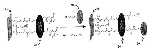
Figures lA and 1B
illustrate a method of making such a slide. First, a molecular monolayer of
BSA is attached to
the surface of a glass slide. Specifically shown in Fig. lA, a glass slide 10
with hydroxyl groups
3o is silanated with aminopropyl triethoxy silane (step 1) before being
activated with N,N'-
disuccinimidyl carbonate (step 2). The activated amino group on the slide in
turn forms covalent
bonds with linlcer 20, which is BSA (step 3). Then, the surface of the BSA is
activated with
CA 02418105 2003-02-03
WO 02/12893 PCT/USO1/24264
-9-
N,N'-disuccinimidyl carbonate (step 4), resulting in activated carbamate and
ester, such as a N-
hydroxy succinimide (NHS) group. Referring to FIG. 1 B, the activated lysine,
aspartate, and
glutamate residues on the BSA react readily with the surface amines on the
binding element 30,
which is a capture protein here (step 5) to form covalent urea or amide
linlcages. Any remaining
reactive groups on BSA are subsequently quenched with glycine (step 6). The
result is a binding
element 30 (a capture protein here) immobilized to a support 10 through a
linlcer 20 (a BSA
molecule here). In contrast to the BSA-blocked slides with aldehyde
functionality, proteins or
peptides arrayed on BSA-NHS substrates are displayed on top of the BSA
monolayer, rendering
them accessible to macromolecules in solution. .
l0 III. Binding Elements
The binding elements of the present invention may be chosen from any of a
variety of
different types of naturally occurring or synthetic molecules, including those
having biological
significance ("biomolecules").
For example, the binding elements may include naturally occurring molecules or
15 molecule fragments such as nucleic acids, nucleic acid analogs (e.g.,
peptide nucleic acid),
polysaccharides, phospholipids, capture proteins including glycoproteins,
peptides, enzymes,
cellular receptors, and immunoglobulins (e.g., antibodies, antibody
fragments,) antigens,
naturally occurring ligands, other polymers, and combinations of any of the
above. And it is also
contemplated that natural product-like compounds, generated by standard
chemical synthesis or
2o from split-and-pool library or parallel syntheses, may be utilized as
binding elements.
A. Antibodies and Antibody Fragments
Antibodies and antibody fragments are preferred candidates for binding
elements. These
include antigen-binding fragments (Fabs), Fab' fragments, pepsin fragments
(F(ab')2 fragments),
scFv, Fv fragments, single-domain antibodies, dsFvs, Fd fragments, and
diabodies, as well as
25 full-length polyclonal or monoclonal antibodies. Antibody-lilce fragments,
such as modified
fibronectin, CTL-A4, and T cell receptors are contemplated here as well. Once
the microarray
has been formed, the antigen binding domains of the antibodies or antibody
fragments may be
utilized to screen for molecules with the specific antigenic determinants
recognized by the
antibodies or antibody fragments.
3o In a preferred embodiment, to study cellular translocation events and cell
surface
expression, phage-displayed scFv that trigger cell internalization of a
surface receptor can be
directly selected from large non-immune phage libraries by recovering and
amplifying phage
CA 02418105 2003-02-03
WO 02/12893 PCT/USO1/24264
-10-
particles from within the cells. See Becerril et al. (1999), Biochem Bio~hYs
Res Commun.
255(2): 386-93, the entire disclosure of which is incorporated by reference
herein.
B. Rece toys
Naturally occurring biological receptors, ~or:~synthetically or recombinantly
modified
variants of such receptors, also may be used as the binding elements of the
invention. Classes of
receptors that can be used as binding elements include extracellular matrix
receptors, cell-surface
receptors and intracellular receptors. Specific examples of receptors include
fibronectin
receptors, fibrinogen receptors, mannose 6-phosphate receptors, erb-B2
receptors, and EGF
(epidermal growth factor) receptors.
to C. Receptor Li: ands
. Similarly, naturally occurring biological receptor ligands, or synthetically
or
recombinantly modified variants of such ligands, also may be used as binding
elements to screen
for their specific binding partners, or for other, non-natural binding
partners. Classes of such
ligands include hormones, growth factors, neurotransmitters, antigens and can
be phage-
displayed.
D. Modifications for Coupling to Substrate/Linlcers
As will be apparent to those of skill in the art, the binding elements may be
modified in
order to facilitate attachment, through covalent or non-covalent bonds, to the
reactive groups on
the surface of the substrate, or to the second reactive groups of a linker
attached to the substrate.
2o As examples of such modifications, nucleophilic S-, N- and O- containing
groups may be added
to facilitate attachment of the binding element to the solid support via a
Michael addition
reaction to the linker.
To preserve the binding affinity of an binding element, it is preferred that
the binding
element is modified so that it binds to the support substrate at a region
separate from the region
responsible for interacting with the binding element'vs-rngnate ligand. If the
binding element
binds its ligand at a first terminus, attaching the binding element to the
support at a second or
opposite terminus, or somewhere in between the termini may be such a solution.
In a preferred
embodiment, where the binding element is an scFv, the present invention
provides a modification
method such that the scFv can be attached to the surface of a glass slide
through binding with an
3o electrophilic linker, such as a maleimide group, without interfering with
the scFv's antigen-
binding activity. According to this method which is detailed in Example C (i),
an scFv is first
engineered so that its carboxy-terminus includes a cysteine residue which can
then form a
CA 02418105 2003-02-03
WO 02/12893 PCT/USO1/24264
-11=
covalent bond with an electrophilic linker such as the maleimide group.
Similarly, a binding
element's N-terminus can be engineerd to include a reactive group for
attachment to the support
surface.
E. Couplin , to Substrates/Liucers
Methods of coupling the binding element to the reactive end groups on the
surface of the
substrate or on the linker include reactions that form linkage such as
thioether bonds, disulfide
bonds, amide bonds, carbamate bonds, urea linkages, ester bonds, carbonate
bonds, ether bonds,
hydrazone linkages, Schiff base linkages, and noncovalent linkages mediated
by, for example,
ionic or hydrophobic interactions. The form of reaction will depend, of
course, upon the
... ,.,,,,..-,.~..
to available reactive groups on both the substrate/linlcer and binding
element.
As discussed in the Examples section below, a Michael addition may be employed
to
attach compounds to glass slides, and plain glass slides may be derivatized to
give surfaces that
are densely functionalized with maleimide groups. Compounds containing thiol
groups, such as
an scFv modified to include a cysteine at the carboxy-terminus, may then be
reacted with the
15 maleimides to form a thioether linkage.
IV. Formation of Microarrays
' In one aspect, the present invention provides methods for the generation of
arrays,
including high-density microarrays, of binding elements immobilized on a
substrate directly or
via a linker. According to the methods of the present invention, extremely
high density
2o microarrays, with a density over 100, preferably over 1000, and further
preferably over 2000
spots per cm2, can be formed by attaching a biomolecule onto a support surface
which has been
functionalized to create a high density of reactive groups or which has been
functionalized by the
addition of a high density of linkers bearing reactive groups.
A. Spotting
25 The microarrays of the invention may be produced by a number of means,
including
"spotting" wherein small amounts of the reactants are dispensed to particular
positions on the
surface of the substrate. Methods for spotting include, but are not limited
to, microfluidics
printing, microstamping (see, e.g., U.S. Pat. No. 5,515,131 and U.S. Pat. No.
5,731,152),
microcontact printing (see, e.g., PCT Publication WO 96/29629) and inkjet head
printing.
3o Generally, the dispensing device includes calibrating means for controlling
the amount of sample
deposition, and may also include a structure for moving and positioning the
sample in relation to
the support surface.
CA 02418105 2003-02-03
WO 02/12893 PCT/USO1/24264
-12-
(il Volume/Snot Size
The volume of fluid to be dispensed per binding element in an array varies
with the
intended use of the array, and available equipment. Preferably, a volume
formed by one
dispensation is less than 100 nL, more preferably~~~ss~than 10 nL, and most
preferably about lnL.
The size of the resultant spots will vary as well, and in preferred
embodiments these spots are
less than 20,000 ~,m in diameter, more preferably less than 2,000 ~.m in
diameter, and most
preferably about 150-200 ~,m in diameter (to yield about 1600 spots per square
centimeter).
(ii Viscosit~dditives
The size of a spot in an array corresponding to a single binding element spot
may be
to reduced through the addition of media such as glycerol or trehalose that
increase the viscosity of
the solution, and thereby inhibit the spreading of the solution. Hydrophobic
boundaries on a
hydrophilic substrate surface can also serve to limit the size of the spots
comprising an array.
Adding a humectant to the solution of the binding element may also effectively
prevent
the dehydration of the microarrays, once they are.created on the surface of
the substrate. Because
15 dehydration can result in chemical or stereochemical changes to binding
elements, such as
oxidation or, in the case of proteins, denaturation, the addition of a
humectant can act to preserve
and stabilize the microarray and maintain the functionality of binding
elements such as scFv. For
example, in some preferred embodiments, scFv are coupled to maleimide-
derivatized glass in
phosphate-buffered saline (PBS) solutions with 40% glycerol. The glycerol
helps maintain
20 continued hydration which, in turn, helps to prevent denaturation.
(iii) Blocking A ents
Solutions of blocking agents may be applied to the microarrays to prevent non-
specific
binding by reactive groups that have not bound to a binding element. Solutions
of bovine serum
albumin (BSA), casein, or nonfat mills, for example, may be used as blocking
agents to reduce
25 background binding in subsequent assays.
(iv) Robotics
In preferred embodiments, high-precision, contact-printing robots are used to
pick up
small volumes of dissolved binding elements from the wells of a microtiter
plate and to
repetitively deliver approximately 1 nL of the solutions to defined locations
on the surfaces of
3o substrates, such as chemically-derivatized glass microscope slides.
Examples of such robots
include the GMS 417 Arrayer, commercially available from Affymetrix of Santa
Clara, CA, and
a split pin arrayer constructed according to instructions downloadable from
CA 02418105 2003-02-03
WO 02/12893 PCT/USO1/24264
-13-
http://cmgm.stanford.edu/pbrown. The chemically-derivatized glass microscope
slides are
preferably prepared using custom slide-sized reaction vessels that enable the
uniform application
of solution to one face of the slide as shown and discussed in the Examples
section. This results
in the formation of microscopic spots of compounds on the slides. It will be
appreciated by one
of ordinary skill in the art, however, that the current invention is not
limited to the delivery of
1 nL volumes of solution, to the use of particular robotic devices, or to the
use of chemically
derivatized glass slides, and that alternative means of delivery can be used
that are capable of
delivering picoliter or smaller volumes. Hence, in addition to a high
precision array robot, other
means for delivering the compounds can be used, including, but not limited to,
ink jet printers,
to piezoelectric printers, and small volume pipetting robots.
B. In Situ Photochemistry
In forming arrays or microarrays of molecules on the surface of a substrate,
in situ
photochemistry maybe used in combination with photoactivatable reactive
groups, which may be
present on the surface of the substrate, on linkers, or on binding elements.
Such photoactivatable
groups are well known in the art.
C. Labeling
Binding elements may be tagged with fluorescent, radioactive, chromatic and
other
physical or chemical labels or epitopes. For certain preferred embodiments
where quantified
labeling is possible, this yields great advantage for later assays.
In a preferred embodiment, a fluorescent dye containing a hydroplulic polymer
moiety
such as polyethyleneglycol is used.
V. Samples for Assays
Upon formation of microarrays of binding elements on the solid support, large
quantities
of samples may be applied to the support surface for binding assays. Examples
of such samples
are as follows:
A. Body Fluids/Tissue and Biopsy Samples
Samples to be assayed using the microarrays of the present invention may be
drawn from
various physiological, environmental or artificial sources. In particular,
physiological samples
such as body fluids of a patient or an organism may be used as assay samples.
Such fluids
3o include, but are not limited to, saliva, mucous, sweat, whole blood, serum,
urine, genital fluids,
fecal material, marrow, plasma, spinal fluid, pericardial fluids, gastric
fluids, abdominal fluids,
peritoneal fluids, pleural fluids and extraction from other body parts, and
secretion from other
CA 02418105 2003-02-03
WO 02/12893 PCT/USO1/24264
-14-
glands. Alternatively, biological samples drawn from cells grown in culture
may be employed.
Such samples include supernatants, whole cell lysates, or cell fractions
obtained by lysis and
fractionation of cellular material.
B. Cell Extracts
Extracts of cells and fractions thereof, including those directly from a
biological entity
and those grown in an artificial environment, can also be used to screen for
molecules in the
lysates that bind to a particular binding element.
C. Normal v. Diseased Sam les
Any of the above-described samples may be derived from cell populations from a
normal
to or diseased biological entity.
D. Treated v. Untreated Samples
Any of the above-described samples may be derived from cell populations which
have or
have not been treated with compounds or other treatments which are believed or
suspected of
being either deleterious or beneficial, and differences between the treated
and untreated
15 populations may be used to assess the effects of the treatment.
E. Labeling
Specific molecules in a given sample may be modified to enable later detection
by using
techniques known to one of ordinary skill in the art, such as using
fluorescent, radioactive,
chromatic and other physical or chemical labels. In a preferred embodiment, a
fluorescent dye
2o containing a hydrophilic polymer moiety such as polyethyleneglycol (e.g.
fluorescin-PEG2000-
NHS) is used. Labeling can be accomplished through direct labeling of analytes
in the sample, or
through labeling of an affinity tag that recognizes an analyte (indirect
labeling). Direct labeling
of sample analytes with different fluorescent dyes makes it possible to
conduct multiple assays
from the same spot (e.g., measuring target protein's expression level and
phosphorylation level).
25 When the analyte is a phage-displayed ligand, the pliage may be pre-labeled
for detecting binding
between the ligand and the microarray of binding elements.
Under the direct-labeling approach, sample over-labeling has long been
recognized as a
serious problem. Over-labeling of proteins can cause aggregation of protein
conjugate, which
tends to result in non-specific staining; it can also reduce antibody's
specificity for its antigen by
3o disrupting antibody's epitope-recognition function, causing loss of signal.
It is well lcnown in the
art that, to mitigate over-labeling, one need to either shorten reaction time
for the labeling
process or increase substrate:label ratio. A solution to over-labeling is to
first digest a whole
CA 02418105 2003-02-03
WO 02/12893 PCT/USO1/24264
-15-
protein into peptides and then label the termini of the peptides, which avoids
labeling any
internal epitopes. Accordingly, the labeling process may proceed to completion
without one
having to worry about over-labeling and thus giving a researcher more complete
control over the
labeling process. Moreover, if the potential labeling sites on a peptide is
lcnown, it is possible to
quantify labeled peptide once the peptide is captured through affinity
reagents that recognize an
internal epitope. An application of this method would be to quantify labeled
peptides digested
from whole proteins in cell extracts for quantitative analysis of protein
expression levels.
In a preferred embodiment, whole proteins are digested with trypsin before
subjected to
labeling by a succinimidyl ester dye such as Cy3,. Cy5 or an Alexa dye. A
succinimidyle ester
1 o dye labels primary amines, such as the one in lysine. Trypsin cleaves
after lysines and generates
peptides with lysines at their C-terminus. Therefore, peptides resulting from
trypsin digestion
fall into two categories: those without lysine and having a primary amine at
the N-terminus, and
those with a lysine at the C-terminus and hence primary amines at both
termini. None of the
peptide would have any internal lysine. As a result, a succinimidyl ester dye
will only label
tryptic peptides at their termini without labeling any internal epitope.
In an alternative embodiment, one may use a protease other than trypsin to
digest a whole
protein and still use a succinimidyl ester dye for labeling as long as the
peptide to be captured
does not contain an internal lysine. That way, labeling will still only occur
at a terminus of the
selected peptide. Such a peptide may be used as a preferential panning
peptide. To take
_ . ~.,~::. . .~ .
2o advantage of a preferential panning peptide, an imrriunoglobulin is first
raised against the
peptide. Second, a sample, e.g., from a whole cell lysate, is digested with a
protease or a
combination of proteases that will generate that specific panning peptide,
resulting in a library of
peptides. These peptides are then labeled to completion with a succinimidyl
ester dye. A large
excess of reactive labeling reagent may be used to ensure complete labeling of
the non-lysine
containing peptide. Then, the labeled peptides are applied to the
immunoglobulin for capture.
Because the amount of labeling on a preferential panning peptide is known, one
can
quantify the amount of such peptide in a given sample through the amount of
label signals
detected after affinity capture. Once the number of such panning peptides
resulting from the
protease digestion of one target protein is known, that number can be easily
translated into the
3o amount of the target protein in the sample. ~ Amino"acids other than lysine
can also be targeted for
use with this method. For example, proteins with limited number of natural or
added cysteine
f
may be selected or constructed to be labeled, via a reduced thiol with
maleimide-coupled dye
CA 02418105 2003-02-03
WO 02/12893 PCT/USO1/24264
-16-
such as maleimide-coupled Alexa 488 (commercially available from Molecular
Probes of
Eugene, Oregon).
Indirect labeling of an antigen. analyte may be achieved by using a second
antibody or
antibody fragment that has been labeled for subsequent°detection (e.g.,
with radioactive atoms,
fluorescent molecules) in a sandwiched fashion. Iri a preferred embodiment, an
antigen that
binds to a microarray of antibodies is detected through a second fluorescently
labeled antibody to
the antigen, obviating the need for labeling the antigen. In a further
preferred embodiment, the
second antibody is a labeled phage particle that displays an antibody
fragment. Standaxd phage
display technology using phages such as M13 may be used to produce phage
antibodies including
to antibody fragments such as scFv. This allows relatively easy and fast
production of reagents for
sandwich detection from phage display antibody libraries. To ensure that the
phage antibodies
recognize an epitope different from the one that the immobilized capture
antibody recognizes on
the antigen, selection from phage display libraries may be carried out in the
following way: (1)
tubes are coated with the same antibody that is immobilized in microaxray for
capture purpose,
. .. ":: "~ ,..
(2) the tube is blocked and the antigen is added and captured by the coated
antibody, (3) after
washing, phage antibody libraries may be panned in the~tubes. The isolated
phage antibodies (or
polyclonal phage antibody) will only bind epitopes distinct from the epitope
the capture antibody
recognizes, and are thus ideal for the sandwich detection approach.
F. Contact time
Binding assays can be performed by exposing samples to the surface prepared
according
to methods described above. Such a surface is first exposed to a sample
solution and then
incubated for a period of time appropriate for each specific assay, which
largely depends on the
time needed for the expected binding reactions. This process can be repeated
to apply multiple
samples either simultaneously or sequentially. Sequential application of
multiple samples
generally requires washes in between.
VI. Binding Assays
A surface prepared according to the methods described above can be used to
screen for
molecules in a sample that have high affinity for the binding elements
attached to the surface.
3o Specific binding may be detected and measured in a number of different
ways, depending on the
way the target molecules in the sample are labeled, if at all. A common
example is to use the
CA 02418105 2003-02-03
WO 02/12893 PCT/USO1/24264
17-
technique of autoradiography to detect binding of molecules pre-labeled with
radioactive
isotopes.
In a preferred embodiment, fluorescent dyes (CYS) were used to label proteins
in a given
sample before the sample was applied to a slide surface printed with
microarrays of functional
scFv. After incubation and washes, the slide surface was then dried and imaged
on a molecular
dynamics STORM or ArrayWorx~ optical reader fiom Applied Precision of Seattle,
WA.
In another preferred embodiment, secondary antibodies labeled with
fluorochromes such
as CY3 were used for later detection of a primary antibody participating in
the binding.
Various detection methods known in the art such as mass spectrometry, surface
plasmon
l0 resonance, and optical spectroscopy, to name a few, can be used in this
invention to allow
detection of binding even if binding targets are not labeled at all.
VII. Analysis of AssayResults
A. Detecting Presence/Absence in Samples
This invention can be used to confirm the presence or the absence, in a
biological sample,
15 of a binding partner to a molecule of interest.
B. Determining Ratios Between Samples
Ratios of gene and protein expression in different cell populations, such as
between a
normal and a diseased state, can be calculated for comparison.
VIII. Applications/Utilities
20 Because the molecules of biological significance that can be studied by
this invention
include, but are not limited to, those involved in signal transduction,
apoptosis, dimerization,
gene regulation, cell cycle and cell cycle checkpoints, and DNA damage
checkpoints, the present
invention has broad applications in the research of biological sciences and
medicine.
As will also be appreciated by one of ordinary slcill in the art, protein
arrays may also be
25 useful in detecting interactions between the proteins and alternate classes
of molecules other than
biological macromolecules. For example, the arrays of the present invention
may also be useful
in the fields of catalysis, materials research, information storage,
separation sciences, to name a
few.
A. Target Discovery
30 It will be appreciated by one of ordinary slcill in the art that the
generation of arrays of
proteins having extremely high spatial densities facilitates the detection of
binding and/or
CA 02418105 2003-02-03
WO 02/12893 PCT/USO1/24264
-18-
activation events occurring between proteins of a defined set and biological
macromolecules.
Thus, the present invention provides, in one aspect, a method for identifying
molecular partners
and discovering binding targets for macromolecules of biological significance.
The partners may
be proteins that bind to particular macromolecules of interest and are capable
of activating or
inhibiting the biological macromolecules of interest. In general, this method
involves (1)
providing an array of one or more proteins, as described above, wherein the
array of proteins has
a density of at least 1,000 spots per cm2 (2) contacting the array with one or
more types of
biological macromolecules of interest; and (3) determining the interaction
between specific
proteins and macromolecule partners.
to In a particularly preferred embodiment the inventive arrays are utilized to
identify
compounds for chemical genetic research. In classical~genetics, either
inactivating (e.g., deletion
or "knock-out") or activating (e.g., oncogenic) mutations in DNA sequences are
used to study the
function of the proteins that are encoded by these genes. Chemical genetics
instead involves the
use of small molecules that alter the function of proteins to which they bind,
thus either
inactivating or activating protein function. This, or course, is the basis of
action of most
currently approved small molecule drugs. The present invention involved the
development of
"chip-like" technology to enable the rapid detection of interactions between
small molecules and
specific proteins of interest. The methods and composition of the present
invention can be used
to identify small molecule ligands for use in chemical genetic research. One
of ordinary slcill in
2o the art will realize that the inventive compositions and methods can be
utilized for other purposes
that require a high density protein format.
B. Signal Transduction
Another preferred embodiment of the binding assays performed in this invention
is to
study modulation of protein-protein interaction by small molecules. These
assays measure either
the facilitation or competition for cognate binding by different molecules in
order to help
understand aspects of binding dynamics under varying conditions. In an
exemplary embodiment,
a capture protein is attached on a support surface in microarray, cognate
ligands are added to
bind to the capture protein. The binding between the capture protein and its
cognate ligand is
monitored and compared in the presence or absence of a small molecule that may
be a drug
candidate. In a preferred embodiment, various capture proteins's interaction
with various ligands
affected by various small molecules are investigated in a mufti-Alex fashion
on a microarray chip.
CA 02418105 2003-02-03
WO 02/12893 PCT/USO1/24264
-19=
Protein interactions often occur through domains that are sometimes called
binding
motifs. It is in these regions that small molecules that are effective at
regulating protein
interactions are most likely to work. However, proteins within a family tend
to share
homologous sequences that contribute to forming binding motifs and proteins
that contain these
motifs often have similar functions. A problem in screening for drugs that
regulate such protein
functions is obtaining specificity in these screens as the targets among the
binding motif family
of proteins are similar in structure, and have similar binding features. The
protein microarray
technology disclosed here permits efficient and easily repeatable steps for
determine specificity
of small molecules for regulating large numbers of motif containing protein
family members, and
l0 will greatly facilitate the process of drug screening:
In an exemplary embodiment, regulation of the Bcl-2 family, known to affect
cell
apoptosis, is studied. These proteins share homology to combinations of four
Bcl-2 homology
regions (BH1- 4). The Bcl-2 family proteins function to either protect cells
against apoptosis or
to promote apoptosis by regulating membrane behavior and ion channel function
at the
mitochondria and the endoplasmic reticulum. The anti-apoptotic family members,
Bcl-2, Bcl-XL,
and Mcl-1 contain all four domains. The largest group of pro-apoptotic
members, Bad, Bilc, Bid,
Bag-l, HRK, and Noxa contain only BH-3 domains, while pro-apoptotic proteins
Bax and
(Multidomain pro-apoptotic proteins) contain BH-1, BH-2, and BH-3 domains.
Methods of the invention can be used to screen for small molecules that
regulate the
2o function of an entire family of apoptosis-regulating proteins. Such a small
molecule may mimic
the function of a BH-3 protein and serve as a drug candidate. Referring to
FIG. 3A and 3B,
recombinant fusion proteins from the Bcl-2 family of apoptosis regulating
proteins may be
prepared by standard methods and printed in microarrays as binding element 30
on either BSA-
NHS glass slides or an aldehyde derivatised glass slide 10 as described
earlier through a linker
20. Ligands 80 for these proteins such as a full length Bcl-XL protein may be
added in the
absence or presence of a small molecule 90 such as a BH-3 containing peptide
from the Bcl-2
family protein BAK or a small molecule that mimics a BH-3 containing peptide.
The ligand 80
may be labeled with a fluorescent dye (e.g. CYS). Concentration of the printed
proteins, the
ligands, or the small molecule may be varied, by itself or in combinations
with others. The slides
may then be read using an optical reader such as the Arrayworx scanner and/or
confirmed
through mass spectrometry using commerically available mass spectrometry
chips. The increase
or decrease in the signal obtained from bound ligand can be used to chart the
regulatory roles of
CA 02418105 2003-02-03
WO 02/12893 PCT/USO1/24264
-20-
the small molecule, whether it is up-regulatory or down-regulatory. Using the
method of the
invention, multiple capture molecules, multiple ligands and multiple small
molecules can be
screened side by side on a single array support (e.g. a 96 well plate),
greatly increasing efficiency
..
in drug screening. A more detailed example can be found in the Example Section
E (iii).
Another example of the invention's application in studying signal transduction
is to
screen for small molecules that inhibit protein-protein binding in the
apoptotic pathway through
the BH-4 region of multidomain-containing BCl-2 family members.
C. Protein Expression
To date, there are no published reports on microarray-based detection of
proteins in
l0 labeled cell extracts. Labeling and detection of cell surface proteins
would allow parallel
profiling of multiple cell surface antigens. State of the art in cell surface
molecule profiling is by
flow cytometry or fluorescence microscopy, currently allowing 2-5 different
antigens to be
profiled in a single sample. Antibody arrays in theory allow the detection of
an unlimited number
of antigens. Furthermore, antibody arrays have the: potential for detecting
intracellular proteins
15 and protein modifications such as phosphorylation in parallel with
expression.
In an exemplary embodiment, monoclonal antibodies to cell surface proteins
such as c-
ErbB2, EGFR, and transferrin receptor are arrayed on a BSA-NHS slide by a GMS
417 arrayer.
Live cells from a cancerous cell line such as the epidermoid carcinoma cell
line A-431 or breast
cancer cell line SIB-BR-3 may be used as sample cells. Cell surface proteins
are preferably
20 labeled with a dye that contains a hydrophilic polymer moiety such as a
polyethyleneglycol,
which has shown good specificity, low baclcground, and does not label proteins
inside cells. An
example of such a dye is fluorescein-PECr2000-NHS dye available from
Shearwater. Following
labeling and wash, cells are lysed (e.g., in SDS). Total labeled proteins are
then incubated on the
antibody microarray for binding to occur before the slides are scanned by an
optical reader. As a
..... .- ~.:,.V. .
25 result, it was confirmed that the A-431 cell line over-expresses EGFR but
not ErbB2. Lilcewise,
it was confirmed that the SK-BR-3 cell line over-expresses ErbB2, but not
EGFR.
D. Post-translational Modification
Protein function is often regulated by post-translational modifications such
as the addition
of sugar complexes, lipid anchors such as provided by myristoylation, geranyl-
geranylation or
3o farnesylation, or by phosphorylation to mention a few. The regulation of
protein function by
phoshorylation or dephosphorylation is central in cell signal transduction.
CA 02418105 2003-02-03
WO 02/12893 PCT/USO1/24264
-21 =
Methods of the present invention can be used to study post-translational
events or to
identify phosphorylation sites. In a preferred embodiment, antibody fragments
such as scFv are
printed on Matrix-Assisted Laser DesportionlIonization (MALDI) chips for
detecting
phosphorylation of known and suspected phosphorylation sites in proteins.
Coupling proteins to
reactive surface MALDI mass spectrometry surfaces was described in U.S. Patent
6,020,208, and
incorporated herein by reference. The chip is commercially available from
Ciphergen
Biosystems Freemont, CA. In an exemplary embodiment, phosphospecific
antibodies against the
apoptotic proteins~Bcl-2, Bad, and caspase 9 are coupled to reactive surface
MALDI chips, and
are used for selective capture of phosphorylated fragments of these proteins.
The chip can be
to analyzed for mass using time of flight mass spectrometry.
Methods of the present invention further provide a new way to detect the
occurrence of a
phosphorylation event on a known or unknown phospho-accepting residue using
recombinant
single chain antibodies (scFv) coupled with mass spectrometry. This method has
been termed
proximal phospho-affinity mapping, and serves as an alternative method that
does not rely on the
use of IMAC or the use of phospho-specific antibodies, which are notoriously
difficult to make.
Referring to FIG. 2, an embodiment of this method uses recombinant single
chain
antibodies (scFv), polyclonal, or monoclonal antibodies 30 that are designed
to recognize, instead
of a phorsphorylation site 70 itself, an epitope 50 on the same antigen that
is in proximity to the
phosphorylation site 70, whether site 70 is confirmed or just suspected for
phosphorylation. The
. ;,F,.,
2o epitope 50 may be as close as 5-10 amino acids away, as long as the
distance between the epitope
SO and the phosphorylation site 70 is such that antibody recognition is not
hindered by a
phorsphoiylation event. Such an antibody or antibody fragment 30, which is
coupled to a
support surface 10 through a linker 20, will recognize the antigen 60 (e.g. a
tryptic peptide)
whether or not the antigen is phosphorylated. In an exemplary embodiment,
peptides are
generated using proteases such as trypsin or V8, or by non-enzymatic methods,
such as CNBr.
This yields peptide fragments that can be identified by their unique sizes.
Among these
fragments are the target fragments 60 that contains known or predicted
phosphorylation sites.
Single chain antibodies or traditional antibodies are panned or immunized
against synthetic
peptides that correspond to an epitope region 50 that is close to the
phosphorylation site 70 in the
3o tryptic fragment 60 using standard panning procedures: ° The epitope
50 may consist of as few as
3-7 amino acids. The antibody or antibody fragment that are generated may be
used as capture
molecule coupled to MALDI reactive chips. The chips may then be used to detect
characteristic
CA 02418105 2003-02-03
WO 02/12893 PCT/USO1/24264
-22-
mass shift indicative of phosphorylation. Since this method enables parallel
purification/identification and analysis of phosphorylation, it offers a
valuable detection tool for
phosphorylation screening. And because the antibody or antibody fragment
generated according
.. .~~." ..e.,
to this method recognizes the target peptide in both the phosphorylated and
unphosphorylated
state, this method is also useful in studying events and conditions that
affect phosphorylation.
In a particularly preferred embodiment, the peptide 60 is selected in the
following way:
first, kinase substrate consensus sequences are located in the target protein
through searches
conducted in a database that contains protein sequence information. Then, a
peptide containing
such consensus sequence is selected through comparing the digestion maps of
various proteases--
1o peptides of about 20 amino acids are preferred. Last, an epitope other than
the leinase substrate
consensus sequences on the selected peptide is chosen for raising an antibody
or antibody
fragment.
E. Cellular Organelle
Methods of the invention can also be~ usedao capture cellular organelles
organelles from
whole cell extracts or from fractions of whole cell extracts. In a preferred
embodiment, an
antibody that recognizes a voltage dependent anion channel ("VDAC") receptor
uniquely
associated with the mitochondrial membrane is printed as described earlier to
capture Green
Fluorescent-coupled cytochrome C expressing mitochondria. Dyes that have
potentiometric
quality can be used to specifically label mitochondria that have intact
voltage gradient. The
2o detection of captured mitochondria or other organelles from cells at
different states can be used
to indicate occurrence of apoptosis or other cellular events.
F. Others
Methods of the invention may also be used for other applications such as
tissue typing,
disease diagnosis, and evaluation of therapeutics. Biological samples from
patients that may
,... . ~.~ ,. .
reveal genetic disorders (PCT patent publication No. 89/11548, incorporated
herein by
reference), may be used in the present invention. Lilcewise, this invention
can be used to detect
abnormality in protein expressions, the existence of antigens or toxins in a
given sample.
Further, methods of the invention can also be used to evaluate responses from
organisms, tissues
or individual cells to exposure to drugs, pharmaceutical lead compounds, or
changes in
3o environmental factors.
CA 02418105 2003-02-03
WO 02/12893 PCT/USO1/24264
.. . . . . , . ~f- a"~ a .: ~~...r ~,~ iuw~ .,as" s~- ~,..,. -er~~:.." ~~.na: -
~
_ 23 '_
EXAMPLES
A. Substrate Surface Preparation
Vii) Method of stripping Mass slide and re-paclcin~ with reactive groups
An example of this preferred method is as follows: first, a plain glass slide
(VWR
Scientific Products, for instance) is cleaned in a piranha solution (70:30 v/v
mixture of
concentrated H2S04 and 30% H202) for 12 hours at room temperature. (Caution:
"piranha"
solution reacts violently with several organic materials and should be handled
with extreme
care). After thorough rinsing with water, the slides is treated with a silane
solution, such as a 3%
solution of 3-aminopropyltriethoxysilane in 95% ethanol. And before treating
the slides, the
1 o silane solution may be stirred for at least 10 minutes to allow hydrolysis
and silanol formation.
The slide is then briefly dipped in ethanol or like solutions and centrifuged
to remove excess
silanol. The adsorbed silane layer is then cured (e.g., one hour at
115°C). After cooling, the slide
is washed in ethanol or like solutions to remove uncoupled reagent.
A simple, semi-quantitative method can be used to verify the presence of amino
groups
15 on the slide surface. An amino-derivatized slide is washed briefly with 5
mL of 50 mM sodium
bicarbonate, pH 8.5. The slide can then be dipped in 5 mL of 50 mM sodium
bicarbonate, pH
8.5 containing 0.1 mM sulfo-succinimidyl-4-0-(4,4'-dimethoxytrityl)-butyrate
(s-SDTB; Pierce,
Roclcford, IL) and shaken vigorously for 30 minutes. (The s-SDTB solution may
be prepared by
dissolving 3.03 mg of a s-SDTB in 1 mL of DMF and diluting to 50 mL with 50 mM
sodium
. , ..~., ~.,... .
20 bicarbonate, pH 8.5). After a 30-minute incubation, the slide can then be
washed with 20 mL of
distilled water and subsequently treated with 5 mL of 30% perchloric acid. The
development of
an orange-colored solution will indicate that the slide has been successfully
derivatized with
amines; no color change has been seen for untreated glass slides. Quantitation
of the 4,4'-
dimethoxytrityl cation (E49snm 70,000 M'lcrri 1) released by the acid
treatment has indicated an
25 approximate density of 2 amino groups per nm2.
B. Addition of Linkers to Substrates
(i) BSA as linker
BSA-NHS slides, displaying activated amino and carboxyl groups on the surface
of an
immobilized layer of bovine serum albumin (BSA), were fabricated as. follows:
10.24 g N,N-
30 disuccinimidyl carbonate (100 mM) and 6.96 m1 lV;N diisopropylethylamine
(100 mM) were
dissolved in 400 ml anhydrous N,N dimethylformamide (DMF). Thirty polylysine
slides, such as
CMT-GAP slides (Corning Incorporated, Corning, NY), displaying amino groups on
their
CA 02418105 2003-02-03
WO 02/12893 PCT/USO1/24264
v p3~.~. ~~ .. ~,~, .,~, ~, ~,~~. _ ....m ~ 3~..... 3.... ,~
-24-
surface, were immersed in this solution for 3 hr at room temperature. These
slides were rinsed
twice with 95% ethanol and then immersed in 400 ml of phosphate buffered
saline (PBS), pH 7.5
containing 1% BSA (w/v) for 12 hr at room temperature. Slides were further
rinsed twice with
ddH20, twice with 95% ethanol, and centrifuged at 200 g for 1 min to remove
excess solvent.
Slides were then immersed in 400 ml DMF containing 100 mM N,N'-disuccinimidyl
carbonate
and 100 mM N,N diisopropylethylamine for 3 hr at room temperature. Slides were
rinsed four
times with 95% ethanol and centrifuged as above to yield BSA-NHS slides.
Slides were stored
in a desiccator under vacuum at room temperature for up to two months without
noticeable loss
of activity.
(ii) A malemide group as liu~er
Maleimide-derivatised slides were manufactured as follows: after the surface
of a plain
glass slide was "packed" (re-silanated, for instance) as described in the
Example A(i), the
resulting slides were transferred to slide-sized polydimethylsiloxane (PDMS)
reaction vessels.
One face of each slide was treated with 20 mM N succinimidyl 3-maleimido
propionate in 50
mM sodium bicarbonate buffer, pH 8.5, for three hours. (This solution was
prepared by
,;
dissolving the N succinimidyl 3-maleimido propionate in DMF and then diluting
10-fold with
buffer). After incubation, the plates were washed several times with distilled
water, dried by
centrifugation, and stored at room temperature under vacuum until further use.
The resulting
slide surface was equipped with a maleimide end.
2o C. Preparation of Binding Elements
~i1 Production and purification of cysteine-ta~~ed scFv
The scFv C6.5 binds to the extracellular region of the human tumor antigen c-
erbB-2 with
a Kd of 1.6 X 10'1° M. This antibody was isolated using affinity driven
selection as described in
Schier et al. (1996), J. Mol. Biol. 255(1):28-43. , , , . .
The gene for the scFv C6.5 was then subcloned into a pUC-119-(Hexa-His)-Cys
expression vector, which results in the addition of a hexa-His tag followed by
a single cysteine to
the COOH-terminus of the scFv. The protein was expressed and purified using
immobilized
metal affinity chromatography (IMAC). Binding affinity mutants of C6.5 were
made by
mutagenizing the complementary binding region (CDR), and the affinity
constants of the
3o derivative mutants [C6.SML 3-4(Kd=3.4x10'9) and C6.SG98 (Kd=1.6x10⁻⁹)],
were determined
using BiaCore (described in Schier et al 1996b). The cysteine tagged scFv
C6.5, C6.SML3-4,
and C6.5 G98. were used to demonstrate ligand capture by scFv which have been
chemically
CA 02418105 2003-02-03
WO 02/12893 PCT/USO1/24264
- 25 -
coupled to glass surfaces. The reduced sulfhydryl of the COOH terminal
cysteine of these scFv
yields a thiol that can be used to couple the scFv to glass surfaces that have
been functionalized
with maleimide groups.
(ii) Reducing an scFv for conjugation to a maleimide linker
Purified scFv were reduced with SmM cysteamine (SIGMA) for 1 hour at
25°C and
exchanged into phosphate buffered saline(PBS), pH7.0 using a P10 spin column.
D. Assays Emplo~g Microarra~
(i) Scarmin~ slides for fluorescence . ,
Slides were scanned using an Array WoRoxTM slide scanner (AppliedPrecision,
Issaquah,
to WA). Slides were scanned at a resolution of 5 ~,m per pixel. Double filters
were employed for
both the incident and emitted light. Fluorescein fluorescence was observed
using a FITC/FITC
excitationlemission filter set, Cy3 fluorescence was observed using a Cy3/Cy3
excitation/emission filter set, and Cy5 fluorescence was observed using a
Cy5/Cy5
excitation/emission filter set.
15 E. Applications of Microanays
(i) Affinity capture of labeled peptides on scFv modified glass surfaces.
Steady state trypsin cleavage of cell surface proteins was performed on SKBR3
(human
breast carcinoma) or SKOV3 cells at 4°C using TPCK-treated trypsin.
Tryptic digests were
examined using MALDI mass spectrometry, which, is shown in FIG. 4A for SKOV3
cells. About
20 0.5 ~.1 of the digest was loaded onto a MALDI surface and embedded with
matrix consisting of
cinnamic acid saturated 50% acetonitryl, 0.5% Triflour, and acetic acid.
Digests were treated
with protease inhibitors and incubated with 1 ~,g of purified 6x His-scFv
against the transferrin
receptor ecto-domain. The scFv-peptide complex was purified from the digests
using Ni-NTA
sepharose beads. The beads were washed and then were embedded in cinnamic acid
matrix as
25 described above. The matrix eluted peptides were analyzed for mass
spectrometry, as shown in
FIG. 4B. The epitope containing tryptic peptide was identified using the
pepident program from
the EXPASY suite. For the control experiment HA-tagged transferrin receptor
expressed in
CHO cells was immuno-precipitated using anti-HA IgG coupled to sepharose
beads. The
purified protein was displaced from the beads using HA-peptide and then
digested with
3o immobilized TPCK-treated trypsin. The scFv epitope-containing peptide was
purified using the
H7 scFv and analyzed for mass as above and is shown in FIG. 4C. The
transfected transferrin
CA 02418105 2003-02-03
WO 02/12893 PCT/USO1/24264
ar.. a~..~. .a ,~ 9au:~~ .~" »:~; ~~:.~ a,,.~-r~-3v.:~, ~~:,:, _.~~_
- 26 -
protein contain an HA epitope sequence on it's amino terminal (intracellular
domain). This tag
serves as a control for extracellular-specific labeling.
Trypsin digests of the purified transferrin receptor and of the cell surface
proteins were
labeled with the primary amine reactive dye NHS-CY-5 and~dialyzed against PBS.
The labeled
peptides were then diluted to a concentration of 0.f mg/ml in PBS with lOmg/ml
BSA and
0.05% Tween 20 and incubated on the surfaces of glass slides which had been
derivatized with
the scFv against the transferrin receptor (H7). Incubations were performed
overnight in a
humidified chamber at 4°C. Binding of CY-5 labeled peptide was
determined using a
fluorescence scanner. FIG. 4D shows the result of the experiment where the
transferrin receptors
1o are shown to bind to the H7 scFv of varying concentrations. Because the HA
epitope was on an
intracellular domain, the anti-HA IgG serves a negative control here.
(ii) Functionality testing of scFv coupled to maleimide-derivatized glass
slides
Spots on a maleimide-derivatized slide surface were outlined with a
hydrophobic pen to
lceep samples from spreading and l.Oq,g of scFv reduced as described in
Example C (ii) was then
allowed to couple to the glass surfaces for 12 hours at 4°C in a
humidity chamber. The thiol-
containing terminal cysteines readily attach to the maleimide groups,
presumably by a thioether
liucage. Monoclonal antibodies to cytochrome-c and Bcl-2, and scFv without
terminal cysteines
were treated with 2-iminothiolane~HCI (Traut's reagent) to introduce
sulfhydryl residues at
surface-exposed lysines. These antibodies were then reduced as described above
and used as
controls. After coupling, the spots were rinsed 3X with PBS containing 2% BSA,
0.05% Tween
20, and 1.0 mM (3-mercaptoethanol for 15 minutes at 25°C. Cognate
Iigand or negative control
were added to the appropriate spots at concentrations ranging from 10.0 pM to
0.01 pM in PBS
containing 2%BSA, 0.05%, Tween-20 and allowed to incubate for 2 hours in a
humidity chamber
at 4°C.
In some cases, 40% glycerol is added to the~spotting mixture to facilitate the
microarraying of the scFv's, because the samples will not dry out even when
spotted in sub-
microliter volumes. For scFv C6.5 and scFv F5, 40% glycerol had no adverse
effect on the
function of the scFv binding.
The cognate ligand for scFvC6.5 is the purified erbB-2 receptor. The
recombinant
3o ectodomain of erbB-2 was expresssed and purified from CHO cells using
standard techniques.
NHS-CYS monofunctional dye (AMERSHAM) was used to label the protein at a final
molax
dye/protein ratio of 5Ø The labeling reaction was carried out in O.1M sodium
carbonate buffer
CA 02418105 2003-02-03
WO 02/12893 PCT/USO1/24264
-27=
for 30 minutes at 25°C and exchanged into PBS using a P10 spin column.
Other proteins used as
controls (Bcl-2, cytochrome-c, and BSA) were similarly labeled with CYS as
described. Labeled
proteins were examined for immunogenicity by immuno-precipitation either with
phage
generated antibody or monoclonal antibodies and were then used as ligands to
glass coupled
scFv. The erbB-2 proteins were incubated in a range of 1 uM to 1 pM in PBS
Tween 20 with 2%
BSA for 2 hours at 25°C in a humidity chamber. CYS labeled erbB2 was
used as a negative
control.
After incubation, samples were washed 3 X 2 minutes with PBS, 0.05% Tween 20
and
1 X with PBS. Samples were allowed to dry. and theu.imaged on a molecular
dynamics STORM
to using the excitation at 640 nm.
(iii) Small molecules in Signal Transduction
Recombinant fusion proteins from the Bcl-2 family of apoptosis regulating
proteins were
prepared by standard methods and printed on either BSA-NHS glass slides or an
aldehyde
derivatised glass slide. Proteins were printed at concentrations ranging from
200 to 20
micrograms per milliliter in a buffer containing 40% glycerol. Printing was
performed as
described using~the GMS 417 ring and pin printer. Plates were loaded with the
capture protein
samples; 96 well plates for printing with the GMS417 printer. Proteins were
allowed to incubate
on the reactive slides for 12 hours under slightly hydrated conditions at
4°C. After the binding
reaction went to completion the slides were rinsed with PBS and variations of
the cognate ligand
labeled with fluorescent dyes. Detection was performed using the Arrayworx
optical reader.
The printed proteins were GST fusions of Bcl-XL and BAX and a 6 x histidine-
tagged-
Bcl-XL. Ligands for these proteins were the full length Bcl-XL protein and the
BH3 containing
peptide from the Bcl-2 family protein BAIL. The peptides were labeled with
Alexa 488 and the
full length protein was labeled with CYS. The volume of liquid delivered from
the GMS printer
is 50-70 pL per stroke repeated 5 times. Protein delivered ranged from 350 pg
to 350 fg of
protein per spot. After printing, proteins were allowed to incubate for 12
hours at 4 degree in a
humidity chamber. The slides were then washed with PBS and blocked with PBS
with 10%
BSA for 5 minutes. To determine the reactivity of the surfaces and the
coupling efficiency of the
proteins, the presence of the GST-fusion proteins were monitored using labeled
anti-GST-tag
antibody at 1 ng/ml.
Labeled protein ligands were incubated in a volume of 401 contained in an area
of 1 cm2
by a hydrophobic barrier.
CA 02418105 2003-02-03
WO 02/12893 PCT/USO1/24264
-28-
The slides were then rinsed and read using the Arrayworx scanner. In addition,
As shown
in FIG. 5, which is a mass spectrometry profile, binding of a ligand by a Bax-
GST protein is
confirmed on the left, while non-binding by a GST protein is shown on the
right.
FIG. 6 confirms the ability of an unlabelled small molecule (a BH3 peptide
here) to
compete a labeled ligand (Bcl-XL here) off the capture molecule (Bax-GST
fusion protein). As
shown in the four mass spectrometry profiles, with an increasing amount of the
BH3 peptide,
lesser binding between labeled ligand and the capture protein was observed.
This confirmed that
the interaction between the capture protein and the ligand was indeed
attributable to the BH-3
domain. The same type of experiment was carried out using a small molecule
that has been
to identified as specifically enhancing BH3 protein-protein interaction, and
enhancement in ligand
(Bcl-XL) binding by a capture molecule (Balc peptide) was observed as
expected.
These experiments were then repeated using several peptides of the BH3 family
as
ligands to compete with three drugs known to affect Bcl-2 family member
function at various
concentrations. Bcl-XL was printed on BSA-NHS glass slides as capture proteins
in each case.
15 The detected fluorescence of the labeled ligand captured on the slide were
shown in columns in
FIGS. 7A and 7B, different drugs showed differential specificity for the two
ligands from the
same family. For Balc (FIG.7A), inhibitory effects were seen in virtually all
the cases, while for
Bid (FIG. 7B), PNAS or a relatively low concentration of anitmycin does not
seem to inhibit its
binding. This experiment can be useful in mapping out a drug candidate's
specificity regarding
20 each member of a large family of target proteins.
(iv) Cell Surface Protein Expression
Monoclonal and scFv antibodies were printed on glass microarrays for detection
of cell
surface antigen expression in cancer cell lines. Antibodies to c-ErbB2, EGFR,
and transferrin
receptor were printed on BSA-NHS activated glass slides. With the monoclonal
antibodies, less
25 than 2 ng/mL of recombinant antigen labeled with~fluorescent dye was
detected. For antigen
detection in cell extracts, the cell surfaces of cancer cell lines were
labeled with fluorescence
using NHS-based dyes. This allowed the detection of differential cell surface
expression of c-
ErbB2 and EGFR on several cancer cell lines. The transferrin receptor was not
detected using the
direct labeling approach; however, when a micro-sandwich approach was
employed, also the
3o transferrin receptor was detected.
Monoclonal antibodies to c-ErbB2, EGFR, and transferrin receptor (TfR) were
arrayed on
a GMS 417 arrayer. The antibodies were spotted in 40% glycerol to prevent
drying out of the
CA 02418105 2003-02-03
WO 02/12893 PCT/USO1/24264
_ 29
spots onto BSA-NHS slides. Antibodies were allowed to react with the slide
overlught in the
cold. The resulting spot size was about 150 micrometer with a spacing of 375
micrometer (center
to center).
Slides were bloclced for 30 minutes in 0.5 M glycine and then in BSA for
another 30
minutes before samples were added. When multiple samples were processed on a
single slide,
groups of antibody spots were separated by drawing with a hydrophobic pen to
allow up to 24
samples to be processed per slide. Alternatively, the groups of antibody spots
were separated
using an adhesive Teflon mask allowing 50 or more samples to be processed per
slide.
The samples were usually labeled with Cy3 or Cy5-NHS dyes for one hour at room
l0 temperature and un-reacted dye is removed by gel filtration. The cell lines
used in this study were
the breast adenocarcinoma cell line SKBR3 and the epidermoid carcinoma cell
line A-431. Cell
surfaces were labeled using the dye, fluorescein-PEG2000-NHS (Shearwater), at
10 mg/mL in
PBS for two hours on ice and un-reacted dye was removed by washing the cells
before
solubilizing in 0.25% SDS in TBS. Recombinant protein antigens were incubated
in 2% BSA in
0.1% tween-PBS. Cell lysates were incubated in the lyses buffer without BSA.
Following
incubation with the samples for two to three hours, the slides were washed
4x10 times: 20 times
in TPBS, then 20 times in PBS, by rapid submersion in a bealcer containing the
wash buffer. The
fluorescence was detected using the ArrayWoRx slide reader.
Sensitivity:
2o Microarrays were incubated with serial dilutions of ErbB2 labeled with
alexa488 and
EGFR labeled with CyS. After washing, the slide was scanned on the ArrayWoRx.
As shown in
Figure 8, except for TfR antibody #3, all the antibodies were able to capture
ErbB2, TfR, and
EGFR respectively. Protein capture was detected at a dilution as low as 1.6
ng/mL,.
Detection of cell surface antigens:
The breast adenocarcinoma cell line SKBR3, and the epidermoid carcinoma cell
line A-
431, were grown to confluence and the cell surface labeled with the dye
fluorescein-PEG2000-
NHS. Following labeling, un-reacted dye was removed by washing the cells and
the cells were
lysed in 0.25% SDS. Total labeled protein (corresponding to about 50,000
cells) was then
incubated on the antibody microarray for two hours and the slides scanned on
the ArrayWoRx.
3o As shown in FIG. 9, the A-431 cell line over-expresses,EGFR, but not ErbB2;
and the SK-BR-3
cell line over-expresses ErbB2, but only expresses low levels of EGFR. This
differential
CA 02418105 2003-02-03
WO 02/12893 PCT/USO1/24264
- 30 -
expression of the two receptors in the two cell lines is confirmed by by flow
cytometry (e.g.,
>106 EGFR receptors per cell in A-431 cells).
In a different approach, the cell proteins were not labeled directly with
fluorescence.
Tnstead, instead, antigen binding to the array was ,dete,cted with a second
fluorescent-labeled
s antibody to the antigen. The sensitivity of this "sandwich" detection
approach was similar to
what was observed for the directly labeled recombinant antigens.
In one experiment, antibodies were printed as before in microarrays and
incubated with
unlabeled antigens for two hours. Binding was detected with a second antibody
to the antigen
labeled with Cy5 (for detecting EGFR) or Cy3 (for detecting TfR). Results are
shown in FIG.
l0 10: monoclonal antibodies as listed in the legend exhibits good sensitivity
at about 25 ng/mL.
The same sandwich approach was performed using phage displayed antibody such
as
scFv FS labeled with CyS.
For detection of antigens in cell extracts, cell lines (A431 or SKBR-3) were
lysed in
0.25% SDS and extracts were incubated with the antibody array for two hours.
After washing,
15 bound antigen was detected with fluorescent monoclonal antibodies (for EGFR
and TfR) or
phage antibody (for ErbB2). As shown in FIG. 1 l, using the sandwich approach,
all three
antigens, EGFR, ErbB2, or TfR, were detected in both cell lysates. The anti-
EGFR antibodies
detected the differential expression of ErbB2 in the A431 and SK-BR-3 cell
lines (>10 fold
difference). Lilce wise, the anti-ErbB2 phage antibody detected the difference
in expression of
2o ErbB2 in the two cell lines. As expected, in the case of transferrin
receptor expression, no major
difference in expression was detected between the two cell lines.
All documents, patents, publications cited above in the specification are
herein
incorporated by reference. Various modifications and variations of the present
invention will be
apparent to those skilled in the art without departing from the scope and
spirit of the invention.
25 Although the invention has been described in conii~ction with specific
preferred embodiments, it
should be understood that the invention as claimedshould not be unduly limited
to such specific
embodiments. Indeed, various modifications of the described modes for canying
out the
invention which are obvious to those skilled in the art are intended to be
within the scope of the
invention.