Note: Descriptions are shown in the official language in which they were submitted.
CA 02421005 2003-02-21
WO 02/15768 PCT/USO1/26360
- 1 -
SYSTEMS AND METHODS FOR APPLYING
ULTRASONIC ENERGY TO THE THORACIC CAVITY
AND OTHER TARGETED BODY REGIONS
Related Application
This application is a continuation-in-part of co-
pending United Stated Patent Application Serial No.
09/645, 662, filed August 24, 2000, and entitled "Systems and
Methods for Enhancing Blood Perfusion Using Ultrasound
Energy," which is incorporated herein by reference.
Field of the Invention
This invention relates to systems and methods for
increasing blood perfusion, e.g., in the treatment of
myocardial infarction, strokes, and vascular diseases.
Background of the Invention
High frequency (5 mHz to 7 mHz) ultrasound has
been widely used for diagnostic purposes. Potential
therapeutic uses for ultrasound have also been more recently
suggested. For example, it has been suggested that high
power, lower frequency ultrasound can be focused upon a
blood clot to cause it to break apart and dissolve. The
interaction between lower frequency ultrasound in the
presence of a thrombolytic agent has also been observed to
assist in the breakdown or dissolution of thrombi. The
effects of ultrasound upon enhanced blood perfusion have
also been observed.
While the therapeutic potential of these uses for
ultrasound has been recognized, their clinical promise has
yet to be fully realized. Treatment modalities that can
apply ultrasound in a therapeutic way are designed with the
premise that they will be operated by trained medical
CA 02421005 2003-02-21
WO 02/15768 PCT/USO1/26360
- 2 -
personnel in a conventional fixed-site medical setting. They
assume the presence of trained medical personnel in a non-
mobile environment, where electrical service is always
available. Still, people typically experience the effects of
impaired blood perfusion suddenly in public and private
settings . These people in need must be transported from the
public or private settings to the fixed-site medical
facility before ultrasonic treatment modalities can begin.
Treatment time (which is often critical in the early stages
of impaired blood perfusion) is lost as transportation
occurs. Even within the fixed-site medical facility, people
undergoing treatment need to be moved from one care unit to
another. Ultrasonic treatment modalities must be suspended
while the person is moved.
Summary of the Invention
One aspect of the invention provides systems and
methods for applying ultrasound energy to the thoracic
cavity.
In one embodiment, an ultrasound energy applicator
is used that comprises a housing sized for placement in
acoustic contact with the thorax. An ultrasound transducer
is carried by the housing to generate ultrasound energy at
a prescribed fundamental therapeutic frequency laying within
a range of fundamental therapeutic frequencies not exceeding
2 5 about 500 kHz. An ultrasonic coupling region is carried by
the housing. The coupling region is adapted, in use, to
contact skin. The coupling region is also sized to
transcutaneously conduct ultrasound energy in a diverging
beam that substantially covers an entire heart. The
3 0 applicator further includes an assembly worn on the thorax,
which is adapted to be affixed to the housing. The assembly
stabilizes placement of the housing on the thorax during
transcutaneous conduction of ultrasound energy.
In one embodiment, an applicator is used having an
35 ultrasonic coupling region that is adapted, in use, to
CA 02421005 2003-02-21
WO 02/15768 PCT/USO1/26360
- 3 -
contact skin. The coupling region has an effective diameter
(D) to transcutaneously conduct ultrasound energy at the
prescribed fundamental therapeutic frequency by the
transducer. According to this aspect of the invention, the
ultrasound transducer has an aperture size (AP)~not greater
than about 5 wavelengths. The quantity AP is expressed as
AP = D/WL, where WL is the wavelength of the fundamental
frequency.
In one embodiment, the systems and methods use an
ultrasound applicator that is sized to be placed in acoustic
contact with the individual to transcutaneously apply
ultrasound energy to the thoracic cavity. The systems and
methods couple an electrical signal generating machine to
the ultrasound applicator. The electrical signal generating
machine includes a controller to generate electrical signals
to operate the ultrasound applicator during a treatment
session to produce ultrasound energy in pulses at (i) a
prescribed pulse repetition frequency (PRF), (ii) a
prescribed fundamental therapeutic frequency laying within
a range of fundamental therapeutic frequencies not exceeding
about 500 kHz, and (iii) at a duty cycle (DC) of about 50%
or less. According to this aspect of the invention, the
duty cycle (DC) is expressed as DC = PD divided by 1/PRF,
where PD is the amount of time for one pulse.
2 5 Another aspect of the invention provides systems
and methods for applying ultrasound energy to a body region.
The systems and methods provide an ultrasound applicator
including a housing, an ultrasound transducer carried by the
housing, and a chamber sized to hold an acoustic coupling
media subject to a pressure in acoustic communication with
the ultrasound transducer. The systems and methods generate
electrical signals to operate the ultrasound transducer to
output acoustic energy at a selected intensity level. The
systems and methods sense at least one system parameter and
compare the sensed system parameter to a desired level. The
CA 02421005 2003-02-21
WO 02/15768 PCT/USO1/26360
- 4 -
systems and methods vary the pressure in the chamber based,
at least in part, upon the comparison.
In one embodiment, the system parameter includes
impedance. In this arrangement, the systems and methods can
vary pressure in the chamber based, at least in part, upon
variance between the sensed impedance and a desired
impedance level.
In one embodiment, the systems and methods select
the desired level based upon the selected intensity level.
In one embodiment, the systems and methods vary
pressure in the chamber to maintain an essentially constant
acoustic output.
In one embodiment, the acoustic coupling media
within the chamber conducts heat from the ultrasound
transducer.
Other features and advantages of the inventions
are set forth in the following specification and attached
drawings.
Brief Description of the Drawings
Fig. 1 is a perspective view of a system for
transcutaneously applying ultrasonic energy to affect
increased blood perfusion;
Fig. 2 is an enlarged side perspective view of an
ultrasonic applicator that forms a part of the system shown
2 5 in Fig. 1;
Fig. 3 is a side section view, with parts broken
away and in section of the applicator shown in Fig. 2;
Fig. 4 is an enlarged side perspective view of an
alternative embodiment of an ultrasonic applicator having an
3 0 ultrasonic conductive pad that can be joined to the
applicator for use as part of the system shown in Fig. 1;
Fig. 5 is a view of the applicator shown in Fig.
2 held by a stabilization assembly in a secure position
overlaying the sternum of a patient, to transcutaneously
35 direct ultrasonic energy toward the vasculature of the
CA 02421005 2003-02-21
WO 02/15768 PCT/USO1/26360
- 5 -
heart;
Fig. 6 is a view of the applicator shown in Fig.
2 held by another type of stabilization assembly on the
thorax of a patient to transcutaneously direct ultrasonic
energy toward the vasculature of the heart;
Fig. 7 is an enlarged side perspective view of an
ultrasonic applicator of the type shown in Fig. 2 used in
association with an ultrasonic material externally applied
to the skin;
Fig. 8 is an enlarged side perspective view of an
ultrasonic applicator of the type shown in Fig. 2 used in
association with a patch externally applied to the skin to
create a clean ultrasonic interface;
Fig. 9 is a schematic view of an ultrasonic
applicator of the type shown in Fig. 2 positioned to
transcutaneously apply ultrasonic energy to the heart in the
thoracic cavity, showing a desired degree of ultrasonic
energy beam divergence that applies ultrasonic energy
substantially to the whole heart;
2 0 Fig. 10 is a side elevation view of an ultrasonic
applicator having a flexible ultrasound radiating surface
that can conform evenly to a skin surface region,
eliminating gaps between the radiating surface and the skin,
to thereby mediate localized conductive heating effects
2 5 during use;
Fig. 11 is a side section view of an ultrasonic
application of the type shown in Fig. 10, and also showing
and interior well region surrounding the transducer face for
collecting air to further mediate localized conductive
3 0 heating effects during use;
Fig. 12 is a view of another embodiment of an
ultrasonic applicator usable in association with the system
shown in Fig. 1, the applicator being shaped to apply
ultrasonic energy to the vasculature in the heart without
35 passage through adjacent organs like the lungs, the system
CA 02421005 2003-02-21
WO 02/15768 PCT/USO1/26360
- 6 -
also including an assembly to administer a therapeutic agent
in conjunction with the application of ultrasonic energy;
Fig. 13 is a schematic view of a system for
achieving different localized systemic treatments in
different regions of the body, one of which involves the use
of the system shown in Fig. 1;
Fig. 14 is a perspective view of a cooling module
and associated heat exchange cassette that the system shown
in Fig. 1 can incorporate;
Fig. 15 is a side schematic view of the cooling
module and heat exchange cassette shown in Fig. 14;
Fig. 16 is a side schematic view of another
embodiment of a cooling module and heat exchange cassette
that the system shown in Fig. 1 can incorporate;
Fig. 17 is a schematic view of a controller that
can be used in conjunction with the system shown in Fig. 1,
which combines power control and media management control to
maintain an essentially constant acoustic output for the
ultrasound applicator; and
Fig. 18 is a plan view of a kit, in which all or
some of the disposable components of the system shown in
Fig. 1 can be packaged before use, along with instructions
for using the components to achieve the features of the
invention.
The invention may be embodied in several forms
without departing from its spirit or essential
characteristics. The scope of the invention is defined in
the appended claims, rather than in the specific description
preceding them. All embodiments that fall within the
meaning and range of equivalency of the claims are therefore
intended to be embraced by the claims.
Description of the Preferred Embodiments
The various aspects of the invention will be
described in connection with the therapeutic indication of
providing increased blood perfusion by the transcutaneous
CA 02421005 2003-02-21
WO 02/15768 PCT/USO1/26360
application of ultrasonic energy. That is because the
features and advantages of the invention are well suited to
this therapeutic indication. Still, it should be appreciated
that many aspects of the invention can be applied to achieve
other diagnostic or therapeutic objectives as well.
Furthermore, in describing the various aspects of
the invention in the context of the illustrated embodiment,
the region targeted for an increase in blood perfusion is
the thoracic cavity (i.e., the space where the heart and
lungs are contained). Tt should be appreciated, however,
that the features of invention have application in other
regions of the body, too, for example, in the arms, legs, or
brain.
I. System for Providing Noninvasive Ultrasound-Assisted
Blood Perfusion
Fig. 1 schematically shows a compact, portable
therapeutic system 10 that makes it possible to treat a
person who needs or who is likely to need an increase in the
flow rate or perfusion of circulating blood.
The system 10 includes durable and disposable equipment
and materials necessary to treat the person at a designated
treatment location. Tn use, the system 10 affects increased
blood perfusion by transcutaneously applying ultrasonic
energy.
As Fig. 1 shows, the system 10 includes at the
treatment location an ultrasound generating machine 16. The
system 10 also includes at the treatment location at least
one ultrasound applicator 18, which is coupled to the
machine 16 during use. As Figs. 4 and 5 show, the system 10
also includes an assembly 12 for use with the applicator 18
to stabilize the position of the applicator 18 on a patient
for hands-free use. In the illustrated embodiment (see
Figs. 4 and 5), the applicator 18 is secured against
movement on a person's thorax, overlaying the sternum, to
direct ultrasonic energy toward the vasculature of the
CA 02421005 2003-02-21
WO 02/15768 PCT/USO1/26360
_ g _
heart.
The location where treatment occurs can vary. It can be
a traditional clinical setting, where support and assistance
by one or more medically trained care givers are immediately
available to the person, such as inside a hospital, e.g., in
an emergency room, catheter lab, operating room, or critical
care unit. However, due to the purposeful design of the
system 10, the location need not be confined to a
traditional clinical setting. The location can comprise a
mobile setting, such as an ambulance, helicopter, airplane,
or like vehicle used to convey the person to a hospital or
another clinical treatment center. The location can even
comprise an everyday, public setting, such as on a cruise
ship, or at a sports stadium or airport, or a private
setting, such as in a person's home, where the effects of
low blood perfusion can arise.
By purposeful design of durable and disposable
equipment, the system 10 can make it possible to initiate
treatment of a reduced blood perfusion incident in a non-
clinical, even mobile location, outside a traditional
medical setting. The system thereby makes effective use of
the critical time period before the person enters a hospital
or another traditional medical treatment center.
The features and operation of the system 10 will now be
described in greater detail.
A. The Ultrasound Generator
Fig. 1 shows a representative embodiment of a machine
1~. The machine 16 can also be called an "ultrasound
generator." The machine 16 is intended to be a durable item
capable of long term, maintenance free use.
As shown in Fig. 1, the machine 16 can be variously
sized and shaped to present a lightweight and portable unit,
presenting a compact footprint suited for transport, e.g.,
mounted on a conventional pole stand 14, as Fig. 1 shows.
This allows the machine 16 to accompany the patient from one
CA 02421005 2003-02-21
WO 02/15768 PCT/USO1/26360
- 9 -
location to another. The machine 16 can alternatively be
sized and shaped to be mounted at bedside, or to be placed
on a table top or otherwise occupy a relatively small
surface area. This allows the machine 16 to travel with the
patient within an ambulance, airplane, helicopter, or other
transport vehicle where space is at a premium. This also
makes possible the placement of the machine 16 in a non-
obtrusive way within a private home setting, such as for the
treatment of chronic angina.
In the illustrated embodiment, the machine 16 includes
a chassis 22, which can be made of molded plastic or metal
or both. The chassis houses a module 24 'for generating
electric signals. The signals are conveyed to the
applicator 18 by an interconnect 30 to be transformed into
ultrasonic energy. A controller 26, also housed within the
chassis 22 (but which could be external of the chassis 22,
if desired), is coupled to the module 24 to govern the
operation of the module 24. Further details regarding the
controller 26 will be described later.
The machine 16 also preferably includes an operator
interface 28. Using the interface 28, the operator inputs
information to the controller 26 to affect the operating
mode of the module 24. Through the interface 28, the
controller 26 also outputs status information for viewing by
the operator. The interface 28 can provide a visual readout,
printer output, or an electronic copy of selected
information regarding the treatment. The interface 28 is
shown as being carried on the chassis 22, but it could be
located external of the chassis 22 as well. Further details
regarding the interface 28 will be described later.
The machine 16 includes a power cord 30 for coupling to
a conventional electrical outlet, to provide operating power
to the machine 16. The machine 16 also preferably includes
a battery module 34 housed within the chassis 22, which
enables use of the machine 16 in the absence or interruption
CA 02421005 2003-02-21
WO 02/15768 PCT/USO1/26360
- 10 -
of electrical service. The battery module 34 can comprise
rechargeable batteries, that can be built in the chassis 22
or, alternatively, be removed from the chassis 22 for
recharge. Likewise, the battery module 34 can include a
built-in or removable battery recharger 36. Alternatively,
the battery module 34 can comprise disposable batteries,
which can be removed for replacement.
Power for the machine 16 can also be supplied by an
external battery and/or line power module outside the
chassis 22. The battery and/or line power module is
releasably coupled at time of use to the components within
the chassis 22, e.g., via a power distribution module within
the chassis 22.
The provision of battery power for the machine 16 frees
the machine 16 from the confines surrounding use of
conventional ultrasound equipment, caused by their
dependency upon electrical service. This feature makes it
possible for the machine 16 to provide a treatment modality
that continuously "follows the patient," as the patient is
2 0 being transported inside a patient transport vehicle, or as
the patient is being shuttled between different locations
within a treatment facility, e.g., from the emergency room
to a holding area within or outside the emergency room.
In a representative embodiment, the chassis 22 measures
2 5 about 12 inches x about 8 inches x about 8 inches and weighs
about 9 pounds.
B. The Ultrasound Applicator
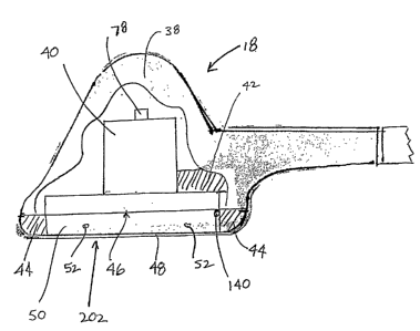
As best shown in Figs. 2 and 3, the applicator 18 can
also be called the "patient interface." The applicator 18
30 comprises the link between the machine 16 and the treatment
site within the thoracic cavity of the person undergoing
treatment. The applicator 18 converts electrical signals
from the machine 16 to ultrasonic energy, and further
directs the ultrasonic energy to the targeted treatment
35 site.
CA 02421005 2003-02-21
WO 02/15768 PCT/USO1/26360
- 11 -
Desirably, the applicator 18 is intended to be a dis-
posable item. At least one applicator 18 is coupled to the
machine 16 via the interconnect 30 at the beginning a
treatment session. The applicator 18 is preferably decoupled
from the interconnect 30 (as Fig. 2 shows) and discarded
upon the completing the treatment session. However, if
desired, the applicator 18 can be designed to accommodate
more than a single use.
As Figs. 2 and 3 show, the ultrasound applicator 18
includes a shaped metal or plastic body 38 ergonomically
sized to be comfortably grasped and manipulated in one hand.
The body 38 houses at least one ultrasound transducer 40
(see Fig. 3).
The body 38 can include a heat sink region 42 placed
about the transducer 40, to conduct heat generated by the
transducer or transducers during operation, to minimize
heating effects. As will be described later, impedance
matching or active cooling can also be achieved to prevent
or counter heating effects.
2 0 Preferably, the plastic body 38 includes a stand-off
region 44 or skirt extending from the front mass or face 46
of the transducer 40. The skirt region 44 enables spacing
the transducer face 46 a set distance from the patient's
skin. The skirt region 44 prevents direct contact between
the transducer face 46 and the person's skin. In a preferred
arrangement, the skirt region 44 is formed of a soft
material, such as foam.
In a preferred embodiment, the front mass 46 of the
transducer 40 measures about 2 inches in diameter, whereas
3 0 the acoustic contact area 202 formed by the skirt region 44
measures about 4 inches in diameter. An applicator 18 that
presents an acoustic contact area 202 of significantly
larger diameter than the front mass of the transducer 40
(e.g. , in a ratio of at least 2:1) reduces overall weight and
3 5 makes possible an ergonomic geometry (like that shown in
CA 02421005 2003-02-21
WO 02/15768 PCT/USO1/26360
- 12 -
Fig. 2) that enables single-handed manipulation during set-
up, even in confined quarters, and further provides'(with the
assembly 12) hands-free stability during use. In a
representative embodiment, the applicator 18 measures about
4 inches in diameter about the skirt region 44, about 4
inches in height, and weighs about one pound.
The material 48 defines a bladder chamber 50 between it
and the transducer face 46. The bladder chamber 50
accommodates a volume of an acoustic coupling media liquid,
e.g., liquid, gel, oil, or polymer, that is conductive to
ultrasonic energy, to further cushion the contact between
the applicator 18 and the skin. The presence of the acoustic
coupling media also makes the acoustic contact area 202 of
the material 48 more conforming to the local skin
topography.
The material 48 and bladder chamber 50 can together
form an integrated part of the applicator 18. Alternatively,
as shown in Fig. 4, the material 48 and bladder chamber 50
can be formed by a separate molded component, e.g., a gel or
liquid filled pad 200, which is not an integral part of the
applicator 18, but which is supplied separately. In this
arrangement, the separate component 200 can be releasably
attached, e.g., by an adhesive strip 204 or the like on the
pad 200, to the transducer face 46 or to the skirt 44, if
present, at instant of use. A molded gel filled pad
adaptable to this purpose is the AQUAFLEX° Ultrasound Gel
Pad sold by Parker Laboratories (Fairfield, New Jersey).
As will be described later, an acoustic coupling media
may be circulated through ports 52 ( see Fig . 3 ) into and out
of the bladder chamber 50, to conduct heat from the bladder
chamber 50 and/or perform a function to maintain a desired
impedance value.
The interconnect 30 carries a distal connector 54 (see
Fig. 2), designed to easily plug into a mating outlet 56 in
the transducer 40. A proximal connector 58 on the
CA 02421005 2003-02-21
WO 02/15768 PCT/USO1/26360
- 13 -
interconnect 30 likewise easily plugs into a mating outlet
60 on the chassis 22 (see Fig. 1), which is itself coupled
to the controller 26. In this way, the applicator 18 can be
quickly connected to the machine 16 at time of use, and
likewise quickly disconnectedfor discard once the treatment
session is over. Other quick-connect coupling mechanisms can
be used. It should also be appreciated that the interconnect
30 can be hard wired as an integrated component to the
applicator 18 with a proximal quick-connector 58 to plug
into the chassis 22, or, vice versa, the interconnect 30 can
be hard wired as an integrated component to the chassis 22
with a distal quick-connector 54 to plug into the applicator
18.
As Fig. 5 shows, a stabilization assembly 12 allows the
operator to temporarily but securely mount the applicator 18
against an exterior skin surface for use. In the illustrated
embodiment, since the treatment site exists in the thoracic
cavity, the attachment assembly 54 is fashioned to secure
the applicator 18 on the person's thorax, overlaying the
sternum or breastbone, as Fig. 5 shows.
Just as the applicator 18 can be quickly coupled to the
machine 16 at time of use, the stabilization assembly 12
also preferably makes the task of securing and removing the
applicator 18 on the patient simple and intuitive. Thus, the
stabilization assembly 12 makes it possible to secure the
applicator 18 quickly and accurately in position on the
patient in cramped quarters or while the person (and the
system 10 itself) is in transit.
The stabilization assembly 12 can be variously
3 0 constructed. In the embodiment shown in Fig. 5, the
stabilization assembly 12 comprises a sling 62 worn on the
back of the patient between the waist and shoulders. The
sling 62 carries a shoulder loop 64 and a waist loop 66.
The loops 64 and 66 are made of a stretchable, elastic
material. The loops 64 and 66 can be stretched to hook into
CA 02421005 2003-02-21
WO 02/15768 PCT/USO1/26360
- 14 -
flanges 68 formed on the body 38 of the applicator 18 (also
shown in Fig. 2) . The stretchable loops 64 and 66 allow for
a rapid mounting and removal of the applicator 18 on the
thorax of the patient. The stretchable loops 64 and 66 also
securely hold the applicator 18 in a stable position on the
patient, even in the midst of a dynamic and mobile
environment.
As Fig. 5 shows, the stabilization assembly 12
preferably occupies only a relatively small area on the
chest. The stabilization assembly 12 (and the compact size
of the applicator 18 itself) allow other devices, e.g., a
twelve lead ECG electrode device, to be placed on the chest
at the same time the applicator 18 is being used.
In another embodiment (see Fig. 6), the stabilization
assembly 12 comprises halter straps 70 and 72 worn about the
chest and shoulders of the patient. The straps 70 and 72
are made of quick release material, e.g., from Velcro''
material. The straps can be easily passed through rings 74
formed in the body 38 of the applicator 18, and doubled back
upon themselves to be secured together. This arrangement,
like the arrangement shown in Fig. 5, allows for rapid
placement and removal of the applicator 18 on the thorax
(sternum) of the patient. Also, like the stabilization
assembly 12 shown in Fig. 5, the assembly 12 shown in Fig.
6 also does not to impede the placement of other treatment
devices on the thorax simultaneously with the applicator 18.
For added comfort in either embodiment of the
stabilization assembly 12, the sling 62 or halter strips
70/72 can be attached to a flexible back piece (not shown)
worn on the patient's back. The back piece can comprise,
e.g., a flexible cloth or plastic sheet or pad, formed in
the manner of the back half of a vest . The slings 62 or
halter straps 70/72 are sown or buckled to the back piece
and extend forward about the shoulders and chest of the
patient, to be coupled to the applicator 18 in the fashion
CA 02421005 2003-02-21
WO 02/15768 PCT/USO1/26360
- 15 -
shown Figs. 5 and 6 show. The sling 62 or halter straps
70/72 transfer the weight of the applicator 18 to the back
piece. The back piece distributes the weight borne by the
sling 62 or halter straps 70/72 in a uniform manner across
the patient's back.
If desired (see Fig. 7), an external ultrasound
conducting material 78 can also be applied directly to the
skin of the person, to provide acoustic coupling between the
applicator 18 and the treatment site. The external material
1,0 78 can comprise, e.g., a gel material (such as AQUASONIC~
100, by Parker Laboratories, Inc., Fairfield, N.J.). The
external material 78 can possess sticky or tacky properties,
to further enhance the securement of the applicator 18 to
the skin.
Alternatively or in combination with a gel material 78
(see Fig. 8), an adherent patch 206 can be secured on the
individual skin. 'The patch 206 forms a clean interface
surface between the acoustic contact area 202 of the
applicator 18 and the individual's skin. The patch 206
keeps the interface surface free from body hair,
perspiration, and other materials that can interfere with
the direct transcutaneous transmission of ultrasonic energy.
The applicator 18 can be formed in various shapes for
ease of storage, handling, and use. As Figs. 2 and 3 show,
the applicator 18 can comprise generally discus or hockey
puck shape. As Fig. 9 shows, the applicator 18 can be
shaped in a more elliptical or elongated fashion that aligns
with the axis of the sternum or heart, for example. In this
arrangement, passage of ultrasonic energy into adjacent
organs, e.g., the lungs, is minimized.
C. Aperture (Directivity)
Desirably, when used to apply ultrasonic energy
transcutaneously in the thoracic cavity to the heart, the
transducer face 46 is sized to deliver ultrasonic energy in
a desired range of fundamental frequencies to substantially
CA 02421005 2003-02-21
WO 02/15768 PCT/USO1/26360
- 16 -
the entire targeted region. Generally speaking, the
fundamental frequencies of ultrasonic energy suited for
transcutaneous delivery to the heart in the thoracic cavity
to increase blood perfusion can lay in the range of about
500 kHz or less . Desirably, the fundamental frequencies for
this indication lay in a frequency range of about 20 kHz to
about 100 kHz, e.g., about 27 kHz.
Within this range of fundamental frequencies (see Fig.
9), the transducer face 46 of the applicator 18 should be
sized to percutaneously transmit the energy in a diverging
beam 208 which substantially covers the entire heart and
coronary circulation 218. The applicator 18 may comprise a
single transducer (as Fig. 9 shows) or an array of
transducers that together form an acoustic contact area 202.
. Normal hearts vary significantly in size and distance
from skin between men and women, as well as among
individuals regardless of sex. Typically, for men, the size
of a normal heart ranges between 8 to 11 cm in diameter and
6 to 9 cm in depth, and the weight ranges between 300 to 350
grams. For men, the distance between the skin and the
anterior surface of the heart (which will be called the
"subcutaneous depth" of the heart) ranges between 4 to 9 cm.
Typically, for women, the size of a normal heart ranges
between 7 to 9 cm in diameter and 5 to 8 cm in depth, and
the weight ranges between 250 to 300 grams. For women, the
subcutaneous depth of the heart ranges between 3 to 7 cm.
The degree of divergence or "directivity" of the
ultrasonic beam 208 transmitted percutaneously through the
acoustic contact area 202 is a function of the wavelength of
3 0 the energy being transmitted. Generally speaking, as the
wavelength increases, the beam divergence (shown generally
as BD in Fig. 9) becomes larger (given a fixed aperture
size). If the beam divergence BD at the subcutaneous depth
of the heart 210 is less than beam area of the heart 210
(shown as H in Fig. 9), the ultrasonic energy will not be
CA 02421005 2003-02-21
WO 02/15768 PCT/USO1/26360
- 17 -
delivered to substantially the whole heart. Therefore, the
beam divergence BD should desirably be essentially equal to
or greater than the targeted beam area H at the subcutaneous
depth of the heart 210.
Within the desired range of fundamental frequencies of
20 kHz to 200 kHz, the beam divergence can be expressed in
terms of an aperture size measured in wavelengths. The
aperture size (AP) can be expressed as a ratio between the
effective diameter of the transducer face 46 (D) and the
wavelength of the ultrasonic energy being applied (WL), or
AP = D/WL. For example, a transducer face 46 having an
effective diameter (D) of 4 cm, transmitting at a
fundamental frequency of about 48 kHz (wavelength (WL) of 3
cm), can be characterized as having an aperture size of 4/3
wavelengths, or 1.3 wavelengths. The term "effective
diameter" is intended to encompass a geometry that is
"round," as well as a geometry that is not "round", e.g.,
being elliptical or rectilinear, but which possesses a
surface area in contact with skin that can be equated to an
2 0 equivalent round geometry of a given effective diameter.
For the desired range of fundamental frequencies of 20
kHz to about 100 kHz, transducer faces 46 characterized by
aperture sizes laying within a range of 0.5 to 5
wavelengths, and preferably less than 2 wavelengths, possess
the requisite degree of beam divergence to transcutaneously
deliver ultrasonic energy from a position on the thorax, and
preferably on or near the sternum, to substantially an
entire normal heart of a man or a woman.
Of course, using the same criteria, the transducer face
46 can be suitably sized for other applications within the
thoracic cavity or elsewhere in the body. For example, the
transducer face 46 can be sized to delivery energy to beyond
the heart and the coronary circulation, to affect the
pulmonary circulation.
D. Reduced Localized Cavitational-Cause Heating
CA 02421005 2003-02-21
WO 02/15768 PCT/USO1/26360
- 18 -
In addition to desirably possessing the characteristic
of coupling energy to substantially the entire targeted
tissue region, the acoustic contact area 202 desirably is
configured to minimize localized skin surface heating
effects.
Localized skin surface heating effects may arise by the
presence of air bubbles trapped between the acoustic contact
area 202 and the individual's skin. In the presence of
ultrasonic energy, the air bubbles vibrate, and thereby may
cause cavitation and attendant conductive heating effects at
the skin surface. To minimize the collection of air bubbles
along the acoustic contact area 202, the acoustic contact
area 202 desirably presents a flexible, essentially flat
radiating surface contour where it contacts the individual's
skin (as Fig. 3 shows), or a flexible, outwardly bowed or
convex radiating surface contour (i . a . , curved away from the
transducer face 46) where it contacts with or conducts
acoustic energy to the individual's skin (as Figs. 10 and 11
show) . Either a flexible flat or convex surface contour can
"mold" evenly to the individual's skin topography, to
thereby mediate against the collection and concentration of
air bubbles in the contact area 202 where skin contact
occurs. In comparison, an inwardly bowed or concave contact
area 202 (i.e., curved toward the transducer face 46) is
2 5 more prone to air bubble collection in the region of skin
contact, and thereby may be more subject to cavitation-
caused localized skin surface heating.
To further mediate against cavitation-caused localized
skin surface heating (see Fig. 11), the interior of the
3 0 bladder chamber 50 can include a recessed well region 212
surrounding the transducer face 46. The well region 212 is
located at a higher gravity position than the plane of the
transducer face 46. Air bubbles 214 that may form in fluid
located in the bladder chamber 50 are led by gravity to
35 collect in the well region 212 away from the ultrasonic
CA 02421005 2003-02-21
WO 02/15768 PCT/USO1/26360
- 19 -
energy beam path. A convex contact area 202 (as shown in
Fig. 11) further enhances the gravity-assisted collection of
air bubbles 214 in the well region 212, as shown by arrows
216 in Fig. 11. The air bubbles 214, to the extent they
form, are kept away from the region of skin contact and out
of the path of the ultrasonic energy beam. To minimize the
possibility of air bubbles being present in the ultrasonic
beam, the transducer face 46 may also be convex in shape (as
Fig. l1 shows).
II. Use Of the System With a Therapeutic Agent
As Fig. 12 shows, the system 10 can further include at
the treatment location a delivery system 32 for introducing
a therapeutic agent 20 in conjunction with the use of the
applicator 18 and machine 16. In this arrangement, the
effect of increased blood perfusion caused by the
application of ultrasonic energy can also be enhanced by the
. therapeutic effect of the agent 20, or vice versa.
Application of ultrasound within the range of fundamental
frequencies of about 20 kHz to about 100 kHz at a power
2 0 density equal to or less than about 3 W/cm2 and at a maximum
total power output between 15 W and 150 W increases coronary
vessel diameter approximately 10%, which results in a 460
increase in blood flow.
A. Use with a Thrombolytic Agent
For example, the therapeutic agent 20 can comprise a
thrombolytic agent. In this instance, the thrombolytic
agent 20 is introduced into a thrombosis site (using the
delivery system 32) , prior to, in conjunction with, or after
the application of ultrasound. The interaction between the
applied ultrasound and the thrombolytic agent 20 is observed
to assist in the break-down or dissolution of the thrombi,
compared with the use of the thrombolytic agent 20 in the
absence of ultrasound. This phenomenon is discussed, e.g.,
in Carter United States Patent 5,509,896; Siegel et al
United States Patent 5,695,460; and Lauer et al United
CA 02421005 2003-02-21
WO 02/15768 PCT/USO1/26360
- 20 -
States Patent 5,399,158, which are each incorporated herein
by reference.
The process by which thrombolysis is affected by use of
ultrasound in conjunction with a thrombolytic agent 20 can
vary according to the frequency, power, and type of
ultrasonic energy applied, as well as the type and dosage of
the thrombolytic agent 20. The application of ultrasound has
been shown to cause reversible changes to the fibrin
structure within the thrombus, increased fluid dispersion
into the thrombus, and facilitated enzyme kinetics. These
mechanical effects beneficially enhance the rate of
dissolution of thrombi. In addition, cavitational disruption
and heating/streaming effects can also assist in the
breakdown and dissolution of thrombi.
The type of thrombolytic agent 20 used can vary. The
thrombolytic agent 20 can comprise a drug known to have a
thrombolytic effect, such as t-PA, TNKase, or RETAVASE.
Alternatively (or in combination), the thrombolytic agent 20
can comprise an anticoagulant, such as heparin; or an
antiplatelet drug, such as a GP IIb IIIa; or a fibrinolytic
drug; or a non-prescription agent having a known beneficial
effect, such as aspirin. Alternatively (or in combination),
the thrombolytic agent 20 can comprise microbubbles, which
can be ultrasonically activated; or microparticles, which
can contain albumin.
The thrombolytic syndrome being treated can also vary,
according to the region of the body. For example, in the
thoracic cavity, the thrombolytic syndrome can comprise
acute myocardial infarction, or acute coronary syndrome.
3 0 The thrombolytic syndrome can alternatively comprise suspect
myocardial ischemia, prinzmetal angina, chronic angina, or
pulmonary embolism.
The thrombolytic agent 20 is typically administered by
the delivery system 32 intravenously prior to or during the
3 5 application of ultrasonic energy. The dosage of the
CA 02421005 2003-02-21
WO 02/15768 PCT/USO1/26360
- 21 -
thrombolytic agent 20 is determined by the physician
according to established treatment protocols.
It may be possible to reduce the typical dose of
thrombolytic agent 20 when ultrasonic energy is also
applied. It also may be possible to use a less expensive
thrombolytic agent 20 or a less potent thrombolytic agent 20
when ultrasonic energy is, applied. The ability to reduce
the dosage of thrombolytic agent 20, or to otherwise reduce
the expense of thrombolytic agent, or to reduce the potency
of thrombolytic agent, when ultrasound is also applied, can
lead to additional benefits, such as decreased complication
rate, an increased patient population eligible for the
treatment, and increased locations where the treatment can
be administered (i.e., outside hospitals and critical care
settings, such as in ambulances, helicopters, other public
settings, as well as in private, in-home settings).
B. Use With an Angiogenic Agent
Treatment using ultrasound alone can stimulate
additional capillary or microcirculatory activity, resulting
in an angiogenesis effect. This treatment can be used as an
adjunct to treatment using angiogenic agents released in the
coronary circulation to promote new arterial or venous
growth in ischemic cardiac tissue or elsewhere in the body.
In this instance, the therapeutic agent 20 shown in Fig. 12
can comprise an angiogenic agent, e.g., Monocyte
Chemoattractant Protein-1, or Granulocyte-Macrophage Colony-
Stimulating-Factor.
It is believed that the angiogenic effects of these
agents can be enhanced by shear-related phenomena associated
with increased blood flow through the affected area.
Increased blood perfusion in the heart caused by the
application of ultrasound energy can induce these shear
related phenomena in the presence of the angiogenic agents,
and thereby lead to increased arterial-genesis and/or
vascular-genesis in ischemic heart tissue.
CA 02421005 2003-02-21
WO 02/15768 PCT/USO1/26360
- 22 -
III. Use of the System with Other Treatment Applications
The system 10 can be used to carry out other
therapeutic treatment objectives, as well.
For example, the system 10 can be used to carry out
cardiac rehabilitation. The repeated application of
ultrasound over an extended treatment period can exercise
and strengthen heart muscle weakened by disease or damage.
As another example, treatment using ultrasound can
facilitate an improvement in heart wall motion or function.
The system 10 may also be used in associated with other
diagnostic or therapeutic modalities to achieve regional
systemic therapy. For example, Fig. 13 shows a composite
system 220 for achieving regional systemic therapy. The
composite system 220 includes a first selected treatment
modality 218, which is applied to the body to achieve a
desired systemic effect (for example, the restriction of
blood flow). The composite system 220 includes a second
selected treatment modality, which comprises the ultrasound
delivery system 10 previously described. The system 10 is
operated before, during, or after the treatment modality
218, at least for a period of time, to transcutaneously
apply ultrasonic energy to a selected localized region of
the body (e. g., the thoracic cavity) to achieve a different,
and perhaps opposite, localized system result, e.g.,
increased blood perfusion.
For example, an individual who has received a drug that
systemically restricts blood flow may experience a need for
increased blood perfusion to the heart, e.g., upon
experiencing a heart attack. In this situation, the
ultrasound delivery system 10 can be used to locally apply
ultrasound energy to the thoracic cavity, to thereby locally
increase blood perfusion to and in the heart, while systemic
blood perfusion remains otherwise lowered outside the
thoracic cavity due to the presence of the flow-restricting
drug in the circulatory system of the individual.
CA 02421005 2003-02-21
WO 02/15768 PCT/USO1/26360
- 23 -
As another example, a chemotherapy drug may be
systemically or locally delivered (by injection or by
catheter) to an individual. The ultrasound delivery system
can be used to locally supply ultrasound energy to the
5 targeted region, where the tumor is, to locally increase
perfusion or uptake of the drug.
The purposeful design of the durable and disposable
equipment of the system 10 makes it possible to carry out
these therapeutic protocols outside a traditional medical
10 setting, such as in a person's home.
IV. Exemplary Treatment Modalities '
As is apparent, the system 10 can accommodate diverse
modalities to achieve desired treatment protocols and
outcomes. These modalities, once identified, can be
preprogrammed for implementation by the controller 26.
A. Controlling Output Frequency
Depending upon the treatment parameters and outcome
desired, the controller 26 can operate a given transducer 40
at a fundamental frequency below about 50 kHz, or in a
fundamental frequency range between about 50 kHz and about
1 MHz, or at fundamental frequencies above 1 MHz.
A given transducer 40 can be operated in either a
pulsed or a continuous mode, or in a hybrid mode where both
pulsed and continuous operation occurs in a determined or
random sequence at one or more fundamental frequencies.
The applicator 18 can include multiple transducers 40
(or multiple applicators 18 can be employed simultaneously
for the same effect), which can be individually conditioned
by the controller 26 for operation in either pulsed or
continuous mode, or both. For example, the multiple
transducers 40 can all be conditioned by the controller 26
for pulsed mode operation, either individually or in
overlapping synchrony. Alternatively, the multiple
transducers 40 can all be conditioned by the controller 26
for continuous mode operation, either individually or in
CA 02421005 2003-02-21
WO 02/15768 PCT/USO1/26360
- 24 -
overlapping synchrony. Still alternatively, the multiple
transducers 40 can be conditioned by the controller 26 for
both pulsed and continuous mode operation, either
individually or in overlapping synchrony.
One or more transducers 40 within an array of
transducers 40 can also be operated at different fundamental
frequencies. For example, one or more transducers 40 can be
operated at about 25 kHz, while another one or more
transducers 40 can be operated at about 100 kHz. More than
two different fundamental frequencies can be used, e.g.,
about 25 kHz, about 50 kHz, and about 100 kHz.
Operation at different fundamental frequencies provides
different effects. For example, given the same power level,
at about 25 kHz, more cavitation effects are observed to
dominate, while above 500 kHz, more heating effects are
observed to dominate.
The controller 26 can trigger the fundamental frequency
output according to time or a physiological event (such as
ECG or respiration).
2 0 B. Controlling Output Power Parameters
Also depending upon the treatment parameters and
outcome desired, the controller 26 can operate a given
transducer 40 at a prescribed power level, which can remain
fixed or can be varied during the treatment session. The
controller 26 can also operate one or more transducers 40
within an array of transducers 40 (or when using multiple
applicators 18) at different power levels, which can remain
fixed or themselves vary over time. Power level adjustments
can be made without fundamental frequency adjustments, or in
combination with fundamental frequency adjustments.
The parameters affecting power output take into account
the output of the signal generator module 24; the physical
dimensions and construction of the applicator 18; and the
physiology of the tissue region to which ultrasonic energy
3 5 is being applied. In the context of the illustrated
CA 02421005 2003-02-21
WO 02/15768 PCT/USO1/26360
- 25 -
embodiment, these parameters include the total output power
(PTOrai) (expressed in watts -- W) provided to the transducer
40 by the signal generator module 24; the intensity of the
power (expressed in watts per square centimeter -- W/cmz) in
the effective radiating area of the applicator 18, which
takes into account the total power PTOtal and the area of the
material 48 overlaying the skirt 44; and the peak
rarefactional acoustic pressure (Ppeak(Neg) ) (expressed in
Pascals -- Pa) that the tissue experiences, which takes into
consideration that the physiological tolerance of animal
tissue to rarefactional pressure conditions is much less
than its tolerance to compressional pressure conditions.
PPeak(Neg) Can be derived as a known function of W/cmz .
In a preferred embodiment, the applicator 18 is sized
to provide an intensity equal to or less than 3 W/cmz at a
maximum total power output of equal to or less than 200 W
(most preferably 15 W s PTOta~ <- 150 W) operating at a
fundamental frequency of less than or equal to 500 kHz.
Ultrasonic energy within the range of fundamental
frequencies specified passes through bone, while also
providing selectively different cavitational and mechanical
effects (depending upon the frequency), and without
substantial heating effects, as previously described. Power
supplied within the total power output range specified meets
the size, capacity, and cost requirements of battery power,
to make a transportable, "follow the patient" treatment
modality possible, as already described. Ultrasound
intensity supplied within the power density range specified
keeps peak rarefactional acoustic pressure within
physiologically tolerable levels. The applicator 18 meeting
these characteristics can therefore be beneficially used in
conjunction with the transportable ultrasound generator
machine 16, as described.
As stated above, the controller 26 can trigger the
output according to time or a physiological event (such as
CA 02421005 2003-02-21
WO 02/15768 PCT/USO1/26360
- 26 -
ECG or respiration).
C. Pulsed Power Mode
The application of ultrasonic energy in a pulsed power
mode can serve to reduce the localized heating effects that
can arise due to operation of the transducer 40.
During the pulsed power mode, ultrasonic energy is
applied at a desired fundamental frequency or within a
desired range of fundamental frequencies at the prescribed
power level or range of power levels (as described above, to
achieve the desired physiologic effect) in a prescribed duty
cycle (DC) (or range of duty cycles) and a prescribed pulse
repetition frequency (PRF)(or range of pulse repetition
frequencies).
The selection of the desired pulse repetition frequency
(PRF)can be governed by practical reasons, e.g., to lay
outside the human audible range, i.e., less than about 500
Hz. Desirably, the pulse repetition frequency (PRF) is
between about 20 Hz to about 50 Hz (i.e, between about 20
pulses a second to about 50 pulses a second).
The duty cycle (DC) is equal to the pulse duration (PD)
divided by one over the pulse repetition frequency (PRF).
The pulse duration (PD) is the amount of time for one pulse.
The pulse repetition frequency (PRF) represents the amount
of time from the beginning of one pulse to the beginning of
the next pulse. For example, given a pulse repetition
frequency (PRF) of 30 Hz (30 pulses per second) and a duty
cycle of 25% yields a pulse duration (PD) of approximately
8 msec. At these settings, the system outputs an 8 msec
pulse followed by a 25 msec off period 30 times per second.
Given a pulse repetition frequency (PRF) selected at 27
Hz and a desired fundamental frequency of 27 kHz delivered
in a power range of between about 15 to 20 watts, a duty
cycle of about 50% or less meets the desired physiologic
objectives in the thoracic cavity, with less incidence of
localized conductive heating effects compared to a
CA 02421005 2003-02-21
WO 02/15768 PCT/USO1/26360
- 27 -
continuous application of the same fundamental frequency and
power levels over a comparable period of time. Given these
operating conditions, the duty cycle desirably lays in a
range of between about 10% and about 25%.
D. Cooling
The controller 26 can also include a cooling function.
During this function, the controller 26 causes an acoustic
coupling media (e.g., water or saline or another fluid or
gel) to circulate at or near the ultrasound applicator 18.
The circulation of the acoustic coupling media conducts heat
that may arise during the formation and application of
ultrasonic energy.
In one embodiment, the machine 16 carries out this
function using a acoustic coupling media handling module 80
on the machine 16 (see Fig. 14). The module 80 operatively
engages a pumping and heat exchange cassette 84 coupled to
the applicator 18.
In the embodiment shown in Fig. 14, the module 80 is
physically located within a cavity 82 formed in the machine
16. Access to the cavity 82 is governed by a hinged door
120 (shown closed in Fig. 1 and opened in Fig. 14). The
cassette 84 is received in the cavity 82 when the door 120
is opened and enclosed within the cavity 82 for use when the
door 120 is subsequently closed. Opening the door 120 after
use allows the operator to remove and dispose of the
cassette 84.
Alternatively, the cavity 82 can be free of a closure
door 120, and the cassette 82 directly plugs into the cavity
82. In this arrangement, the top surface of the cassette 84
serves as a closure lid.
In the illustrated embodiment (see Fig. 14), the
cassette 84 comprises a molded plastic assembly that is
integrally connected by tubing 86 to the applicator 18. In
this arrangement, the cassette 84 forms a pre-connected unit
of the disposable components of the system 10.
CA 02421005 2003-02-21
WO 02/15768 PCT/USO1/26360
- 28 -
Alternatively, the cassette 84 and tubing 86 could form a
separate component that is connected to the applicator l8 at
time of use. In this arrangement, the cassette 84 and tubing
86 still preferably comprise a single use, disposable unit.
In the illustrated embodiment, the tubing 86 includes
two media flow lumens 88 and 90 (although individual tubing
lengths can also be used). In the embodiment shown in Fig.
14, the cassette 84 includes an internal pumping mechanism
92, such as a diaphragm pump or centrifugal pump. Fig. 15
also diagrammatically shows this arrangement.
The cassette 84 also includes an internal heat exchange
circuit 94 coupled to the pumping mechanism 92. The pumping
mechanism 92, when operated, circulates media through the
lumens 88 and 90 and the heat exchange circuit 94. Media is
thereby circulated by the pumping mechanism 92 in a closed
loop from the cassette 84 through the lumen 88 and into the
bladder chamber 50 of the applicator 18 (through one of the
ports 52), where heat generated by operation of the
transducer 40 is conducted into the media. The heated media
is withdrawn by the pumping mechanism 92 from the bladder
chamber 50 through the other lumen 90 (through the other
port 52) into the cassette 84. Preformed interior media
paths in the cassette 84 direct the media through the heat
exchange circuit 94, where heat is conducted from the media.
The circulating media can be supplied by a bag 96 that
is coupled to the tubing 86 at time of use or,
alternatively, that is integrally connected to the cassette
during manufacture. Still alternatively, the media channels
of the cassette 84 and the tubing 86 can be charged with
3 0 media during manufacture.
In this arrangement (see, in particular, Fig. 15), the
module 80 includes an internal electric motor 98 having a
drive shaft 100. The motor drive shaft 100 is keyed to
operatively engage the driver 208 of the pumping mechanism
92 when the cassette 84 is fitted into the cavity 82.
CA 02421005 2003-02-21
WO 02/15768 PCT/USO1/26360
- 29 -
Operation of the motor 98 drives the pumping mechanism 92 to
circulate media to cool the applicator 18.
Also in the illustrated embodiment (see Fig. 15), the
cassette 84 includes an externally exposed heat conducting
plate 102: The plate 102 is coupled in heat conducting
association with the heat exchange circuit 94. When the
cassette 84 is fitted within the cavity 82 of the module 80,
the heat conducting plate 102 on the cassette 84 contacts a
heat conducting plate 104 in the module 80. The plate 104
is cooled by an interior fan 106 in the module 80, to
withdraw heat from the heat exchange circuit 94 of the
cassette 84. In this way, media is cooled as it circulates
through the cassette.
In the embodiment shown in Fig. 15, no media circulates
within the module 80 itself. The closed loop flow of media
is all external to the machine 16.
In an alternative arrangement (see Fig. 16), the
cassette 84 does not include an on-board pumping mechanism.
Instead, the module 80 includes an interior pump 110 that
couples to ports 112 that communicate with the interior
media paths of the cassette 84. In this arrangement, the
pump 110 conveys media into and through the module 84 to
circulate media through the heat exchanger circuit 94 of the
cassette 84 in the manner previously described.
Other arrangements are also possible. For example, the
cooling function can be implemented by a conventional
peristaltic pump head mounted outside the chassis 22. The
pump head couples to external tubing coupled to the
applicator 18 to circulate media through the cassette. Still
alternatively, the media handling module 80 can comprise a
separate unit that can be remotely coupled to the machine 16
when cooling is desired.
Alternatively, the cassette can communicate with a
separate bladder placed about the applicator 18 to achieve
localized cooling.
CA 02421005 2003-02-21
WO 02/15768 PCT/USO1/26360
- 30 -
E. Maintaining Acoustic Output
Acoustic output of the system can be maintained by
sensing one or more system parameters, comparing the sensed
parameters to a desired level, and adjusting the system to
maintain the desired level . For example, a system parameter
that can be sensed is impedance. Based upon the impedance
level, the controller 26 operates the acoustic coupling
media handling module 80 to achieve an ultrasonic energy
control function; namely, by maintaining the impedance and
thus the acoustic output (AO) of the transducer 40
essentially constant at the fundamental frequency applied.
For instance, for a given power output, there is a
desired range of impedance values. As Fig. 17 shows, the
controller 26 receives as input from the operator the
fundamental frequency selected for operation. The controller
26 determines, e.g., through preprogrammed logic or look-up
tables, what the corresponding impedance value or range of
values are.
As Fig. 17 also shows, the controller 26 also receives
as input a targeted power (P) at which the selected
fundamental frequency is to be applied. Knowing targeted
power (P) and impedance (IMP) for the selected fundamental
frequency, the controller 26 derives a targeted acoustic
output (AO). The controller 26 operates to maintain the
targeted acoustic output essentially constant during
operation.
Under control of the controller 26, the transducer 40
outputs acoustic energy. The transducer also senses actual
impedance, which the controller 26 receives an input.
The controller 26 periodically compares the sensed
actual impedance to the targeted minimum impedance. If the
sensed actual impedance varies from the targeted minimum
impedance, the controller 26 commands operation of the media
handling module 80 to adjust pressure within the bladder 50
to minimise the variance. In this way, the controller 26 is
CA 02421005 2003-02-21
WO 02/15768 PCT/USO1/26360
- 31 -
able to maintain an essentially constant acoustic output at
an essentially constant electrical output, without direct
sensing of acoustic output. The controller 26 can, if
desired, adjust electrical output to maintain an essentially
constant acoustic output, as the variance is eliminated and
the impedance returns to the desired target minimum value.
F. Monitoring and Displaying Output
The controller 26 can implement various output
monitoring and feedback control schemes. For example, the
controller 26 can monitor ultrasonic output by employing one
or more accelerometers 78 (see Fig. 3) (or other types of
displacement or compression feedback components) on or
within the applicator 18. The ultrasonic output that is
monitored in this way can comprise fundamental frequency,
total power output, power density, acoustic pressure, or
Mechanical Index (MI). The controller 26 can also monitor
temperature conditions using one or more temperature sensors
140 or thermistors on the applicator 18.
Implementing feedback control schemes, the controller
26 can also execute various auto-calibration schemes. The
controller 26 can also implement feedback control to achieve
various auto-optimization schemes, e.g., in which power,
fundamental frequency, and/or acoustic pressure outputs are
monitored and optimized according to prescribed criteria to
meet the desired treatment objectives and outcomes.
The controller 26 can also implement schemes to
identify the nature and type of applicator when coupled to
the machine. These schemes can also include functions that
register and identify applicators that have undergone a
prior use, to monitor and, if desired, prevent reuse, store
treatment data, and provide serial number identification.
This function can be accomplished using, e.g., analog
electrical elements (e. g., a capacitor or resistor) and/or
solid state elements (micro-chip, ROM, EEROM, EPROM, or non
volatile RAM) within the applicator 18 and/or in the
CA 02421005 2003-02-21
WO 02/15768 PCT/USO1/26360
- 32 -
controller 26.
The controller 26 can also display the output in
various text or graphical fields on the operator interface
28. For example, the controller 26 can conveniently display
on the interface a timer, showing the time of treatment; a
power ON indicator; a cooling ON indicator; and ultrasonics
ON indicator; and other data reflecting information helpful
to the operator, for example, the temperature, fundamental
frequency, the total power output, the power density, the
acoustic pressure, and/or Mechanical Index.
The controller 26 can also include an internal or
external input device to allow the operator to input
information (e. g., the patient's name and other
identification) pertaining to the treatment session. The
controller 26 can also include an internal or external
storage device to allow storage of this information for
output to a disk or a printer in a desired format, e.g.,
along with operating parameters such as acoustical
intensity, treatment duration, etc.
2 0 The controller 26 can also provide the means to link
the machine 16 at the treatment location in communication
with one or more remote locations via, e.g., cellular
networks, digital networks, modem, Internet, or satellites.
V. Integrated Function
The machine Z6 and associated applicator 18 can form a
part of a free standing system 10, as the previous drawings
demonstrate. The machine 16 can also be integrated into
another functional device, such as an ECG apparatus, a
defibrillator apparatus, a diagnostic ultrasound apparatus,
or another other diagnostic or therapeutic apparatus. In
this arrangement, the former functionality of the diagnostic
or therapeutic device is augmented by the added ability to
provide noninvasive ultrasound-induced increased blood
perfusion and/or thrombolysis.
VI. Supplying the System
CA 02421005 2003-02-21
WO 02/15768 PCT/USO1/26360
- 33 -
As before explained, the machine 16 is intended to be
a durable item capable of multiple uses.
One or more of the disposable components of the system
10, which are intended for single use, can be separately
supplied in a kit 114. For example, in one embodiment (see
Fig. 12), the kit 114 can include, contained within in a
sealed, tear-apart package 116, the applicator 18 and
instructions 118 for using the applicator 18 in association
with the machine 16 to transcutaneously apply ultrasonic
energy to enhance blood perfusion. In this regard, the
instructions 118 may set forth all or some of the method
steps, described above. The instructions 118 may also
comprise the method steps to transcutaneously apply
ultrasonic energy in association with the administration of
a thrombolytic agent.
Additional elements may also be provided with the
applicator 18 in the kit 114, such as the, patient
stabilization assembly 12, the heat exchanging cassette 84
and associated tubing 86, and exterior ultrasound conducting
2 0 material 78 . These and other additional elements may also be
packaged separately.
The instructions 118 can comprise printed materials.
Alternatively, the instructions 118 can comprise a recorded
disk or media containing computer readable data or images,
a video tape, a sound recording, and like material.
Various features of the invention are set forth in the
following claims.