Note: Descriptions are shown in the official language in which they were submitted.
CA 02421352 2003-03-07
-1-
VESSEL EVALUATION METHODS, APPARATUS, COMPUTER
READABLE MEDIA AND SIGNALS
BACKGROUND OF THE INVENTION
1. Field of Invention
The present invention relates to vessels, and more particularly to methods,
apparatus,
computer-readable media and signals for evaluating a vessel.
2. Description of Related Art
Many applications benefit from the evaluation of a vessel. For example, where
the
vessel includes a coronary artery or other blood-carrying vessel, coronary
artery
diseases or other vessel disease can result in the narrowing or alteration of
the shape
of a vessel by various disease processes. Such narrowing or alteration in the
shape of
a vessel can be diagnostic of a disease process and dictate the approach or
approaches
that are desirable to follow in the treatment of a patient or other subject.
Blockages or
narrowing (stenosis) of vessels can lead to reduced blood flow to tissues and
subsequently reduced oxygen delivery to those tissues, potentially causing
many
serious medical problems, such as heart attacks or strokes, for example.
Blockages
and narrowing can occur for many reasons.
Atherosclerosis, or hardening of the arteries, may cause a number of diseases.
These
diseases stem from the loss of normal function of the blood vessels caused by
the
presence of plaques that may gradually encroach on the lumen of the artery. As
a
result, atherosclerotic vessels may be unable to provide adequate flow of
blood to a
particular downstream organ, resulting in ischemia. In atherosclerosis,
vessels
develop plaques or atheromas within the intima of the diseased arteries. A
variety of
cell types accumulate in the developing atherosclerotic plaque, including
modified
smooth muscle cells, monocytes/macrophages, and T lymphocytes. The presence of
leukocytes in human atherosclerotic plaques can lead to subacute or chronic
inflammation. Monocytes and T lymphocytes from the bloodstream invade the
damaged arterial wall, which can lead to an accumulation and the formation of
an
CA 02421352 2003-03-07
-2-
early lesion. An advanced atherosclerotic lesion generally consists of a
cholesterol-
and lipid-rich core that contains lipid-laden macrophages and is covered by a
fibrous
cap of connective tissue. There have also been associations between common
bacterial pathogens, like chlamydiae, and atherosclerosis.
In most diagnostic testing, the presence or absence of an abnormality and the
severity
of the abnormality can be indicative of a disease process. However, the
effectiveness
of diagnostic tests may be limited by the sensitivity of the testing method in
detecting
an abnormality; and may also be limited by the ability to interpret the
results. In
conventional coronary angiography, for example, where the vessel is a coronary
artery, the traditional diagnostic parameter is the Percent Diameter Stenosis
(%DS)
value, which effectively measures the diameter of the artery at a site of a
lesion or
other obstruction; relative to a reference diameter measurement at an
"unobstructed"
site within the same artery, adjacent the obstruction. A %DS value that is low
or
I S close to zero indicates virtually no obstruction or stenosis at the lesion
site, whereas at
the other extreme, a %DS value close to 100% would indicate almost total
blockage at
the lesion site. However, the reliability of the conventional %DS value as an
indicator
of actual stenosis is dependent upon the ability of the diagnostician to
correctly
identify a "healthy" nearby location that is unobstructed, at which the
reference
diameter measurement can be obtained
In practice, the diagnostician uses the angiographic images to visually
distinguish
between the lesion or diseased site and the healthy, unobstructed site,
selecting the
location of a visible lesion or focal stenosis for the lesion site
measurement; and
selecting a nearby location having no such visible lesion or focal stenosis
for the
reference diameter measurement. However, examining the results of invasive
methods such as intravascular ultrasound, the present inventors have found
that many
areas that appear to be normal in the angiographic images and therefore appear
to be
suitable for the reference diameter measurements, are in fact affected by
atheroma.
'The atheroma accumulation induces outward expansion or "centrifugal
remodeling"
of the artery, thereby preserving the lumen of the vessel and rendering the
angiographic appearance of the lumen as "normal", when in fact it is diseased.
This
CA 02421352 2003-03-07
-3-
camouflaging effect is particularly prevalent at the early stages of atheroma.
This
misleads the diagnostician into improperly selecting a diseased artery site as
a
measurement site for the supposedly normal, unobstructed reference diameter,
which
adversely affects the diagnostic value of the angiogram with respect to
detection of
atherosclerosis, especially in its early stages. Typically, such an error
results in the
%DS value being lower than it would have been if a truly healthy site had been
used
for the reference value, thereby resulting in a likelihood that the %DS value
will fail
to reveal an underlying stenosis caused by early-stage atherosclerosis.
Although intravascular ultrasound can detect some such diseased sites that
appear
visually normal in angiographic images, intravascular ultrasound is an
invasive
method, and is typically applied only as an adjunct to angiography.
Intravascular
ultrasound is typically not suitable for the general population of patients
undergoing
angiography, especially those that do not have any other need for the
insertion of
large hardware (such as that required to perform percutaneous coronary
intervention
such as balloon/stent angioplasty) into an apparently normal-looking artery.
Other methods, such as carotid ultrasound, computed axial tomography, or
magnetic
resonance imaging, may assist in screening for detection of early-stage
atheroma.
However, these relatively new techniques are not expected to displace current
measurement techniques such as angiography. These techniques are also
expensive,
and typically do not diminish the number of patients undergoing angiography.
Indeed, wider spread use of these other methods may increase the need to
proceed to
angiography.
Accordingly, there is a need for an improved way of evaluating a vessel.
SUMMARY OF THE INVENTION
In accordance with one aspect of the invention, there is provided a method of
evaluating a vessel. The method includes receiving at least one measurement of
a
physical dimension of the vessel, and producing an indication of abnormality
in the
CA 02421352 2003-03-07
-4-
vessel, in response to the at least one received measurement and at Ieast one
population-based parameter for the vessel.
In such an embodiment, as the indication of abnormality is produced in
response to
the measurement and a population-based parameter for the vessel, the
difficulties and
errors that tend to result in conventional techniques from incorrectly
selecting an
inappropriate reference parameter for the vessel are avoided.
Receiving may include receiving at least one measurement of a physical
dimension of
a segment of the vessel. The segment may be defined between an upstream end
and a
downstream end thereof, and receiving may include receiving at least one
measurement of a diameter at a location in the segment. For example, receiving
may
include receiving measurements of a proximal diameter at a Location proximal
to the
upstream end, a distal diameter at a location distal from the upstream end,
and a
reference diameter indicative of a diameter at one or more reference locations
in the
segment. Receiving may further include receiving a measurement of a diameter
of the
segment at a location of a lesion thereof.
Receiving may include receiving a first diameter measurement of the vessel.
This
may include receiving a measurement of a diameter of the vessel at a location
of a
lesion thereof, and/or a reference diameter measurement indicative of a
diameter of
the vessel at one or more reference locations thereof, for example.
Producing the indication of abnormality may include producing an indication of
stenosis of the vessel, in response to the physical dimension measurement and
a
population-based reference dimension for the vessel. Producing an indication
of
stenosis may include producing a population-based percent stenosis value, in
response
to a ratio of the physical dimension measurement to the population-based
reference
dimension. For example, producing may include setting the population-based
percent
stenosis value equal to 100 times a difference between unity and a ratio of
the
physical dimension measurement to the population-based reference dimension.
For
example, the physical dimension measurement may include the first diameter
CA 02421352 2003-03-07
-5-
measurement, and the population-based reference dimension may include a
population-based reference diameter for the vessel. In such a case, producing
the
population-based percent stenosis value may include producing a population-
based
percent diameter stenosis value.
Producing an indication of stenosis may include identifying a confidence
interval for
the stenosis of the vessel, in response to the first diameter measurement, the
population-based reference diameter, and an error value associated with the
population-based reference diameter. Identifying the confidence interval may
include
identifying a lower confidence interval boundary equal to unity minus a ratio
of the
first diameter measurement to a difference between the population-based
reference
diameter and a constant multiplied by the error value. Similarly, identifying
the
confidence interval may include identifying an upper confidence interval
boundary
equal to unity minus a ratio of the first diameter measurement to a sum of the
population-based reference diameter and a constant multiplied by the error
value.
Producing an indication of stenosis may include producing a comparison value
relating the population-based reference dimension, the physical dimension
measurement, and an error value associated with the population-based reference
dimension. This may include setting the comparison value equal to a ratio of a
difference between the population-based reference dimension and the physical
dimension measurement to the error value.
The method may include producing a plurality of such comparison values, each
comparison value corresponding to a respective one of a plurality of segments
of the
vessel. In such a case, the method may further include producing an average
comparison value for the plurality of segments.
Producing the indication of abnormality may include producing a Z-score in
response
to the physical dimension measurement, a population-based average reference
dimension and an error value associated therewith.
CA 02421352 2003-03-07
-6-
Receiving may include receiving first and second physical dimension
measurements
of the vessel at first and second respective locations thereof. Producing may
include
identifying a shape characteristic of the vessel. Identifying the shape
characteristic
may include identifying a tapering of the vessel in response to the first and
second
physical dimension measurements. Identifying the shape characteristic may
include
producing a tapering comparison value in response to the tapering of the
vessel and a
population-based average tapering value. This may include setting the tapering
comparison value equal to a ratio of a difference between the tapering and the
population-based average tapering value, to an error value associated with the
population-based average tapering value. Advantageously, in this regard, the
present
inventors have found that atheroma tends to accumulate preferentially at
branch
points, and have found that such shape characteristics, and in particular such
tapering
comparison values, may provide an indication as to whether the actual tapering
of the
vessel at a given location is normal, or whether it is significantly different
than the
normal or natural expected tapering of the vessel at that location. An
abnormal
amount of tapering may provide an indication of underlying atheroma, even if
the
vessel visually appears to be smooth and healthy in angiographic or other
vascular
images of the vessel.
The method may further include notifying a user as to whether the indication
indicates
presence or absence of an apparent abnormality of the vessel. Notifying may
include
notifying the user of the absence of an apparent abnormality of the vessel
when the
indication of abnormality is within a first pre-defined range.
Conversely, notifying may include notifying the user of the presence of an
apparent
abnormality of the vessel when the indication of abnormality is outside the
first pre-
defined range. This may include notifying the user of a possible presence of
an
abnormality of the vessel when the indication of abnormality is outside the
first pre-
defined range and within a second pre-defined range. This may further include
notifying the user of a probable presence of an abnormality of the vessel when
the
indication of abnormality is outside the second pre-defined range.
CA 02421352 2003-03-07
Notifying may include highlighting a display of the indication of abnormality.
Highlighting may include highlighting the display in a first color when the
indication
indicates the presence of an apparent abnormality. Similarly, highlighting may
include highlighting the display in a second color when the indication
indicates the
absence of an apparent abnormality. Highlighting may further include
highlighting
the display in a third color when the indication indicates a possible presence
of an
abnormality.
In accordance with another aspect of the invention, there is provided an
apparatus for
evaluating a vessel. The apparatus includes a processor circuit configured to
receive
at least one measurement of a physical dimension of the vessel: The processor
circuit
is configured to produce an indication of abnormality in the vessel, in
response to the
at least one received measurement and at Least one population-based parameter
for the
vessel.
The processor circuit may be further configured to carry out the various
methods
described herein. The processor circuit may be in communication with one or
more
output devices, one or more input devices, one or more memory and/or storage
devices or media, a network, and remote devices connected to the network such
as a
database for example, if desired.
In accordance with another aspect of the invention; there is provided an
apparatu for
evaluating a vessel. The apparatus includes means for receiving at least one
measurement of a physical dimension of the vessel. The apparatus also includes
means for producing an indication of abnormality in the vessel, in response to
the at
least one received measurement and at least one population-based parameter for
the
vessel.
The apparatus may further include means for performing the various other
functions
disclosed herein.
CA 02421352 2003-03-07
_g_
In accordance with another aspect of the invention, there is provided a
computer
readable medium storing codes for directing a processor circuit to receive at
least one
measurement of a physical dimension of the vessel, and to produce an
indication of
abnormality in the vessel, in response to the at least one received
measurement and at
least one population-based parameter for the vessel.
In accordance with another aspect of the invention, there is provided a signal
embodied in a communications medium. The signal includes a first code segment
for
directing a processor circuit to receive at least one measurement of a
physical
dimension of the vessel. The signal further includes a second code segment for
directing the processor circuit to produce an indication of abnormality in the
vessel, in
response to the at least one received measurement and at least one population-
based
parameter for the vessel.
In accordance with another aspect of the invention, there is provided a signal
embodied in a carrier wave. The signal includes a first code segment for
directing a
processor circuit to receive at least one measurement of a physical dimension
of the
vessel. The signal further includes a second code segment for directing the
processor
circuit to produce an indication of abnormality in the vessel, in response to
the at least
one received measurement and at least one population-based parameter for the
vessel.
Other aspects and features of the present invention will become apparent to
those
ordinarily skilled in the art upon review of the following description of
specific
embodiments of the invention in conjunction with the accompanying figures.
BRIEF DESCRIPTION OF THE DRAWINGS
In drawings that illustrate embodiments of the invention,
Figure 1 is a block diagram of an apparatus for vessel evaluation according to
a
first embodiment of the invention;
CA 02421352 2003-03-07
-9-
Figure 2 is a cross-section of a vessel evaluated by the apparatus shown in
Figure
1;
Figure 3 is a block diagram of a processor circuit of the apparatus shown in
Figure
1;
Figure 4 is a tabular representation of population-based parameters for the
vessel
shown in Figure 2, stored and used by the processor circuit shown in
Figure 3;
Figures SA-SB are a flow chart of a vessel evaluation routine executed by the
processor
circuit shown in Figure 3;
Figure 6 is a screenshot of an output report produced by the processor circuit
shown in Figure 3, including an indication of abnormality of the vessel
shown in Figure 2; and
Figure 7 is a screenshot of a combined graphical interface and output report
produced by the processor circuit shown in Figure 3, including
indications of abnormality of the vessel shown in Figure 2, according to a
second embodiment of the invention.
DETAILED DESCRIPTION
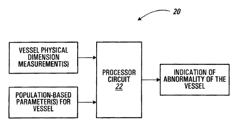
Referring to Figure 1, an apparatus for evaluating a vessel according to a
first
embodiment of the invention is shown generally at 20. In this embodiment, the
apparatus 20 includes a processor circuit 22, which is configured to receive
at least
one measurement of a physical dimension of a vessel. The processor circuit 22
is
configured to produce an indication of abnormality in the vessel, in response
to the at
least one received measurement and at least one population based parameter for
the
vessel.
CA 02421352 2003-03-07
-10-
VESSEL
Referring to Figures 1 and 2, in this embodiment the vessel to be evaluated by
the
apparatus 20 shown in Figure 1, is shown generally at 50 in Figure 2. In the
present
embodiment, the vessel 50 includes a coronary artery of a mammal; which in
this
embodiment includes a human. Alternatively, other types of vessels may be
substituted.
In this embodiment, the vessel 50 includes a plurality of coronary artery
segments,
one of which is shown at 52 in Figure 2. The coronary artery segment 52 is
defined
between an upstream end 54 and a downstream end 56 thereof. More particularly,
the
upstream end 54 is defined at an intersection of the segment 52 with an
upstream
branch 58, and the downstream end 56 is defined at an intersection of the
segment 52
with a downstream branch 60. The upstream and downstream branches 58 and 60
act
as anatomical landmarks to locate the segment 52. In the illustrative example
shown
in Figure 2, blood is pumped through the coronary artery segment 52 by a heart
(not
shown), in a direction flowing from the upstream end 54 toward the downstream
end
56.
In the present embodiment, the vessel 50 has a plurality of internal physical
dimensions, including a proximal diameter 62 at a location proximal to the
upstream
end 54, a distal diameter 64 at a location distal from the upstream end 54,
and a
reference diameter 66 indicative of a diameter at one or more reference
locations in
the segment. In this embodiment, the segment 52 also includes a focal
abnormality or
lesion 68. The segment 52 has an internal diameter 70 at a location of the
lesion 68.
In this specification, the term "diameter" means the distance from any point
on the
periphery of a surface, body or space to the opposite point. Thus, in the case
of the
coronary artery segment 52 shown in Figure 2, the term "diameter" in
connection with
the proximal diameter 62, the distal diameter 64, the reference diameter 66
and the
diameter 70 at the location of the lesion 68, means the length of a straight
line
extending from one point on an internal wall of the segment 52, through a
central axis
(not shown) of the segment, to an opposite point on an opposite side of the
internal
CA 02421352 2003-03-07
-11-
wall of the segment 52, the straight line lying in a plane normal to the
central axis of
the segment. The term "diameter" does not necessarily connote either
circularity or
symmetry of a cross-section of the segment 52, which may be naturally or
unnaturally
deformed from such circularity or symmetry in a given case, depending upon the
particular vessel in question.
In the present embodiment, the reference diameter 66 may represent an internal
diameter of the segment 52 at a single location interposed between the
proximal
diameter and the distal diameter other than the location of the lesion 68, or
alternatively, may include an average of a plurality of such diameters, for
example.
Such an average reference diameter may be an average of multiple contiguous
measurements in the reference area of the segment, for example. The reference
diameter 66 preferably does not include any measurement at the location of the
lesion
68 or any other focal (visible) abnormality in an image of the segment 52.
As noted, in the present embodiment the diameter 70 is the internal diameter
of the
segment 52 at the location of the lesion 68. Alternatively, if no lesion is
visible in an
angiographic or other image of the segment 52, the diameter 70 may include the
minimum lumen diameter of the segment 52. Alternatively, the measurement of
the
diameter 70 may be omitted entirely in such cases.
In this embodiment, the vessel 50 includes a plurality of segments such as the
segment 52 shown in Figure 2. More particularly, in the present embodiment the
vessel 50 includes a left main artery segment (LM); a proximal left anterior
descending artery segment (PLAD); a middle left anterior descending artery
segment
(MLAD); a distal left anterior descending artery segment (DLAD); a diagonal
artery
segment (DIAG); a proximal circumflex artery segment (PLCX); a distal
circumflex
artery segment (DLCX); a marginal artery segment (OM); an intermediate artery
segment (INT); a proximal right coronary artery segment (PRCA); a middle right
coronary artery segment (MRCA); a distal right coronary artery segment (DRCA);
and a right posterior descending artery segment (RPDA). Alternatively, other
types of
CA 02421352 2003-03-07
-12-
segments may be substituted. More generally, other types of vessels may be
substituted.
APPARATUS
Referring to Figures 1, 2 and 3, the processor circuit of the apparatus 20 is
shown
generally at 22 in Figure 3. In this embodiment, the processor circuit 22
includes a
microprocessor 23, which may be housed in a general purpose or special purpose
computer (not shown), for example. More generally, however, in this
specification,
the term "processor circuit" is intended to broadly encompass any type of
device or
combination of devices capable of performing the methods and functions
described
herein, including (without limitation) other types of microprocessors,
microcontrollers, other integrated circuits, other types of circuits or
combinations of
circuits, logic gates or gate arrays or programmable devices of any sort, for
example,
either alone or in combination with other such devices located at the same
location or
remotely from each other, for example. Additional types of processor circuits
will be
apparent to those ordinarily skilled in the art upon review of this
specification, and
substitution of any such other types of processor circuits is considered not
to depart
from the scope of the present invention as defined by the claims appended
hereto.
In this embodiment, the microprocessor 23 is in communication with a random
access
memory (RAM) 24, which may be either separate from or integral with the
microprocessor, or which may include a combination of onboard and external
RAM.
In this embodiment, the microprocessor 23 is also in communication with a
storage
medium 26, which in this embodiment includes a hard disk drive, although
alternatively, other types of storage media may be substituted.
In the present embodiment, the microprocessor 23 is also in communication with
an
input/output (I/O) interface 28, through which the microprocessor is in
communication with one or more input devices 30 and one or more output devices
32.
More particularly, in this embodiment the input devices 30 include a keyboard
and a
mouse, and the output devices 32 include a display monitor, a printer, and a
CA 02421352 2003-03-07
-13-
removable media data recorder for recording information on a removable medium
such as a compact disc or a floppy diskette, for example. The microprocessor
23 is
also in communication, via the I/O interface 28, with a network 34, which in
this
embodiment includes the public Internet. The processor circuit 22 is thus able
to
communicate with other devices that are in communication with the network 34,
such
as a remote database 36, for example. The microprocessor 23 may also be in
communication via the I/O interface with additional devices (not shown). For
example, the microprocessor may be in communication with a media interface
device
such as a CD-ROM drive, a CD-RW drive, a floppy diskette drive, a tape drive,
or
other removable media read or read/write device.
In this embodiment, the storage medium 26 acts a computer readable medium
storing
various codes, including a vessel evaluation routine 101, fog directing the
processor
circuit 22 to carry out the methods and functions disclosed herein.
Alternatively,
however, such codes may be provided by other computer readable media. For
example, removable media such as a compact disc or floppy diskette, or a
transmission medium such as a communications network, may provide such codes.
Generally, any medium capable of providing signals such as that shown at 27,
including code segments for directing the processor circuit 22 to perform the
methods
and functions disclosed herein, may be substituted if desired.
In this embodiment, in addition to storing the functional codes of the vessel
evaluation
routine 101, the storage medium 26 also includes a population based vessel
parameters store 103, for storing at least one population-based parameter for
the
vessel 50 shown in Figure 2. More particularly, in this embodiment the
population-
based vessel parameters store 103 stores a plurality of population-based
parameter
records, each record including a dominance field 106, a gender field 107, a
segment
identification field 108, an average reference diameter field 109, a reference
diameter
standard error field 110, a reference diameter standard deviation field 111,
an average
antegrade tapering field 112, an antegrade tapering standard deviation field
113, an
average retrograde tapering field 114, and a retrograde tapering standard
deviation
field 115, as described in greater detail below in connection with Figure 4.
CA 02421352 2003-03-07
-14-
Alternatively, other types of population-based vessel parameters may be
substituted,
as appropriate for a particular application.
In the present embodiment, the population-based vessel parameters are stored
within
the vessel evaluation routine 101 itself, as a data portion thereof.
Alternatively, if
desired, the population-based vessel parameters store 103 may be provided in a
separate area of the storage medium 26, or may be stored in any other suitable
local or
remote computer-readable medium accessible by the processor circuit 22, such
as the
remote database 36, for example.
In this embodiment, the storage medium 26 also includes an output reports
store 117,
for storing output reports produced by the microprocessor 23 under the
direction of
the vessel evaluation routine 101, as discussed in greater detail below.
In the present embodiment, the vessel evaluation routine 101 configures the
microprocessor 23 to define a plurality of registers in the RAM 24, including
a
dominance register 123, a gender register 125, and a subject identification
register
127. The vessel evaluation routine 101 also configures the microprocessor 23
to
define a vessel evaluation store 131 in the RAM 24. The vessel evaluation
store 131
stores a plurality of vessel evaluation records, each record pertaining to a
particular
corresponding segment of a vessel of a subject identified by the contents of
the
dominance, gender and subject identification registers. In this embodiment,
each
vessel evaluation record in the vessel evaluation store 131 includes a vessel
segment
identification field 132, a proximal diameter field 134, a distal diameter
field 136, a
reference diameter field 138, a minimum lumen diameter field 140, a percent
diameter
stenosis field 150, an antegrade tapering field 152, a retrograde tapering
field 154, an
atheroma burden field 156, a population-based percent diameter stenosis field
158, a
lower confidence interval boundary field 160, an upper confidence interval
boundary
field 162, a reference diameter Z-score field 164, a population-based
antegrade
tapering field 166, and a population-based retrograde tapering field 168. The
contents
of the various fields of the vessel evaluation store 131 are discussed in
greater detail
below in connection with the vessel evaluation routine 101.
CA 02421352 2003-03-07
-15-
VESSEL PHYSICAL DIMENSION MEASUREMENTS
Referring back to Figure 2, in this embodiment, measurements of the physical
dimension or dimensions of interest of the vessel 50 may be obtained by
conventional
methods, if desired. More particularly, in the present embodiment, in which
the
vessel 50 includes the coronary artery segment 52, the measurements of
physical
dimensions of the vessel that are obtained include the proximal diameter 62,
the distal
diameter 64, the reference diameter 66, and the diameter 70 in the location of
the
lesion 68. If the segment 52 does not include the focal abnormality or lesion
68, then
the measurement of the diameter 70 may be omitted.
In this embodiment, the measurements 62, 64, 66 and 70 (if applicable) are
obtained
from standard angiographic images. Such angiographic images may be produced by
a
variety of methods, such as the Judkins technique, for example. In this
embodiment,
to obtain such images, a radio-opaque dye is injected into the subject near
the vessel
50 or segment 52 of interest, and X-ray images of the vessel or segment are
obtained.
For example, where the subject is a human, a thin wire and catheter may be
inserted
into an artery and fed through the artery to the vicinity of the coronary
segment 52 of
interest (often in the vicinity of the heart), at which point the 'radio-
opaque contrast
material is injected into the vessel. Typically, such injection is repeated
more than
once as an X-ray imaging machine is moved relative to the subject's body, to
obtain
X-ray angiographic images from different views. The angiographic images are
then
analyzed using conventional analysis techniques to obtain the desired
measurements.
Most such conventional analysis techniques employ a computer assisted edge-
detection algorithm to quantify the physical dimensions of the vessel 50 or
segment
52 within a given angiographic image. Typically, conventional methods utilize
the
density information registered by the opaque contrast material when injected
into a
vessel. This density information is mathematically analyzed with respect to
first and
second derivatives of the density. Each algorithm utilizes a certain weighting
of the
position of the first and second derivative of the density function to provide
an initial
estimate of the location of the edge of the arteriographic image. The
algorithm then
CA 02421352 2003-03-07
-16-
employs various methods that ensure that the resulting locations are
contiguous and
smooth in producing diameter measurements for a given segment. Typically, the
user
is able to manually adjust the edges located by the automated edge detection
algorithm, to allow the user to manually improve the accuracy of the vessel
diameter
measurements if desired. The resulting vessel diameter measurements are
initially
expressed as numbers of pixels in the image. Once such initial vessel physical
dimension measurements have been obtained, calibration information is then
used to
convert such pixel number measurements into absolute length units. Typically,
this is
achieved by inputting a known size of at least one object present in the
image, such as
the catheter used to inject the radio-opaque dye, for example, to provide a
size scale to
the image. Numerous software packages are available to quantify the dimensions
of
an angiographic image, such as CorTrek~ (by Quinton Instruments Company, USA),
Artrek (from ImageComm System, USA) and the QCA-CMS system (by Medis
Company, the Netherlands), for example. The above exemplary systems have been
reviewed by Mancini et al. ((2001) Can J Cardiol 17(7):785-791).
Although conventional coronary angiographic imaging techniques have been
described, by way of example, for obtaining the vessel physical dimension
measurements 62, 64, 66 and 70, alternatively, any other suitable measurement
techniques, conventional or otherwise, may be substituted to obtain these
values. For
example (without limitation), other types of angiography, radiography,
ultrasound,
magnetic resonance imaging, computed axial tomographic imaging, or vascular
imaging techniques, may be substituted.
In addition, it will be appreciated that for some applications, not all of the
vessel
physical dimension measurements 62, 64, 66 and 70 are required. For example,
it will
be apparent from the following description of the vessel evaluation routine
101 that
some advantageous indications of abnormality may be obtained using only the
reference diameter measurement 66, for example. Similarly, other advantageous
indications of abnormality may be produced using only the diameter 70 in the
vicinity
of the lesion 68. Still other advantageous indications of abnormality may be
obtained
CA 02421352 2003-03-07
-1~-
using the reference diameter 66 and either the proximal diameter 62 or the
distal
diameter 64, for example. More generally, measurements of other types of
physical
dimensions, of the same or other types of vessels may be substituted, if
desired.
POPULATION-BASED PARAMETERS FOR VESSEL
Refernng to Figures 2, 3 and 4, an illustrative sample of the contents of the
population-based vessel parameters store is shown generally at 103 in Figure
4. In
this embodiment, for each record stored in the population-based vessel
parameters
store 103, the dominance field 106 is used to store an identification of the
dominance
of all members of a population group from which the population-based vessel
parameters in the record were obtained. In the present embodiment, in which
the
vessel 50 is a human coronary artery segment 52, it will be appreciated that a
given
subject may have either a right dominant system, a left dominant system, or a
co-
dominant system. As the physical dimensions of a given artery segment may vary
significantly between different dominance types, for the purposes of the
present
embodiment, it is not desirable to compare the vessel physical dimension
measurements for a subject of one dominance type to population-based vessel
parameters obtained from measurements of individuals with a different
dominance
type or with mixed dominance types. Accordingly, in this embodiment the
contents
of each record in the population-based vessels parameters store are based on
measurements obtained from individuals having a single corresponding dominance
type, and the dominance field 106 contents identify that corresponding type as
right
dominant, left dominant, or co-dominant, as the case may be. Similarly, it
will be
appreciated that physical dimensions of a given artery segment may differ
significantly between opposite genders. Accordingly, each record in the
population-
based .vessel parameters store 103 contains population-based parameters
obtained
from a population group of individuals of the same gender, and the gender
field 107
stores an identification of that gender. Similarly, in this embodiment the
segment
identification field 108 stores an identification of the particular coronary
artery
segment to which the record in question relates.
CA 02421352 2003-03-07
-18-
In the present embodiment, the average reference diameter field 109 of each
record
stores an average reference diameter value of the relevant vessel segment.
More
particularly, in this embodiment the average reference diameter is a mean
value of
measurements of the reference diameter 66 shown in Figure 2, obtained from a
statistically significant number of individuals having the system dominance
and
gender specified in the dominance and gender fields 106 and 107 of the record.
The
reference diameter standard error field 110 stores a value representing the
Standard
Error associated with the average reference diameter, and similarly, the
reference
diameter standard deviation field 111 stores the Standard Deviation associated
with
the average reference diameter.
In this embodiment, the average antegrade tapering field 112 stores a value
representing the average tapering or narrowing of the vessel segment 52, in
the
direction of blood flow. More particularly, in this embodiment, for each
individual of
the population-based group to which the record relates, an individual
antegrade
tapering value representing tapering of the downstream end 56 of the segment
52
relative to its middle region is calculated, by dividing the distal diameter
64 by the
reference diameter 66, subtracting the result from unity, and multiplying by
100%.
Thus, the tapering value will be zero if there is no tapering, i.e. if the
distal diameter is
equal to the reference diameter; the tapering value will be positive if there
is
antegrade tapering, i.e., if the distal diameter is narrower than the
reference diameter,
with a value of +100% representing complete blockage (zero diameter) at the
distal
diameter location; and the tapering value will be negative if. there is
antegrade
widening, i.e. if the reference diameter is narrower than the distal diameter,
with a
value of -100% representing complete blockage at the reference value location.
The
average antegrade tapering value of all individuals of the population group to
which
the record relates, or more particularly, the mean of the individual tapering
values of
all such individuals, is then calculated and stored in the average antegrade
tapering
field 112 of the record. The Standard Deviation associated with the average
antegrade
tapering value is stored in the antegrade tapering standard deviation field
113.
CA 02421352 2003-03-07
-19-
Similarly, in this embodiment the average retrograde tapering field 114 stores
a value
representing the average tapering or narrowing of the vessel segment 52, in a
direction
opposite to the direction of blood flow. More particularly, in this
embodiment, for
each individual of the population-based group to which the record relates, an
individual retrograde tapering value representing tapering of the upstream end
54 of
the segment 52 relative to its middle region is calculated, by dividing the-
proximal
diameter 62 by the reference diameter 66, subtracting the result from unity,
and
multiplying by 100%. Thus, the tapering value will be zero if there is no
tapering, i.e.
if the proximal diameter is equal to the reference diameter; the tapering
value will be
positive if there is retrograde tapering, i.e., if the proximal diameter is
narrower than
the reference diameter, with a value of +100% representing complete blockage
(zero
diameter) at the proximal diameter location; and the tapering value will be
negative if
there is retrograde widening, i.e. if the reference diameter is narrower than
the
proximal diameter, with a value of -100% representing complete blockage at the
reference value location. The average retrograde tapering value of all
individuals of
the population group to which the record relates, or more particularly, the
mean of the
individual tapering values of all such individuals, is then calculated and
stored in the
average retrograde tapering field 114 of the record. The Standard Deviation
associated with the average retrograde tapering value is stored in the
retrograde
tapering standard deviation field 115.
In this embodiment, such a record containing an average reference diameter; an
average antegrade tapering value, and an average retrograde tapering value;
along.
with their associated error values, is produced for each combination of
segment,
gender and dominance. Although the illustrative sample of records shown in
Figure 4
includes only records fox population groups with right-dominant systems, it
will be
understood that the population-based vessel parameters store 103 preferably
stores
similar additional records for left-dominant systems, and for co-dominant
systems.
It will be appreciated that one or more of the types of population-based
vessel
parameters shown in Figure 4 may be omitted if desired. For example, as will
be
apparent from the following description of the vessel evaluation routine,
CA 02421352 2003-03-07
-20-
advantageous indications of abnormality may be obtained using only the average
reference diameter values, or using only the antegrade or retrograde tapering
values,
for example. More generally, other types of population-based vessel parameters
may
be substituted if desired, depending upon the application in question.
OPERATION
Referring to Figures 2, 3, SA and 5B, the vessel evaluation routine is shown
generally
at 101 in Figure SA. Generally, the vessel evaluation routine 101 configures
or
programs the processor circuit 22 to receive at least one measurement of a
physical
dimension of the vessel 50, and configures the processor circuit to produce an
indication of abnormality in the vessel, in response to the at least one
received
measurement and at least one population based parameter for the vessel.
In this embodiment, the vessel evaluation routine 101 includes a first block
201 of
codes, which directs the processor circuit 22 to obtain a dominance indication
for a
system in which the vessel 50 is located (for example, in the present
embodiment, in
which the vessel 50 includes the coronary artery segment 52 of a human
subject, the
dominance indication identifies the subject as having a right dominant, left-
dominant,
or co-dominant system). To achieve this, block 201 directs the processor
circuit 22 to
control the output devices 32 to generate and display a graphical user
interface
window, prompting a user (not shown) of the apparatus 20 to use one or more of
the
input devices 30 to specify the dominance of the vessel's system. In response
to
receiving user input identifying the dominance of the system, block 201
directs the
processor circuit to store the dominance indication in the dominance register
123 in
the RAM 24.
Block 203 then directs the processor circuit 22 to obtain a gender indication
identifying the subject as male or female. Block 203 directs the processor
circuit to
control the output devices 32 to generate and display a graphical user
interface
window prompting the user to identify the subject's gender. In response to
receiving
user input from one or more of the input devices 30 identifying the gender,
block 203
directs the processor circuit to store the gender information in the gender
register 125
CA 02421352 2003-03-07
-21-
in the RAM 24. (In this embodiment, the contents of the subject identification
register 127 are obtained and stored only if the user wishes to save the
resulting
output report, as discussed below in connection with block 227.)
Block 205 then directs the processor circuit 22 to obtain an identification of
the
particular segment type of the segment 52 of the vessel 50 that has been
measured for
the subject in question. To achieve this, block 205 directs the processor
circuit to
control the output devices 32 to generate and display a graphical user
interface
window prompting the user of the apparatus 20 to use the input devices 30 to
identify
the segment type. In the present embodiment, the graphical user interface
window
allows the user to select any one of the following coronary artery segment
types: left
main artery (LM}; proximal, left anterior descending artery (PLAD); middle
left
anterior descending artery (MLAD); distal left anterior descending artery
(DLAD);
diagonal artery (DIAG); proximal circumflex artery (PLCX); distal circumflex
artery
(DLCX); marginal artery (OM); intermediate artery (INT); proximal right
coronary
artery (PRCA); middle right coronary artery (MRCA); distal right coronary
artery
(DRCA); and right posterior descending artery (RPDA). Alternatively, if
desired,
other segment types or combinations of segment types may be substituted. Upon
receiving user input representing the selected segment type, block 205 directs
the
processor circuit 22 to create a new vessel evaluation record in the vessel
evaluation
store 131, arid to write an identification of the selected segment type into
the segment
identification field 132 of the newly created record.
In the present embodiment, block 206 then configures the processor circuit 22
to
receive at least one measurement of a physical dimension of the segment 52 of
the
vessel 50. More particularly, in this embodiment the at least one measurement
includes a diameter at a location in the segment. More particularly still, in
this
embodiment the at least one measurement includes the proximal diameter 62, the
distal diameter 64, the reference diameter 66, and the diameter 70 of the
segment at
the location of the lesion 68. To achieve this, block 206 directs the
processor circuit
22 to control the output devices 32 to generate and display a graphical user
interface
window prompting the user of the apparatus 20 to use the input devices 30 to
specify
CA 02421352 2003-03-07
-22-
whether a focal abnormality or lesion was visible in the angiographic or other
image
used to produce the vessel measurements, or in other words, whether a
measurement
of the diameter 70 at the location of the lesion 68 is available. If such a
focal
abnormality or visible lesion was present, block 206 directs the processor
circuit to
control the output devices 32 to generate and display a graphical user
interface
window prompting the user to enter the proximal diameter, distal diameter,
reference
diameter, and minimum lumen diameter (i.e., diameter 70) values for the
segment.
Otherwise, if no lesion or focal abnormality was apparent, the user is
prompted to
enter only the proximal diameter, distal diameter, and reference diameter
values.
Upon receiving user input specifying these values, block 206 directs the
processor
circuit to store these received physical dimension measurement values in the
new
vessel evaluation record created at block 205 above in the vessel evaluation
store 131,
in the proximal diameter field 134, the distal diameter field 136, the
reference
diameter field 138, and the minimum lumen diameter field 140 respectively, as
appropriate. Alternatively, it will be appreciated from the following that
significant
advantages may be obtained even if some such physical dimensions are omitted.
More generally, other types of measurements of physical dimensions of a vessel
may
be substituted, if desired.
Once the measurements of the physical dimensions of the segment 52 have been
received and stored at block 206, block 207 directs the processor circuit 22
to
determine whether evaluations of any additional segments of the vessel 50 are
to be
performed. Block 207 directs the processor circuit 22 to control the output
devices 32
to generate a graphical user interface window prompting the user of the
apparatus 20
to control the input devices 30 to indicate whether or not vessel measurements
are to
be input for any additional segments. If user input is received indicating
that one or
more further segments are to be evaluated, the processor circuit is .directed
back to
blocks 205 and 206 to create one or more further vessel evaluation records in
the
vessel evaluation store 131, each record corresponding to each further
respective
segment of the vessel 50, as described above.
CA 02421352 2003-03-07
-23-
Alternatively, if user input is received indicating that no further vessel
segment
measurements are to be entered, block 209 directs the processor circuit to
address the
first vessel evaluation record in the vessel evaluation store 131.
Block 211 then directs the processor circuit 22 to identify a percent diameter
stenosis
value (non-population based), as well as a non-population-based tapering value
representing a tapering of the vessel, in response to the physical dimension
measurements (which in this embodiment are diameter measurements of the
vessel)
received and stored in the fields 132, 134, 136 and 138 of the currently
addressed
vessel evaluation record. In this embodiment, block 211 directs the processor
circuit
22 to identify the conventional percent diameter stenosis (% DS) of the
segment to
which the record corresponds, as follows:
%DS=(1-[MD/RD])x100%
where:
MD = the diameter 70 at the location of the lesion 68, stored in the minimum
lumen diameter field 140 of the currently addressed vessel evaluation
record; and
RD = the reference diameter 66 stored in the reference diameter field 138 of
the currently addressed vessel evaluation record.
Block 211 then directs the processor circuit 22 to produce antegrade and
retrograde
tapering values (TA and TR) for the segment to which the currently addressed
record
corresponds, as follows:
TA = ( 1- [DD / RD] ) x 100%
TR = ( 1- [PD / RD] ) x 100%
where:
PD = the proximal diameter measurement stored in the proximal diameter
field 134 of the currently addressed vessel evaluation record;
CA 02421352 2003-03-07
-24-
DD = the distal diameter measurement stared in the distal diameter field 136
of the currently addressed vessel evaluation record; and
RD = the reference diameter measurement value stored in the reference
diameter field 13$ of the currently addressed vessel evaluation record.
Block 211 directs the processor circuit 22 to store the percent diameter
stenosis value
(%DS) and the antegrade and retrograde tapering values (TA and TR) in the
percent
diameter stenosis field 150, the antegrade tapering field 152 and the
retrograde
tapering field 154, respectively, of the currently addressed vessel evaluation
record.
In the present embodiment, block 213 then directs the processor circuit 22 to
produce,
as an indication of abnormality of the vessel 50, an indication of stenosis of
the
vessel, in response to a measurement of a physical dimension of the vessel and
a
population-based reference dimension for the vessel. More particularly, block
213
directs the processor circuit to produce, as the indication of stenosis, a
population-
based percent stenosis value, in response to a ratio of the physical dimension
measurement to the population-based reference dimension. More particularly
still,
block 213 configures the processor circuit to set the population-based percent
stenosis
value equal to one hundred times a difference between unity and a ratio of the
physical dimension measurement to the population-based reference dimension. In
this embodiment, the physical dimension measurement includes a first diameter
measurement of the vessel, and the population-based reference dimension
includes a
population-based reference diameter for the vessel. Thus, in the present
embodiment,
the population-based percent stenosis value includes a population-based
percent
diameter stenosis value.
To produce such a population-based percent diameter stenosis value, in this
embodiment; block 213 first directs the processor circuit 22 to select an
appropriate
value to use as the population-based reference diameter in the above
production of the
population-based percent diameter stenosis value. In this regard, the
processor circuit
is directed to locate and address a record in the population-based vessel
parameters
store 103 corresponding to the currently addressed vessel evaluation record
(i.e.
CA 02421352 2003-03-07
-25-
having segment identification field 108 contents matching those of the segment
identification field 132, having gender field 107 contents matching those of
the
gender register 125, and having dominance field 106 contents matching those of
the
dominance register 123). Block 213 directs the processor circuit to compare
the
reference diameter measurement stored in the reference diameter field 138 of
the
currently addressed vessel evaluation record, to the average reference
diameter value
stored in the average reference diameter field 109 of the currently addressed
population-based vessel parameters record. If the reference diameter
measurement is
less than or equal to the average reference diameter value, then the average
reference
diameter value stored in the average reference diameter field 109 is used as
the
population-based reference diameter for the purpose of calculating the
population-
based percent diameter stenosis value.
Conversely; however, if the reference diameter measurement is greater than the
average reference diameter value, this suggests a significant possibility that
the
subject's actual "healthy" artery segment diameter sizes may be larger than
average,
in which case it may not be desirable to compare the subject's vessel segment
diameter measurements to the average reference diameter value, as such a
comparison
may tend to conceal the presence of atheroma or other abnormalities.
Accordingly, in
such a case, the subject's actual reference diameter measurement stored in the
reference diameter field 138 is used as the population-based reference
diameter for the
purpose of calculating the population-based percent diameter stenosis. The
reference
diameter measurement may be considered to be "population-based" in such a
case,
insofar as it is selected in response to a comparison with the population-
based average
reference diameter value. If desired, block 213 may also store a flag (not
shown) in
association with the reference diameter field 138 contents, to serve as a
reminder that
the subject's actual reference diameter measurement, and not the average
reference
diameter field 109 contents, were used to produce the population-based percent
diameter stenosis value.
Block 213 then directs the processor circuit 22 to select an appropriate
physical
dimension measurement of the vessel segment 52 to use as the first diameter
CA 02421352 2003-03-07
-26-
measurement in the above production of the population-based percent diameter
stenosis value. If the minimum lumen diameter field 140 of the currently
addressed
vessel evaluation record has a defined value therein (e.g., received and
stored at block
206 as discussed above}, then the contents of the minimum lumen diameter field
140
are used as the first diameter measurement, for the purpose of producing the
population-based percent diameter stenosis value. If however, no visible
lesion or
focal abnormality existed and therefore no measurement of the diameter 70 at
the
location of such a lesion was obtained and stored in the minimum lumen
diameter
field 140, block 213 directs the processor circuit 22 to compare the contents
(PD) of
the proximal diameter field 134 to the contents (RD) of the reference diameter
field
138 and the contents (DD) of the distal diameter field 136, and to select the
smallest
value stored in any of these three fields of the currently addressed vessel
evaluation
record as the first diameter measurement for the purpose of producing the
population-
based percent diameter stenosis value (PB% DS).
Block 213 then directs the processor circuit 22 to produce the population
based
percent diameter stenosis as follows:
PB % DS = ( 1 - [DF / RpB) ) x 100%
where:
DF = first diameter measurement (DF = contents MD of minimum lumen
diameter field 140 if defined, otherwise DF = lesser of contents PD , DD
and RD of fields 134, 136, 138); and
RPB = population based reference diameter value (RpB =contents RAV of
average reference diameter field 109 if and only if field 109 contents >
reference diameter field 138 contents RD; otherwise RPB = field 138
contents RD).
Block 213 then directs the processor circuit 22 to store the population based
percent
diameter stenosis value in the population-based percent diameter stenosis
field 158 of
the currently addressed vessel evaluation record.
CA 02421352 2003-03-07
-27-
In the present embodiment, block 2I5 then directs the processor circuit 22 to
identify
a confidence interval for the stenosis of the vessel, in response to the first
diameter
measurement, the population based reference diameter, and an error value
associated
with the population based reference diameter. More particularly, in this
present
embodiment block 21S directs the processor circuit 22 to identify a lower
confidence
interval boundary equal to unity minus a ratio of the first diameter
measurement to a
difference between the population-based reference diameter and a constant
multiplied
by the error value. Similarly, block 215 directs the processor circuit to
identify an
upper confidence interval boundary equal to unity minus a ratio of the first
diameter
measurement to a sum of the population-based reference diameter and a constant
multiplied by the error value. To achieve this, in the present embodiment,
block 215
first directs the processor circuit to produce the lower confidence interval
boundary
value, as follows:
Lower C.I. of PB % DS = [1- (DF / [RPB -1.96 6R])] x 100%
where:
DF = first diameter measurement (DF = contents MD of minimum Iumen
diameter field 140 if defined, otherwise DF = lesser of contents PD ; DD
and RD of fields 134, 136, 138); and
RPB = population based reference diameter value (RpB =contents RAV of
average reference diameter field 109 if and only if field 109 contents >
reference diameter field 138 contents RD; otherwise RPM = field 138
contents RD); and
6R = the standard error for RAV, stored in the standard error field 110 (it is
noted that even if, in a given case, RPB = RD rather than RAV, the value
6R nevertheless provides a reasonable standard error range associated
with the selected RPB value):
If the lower conf dence interval boundary value produced above is negative,
block
215 directs the processor circuit to set the lower confidence boundary value
equal to
CA 02421352 2003-03-07
-28-
zero. Block 215 directs the processor circuit 22 to store the resulting lower
confidence interval boundary value in the lower confidence interval boundary
field
160 of the currently addressed vessel evaluation record
Block 215 then directs the processor circuit 22 to produce the upper
confidence
interval boundary value, as follows:
Upper C.I. of PB % DS = [1- {DF / [8P$ + 1.96 a8])] x 100%
where:
I O DF = first diameter measurement (DF = contents MD of minimum lumen
diameter field 140 if defined, otherwise DF = lesser of contents PD , DD
and RD of fields 134, 136, 138); and
RpB = population based reference diameter value (8P$ =contents RAV of
average reference diameter field 109 if and only if field 109 contents >
reference diameter field 138 contents RD; otherwise RPB = field 138
contents RD); and
6R = the standard error for RAV, stored in the standard error field 110.
Block 215 directs the processor circuit 22 to store the upper confidence
interval
boundary value in the upper confidence interval boundary field 162 of the
currently
addressed vessel evaluation record.
It will be appreciated that the selection of ~1.9668 in the confidence
interval values
represents a 95% confidence interval, or in other words, a 95% chance that the
true
population-based percent diameter stenosis value falls within the range
defined
between the upper and lower confidence interval boundary values.
In this embodiment, block 217 then directs the processor circuit 22 to produce
a
comparison value relating the population-based reference dimension, the
physical
dimension measurement, and an error value associated with the population-based
reference dimension. More particularly, in this embodiment block 217 directs
the
CA 02421352 2003-03-07
-29-
processor circuit to set the comparison value equal to a ratio of a difference
between
the population based reference dimension and the physical dimension
measurement to
the error value. More particularly still, in the present embodiment the
physical
dimension measurement includes a first diameter measurement of the vessel, and
the
population-based reference dimension includes a population-based reference
diameter
for the vessel, namely, the contents of the average reference diameter field
109. In the
present embodiment, the comparison value is also referred to as "atheroma
burden".
Block 217 directs the processor circuit 22 to produce the comparison value or
atheroma burden as follows:
atheroma burden = (RAE - DA) / SR
where:
DA = first diameter measurement (DA = contents MD of minimum lumen
diameter field 140 if defined, otherwise DA = contents RD of reference
diameter field 138);
RAV = contents of average reference diameter field 109; and
SR = the standard deviation fox RAV, stored in the standard deviation field
111.
If the comparison value (atheroma burden) value produced above is negative,.
block
217 directs the processor circuit 22 to set the comparison value equal to
zero. Block
217 then directs the processor circuit 22 to store the resulting comparison
value in the
atheroma burden field 15G of the currently addressed vessel evaluation record.
Block 219 directs the processor circuit 22 to identify a shape characteristic
of the
vessel 50. More particularly, in this embodiment block 219 configures the
processor
circuit to produce, as the shape characteristic, a tapering comparison value;
in
response to the tapering of the vessel and a population-based average tapering
value.
More particularly still, in the present embodiment block 219 directs the
processor
circuit to set the tapering comparison value equal to a ratio of a difference
between
the tapering and the population based average _tapering value, to an error
value
associated with the population based average tapering value. To achieve this,
block.
CA 02421352 2003-03-07
-30-
219 directs the processor circuit to . produce such population based tapering
comparison values for both the antegrade and retrograde tapering, as follows:
TAPB - CTA - TAAV) ~ SAAV
TRPB = CTR - TRAY) ~ SlzAv
where:
Tppg = population based antegrade tapering comparison value;
TRpB =population based retrograde tapering camparison value;
TA = subject's antegrade tapering value stored in the antegrade tapering field
152 (produced above at block 211);
TR = subject's retrograde tapering value stored in the retrograde tapering
field 154 (also produced above at block 211);
T~v =population-based average antegrade tapering value = contents of
average antegrade tapering field 112;
T~v =population-based retrograde tapering value = contents of average
retrograde tapering field 114;
SAAV = ~e standard deviation of T,~v = contents of antegrade tapering
standard deviation field 113; and
SRAV = the standard deviation of TRav = contents of retrograde tapering
standard deviation field 115.
Block 219 then directs the processor circuit 22 to store both the antegrade
and
retrograde tapering comparison values in the population-based antegrade
tapering
field 166 and the population-based retrograde tapering field 168 of the
currently
addressed vessel evaluation record, respectively.
In the present embodiment, block 221 then directs the processor circuit 22 to
produce
a Z-score in response to a physical dimension measurement of the vessel, a
population
based average reference dimension and an error value associated therewith.
More
particularly, in this embodiment the physical dimension measurement includes a
first
diameter measurement of the vessel, and the population-based average reference
CA 02421352 2003-03-07
-31-
dimension includes a population-based reference diameter for the vessel. To
produce
such a Z-score, block 221 directs the processor circuit 22 to produce a
reference
diameter Z-score, as follows:
ZR=~RD-RAV~~sR
where:
ZR = reference diameter Z-score;
RD = contents of reference diameter field 138;
RAV = contents of average reference diameter field 109; and
SR = the standard deviation of RAV, stored in the average reference diameter
standard deviation field 111.
Block 221 directs the processor circuit 22 to store the reference diameter Z-
score
value in the reference diameter Z-score field 164 of the currently addressed
vessel
evaluation record.
In this embodiment, block 223 then directs the processor circuit 22 to
determine
whether the vessel evaluation store 131 includes any further vessel evaluation
records
corresponding to further segments of the vessel 50 (such as the segment 52),
in
respect of which physical dimension measurements have been received and stored
at
block 206 but abnormality indications and other evaluation values have not yet
been
produced at blocks 211 through 221 as discussed above. If any such vessel
evaluation
records exist, block 225 directs the processor circuit 22 to address the next
successive
record corresponding to the next successive segment, and the processor circuit
is
directed back to blocks 211 through 221, as described above, until all such
records
have been addressed.
In the present embodiment, in addition to the fields described above of each
record in
the vessel evaluation store 131 corresponding to each respective segment, the
vessel
evaluation store 131 also includes a plurality of subject average fields, foi
maintaining
averages of various measurements and values over all segments of the vessel 50
of the
CA 02421352 2003-03-07
-32-
particular subject identified by the contents of the dominance, gender and
subject
identification registers 123; 125 and 127. More particularly, in this
embodiment the
vessel evaluation store 131 includes a subject average reference diameter
field 139 for
maintaining an average of the contents of the reference diameter fields 138 of
all
vessel evaluation records for the particular subject; a subject average
percent
diameter stenosis field 151 for maintaining an average of the contents of the
percent
diameter stenosis fields 150 of all vessel evaluation records for that
subject; a subject
average comparison value field 157 for maintaining an average of the contents
of the
comparison value or atheroma burden fields 156 of all vessel evaluation
records for
the subject; and a subject average population-based percent diameter stenosis
field
I59 for maintaining an average of the contents of the population-based percent
diameter stenosis fields 158 of all vessel evaluation records for the subject.
Thus, in
the present embodiment the processor circuit is directed to update the
contents of the
subject average fields 139, 151, 157 and 159. Such updating of the subject
average
fields may be carried out as the contents of each vessel evaluation record
field are
created and stored at blocks 206 through 221, or alternatively, such contents
may be
updated periodically, at less frequent intervals. For example, updating may be
carried
out each time block 225 is executed, or alternatively, when blocks 227 through
229
below are executed.
In this embodiment, if at block 223 it was determined that no further vessel
evaluation
records remain to be evaluated at blocks 211 through 221, block 227 directs
the
processor circuit 22 to identify a desired form of output report. To achieve
this, block
227 directs the processor circuit 22 to control the output devices 32 to
generate and
display a graphical user interface window prompting the user of the apparatus
20 to
use the input devices 30 to select the desired form of output report. In the
present
embodiment the graphical user interface window enables the user to opt to
save, view,
or print the output report. Upon receiving user input representing the
selected option,
block 227 directs the processor circuit to temporarily store an indication of
the
selected option in a working register (not shown) of the RAM 24. In addition,
if the
received user input indicates a selection of the "save" option, block 227
directs the
processor circuit 22 to control the output devices 32 to generate and display
a
CA 02421352 2003-03-07
-33-
graphical user interface prompting the user to provide subject identification
information identifying the subject system to which the vessel 50 belongs, via
the
input devices 30. In the present embodiment, in which the vessel50 includes
one or
more coronary artery segments, the subject is a human whose body contains the
coronary artery segments. Accordingly, the subject identification information
requested may include information such as a medical record number, the
subject's
name, the subject's initials and site (location) of the subject, for example.
In this
embodiment, such subject identification information is requested only if the
"save"
option is selected. Alternatively, however, the subject information may be
requested
for all vessels (for example, at block 203 as discussed above). In response to
receiving such subject identification information from the input devices 30,
block 227
directs the processor circuit 22 to store the subject identification
information in the
subject identification register 127 in the RAM 24.
Refernng to Figures 3, SB, 6 and 7, in the present embodiment, block 229 then
configures the processor circuit 22 to generate and output an output report,
such as
either of those shown at 300 in Figure 6 and at 400 in Figure 7, for example.
In this
embodiment, each of the exemplary output reports 300 and 400 is generated in
the
format of a Microsoft Access (TM) database table, although alternatively, any
suitable
output format may be substituted. Generally, the columns of the output reports
shown
in Figures 6 and 7 correspond to the various fields of the records of the
vessel
evaluation store 131 shown in Figure 3. Thus, the output report 300 includes
fifteen
columns 302, 304, 306, 308, 310, 312, 314, 316, 318, 320, 322, 324, 326, 328
and
330, which respectively correspond to the fifteen fields 132, 134, 136, 138,
140, 150,
152, 154, 156; 158, 160, 162, 164, 166 and 168 of the records of the vessel
evaluation
store 131. Likewise, in this embodiment the output report 400 includes fifteen
columns 402, 404, 406, 408, 410, 412, 414, 416, 418, 420, 422, 424, 426, 428
and 430
respectively corresponding to the above-noted vessel evaluation store fields.
In
addition, the output report 400 includes a gender identifier 432 corresponding
to the
gender register 125, a subject identifier 434 corresponding to the subject
identification
register 127, and may also include a dominance identifier (not shown)
corresponding
to the dominance register 123. In this embodiment, the output report 400 also
CA 02421352 2003-03-07
-3q.-
includes subject average fields 436, 438, 440 and 442, corresponding to the
subject
average fields 139, 151, 157 and 159 of the vessel evaluation store 131.
In this embodiment, block 229 configures the processor circuit 22 to control
the
output devices 32 to notify the user of the apparatus 20 as to whether one or
more of
the indications of abnormality is indicative of the presence or absence of an
apparent
abnormality of the vessel. In the present embodiment, this is achieved in a
number of
different ways.
In this regard, block 229 configures the processor circuit 22 to control the
output
devices 32 to notify the user of the absence of an apparent abnormality of the
vessel
when the indication of abnormality is within a first pre-defined range, and to
notify
the user of the presence of an apparent abnormality of the vessel when the
indication
of abnormality is outside the first pre-defined range. For example, in this
embodiment, if any of the comparison values or atheroma burden values in the
column 318 or the column 418 (corresponding to the atheroma burden field 156
of the
vessel evaluation store 131) is between zero and two (it will .be recalled
that this
particular value cannot be negative), the processor circuit is directed to
notify the user
of the absence of an apparent abnormality, and if any such value is greater
than or
equal to two, the processor circuit is directed to notify the user of the
presence of an
apparent abnormality.
Similarly, in this embodiment the Z-score values shown in the columns 326 or
426
(corresponding to the reference diameter Z-score field f64), the population-
based
antegrade tapering values shown in the columns 428 and 328 (corresponding to
the
population-based antegrade tapering f eld 166) and the population-based
retrograde
tapering values shown in the columns 430 and 330 (corresponding to the
population-
based retrograde tapering field 168), are treated in a similar manner. If any
of these
values is between -2 and +2, the processor circuit is directed to notify the
user of the
absence of an apparent abnormality. Conversely, if any of these values is less
than or
equal to -2, or greater than or equal to +2, then the processor circuit is
directed to
notify the user of the presence of an apparent abnormality.
CA 02421352 2003-03-07
-35-
In the present embodiment, in which the output devices 32 include a display
device;
block 229 configures the processor circuit 22 to cause the display device to
highlight a
display of the indication of abnormality. More particularly, the processor
circuit is
configured to cause the display device to highlight the display in a first
color when the
indication indicates the presence of an apparent abnormality. More
particularly still,
in this embodiment the first color is red. Thus, any of the atheroma burden
values,
reference diameter Z-scores, or population-based antegrade or retrograde
tapering
values having an absolute magnitude greater than or equal to two will be
highlighted
in red, to immediately notify the user of an apparent abnormality of the
segment of the
vessel to which the value in question relates. Conversely, in this embodiment,
any
such value having an absolute magnitude less than two will not be highlighted,
thereby effectively notifying the user of the absence of an apparent
abnormality,
through the absence of the red highlighting that would indicate such an
abnormality.
Alternatively, or in addition, if desired, the concept of the .presence of an
apparent
abnormality may be further refined, to distinguish between a possible presence
of an
abnormality, and a probable presence of an abnormality. Thus, in the present
embodiment, for at least some of the indications of abnormality, block 229
configures
the processor circuit 22 to control the output devices 32 to notify the user
of a possible
presence of an abnormality of the vessel when the indication of abnormality is
outside
the first pre-defined range but within a second pre-defined range, and to
notify the
user of the probable presence of an abnormality of the vessel when the
indication of
abnormality is outside the second pre-defined range.
For example, in the present embodiment, if any of the population-based percent
diameter stenosis values in the columns 420 or 320 (corresponding to the
population-
based percent diameter stenosis field 158 in the vessel evaluation store 131)
is within
a first pre-defined range, namely, between zero and 30%, block 229 directs the
processor circuit 22 to control the output devices 32 to notify the user of
the absence
of an apparent abnormality of the segment of the vessel to which the value
relates, by
highlighting the value in another color (in this embodiment, green). If any of
the
CA 02421352 2003-03-07
-36-
population-based percent diameter stenosis values is outside the first pre-
defined
range but within a second pre-defined range (namely, greater than or equal to
30%,
but less than 70%), then block 229 directs the processor circuit 22 to control
the
output devices to notify the user of a possible presence of an abnormality of
the
segment of the vessel to which the value relates, by highlighting the value in
yet
another color, such as amber, for example (alternatively, such inconclusive
values
may be indicated by white highlighting, or by an absence of highlighting, for
example). Finally, if any of the population-based percent diameter stenosis
values is
outside the second pre-defined range (in this embodiment, greater than or
equal to
70%), block 229 directs the processor circuit to notify the user of the
probable
presence of an abnormality of the segment of the vessel to which the value
relates, by
highlighting the relevant value in red. In this embodiment, block 229 further
directs
the processor circuit to employ a similar notification method in relation to
the
conventional percent diameter stenosis values in the columns 412 and 312 of
the
output reports (corresponding to the percent diameter stenosis field 150 of
the vessel
evaluation store 131).
Alternatively, other highlighting schemes, or more generally, other ways of
notifying
a user of the presence or absence of an apparent abnormality of the vessel,
may be
substituted if desired.
In this embodiment, although the foregoing description of notifications of
apparent
abnormalities emphasized use of a display device as the illustrative example
of the
output devices 32, alternatively, if the output report 300 or 400 is to be
printed, such
notifications may be achieved by printing appropriate notifications on the
printed
report, either by highlighting the relevant values as described above, or by
any other
suitable way, such as by automatically producing text or graphical warnings to
direct
the user's attention to any values indicative of the possible or probable
presence of an
apparent abnormality of the vessel, for example.
Similarly, if the output report is merely to be saved in the output reports
store 117,
such notifications may be saved along with the other output report information
shown
CA 02421352 2003-03-07
-37-
in Figures 6 and 7, if desired. Alternatively, rather than saving the
notifications
themselves, the thresholds defining the boundaries of the first and second pre-
defined
ranges may be saved in association with the vessel evaluation routine 101
itself, so
that each time a saved output report is loaded, such notifications are re-
generated (or
not, as the case may be) in accordance with the thresholds stored in
association with
the vessel evaluation routine. Such an approach may facilitate manual or
automatic
updating or changing of the thresholds. In the present embodiment, in either
such
case, the output reports store 117 includes registers, stores and fields (not
shown)
corresponding to all of those of the RAM 24, and block 229 directs the
processor
circuit 22 to save the output report by copying the contents of the various
registers
and records in the RAM 24 into corresponding registers and records in the
output
reports store 117.
Also in this embodiment, block 229 directs the processor circuit 22 to
highlight any
reference diameter value RD in the column 406 or 306 of the output reports 400
and
300, which is greater than the corresponding population-based average
reference
diameter value stored in the average reference diameter field 109 of the
population-
based vessel parameters store 103 record corresponding to the same segment
(i.e., the
record having dominance field 106, gender field 107 and segment identification
field
108 contents matching those of the dominance register 123, gender register 125
and
segment identification field 132). It will be recalled that in such a case, in
view of the
likelihood that the subject's vessel segment dimensions are atypically large,
the
reference diameter value RD may be substituted for the population-based
average
reference diameter RAV for the production of the population-based percent
diameter
stenosis value and associated confidence interval. Accordingly, for some
applications
it may be desirable to highlight the reference diameter values RD for such
segments,
to act as a reminder that a slightly different calculation method was
employed. As
noted above in connection with block 213, a flag may be generated at the time
the
population-based percent diameter stenosis value is produced, to identify any
such
segments; alternatively, block 229 may direct the processor circuit to compare
the
reference diameter measurement value RD to the population-based average
reference
diameter value for this purpose. In this embodiment, block 229 directs the
processor
CA 02421352 2003-03-07
-38-
circuit to highlight the segment in question, by highlighting the relevant
reference
diameter measurement value RD in a fourth color, which in this embodiment is
blue.
Referring to Figure 7, it will be appreciated that the present embodiment is
capable of
notifying a user of apparatus 20 of the presence of apparent abnormalities of
the
vessel 50, in situations where conventional angiographic techniques would fail
to
detect such abnormalities. For example, in the case of the left main (LM)
artery
segment values shown in Figure 7, the conventional percent diameter stenosis
value
shown in the column 412 (corresponding to the percent diameter stenosis field
150
contents for that segment) is zero. Accordingly, the conventional percent
diameter
stenosis value indicates absolutely no stenosis or blockage of the LM segment.
In
contrast, the comparison value shown in the column 418 and the reference
diameter
Z-score shown in the column 426 (corresponding to the contents of the atheroma
burden field 156 and the reference diameter Z-score field 164, respectively,
of the
vessel evaluation store record for the segment in question) both have absolute
magnitudes greater than two, and therefore, each of these values is
highlighted in red,
to notify the user of the apparatus 20 of the presence of an apparent
abnormality in the
LM segment of the vessel 50. Likewise, the population-based percent diameter
stenosis value in the column 420 (corresponding to the population-based
percent
diameter stenosis field 158 of the vessel evaluation store record for the LM
segment)
is outside a first pre-defined range but within the second predefined range
(i.e., greater
than 30% and less than 70%), and is therefore indicative of a possible
presence of an
abnormality of the vessel. In this embodiment, the user is notified of this
possible
presence by an absence of either green highlighting or red highlighting (which
are
associated with the absence and probable presence of an abnormality,
respectively).
ALTERNATIVES
Various alternative ways of obtaining the relevant physical dimension
measurements
of the vessel 50 may be substituted. For example, rather than prompting the
user of
the apparatus 20 to enter specific information in a stepwise manner,
alternatively, the
user may be presented with an interactive combined spreadsheet and output
report
such as that shown at 400 in Figure 7, allowing the user to enter data in the
CA 02421352 2003-03-07
-39-
appropriate fields) as available. In such an embodiment the production of
indications
may be generated automatically as soon as the appropriate input values are
entered
into the appropriate fields of the spreadsheet. Alternatively, the desired
input
information may have been previously stored in a storage medium, in which case
the
processor circuit 22 may be directed to retrieve such information from the
storage
medium. Alternatively, any other suitable ways of obtaining the desired input
information may be substituted.
Although coronary angiographic images and measurement methods were described
as
an exemplary way of producing the measurements of the physical dimensions of
the
vessel 50, other measurement techniques may be substituted. In the exemplified
embodiment quantitative coronary arteriography values were evaluated in
reference to
population based arteriography values from normal patients. Similar vessel
lumen
values may be obtained using a wide array of diagnostic imaging techniques,
selected
from but not limited to magnetic resonance imaging (MRI), computerized axial
tomography (CAT), positron emission tomography (PET), and ultrasound, for
example.
Although the foregoing embodiment employed linear a length measurement
(diameter) of the vessel as an illustrative example of a physical dimension
measurement of the vessel, alternatively, other types of physical dimension
measurements may be substituted. For example, the physical dimension
measurements may include area measurements of the vessel. As a more particular
example, where the vessel includes a coronary artery segment or similar
vessel, each
physical dimension measurement may include a measurement of an internal cross-
sectional area of the vessel. In this regard, it will be appreciated that some
measurement techniques, such as intravascular ultrasound, for example, often
provide
vessel measurements expressed in units of area, representing the cross-
sectional
internal area of an artery or other vessel. Thus, other embodiments of the
invention
may be provided to accommodate these and other alternative physical dimension
measurements.
CA 02421352 2003-03-07
-40-
For example, referring back to Figure SA, in one such alternative embodiment
of the
invention, block 206 of the vessel evaluation routine 101 may be modified to
allow a
user to choose whether to enter linear diameter measurements or cross-
sectional area
. measurements of the vessel. If axes measurements are selected and input by
the user,
the processor circuit 22 may be configured to convert each received area
measurement
A to an equivalent diameter measurement D, on the assumption that the vessel
in
question has a circular cross-sectional area. Thus, as A = ~ _ ~(D/2)2, D =
(4A/~c)°~5.
The remainder of the vessel evaluation routine may then proceed as above,
using the
converted diameter measurement. Alternatively, in another embodiment, a vessel
evaluation routine may receive and directly manipulate such area measurements,
and
may employ population-based reference areas rather than (or in addition to)
population-based reference diameters. Such population-based reference area
measurements may be calculated directly from the population-based reference
diameter measurements on the assumption of circular cross-sections of the
vessel,
taking due care to re-calculate all associated error measurements as required.
Alternatively, such population-based reference area measurements may be
independently obtained, without any necessary assumption as to cross-sectional
shape
of the vessels.
In addition, embodiments of the present invention may be employed to evaluate
and
produce indications of abnormality for vessel types other than coronary
arteries.
Internal carotid and vertebral arteries, for example, may be imaged and
evaluated to
identify abnormalities that could affect blood flow to the brain and may be
useful in
assessing stroke risk. Similarly, the subclavian, brachial and radial
arteries, for
example, may be imaged and evaluated to identify abnormalities that could
affect
blood flow to the arm. Of particular interest may be an evaluation of the
radial artery
and internal mammary artery, in patients preparing for bypass surgery where
the
radial artery or internal mammary artery is being used in the bypass
procedure. In
addition, the iliac, femoral and popliteal arteries, for example, may be
imaged and
evaluated to identify abnormalities that could affect blood flow to the leg of
a patient.
Similarly, embodiments of the invention may be applied to types of vessels
other than
arterial vessels. For example, embodiments of the invention may be useful in
the
CA 02421352 2003-03-07
-41-
venous system for identifying abnormalities. The superior and inferior vena
cava,
superior and inferior sagittal sinus veins, for example, may also be evaluated
to
identify abnormalities for diagnostic and/or treatment purposes.
More generally, while specific embodiments of the invention have been
described and
illustrated, such embodiments should be considered illustrative of the
invention only
and not as limiting the invention as construed in accordance with the
accompanying
claims.