Note: Descriptions are shown in the official language in which they were submitted.
CA 02429246 2008-11-27
IMPLANTABLE JOINT PROSTHESIS
BACKGROUND OF THE INVENTION
1. Field of the Invention
The invention relates to implantable prostheses that are suitable for
replacement of diarthroidal or arthroidal joints by creating an artificial
diarthroidal-
like joint at the site of the implant.
In a particular embodiment, the invention relates to implantable prostheses
serving as replacements for at least a portion of the intervertebral disc
material, i.e., a
spinal disc endoprostheses suitable for implantation in vertebrates, including
humans.
2. Description of Related Art
Many joints in the human body, such as hips, knees, shoulders, etc., are
diarthroidal, meaning that the joints include a joint capsule that is filled
with fluid.
The capsule fluid lubricates the joint, and allows the surfaces of the joint
to move with
a low coefficient of friction. The spine, by contrast, can be considered to be
a series
of joints, some of which (the anterior joint or disc) lack a fluid filled
capsule and are
therefore arthroidal (the spine also contains facet joints that are
diarthroidal). The
interior portion of intervertebral discs are not provided by the body with
significant
blood supply; their homeostasis is enhanced by the diffusion of fluids into
the disc
tissue, thus supplying them with nutrients. This, to some extent, allows the
tissue to
1
CA 02429246 2003-02-06
WO 02/11650 PCT/US01/24791
grow and repair damage done by stress as the joint moves. Despite this
process, in
mature adults, spinal disc tissue degrades continuously over time.
Sufficiently
advanced degeneration can lead to herniation or rupture of the spinal disc.
Herniation of a spinal disc can result in a number of debilitating symptoms,
including intractable pain, weakness, and sensory loss. Treatment of these
symptoms
frequently requires surgical removal of at least a portion of the herniated
disc, a
procedure known as discectomy. Often discectomy alone cannot stop the
progressive
degeneration at the level of disc excision. An additional procedure is often
performed
in conjunction with the discectomy with the objective of fusing together
(arthrodesis)
the vertebral bodies surrounding the affected disc space. This is accomplished
by
removing the cartilaginous endplates by scraping the surfaces of the vertebral
body
and inserting a piece of graft bone, which may be an allograft from a bone
bank, or an
autograft, typically taken from the iliac crest of the patient, or other
suitable material..
The discectomy and arthrodesis procedures can be problematic, however.
Discectomy problems have been described above. The grafting or fusion
procedure
has a variable success rate of about 80%, and even when successful, requires
considerable recovery time before fusion is complete. Perhaps of even greater
concern, successful fusion eliminates normal spinal biomechanics. Range of
motion
at the level of the fusion is ideally eliminated, because the affected
vertebrae have
been effectively joined to form a single bone. Because the patient tries to
maintain
the same overall range of motion of the entire spine, additional stress is
imposed on
the intervertebral discs of the adjacent vertebrae . This, in turn, may lead
to
accelerated degeneration at levels above and below the fusion site, which may
require
additional treatment, including discectomy and fusion. Grafting procedures
carry
2
CA 02429246 2008-11-27
WO 02/11650 PCT/USO1/24791
some risk of tissue rejection and disease transmission if an allograft is
used, and risk
of harvest site morbidity when the patient's own tissue is harvested.
As a result of these difficulties with intervertebral fusion, attempts have
been
made to provide a prosthetic solution to degenerative disc disease that
maintains the
patient's normal spinal biomechanics, allows for shorter recovery times, and
avoids
the complications inherent in harvesting and/or grafting bone tissue. Some of
these
efforts have centered around providing an endoprosthetic intervertebral
implant, as
described in U.S. Patent Nos. 5,865,846, 5,674,296, 5,989,291, 6001,130, and
6,022,376,
Design and construction of such an implant, however, is not simple.
Desirably, the implant should be precisely placed in a prepared intervertebral
space,
and should contain elements that are immobilized with respect to each of the
vertebral
bodies, so that the implant does not migrate or shift, potentially contacting,
abrading,
or otherwise damaging the spinal cord, ligaments, blood vessels, and other
soft tissue.
At the same time, the implant should allow the vertebral bodies to move
relative to
each other in a way that provides the equivalent motion afforded by a healthy
intervertebral disc, and that allows the affected vertebral joint to
participate in the
coordinated overall movement of the spine in a way that closely approximates
the
natural movement of a healthy spinal column. The implant should be
biocompatible,
and avoid the introduction of toxic or harmful components into the patient,
such as
release of wear debris. The implant should also restore normal disc height and
maintain the patient's vertebral lordosis, and should not allow any
significant post-
operative subsidence. The implant should be at least partially constrained by
soft
tissue in and around the intervertebral space, in order to allow a simpler,
more
efficient design. There remains a need for a device which would decrease
patient
3
CA 02429246 2003-02-06
WO 02/11650 PCT/US01/24791
recovery time, and reduce the occurrence of postoperative degeneration at
levels
above and below the implant, as compared with fusion techniques. In addition,
such
an implant would avoid the need for harvesting of autograft bone tissue,
thereby
eliminating morbidity at the harvesting site. Such an implant should also
provide
elasticity and damping sufficient to absorb shocks and stresses imposed on it
in a
manner similar to that of the natural spinal disc.
SUMMARY OF THE INVENTION
This invention satisfies the needs and concerns described above. Other
concerns can arise that are more unique to any joint replacement or
reconstruction,
particularly with respect to device stability, range of motion, and
postoperative
material degradation. In general, in patients undergoing joint replacement,
the
patient's condition and quality of life is improved more by a technique that
provides a
range of motion that more closely approximates the range of motion of a
healthy joint
(assuming that this can be done in a safe manner) than by a technique that
provides a
decreased range of motion. Important parts of accomplishing this goal include
using
an implant design that is highly stable when implanted, and making use of the
soft
tissue associated with the joint (to the extent possible) to stabilize the
implant and
leave restriction of some of the motion of the joint to the soft tissue. This
allows the
implant design to be considerably simpler. Irrespective of the joint being
implanted,
an implant that provides an effectively sealed, fluid filled capsule (i.e., an
artificial
diarthroidal-like joint) will likely provide an added margin of safety because
the
moving surfaces are isolated from the surrounding tissue and body fluids, and
the
environment in which the moving surfaces operate can be engineered and
controlled.
The lubrication effects in such a joint allow it to function more effectively
and
potentially generate less wear debris. Any wear debris that is generated,
however, is
4
CA 02429246 2003-02-06
WO 02/11650 PCT/US01/24791
contained within the implant and will not come into contact with live tissue
or body
fluids. Similarly, tissue ingrowth into the articulating regions of the
implant and
degradation of the implant materials by body fluids are also avoided.
In one aspect, the invention can be viewed as a surgical implant where the
structure of the implant contains cooperating features that allows a joint
into which
the implant has been inserted to closely approximate the biomechanics and
motion of
a healthy joint.
In this aspect, the invention contains two rigid opposing plates or shells,
each
having an outer surface adapted to engage the prepared surfaces of the bones
of a joint
in such a way that frictional forces resist movement of the plates or shells
relative to
the bone surface. The outer surfaces are sufficiently rough that frictional
forces
strongly resist any slippage between the outer surface and the bone surfaces
in the
joint. In addition to providing surface friction at the interface with the
bone, the outer
surfaces may be adapted to allow for bony ingrowth, which acts to further
stabilize
the plates or shells in place over time. The inner surfaces of the plates or
shells are
relatively smooth, and adapted to slide easily with low friction across a
portion of the
outer surface of an elastically deformable, resilient central body disposed
between the
plates or shells. Desirably, the inner surfaces have an average roughness of
about 1 to
about 8 microinches, more particularly less than about 3 microinches. The
central
body has a shape that cooperates with the shape of the inner surface of the
plate or
shell so as to provide motion similar to that provided by a healthy joint.
The surgical implant of the invention provides exceptional stability, because
the roughened outer surfaces of the plates or shells and their geometric shape
supply
sufficient frictional force to keep the implant from slipping from its proper
position on
the surfaces of the bones forming the joint. In addition, the geometry of the
outer
CA 02429246 2003-02-06
WO 02/11650 PCT/US01/24791
surfaces and the prepared surfaces of the bone cooperate to contain the
implant
between the bone surfaces. The smooth inner surfaces of the rigidopposing
plates or
shells are shaped to cooperate and articulate with the shape of the smooth
surface of
the deformable resilient central body to allow relatively unconstrained motion
of the
plates or shells with respect to the resilient central body until the limit of
acceptable
motion is reached. Once the limit of allowable motion is reached, the shape of
the
inner surface of the plate or shell cooperates with the shape of the
deformable resilient
central body to effectively resist any movement beyond the desired motion.
This
allows the motion of a joint containing the implant to closely approximate the
motion
provided in a healthy joint, alleviating undesirable stresses imposed on the
joint or
bone structure, or in the case of a .vertebral implant, on adjacent joints as
well. This,
in turn, reduces the likelihood of further joint degeneration in adjacent
joints.
The deformable resilient central body also provides elasticity and dampening
properties, similar to those provided by healthy joint tissue. It is also
sufficiently
creep-resistant or resistant to plastic deformation to avoid post-operative
loss of disc
space height and to maintain appropriate joint geometry. The surface of the
central
body is hard, in some embodiments harder than the interior, which provides
good
wear resistance. It is also very lubricious, which provides good tribological
properties
in conjunction with the inner surfaces of the rigid plates or shells.
The resulting implant is safe because it can be implanted with precision, and
once implanted, it is stable. It is extremely effective because the geometry
of the
internal surfaces is configured to provide a range of motion that closely
approximates
that provided by healthy joint tissue, thus allowing coordinated movement of
the
spine and reducing stress on adjacent joints.
6
CA 02429246 2003-02-06
WO 02/11650 PCT/US01/24791
In another aspect, the invention relates to an implant that effectively
provides
an artificial diarthroidal-like joint, suitable for use in replacing any
joint, but
particularly suitable for use as an intervertebral disc endoprosthesis. In
this aspect,
the implant contains, in addition to the opposing rigid plates or shells and
deformable,
resilient central body described above, a flexible sleeve or sheath that
extends
between edges of the opposing plates or shells.
The inner surface of this sheath, together with the inner surfaces of the
rigid
plates or shells, defines a cavity surrounding the central body. Most, if not
all, of the
interior space of this cavity can be filled with a fluid lubricant, further
decreasing the
frictional force between inner surfaces of the plates or shell and the surface
of the
central body, again within the constraints of allowable motion.
The flexible sleeve or sheath serves to hold the implant together as a single
unit, making it easier to manipulate during the implant procedure. It also
retains the
lubricant within the implant and provides a contained, sealed environment that
keeps
tissue from entering the interior of the implant, isolates the central body
from possible
attack or degradation by body fluids, and prevents any wear debris that might
be
generated from exiting the implant and migrating into surrounding tissues .
The
implant therefore provides a sealed capsule presenting only biocompatible
surfaces to
surrounding tissues, and keeping wear surfaces internal to the implant and
permanently lubricated. The result is an implant with extremely good
durability,
because the articulating surfaces have been isolated away from the natural
bone
surfaces and placed in a lubricated capsule.
In yet another aspect, the invention provides a vertebral endoprosthesis,
having:
7
CA 02429246 2003-02-06
WO 02/11650 PCT/US01/24791
an upper and a lower rigid, opposed, biocompatible plate or shell, each
comprising:
an outer, rough surface;
an inner, smooth surface; and
an edge between the surfaces;
wherein the inner smooth surface of at least one of the plates or shells
comprises a first motion limiting device;
a deformable, resilient central body disposed between the inner, smooth
surfaces of the upper and lower plates or shells, comprising:
a smooth upper surface adjacent to the inner smooth surface of the
upper plate or shell and a smooth lower surface adjacent to the inner
smooth surface of the lower plate or shell;
a second motion limiting device disposed on at least one of the smooth upper
and lower surfaces adapted to contact the first motion limiting device and
limit the
relative motion of the plate or shell with respect to the central body.
The inner surfaces of the plates or shells can desirably be concave, and
articulate with smooth upper surfaces of the deformable resilient central body
that are
convex. This arrangement creates, in effect, an artificial ball-and-socket-
like joint in
the intervertebral space, which joint is inherently stable under compression.
In a more specific embodiment of this aspect of the invention, the vertebral
endoprosthesis contains:
an upper and a lower rigid, opposed biocompatible concavo-convex shell, each
comprising:
an outer, rough convex surface, comprising a porous coating of a
biocompatible material;
8
CA 02429246 2003-02-06
WO 02/11650 PCT/US01/24791
an inner concave surface, comprising:
a smooth contact area; and
an axial post extending toward the opposing shell; and
an edge between the surfaces, comprising:
a circumferential groove adapted to receive a retaining ring;
a first ridge circumscribing the contact area of the inner
concave surface and extending axially toward the opposing
shell;
an insertion tab extending axially away from the opposing
shell, and comprising an opening adapted to releasably engage
a tool for manipulating, inserting, or removing the
endoprosthesis;
a closable passage between the outer surface and the inner surface of
the shell;
a deformable, resilient central body disposed between the inner, smooth
concave surfaces of the upper and lower shells, comprising:
smooth convex upper and lower surfaces complementary and adjacent
to the smooth contact area of the inner surfaces of the respective upper
and lower shells;
a second ridge circumscribing each of the smooth convex upper and
lower surfaces and adapted to contact the first ridge of the adjacent
shell and limit the relative motion of the shell with respect to the
central body;
9
CA 02429246 2003-02-06
WO 02/11650 PCT/US01/24791
a laterally extending equatorial ridge disposed between the first ridge
of the upper concavo-convex shell and the first ridge of the lower
concavo-convex shell;
an opening in the upper and lower convex contact surfaces adapted to
receive the axial post of the inner surface of each shell;
an elastic sheath or sleeve disposed between the upper and lower shells and
surrounding the central body, comprising an inner surface, an outer surface,
an
upper edge attached to the upper shell, and a lower edge attached to the lower
shell, wherein the inner surface of the sheath and the inner surfaces of the
shells define an enclosing cavity;
an upper retaining ring of a biocompatible material disposed in the
circumferential groove in the upper concavo-convex shell and securing the
upper edge
of the elastic sheath or sleeve to the shell and a lower retaining ring of a
biocompatible material disposed in the circumferential groove of the lower
concavo-
convex shell and securing the lower edge of the sheath or sleeve to the shell.
This endoprosthesis provides the advantages described above with respect to
the more general aspects of the invention, and more specifically provides an
implantable vertebral joint that approximates the disc height and range of
motion of a
healthy intervertebral disc, with significantly increased durability relative
to natural
intervertebral disc material, and without the drawbacks of spinal fusion.
In addition, the concavo-convex geometry of the opposing shells, and the
precise preparation of a mating concave surface in the vertebral body
endplates, into
which the convex outer surfaces of the shells are inset, provide a highly
stable
implanted joint. Coupled with the roughness provided by the porous coating on
the
outer surface of the shell, this inset shape holds the implant firmly in place
so that it
CA 02429246 2003-02-06
WO 02/11650 PCT/US01/24791
cannot migrate and come into contact with nerves or blood vessels, and so that
the
desired bony ingrowth can occur. The convex outer surface also provides
additional
surface area that contacts cancellous bone, increasing both the opportunity
for bony
ingrowth and the frictional force holding the shells in place. The mating of
the
concave inner surfaces of the shells with the curved shape of the central body
provides a simple ball-and-socket-like system that is inherently highly stable
under
compression, as it will be when implanted. The embodiment of the invention
using
concavo-convex shells and a convex surface on the deformable central body
therefore
provides immediate mechanical stability.
Because the range of motion provided by the implant closely approximates
that of a healthy disc, post operative adjacent level disc degeneration is
minimized or
avoided entirely. In addition, the implant does not significantly constrain
joint
torsion, but instead relies on the remaining soft tissue (e.g., remaining disc
annulus,
ligaments, etc.) in and around the implanted joint to provide appropriate
brsional
constraint. Neither the shapes of the plates or shells or of the central body,
or of the
central retaining posts or central axial opening restrict the torsional
movement of the
shells relative to the central body (i.e., the rotation of the shells or of
the central body
about a central axis. This is of benefit because it significantly decreases
the stress
imposed on the interface between the bone surfaces and the outer surfaces of
the
implant, making movement of these implant surfaces relative to the bone less
likely.
This, in turn, increases the likelihood of bony ingrowth instead of fibrous
tissue
formation, and therefore increases long-term stability.
11
CA 02429246 2003-02-06
WO 02/11650 PCT/USO1/24791
BRIEF DESCRIPTION OF DRAWINGS
The invention can be more clearly understood by reference to the following
drawings, which illustrate specific embodiments thereof, and which are not
intended
to limit the scope of the appended claims.
FIG. 1 is a perspective drawing of an intervertebral endoprosthesis in
accordance with a specific embodiment of the invention.
FIG. 2 is an elevational view of the intervertebral endoprosthesis shown in
FIG. 1.
FIG. 3 is a top plan view of the intervertebral endoprosthesis shown in FIG. 1
and 2.
FIG. 4 is an isometric cross sectional view of the intervertebral
endoprosthesis
shown in FIG. 1, 2, and 3.
FIG. 5 is a plan view of an implant plug and plug installation tool used to
insert a plug into an intervertebral endoprosthesis.
FIG. 6 is a sectional view of the intervertebral endoprosthesis shown in FIG.
1-4.
FIG. 7 is an exploded perspective view of the intervertebral endoprosthesis
shown in FIG. 1-4 and 6.
FIG. 8 is a plan view (A) and sectional view (B) of one embodiment of an
intervertebral endoprosthesis of the invention undergoing lateral bending.
FIG. 9 is a plan view (A) and sectional view (B) of one embodiment of an
intervertebral endoprosthesis of the invention undergoing translation.
FIG. 10 is a plan view (A) and sectional view (B) of one embodiment of an
intervertebral endoprosthesis of the invention undergoing lateral bending.
12
CA 02429246 2003-02-06
WO 02/11650 PCT/US01/24791
FIG. 11 is a plan view (A) and sectional view (B) of one embodiment of an
intervertebral endoprosthesis of the invention undergoing translation.
The invention can be more clearly understood by reference to some of its
specific embodiments, described in detail below, which description is not
intended to
limit the scope of the claims in any way.
DETAILED DESCRIPTION OF SPECIFIC EMBODIMENTS
In broad aspect, the size and shape of the implant are substantially variable,
and this variation will depend upon the joint geometry. Moreover, implants of
a
particular shape can be produced in a range of sizes, so that a surgeon can
select the
appropriate size prior to or during surgery, depending upon his assessment of
the joint
geometry of the patient, typically made by assessing the joint using CT, MRI,
fluoroscopy, or other imaging techniques.
The rigid opposing plates or shells can be made of any rigid, biocompatible
material, but are generally made of a biocompatible metal, such as stainless
steel,
cobalt chrome, ceramics, such as those including A12O3 or Zr2O3, or titanium
alloy.
ASTM F-136 titanium alloy has been found to be particularly suitable. As
indicated
above, the outer surface of the rigid opposing plates or shells are rough, in
order to
restrict motion of the shells relative to the bone surfaces that are in
contact with the
plates. This is particularly important in the time period just after
implantation (the
"acute" phase of healing), since excessive movement of the implant relative to
the
bone can result in the formation of fibrous tissue between the bone and the
implant,
rather than the bony ingrowth, which is desirable for long term implant
stability (i.e.,
during the "chronic" phase of healing). It has been discovered that a porous
coating
formed from nonspherical sintered beads provides very high friction between
the
outer surface of the shell and the bone, as well as providing an excellent
interaction
13
CA 02429246 2003-02-06
WO 02/11650 PCT/USO1/24791
with the cancellous bone of the joint, increasing the chances of bony
ingrowth. One
example of a suitable nonspherical sintered bead coating is that made of pure
titanium, such as ASTM F-67. The coating can be formed by vacuum sintering.
At least a portion of the inner surface of each plate or shell is smooth, and
of a
shape that complements and articulates with the shape of at least a portion of
the
central body. This smoothness and correspondence in shape provides
unconstrained
movement of the plate or shell relative to the central body, provided that
this
movement occurs within the allowable range of motion.
The structural features of the shapes of the inner surface of the plate or
shell
and the central body that interact to limit the movement to this allowable
range will
necessarily vary to some extent, based on the joint in which the implant will
be used.
As an example, the edge of the plate or shell can be extended toward the
central body,
so as to for a wall that, under shear, can contact a ridge or shoulder formed
in the
surface of the central body. This will allow for unconstrained motion of the
plate or
shell except in a direction that will bring the extension into contact with
the ridge. By
forming the extension around the entire edge of the shell, and by forming a
ridge or
shoulder that encloses a portion of the surface of the central body,
translational,
flexural, extensional, and lateral motion of the plate or shell relative to
the central
body can be constrained in all directions. Those of skill in the art will
recognize that a
bead or ridge at other locations on the inner surface of the plate or shell
will serve a
similar purpose, and that the location of this bead or ridge, as well as the
ridge or stop
on the central body, can be varied between implants for different joints, in
order to
obtain the desired range of motion for that particular joint.
The plates may be identical, which is desirable for ease of manufacture, or
may be of different design (shape, size, and/or materials) to achieve
different
14
CA 02429246 2003-02-06
WO 02/11650 PCT/US01/24791
mechanical results. For example, differing plate or shell sizes may be used to
more
closely tailor the implant to a patient's anatomy, or to shift the center of
rotation in the
cephalad or caudal direction.
In a more particular embodiment, the inner surface of the shell and the outer
surface of the central body can contain complementary structures that will
function as
an expulsion stop, so that the central body cannot be expelled from between
the
opposing plates or shells when the plates or shells are at maximum range of
motion in
flexion/extension. Examples of such structures include a post and
corresponding hole
to receive the post. The hole can have a diameter sufficiently large that
relative
motion between the shells and central body is unconstrained within the
allowable
range of motion, but that will nevertheless cause the post to arrest the
central body
before it is expelled from the implant under extreme compression.
Alternatively, the
diameter of the post may be such that it limits the translational movement of
the
central body during normal motion of the spine by contacting the surface of
the hole
in the central body at the limit of the allowable range of motion for the
device. The
elastically deformable, resilient central body may also vary somewhat in
shape, size,
composition, and physical properties, depending upon the particular joint for
which
the implant is intended. The shape of the central body should complement that
of the
inner surface of the shell to allow for a range of translational, flexural,
extensional,
and rotational motion, and lateral bending appropriate to the particular joint
being
replaced. The thickness and physical properties of the central body should
provide for
the desired degree of elasticity or damping. Accordingly, an elastomeric
material is
typically used for the central body. However, the central body should be
sufficiently
stiff to effectively cooperate with the shell surfaces to limit motion beyond
the
allowable range. The surface of the central body should be sufficiently hard
to
CA 02429246 2003-02-06
WO 02/11650 PCT/US01/24791
provide acceptable wear characteristics. One way to achieve this combination
of
properties is to prepare a central body having surface regions that are harder
than the
material of the central body closer to its core. The central body is therefore
desirably
a biocompatible elastomeric material having a hardened surface. Polyurethane-
containing elastomeric copolymers, such as polycarbonate-polyurethane
elastomeric
copolymers and polyether-polyurethane elastomeric copolymers, generally having
durometer ranging from about 80A to about 65D (based upon raw, unmolded resin)
have been found to be particularly suitable for vertebral applications. If
desired, these
materials may be coated or impregnated with substances to increase their
hardness or
lubricity, or both. Examples of suitable materials are provided in more detail
below.
The shape of the central body may also be designed to prevent contact
between the edges of the rigid opposing shells during extreme motion of the
implant.
For example, a ridge or lip in the region of the central body between the
shells and
extending laterally can provide a buffer, preventing contact between the
shells. This
prevents friction and wear between the shells, thereby avoiding the production
of
particulates, which could cause increased wear on the internal surfaces of the
implant.
In a particular embodiment, one or both of the rigid opposing shells can be
provided with an opening therein, in the form of a passage between the outer
and
inner surfaces. When the implant is partially assembled, i.e., the deformable
resilient
central body has been disposed between the rigid opposing shells, and the
sheath has
been attached to the edges of the shells, the passage can be used to introduce
liquid
lubricant into the implant. The passage can then be closed off (e.g., by
filing it with
an appropriately sized plug), thereby providing a sealed, lubricant filled
inner cavity.
Attachment of the sheath to the rigid, opposing shells can be accomplished in
a variety of ways. Typically the rigid opposing shell is made from a
biocanpatible
16
CA 02429246 2003-02-06
WO 02/11650 PCT/US01/24791
metallic alloy, e.g., a titanium alloy, while the sheath is typically made
from an
elastomeric polymeric material, such as segmented polyurethane. Attachment of
the
sheath to the shell can be accomplished by providing the edge of the rigid
shell with a
circumferential groove (the term "circumferential" in this context does not
imply any
particular geometry). The groove is of a shape and depth sufficient to accept
a
retaining ring, typically made of a biocompatible weldable wire, such as
stainless steel
or titanium. The sheath can be disposed so that it overlaps the
circumferential groove,
and the retaining ring formed by wrapping the wire around the groove over the
overlapping portion of the sheath, cutting the wire to the appropriate size,
and welding
the ends of the wire to form a ring. Laser welding has been found to be
particularly
suitable in this regard.
The invention as described above can be used as a prosthetic implant in a wide
variety of joints, including hips, knees, shoulders, etc. The description
below focuses
on an embodiment of the invention wherein the implant is a spinal disc
endoprosthesis, but similar principles apply to adapt the implant for use in
other
joints. Those of skill in the art will readily appreciate that the pa-ticulars
of the
internal geometry will likely require modification from the description below
to
prepare an implant for use in other joints. However, the concept of using a
core body
having geometric features adapted to interact with inner surfaces of opposing
shells to
provide relatively unconstrained movement of the respective surfaces until the
allowable range of motion has been reached, and the concept of encasing these
surfaces in a fluid filled capsule formed by the opposing shells and a
flexible sheath,
are applicable to use in any joint implant.
Reference is made below to the drawings, which shall now be used to illustrate
a specific embodiment of the present invention, namely a spinal disc
endoprosthesis.
17
CA 02429246 2003-02-06
WO 02/11650 PCT/US01/24791
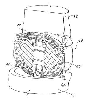
As can be seen best in the exploded view shown in FIG. 7, in accordance with
this
preferred embodiment, the present invention includes four main components: two
shells 20, 40, a central body 60, and a sheath 70. The complete assembly of
the
device is shown in FIGS. 4 and 6, wherein the central body 60 is bracketed
between
shells 20, 40. The flexible sheath 70 extends between the two opposing shells
20, 40,
and encapsulates the central body 60. As described in further detail below,
the
geometric configuration of the shells 20, 40, the central body 60, and the
sheath 70,
are complementary. As such the geometric configuration of these components
cooperate to (1) join the components into a unitary structure, and (2) define
important
functional features of the device.
Preferably, shells 20, 40 are cup-like so as to include an outer convex
surface
23 and an inner concave surface 21, 41. The outer surfaces 23 can be coated
with a
nonspherical sintered bead coating 22, 42, or with some other coating that
will
promote bony ingrowth. The inner surfaces 21, 41 (shown in FIG. 6) are
preferably
very smooth, and may be machined or polished.
The shells, 20, 40 include a number of geometric features that as described in
further detail below cooperate with other components of the devices.
Specifically,
these features include a central retaining post 27, 47, an outer
circumferential groove
82, 84, and radial stop or an extension 86, 88. The central retaining post 27,
47
extends axially from inner surfaces 21, 41. In addition, each shell 20, 40
includes an
edge 73, 74, respectively. The outer circumferential grooves 82, 84 extend
into the
edges 73, 73 of the shells 20, 40. As seen best in FIG. 6, the radial stops or
extensions
86, 88 extend from the edge 73, 74 in a direction generally perpendicular to
the
general plane of the shells 20, 40.
18
CA 02429246 2003-02-06
WO 02/11650 PCT/US01/24791
Each shell may also be provided with tabs or flanges 25, 45. The tabs or
flanges extend from a portion of the edge 73, 74 in a direction generally
perpendicular
to the general plane of the shells 20, 40, but in a direction generally
opposite the radial
stops or extensions 86, 88. The tabs or flanges 25, 45 help to prevent
longterm
migration within the disc space, as well as catastrophic posterior expulsion,
and the
resulting damage to the spinal cord, other nerves, or vascular structures.
Insertion
stops 25, 45 may contain openings 26, 46 that can releasably engage an
insertion tool
(not shown). The insertion tool will generally contain flexible prongs to
releasably
engage openings 26, 46. The insertion tool will also generally include a
disengagement block that can press against the side of the implant once it has
been
properly positioned in the intervertebral space and force the openings 26, 46
off of the
prongs of the tool. The shells can be made from any suitable biocompatible
rigid
material. In accordance with a preferred embodiment, the shells are made from
a
titanium alloy, and most preferably the titanium alloy is ASTM F-136. The bead
coating 22, 42, however, is preferably made from ASTM F-67 pure titanium.
As shown best in FIG. 7, central body 60 is a preferably a donut-shaped
structure, and includes a convex upper contact surface 94, a convex lower
contact
surface 96, and a central axial opening 98. In addition, central body member
60
preferably includes an upper shoulder 92 and a lower shoulder 90. Each
shoulder 90,
92 consists of an indentation in the surface of the central body member which
defines
a ledge that extends around the circumference of the central body 60.
The central body 60 is both deformable and resilient, and is composed of a
material that has surface regions that are harder than the interior region.
This allows
the central body to be sufficiently deformable and resilient that the implant
functions
effectively to provide resistance to compression and to provide dampening,
while still
19
CA 02429246 2008-11-27
providing adequate surface durability and wear resistance. In addition, the
material of
the central body has surfaces that are very lubricious, in order to decrease
friction
between the central body and the rigid opposing shells.
The material used to make the central body is typically a slightly elastomeric
biocompatible polymeric material, which may be coated or impregnated to
increase
surface hardness, or lubricity, or both, as described above. Coding may be
done by
any suitable technique, such as dip coating, and the coating solution may be
include
one or more polymers, including those described below for the central body.
The
coating polymer may be the same as or different from the polymer used to form
the
central body, and may have a different durometer from that used in the central
body.
Typical coating thickness is greater than about 1 mil, more particularly from
about 2
mil to about 5 mil. Examples of suitable materials include polyurethanes, such
as
polycarbonates and polyethers, such as ChronothaneTM P 75A or P 55D (P-eth-PU
aromatic, CT Biomaterials); ChronoflexTM C 55D, C 65D, C 80A, or C 93A (PC-PU
aromatic, CT Biomaterials); Elast-EonTM II 80A (Si-PU aromatic, Elatomedic);
BionateTM
55D/S or 80A-80A / S (PC-PU aromatic with S-SME, PTG); CarboSil-10TH 90A (PC-
Si-PU aromatic, PTG); TecothaneTM TT-1055D or TT-1065D (P-eth-PU aromatic,
Thermedics); TecoflexTM EG-93A (P-eth-PU aliphatic, Thermedics); and
Carbothane
PC 3585A or PC 3555D (PC-PU aliphatic, Thermedics).
The last main component of this preferred embodiment of the present
invention is the sheath 70. As show in FIG. 7, the sheath 70 is a tubular
structure, and
is made from a flexible material. The material used to make the sheath is
typically
biocompatible and elastic, such as a segmented polyurethane, having a
thickness
ranging from about 5 to about 30 mils, more particularly about 10-11 mils.
TM
Examples of suitable materials include BIOSPAN-S (aromatic
polyetherurethaneurea)
CA 02429246 2003-02-06
WO 02/11650 PCT/US01/24791
with surface modified end groups, Polymer Technology Group), CHRONOFLEX
AR/LT (aromatic polycarbonate polyurethane with low-tack properties,
CardioTech
International), CHRONOTHANE B (aromatic polyether polyurethane, CardioTech
International), CARBOTHANE PC (aliphatic polycarbonate polyurethane,
Thermedics).
As noted above, the various geometric features of the main components of this
preferred embodiment of the present invention cooperate to join the components
into
a unitary structure. In general, the ends of the sheath 70 are attached to the
shells, and
the central body 60 is encapsulated between the shells 20, 40 and the sheath
70. More
specifically, referring to FIG. 6, preferably the edges of flexible sheath 70
can overlap
the outer circumferential grooves 82, 84 of the shells 20, 40. Retaining rings
71, 72
are then placed over the edges of the sheath 70 and into the circumferential
grooves
82, 84, thereby holding the flexible sheath in place and attaching it to the
shells.
While any suitable biocompatible material can be used for the retaining rings,
titanium or titanium alloys have been found to be particularly suitable. The
retaining
rings are desirably fixed in place by, e.g., welding the areas of overlap
between the
ends of the retaining rings. Because of the high temperatures needed to weld
titanium
and titanium alloys, and because of the proximity of the weld area to both the
flexible
sheath 70 and the central body 60, laser welding is typically used.
As also noted above, the various geometric features of the main components
of the preferred embodiment of the present invention cooperate to define
important
functional features of the device. These features primarily include defining
the
kinematics of motion provided by the device, prohibiting expulsion of the
central
body 60, providing post assembly access to the interior of the device,
providing an
21
CA 02429246 2003-02-06
WO 02/11650 PCT/US01/24791
attachment mechanism for inserting the device, and providing a port for the
insertion
of lubricant into the implant cavity.
The kinematics of the motion provided by the prosthesis are defined primarily
by the geometric interaction of the central body 60 and the shells 20, 40.
Although
the central body is encapsulated within the sheath and the shells, it is not
attached to
these components. Accordingly, the central body 60 freely moves within
enclosed
structure and is only constrained by geometric limitations. As seen best in
FIG. 6, the
concave shape of the inner surfaces 21, 41 of shells 20, 40 complements the
convex
surfaces 94, 96 of central body 60. As the shells 20, 40 glide across the
convex
surfaces 94, 96, relatively unconstrained translational, flexural, or
extensional motion
of shells 20, 40 with respect to central body 60 is achieved. When the desired
limit of
the range of motion is reached, extensions 86, 88 on shells 20, 40 are
designed to
contact shoulders 90, 92 on the central body 60. Specifically, the inner
portion of the
extension forms a circumferential ridge that limits the range of motion of the
shells
20, 40 relative to the central body 60 by contacting central body shoulders
90, 92 at
the end of the allowable range of motion. In an actual vertebral joint, this
occurs at a
joint flexion/extension of about 100, at lateral bending of about 11 ,
and/or at
translation of about 2-3 mm.
As explained above, in one embodiment of the invention, the shells are
concavo-convex, and their inner surfaces mated and articulated with a convex
outer
surface of the deformable resilient central body. The implant also contains a
sheath or
sleeve that is secured to the rims of the shells with retaining rings, and
which, together
with the inner surfaces of the shells, forms an implant cavity. In a
particular aspect of
this embodiment, using a coordinate system wherein the geometrical center of
the
implant is located at the origin, and assigning the x-axis to the anterior
(positive) and
22
CA 02429246 2003-02-06
WO 02/11650 PCT/US01/24791
posterior (negative) aspect of the implant, the y-axis to the right (positive)
and left
(negative) aspect of the implant, and the z-axis to the cephalad (positive)
and caudal
(negative) aspects of the implant, the convex portion of the outer surface and
the
concave portion of the inner surface of the shells can be described as a
quadric
surfaces, such that
x2 2 z2
a2 +b2 +c2 1
where ( a,0,0), (0,+b,0), and (0,0, c) represent the x, y, and z intercepts of
the
surfaces, respectively. Typical magnitudes for a, b, and c are about 11 mm, 30
mm,
and 10 mm, respectively.
The implant is symmetrical about the x -y plane, and is intended to be
implanted in the right-left center of the disc space, but may or may not be
centered in
the anterior-posterior direction. In any event, the implant is not allowed to
protrude in
the posterior direction past the posterior margin of the vertebral body.
As noted above, geometric features also serve to prevent the expulsion of the
central body 60. In particular, this is achieved by the geometric interaction
of the
shells 20, 40 and the central body 60. Shells 20, 40 also contain central
retaining
posts 27, 47 which extend axially from inner surfaces 21, 41 into a central
axial
opening 98 in central body 60 and which stop central body 60 from being
expelled
from the implant during extreme flexion or extension. The diameter of central
axial
opening 98 is somewhat larger than the diameter of central retaining posts 27,
47. In
the coordinate system described above, the central axis of the retaining post
is
typically coincident with the z-axis, but may move slightly to accommodate
various
clinical scenarios. The shape of the post may be any quadric surface. However,
a
truncated tapered elliptical cone is a particularly suitable geometry.
Similarly, the
23
CA 02429246 2003-02-06
WO 02/11650 PCT/US01/24791
geometry of the central axial opening of the central body will correspond to
the
geometry of the retaining post, and will have a similar geometry.
Also described above, the shells contain extensions or walls formed on the
inner surface, for example around the edge of the shell, and that extend
toward the
deformable resilient central body. This extension or wall limits allowable
translation
of the deformable resilient central body with respect to the shell when the
extension
comes into contact with a shoulder formed on the surface of the central body,
e.g.,
under shear loading of the implant. The height of the extension or wall should
be less
than about 2.5 mm in order to allow the full range of desired
flexion/extension and
right/left lateral bending motions.
The resilient deformable central body contains surfaces that are described by
an equation similar to that for the inner surfaces of the shells, and which
articulates
with those inner surfaces. The central body will have a plane of symmetry if
identical
opposing shells are used. As described above, the central body also features
an
equatorial rim that acts as a "soft stop" in the event the patientparticipates
in extreme
activities that result in movements greater than the designed range of
flexion/extension or lateral bending. In such a situation, the central body
will have
translated until the retaining post has contacted the inner surface of the
central axial
opening, and the extension or wall will have contacted the shoulder of the
central
body. Opposite the wall/shoulder contact, the edges of the shells will be in
close
proximity, but will be kept from contacting each other by contact with the
equatorial
rim of the central body. If desired, the thickness of the rim can be varied to
further
limit the range of motion.
Another important characteristic of this preferred embodiment of the present
invention is the provision of a means for accessing the interior of the device
after it
24
CA 02429246 2003-02-06
WO 02/11650 PCT/US01/24791
has been assembled into a unitary structure. This means consists of a central
axial
opening included in the shells 20, 40. Typically, this opening will be
provided
through central retaining posts 27, 47. By providing access to the interior of
the
device, sterilization can be done just prior to implantation of the device.
Sterilization
is preferably accomplished by introducing an ethylene oxide surface sterilant.
Caution should be exercised in using irradiation sterilization, as this can
result in
degradation of the polymeric materials in the sheath or central body,
particularly if
these include polyurethanes.
After sterilization, the central openings can be sealed using plugs 28, 48.
Preferably, only one plug is inserted first. The plug is inserted using
insertion tool
100, shown in FIG. 5, and which contains handle 101 and detachable integral
plug 28,
48. The tool is designed so that plug 28, 48 detaches from the tool when a
predetermined torque has been reached during insertion of the plug. The tool
can then
be discarded.
After one plug has been inserted to one of the shells, a lubricant 80 is
preferably introduced into the interior of the device prior to inserting the
second plug.
To do this a syringe is used to introduce the lubricant into the remaining
central
opening, and the implant is slightly compressed to remove some of the excess
air.
Another insertion tool 100 is then used to insert a plug into that central
opening, and
thereby completely seal the interior of the device from its exterior
environment. In
accordance with the preferred embodiment of the present invention the
lubricant 80 is
saline. However, other lubricants maybe used, for example, hyaluronic acid,
mineral
oil, and the like.
The two shells 20, 40 are virtually identical in shape and composition,
however those of skill in the art will understand that it is possible to use
shells of
CA 02429246 2003-02-06
WO 02/11650 PCT/US01/24791
different sizes (including thicknesses), shapes, or materials, e.g., in order
to provide a
more customized fit to the patient's anatomy, and that this does not depart
from the
spirit and scope of the invention.
The deformable resilient central body is disposed between the opposed shells,
as described above and illustrated in the drawing figures. Its upper and lower
surfaces
articulate with the upper and lower shells, respectively, and have a geometry
that is
similar to that of the shells.
The kinematics of various embodiments of the implant are illustrated in FIG.
8, 9, 10, and 11. FIG. 8A illustrates a an view of an implant having a hollow
central
retaining post and undergoing lateral bending. The range of lateral bending is
limited
to about 11 , as indicated in FIG. 8B, which is a sectional view along line A-
A of
FIG. 8A. Contact of the walls or extensions 86, 88 of the shells with
shoulders 90, 92
of the central body limit the range of motion to that desired. The central
retaining
posts 27, 47 may also contribute to limiting the range of motion by contact
with the
central axial opening of the central body. FIG. 9A illustrates a plan view of
an
implant of the type shown in FIG. 8 undergoing lateral translation. FIG. 9B
shows a
sectional view along line G-G. Again, the contact between walls or extensions
86, 88
of the shells and shoulders 90, 92 of he central body limit the range of
motion to that
desired, and central retaining posts 27, 47 may also contribute. FIG. 10 and
11
provide similar plan and sectional views (along line H-H and I-I,
respectively),
illustrating a different embodiment of the implant (without a hollow central
retaining
post) undergoing lateral bending (FIG. 10) and lateral translation (FIG. 11).
In each
case, the range of motion is limited by contact between walls or extensions
86, 88 of
the shells and shoulders 90, 92 of the central body.
26
CA 02429246 2008-11-27
As described above, the implant is desirably used as an endoprosthesis
inserted between two adjacent vertebral bodies. The implant may be introduced
using
a posterior or anterior approach. For cervical implantation, an anterior
approach is
preferred. The implanting procedure is carried out after discectomy, as an
alternative
to spinal fusion. The appropriate size of the implant for a particular
patient,
determination of the appropriate location of the implant in the intervertebral
space,
and implantation are all desirably accomplished using precision stereotactic
techniques, apparatus, and procedures.
Of course, non-stereotactic techniques can also be used.
In either case, discectomy is used to remove degenerated, diseased disc
material and
to provide access to the intervertebral space. This access is used to remove a
portion
of the vertebral body using a burr or other appropriate instruments, in order
to provide
access to the intervertebral space for a transverse milling device of the type
described
in U.S. Patent 7,331,963.
The milling device is used to mill the surfaces of the superior and inferior
vertebral bodies that partially define the intervertebral space to create an
insertion
cavity having surfaces that (a) complement the outer surfaces of the implant
and (b)
contain exposed cancellous bone. This provides for an appropriate fit of the
implant
with limited motion during the acute phase of implantation, thereby limiting
the
opportunity for fibrous tissue formation, and increases the likelihood for
bony
ingrowth, thereby increasing long-term stability.
The invention has been described above with respect to certain specific
embodiments thereof. Those of skill in the art will understand that variations
from
27
CA 02429246 2003-02-06
WO 02/11650 PCT/US01/24791
these specific embodiments that are within the spirit of the invention will
fall within
the scope of the appended claims and equivalents thereto.
28