Note: Descriptions are shown in the official language in which they were submitted.
CA 02430744 2003-06-05
WO 02/056800 PCT/USO1/47644
IMPLANT FOR ORTHOPEDIC APPLICATIONS
BACKGROUND OF THE INVENTION
1. Field of the Invention
The present invention relates to an implant which is useful for a variety of
orthopedic applications. More particularly, the present invention relates to
an implant
useful for treating bone injuries, defects, etc., such as spinal disorders for
which spinal
fusion is indicated and the repair or replacement of ligaments, tendons and/or
cartilage.
2. Description of Related Art
A variety of implants having application as artificial bone, ligaments,
tendons,
cartilage, and the like, are known. U.S. Patent No. 4,089,071 describes a
material for
making bone endoprostheses featuring a laminated structure of net-like
construction.
U.S. Patent No. 5,092,887 describes an elongated artificial ligament made from
demineralized bone which is said to exhibit compliant elasticity and high
longitudinal
strength. U.S. Patent No. 5,263,984 describes a prosthetic ligament made up of
a
quantity of substantially aligned, elongated filaments each of which is a
biocompatible, resorbable fibril made, e.g., of collagen, elastin, reticulin,
cellulose,
algenic acid or chitosan. U.S. Patent No. 5,711,960 describes an implant,
useful ifzter
alia, as a prosthetic or filling for a defective bone, which utilizes, as a
base material, a
biocompatible bulk structure of a three-dimensionally woven or knitted fabric
of
organic fibers whose surfaces have been biologically activated or inactivated.
U.S.
Patent No. 6,090,998 describes a bone implant, useful for the repair or
replacement of
1
CA 02430744 2008-07-28
ligaments, tendons and joints, which includes at least one mineralized segment
and at
least one demineralized, flexible segment.
Developing cells are known to migrate along surfaces. When the surface is
oriented, the potential exists to somewhat control the direction of growth. It
has been
observed by the inventors in animal studies that fibrous materials provide
better
osteoconduction than particle based materials. Therefore, a material which
guides the
formation of new tissue would have the ability to direct osteoconduction as
well as
other types of tissue growth. Such a material, by directing the formation of
new tissue,
would be expected to demonstrate improved strengthening effects. In addition,
a
fibrous implant, unlike particle-based implants, would tend to remain where
placed in
the body and would resist being dislodged therefrom.
SUMMARY OF THE INVENTION
In accordance with an embodiment of the present invention an implant is
provided which comprises a quantity of flexible, elongated elements at least
some of
which are derived from bone tissue and possess connective tissue-healing
activity, the
elements being arranged in substantially common alignment along their
longitudinal
axis.
2
CA 02430744 2003-06-05
WO 02/056800 PCT/US01/47644
Significant advantages of the implant flow from the substantial alignment of
the elongated members along their longitudinal, or major, axis. Thus, when the
elongated members are thus aligned to provide, e.g., a woven or braided
structure, the
result is an implant which is generally stronger than the elongated members
from
which the implant is made. In addition, the implant can be made to possess
dimensions which could not be achieved with naturally occurring implant
materials
such as whole bone sections.
Still another advantage resides in the ability of a particular implant to
utilize
combinations of different materials as sources for its elongated members.
Selection
from among a large variety of such materials expands the range of biological
and/or
mechanical properties that can be built into a given implant.
The implant of the present invention, unlike conventional metallic implants,
will not stress shield the bone at the implant site. Therefore, any tendency
for already
existing healthy bone to be resorbed at the implant site will be reduced. In
addition,
unlike metallic implants, the implant of this invention will not interfere
with the use
of postoperative plain film X-rays, MRI or CT scans.
The expression "elongated elements" refers to the structural units
constituting
the implant of this invention and having the appearance of filaments, threads,
strips
and similarly elongated configurations. The elongated elements can be separate
units
for their entire length or two or more of the elements can have a common point
of
attachment, e.g., as shown in the implant of Fig. 1 a.
The term "biocompatible" and expressions of like import shall be understood
to mean the absence of unacceptable detrimental biological response, e.g.,
stimulation
of a severe, long-lived or escalating biological response to an implant and is
distinguished from a mild, transient inflammation which accompanies
implantation of
3
CA 02430744 2003-06-05
WO 02/056800 PCT/US01/47644
essentially all foreign objects into a living organism and is also associated
with the
normal healing response. Thus, materials which alone in appropriate quantities
are
generally considered nonbiocompatible can be considered biocompatible within
the
aforestated meaning if present in small enough quantities such that they do
not elicit a
significant level of undesirable or detrimental tissue response.
The expression "connective tissue-healing activity" refers to the ability of
the
implant of the invention to participate in the repair, regeneration, healing,
etc., of
connective tissue, e.g., bone, ligament, tendon or cartilage, by one or more
mechanisms including chondrogenesis, osteoinduction, osteogenesis and
osteoconduction.
The term "chondrogenic" as used herein shall be understood to refer to the
ability of a material or substance to induce or otherwise participate in the
formation of
cartilage.
The term "osteoinductive" as used herein shall be understood to refer to the
ability of a material or substance to recruit cells from the host which have
osteogenic
potential and the ability to form ectopic bone.
The term "osteogenic" as used herein shall be understood to refer to the
ability
of a material or substance to induce new bone formation via the participation
of living
cells from within the substance.
The term "osteoconductive" as used herein shall be understood to refer to the
ability of a material or substance or material to provide surfaces that are
receptive to
the growth of new host bone.
The expression "substantially common alignment" refers to the relative
orientation of the elongated elements constituting the implant and includes
woven,
4
CA 02430744 2003-06-05
WO 02/056800 PCT/US01/47644
knitted, braided, or twisted arrangements of individual elements as well as
subassemblies of several elongated elements formed into yams, twines, strands,
etc.
The term "resorbable" refers to the ability of materials to be broken down by
normal biochemical and/or physical processes such as erosion, dissolution,
etc.
The term "remodeling" refers to the process whereby materials are broken
down and then replaced by host tissue, e.g., by resorption of existing bone
tissue by
osteoclasts and formation of new bone tissue by osteoblasts.
Other advantages of the present invention will become apparent to one skilled
in the art from the following written description and accompanying figures.
BRIEF DESCRIPTION OF THE DRAWINGS
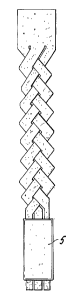
FIGS. 1A-lE are diagrammatic representations of implants in accordance with
the present invention. In FIG. lA, an elongated section of bone is cut or
machined to
provide three relatively wide elongated elements arranged in a braided
pattern. In
FIG. 1B, the elongated section of bone is cut or machined to provide elongated
elements which are formed into yams with the yams subsequently being formed
into
braids. FIGS. 1C-1E schematically depict demineralized bone strips arranged
into
various other structures.
DETAILED DESCRIPTION OF THE INVENTION
The implant of this invention is fabricated in wllole or in part from flexible
elongated elements, advantageously biocompatible in character, e.g.,
connective type
tissues obtained from human and animal tissues and natural and synthetic
fibers
including, but not limited to, demineralized bone, tendon, ligament, collagen,
elastin,
reticulin, cellulose, alginic acid, chitosan, small intestine submucosa, silk,
nonresorbable and resorbable synthetic polymeric fibers, and the like. The
elongated
elements can also be obtained from microorganisms, particularly genetically
5
CA 02430744 2008-07-28
engineered microorganisms such as yeast and bacteria and genetically
engineered
eukaryotic cell cultures such as Chinese hamster ovary cell lines, HeLa cells,
etc. For
example, U.S. Patent Nos. 5,243,038 and 5,989,894 each describe the expression
of
spider silk protein, collagen proteins, keratins, etc., using genetically
engineered
microorganisms and eukaryotic cell lines.
When the elongated elements are fabricated in whole or in part from tissues
such as bone, tendon, ligament, small intestine submucosa tissue, and the
like, such
tissues are first processed to remove any blood and debris that may be
associated
therewith and the tissues are then sterilized employing routine procedures
such as
those described below. The processed tissues are then fashioned into elongated
elements whose dimensions are selected so that when assembled into the
implant, the
latter will have sufficient length to span, and be affixed to, the implant
site, and
sufficient width and thiclaiess to impart such desirable properties as
toughness,
flexibility and strength to the implant.
The elongated tissue elements can be formed into implants having a variety of
configurations such as those shown in FIGS. lA-lE. For example, FIG. lA
schematically depicts one embodiment in which a sheet of bone is further cut
or
machined into tbree elongated elements of about the same width which are then
formed into a braid. FIGS. 1B-1E schematically depict other embodiments
wherein a
section of bone is cut or machined to prbvide a quantity of elongated elements
which
are then assembled into the implants shown.
The overall dimensions of the flexible elongated elements making up the
implant of this invention can vary widely depending on the dimensions of the
site to
which the implant is to be affixed. Typically, these dimensions will range
from about
1 cm to about 1 meter in length, preferably from about 3 cm to about 8 cm in
length,
6
CA 02430744 2003-06-05
WO 02/056800 PCT/US01/47644
from about 0.5 mm to about 30 mm in thickness, preferably from about 2 mm to
about
mm in thickuess, and from about.05 mm to about 150 mm in width, preferably
from about 2 mm to about 10 mm in width.
While fully mineralized bone, tendon, ligament, small intestine submucosa,
5 collagen tissues, etc., in themselves are not particularly osteoinductive,
such tissues
can be rendered osteoinductive by subjecting the tissue to various procedures
and/or
incorporating one or more osteoinductive substances in the tissues. For
example, the
mineral content of bone tissue can be reduced by demineralization, a process
which
results in the removal of the inorganic components of the bone, largely
10 1lydroxyapatite, wliich gives bone its characteristic rigidity and
structural properties.
The resultant demineralized bone is both flexible and osteoinductive. Bone,
tendon,
ligainent, small intestine submucosa and collagen tissues can be rendered
osteoinductive by association with, or incorporation of, various
osteoinductive
materials which include, but are not limited to, growth factors such as bone-
derived
growth factor, bone morphogenic proteins, osteogenic proteins such as OP-1,
hormones, growth hormone, platelet derived growth factor (PDGF), insulin-like
growth factors (IGF-l)(IGF-2), DNA-encoding various therapeutic agents such as
growth factors and hormones, gene activated matrix, i.e., a matrix containing
DNA
encoding therapeutic proteins utilized to promote cell growth, which in turn,
promote
DNA transfer into repair cells, demineralized bone in the form of particles,
powder,
gel, liquid, etc, ceramic powders of calcium phosphate and/or apatite
(hydroxyapatite)
and bioglasses. Bone morphogenic proteins can be obtained from Genetics
Institute,
Inc. (Cambridge, MA) and Stryker Corporation (Kalamazoo, MI) and may also be
prepared by one skilled in the art as described, e.g., in. U.S. Patent Nos.,
5,187,076,
5,366,875, 4,877,864, 5,108,922, 5,116,738, 5,013,649, 5,106,748, W093/00432,
7
CA 02430744 2008-07-28
W094/26893 and W094/26892. All osteoinductive factors are contemplated whether
they are obtained as above or isolated from bone or other human or animal
tissues.
Methods for isolating bone morphogenic protein from bone are described, e.g.,
in U.S.
Patent No. 4,294,753. Methods of preparing demineralized bone powder,
demineralized bone particles, and demineralized bone in the form of a liquid,
and
demineralized bone in the form of a gel are well known in the art as
described, e.g., in
U.S. Patent Nos. 5,314,476, 5,507,813, 5,073,373, and 5,405,390, respectively.
Methods of preparing osteogenic proteins, such as OP- I are described, e.g. in
U.S.
Patent No. 6,048,964. Methods of transferring DNA-encoding therapeutic
proteins into
repair cells utilizing gene activated matrix are described, e.g., in U.S.
Patent No.
5,962,427. Methods of preparing ceramic powders of calcium phosphate and/or
hydroxyapatite are described, e.g., in U.S. Patent Nos. 4,202,055 and
4,713,076.
Methods of preparing bioglasses are described, e.g., in W098/44965. Suitable
methods
of incorporation or association of such osteogenic factors include coating,
immersion
saturation, packing, spraying, e.g., plasma spraying, injecting into the bone
tissue, etc.
When desirable, e.g., for preparing an implant suitable for soft tissue
repair, the
flexible elongated elements constituting the implant can be treated so as to
reduce their
osteoinductive properties. For example, demineralized bone is known to possess
osteoinductive characteristics. When desirable, such characteristics can be
reduced or
eliminated by appropriate further treatment. For example, the osteoinductive
proteins
in the demineralized bone can be denatured, and thus
8
CA 02430744 2008-07-28
deactivated, by reaction with, for example, a chemical denaturant such as
glutaraldehyde or formaldellyde. Demineralized bone treated in this way is
lcnown to
support the formation of fibrous tissue and as such, exhibits connective
tissue-healing
activity although, of course, through a mechanism other than that of
osteoinduction.
The degree of denaturation can be controlled to give the desired physical and
biological properties. Other denaturation methods include irradiation and
tliermal
treatment. Alternatively, osteoinductive proteins can be extracted from the
deniineralized bone employing extractants such as guanidine hydrochloride.
Implants of this invention containing bone or otller tissue material can be
further treated by tanning or other means known in the art to reduce their
antigenicity.
For example, glutaraldehyde treatment (see U.S. Patent No. 5,053,049)
can be used for this purpose.
Employing a milling technique, elongated bone elements ranging in median
length from about 2 up to about 200 mm or more (as in the case of the long
bones), in
median thickness from about 0.05 to about 2mm and in median width from about I
to
about 20 mm can be readily obtained. Another procedure for obtaining the
elongated
bone particles herein, particularly useful for elements of bone of up to about
100 mm
in length, is the bone malling apparatus described in U.S. Patent No.
5,607,269.
Use of this apparatus results.in the production of long, thin bone strips
which
tend to curl lengthwise into tube-like structures.
Depending on the procedure employed for producing the elongate bone
elements, one can obtain a mass of bone elements containing at least about 60
weight
percent, preferably at least about 70 weight percent, and most preferably at
least about
80 weight percent of bone elements possessing a median length of from about 2
to
9
CA 02430744 2003-06-05
WO 02/056800 PCT/US01/47644
about 200 mm or more and preferably from about 10 to about 100 mm, a median
thickness of from about 0.05 to about 2 mm and preferably from about 0.2 to
about 1
mm and a median width of from about 1 mm to about 20 mm and preferably from
about 2 to about 5 mm. These bone elements can possess a median length to
median
thickness ratio of at least about 50:1 up to about 500:1 or more and
preferably from
about 50:1 to about 100:1 and a median length to median width ratio of from
about
10:1 to about 200:1 and preferably from about 50:1 to about 100:1.
If desired, the mass of elongated bone elements can be graded into different
sizes to reduce or eliminate any less desirable size(s) of elements which may
be
present. In overall appearance, the elongated bone elements can be described
as
filaments, fibers, threads, slender or narrow strips, etc. As already noted
and
depending on the manner in which they are produced, these elongated elements
may
have a tendency to curl lengthwise into tube-like structures.
When the implant of this invention is fabricated from bone, the bone is
preferably chosen from a cortical bone such as the femur, tibia, fibula,
radius or ulna.
The bone elements can be obtained from cortical, cancellous and/or
corticocancellous
bone which can be of autogenous, allogenic and/or xenogeneic origin: Porcine
bone is
a particularly advantageous type of xenogeneic bone tissue which can be used
as a
source for the elongated bone elements of this invention.
Following the shaving, milling or other technique whereby they are obtained,
the elongated bone elements are subjected to deinineralization in order to
reduce their
inorganic content and, as may be necessary for a particular embodiment, to
increase
their flexibility. Demineralization of the bone elements will ordinarily
result in
elongated elements of slightly smaller dimensions than those of the
mineralized
elements from which they were obtained.
CA 02430744 2003-06-05
WO 02/056800 PCT/US01/47644
The elongated bone elements can be demineralized in accordance with known
and conventional procedures. The mineral content of bone can be removed to
varying
degrees. The term "fully demineralized" as it applies to an elongated bone
element
refers to a bone element possessing less than about 8, preferably less than
about 1,
weight percent of its original inorganic mineral content. The term "partially
demineralized" as it applies to an elongated bone element means that the bone
element possesses from about 8 to about 90 weight percent of its original
inorganic
mineral content. The term "superficially demineralized" as it applies to an
elongated
bone element refers to a bone element possessing at least 90 weight percent of
its
original inorganic mineral content. The term "demineralized" as it applies to
an
elongated bone element includes any one or combination of the foregoing types
of
demineralized elongated bone elements. The use of superficially, partially or
fully
demineralized bone can, in some embodiments, be particularly advantageous
since
demineralized bone exhibits considerably greater initial osteoinductive
activity than
fully mineralized bone.
Demineralization can precede or follow the cutting, slicing, milling, etc., of
the bone into elongated elements. Thus, a whole section of bone, e.g., a
diaphyseal
shaft, can first be demineralized to the extent desired after which it is
machined to
provide the individual elongated bone elements. Alternatively, the whole bone
can be
subdivided into individual elongated bone elements which are thereafter
demineralized to the desired level.
Of course it will be understood by those skilled in the art that the bone
elements will be demineralized to such an extent that they can be worked to
form the
implant of the invention herein. Therefore, when the bone elements are of such
size
as to be relatively inflexible prior to demineralization, they can be
demineralized to
11
CA 02430744 2008-07-28
the point where they are flexible and capable of being worked, e.g., woven,
braided,
spun, etc. When bone elements are of such dimensions that they are relatively
flexible prior to demineralization, a lesser degree of demineralization may be
appropriate. The extent of demineralization necessary to obtain a bone element
that is
worl:able can be readily deterniined by one skilled in the art employing
routine
experimentation.
Demineralization of the elongated bone elements can be conducted using
conventional procedures that are well lmown in the art, e.g., subjecting the
bone
section to strong acids such as hydrochloric acid as described, e.g., in Reddi
et al.,
Proc. Nat. Acad. Sci. 69:1601-5 (1972). The extent
of demineralization is a function 6f the strength of the acid solution, the
shape of the
bone and the duration of the demineralization treatment. Reference in this
regard may
be made to Lewandrowski et al., J. Biomed. Materials Res. 31:365-372 (1996).
In a preferred demineralization procedure, the elongate bone elements are
subjected to a defatting/disinfecting step which is followed by an acid
deminexalization step. A preferred defatting/disinfectant solution is an
aqueous
solution of ethanol, the ethanol being a good solvent for lipids and the water
being a
good hydrophilic carrier to enable the solution to penetrate more deeply into
the bone
particles. The aqueous ethanol solution also disinfects the bone by'killing
vegetative
microorganisms and viruses. The preferred concentration range of the defatting
solution is from about 60 to about 85 weight percent alcohol and most
preferably
about 70 weight percent alcohol. Following defatting, the bone elements are
immersed in acid over tim.e to effect their demineralization. Acids which can
be
employed in this step include inorganic acids such as hydrochloric acid and
organic
12
CA 02430744 2008-07-28
acids such as peracetic acid. Generally, the concentration of inorganic acid
utilized to
achieve demineralization is from about 0.1N to about 2N and more preferably
from
about 0.2 N to about 1.0 N. The time of exposure to tlie acid is increased for
lower
acid concentrations and decreased for the higher acid concentrations. After
acid
treatment, the demineralized bone elements are rinsed with sterile water for
injection
to remove residual amounts of acid and thereby raise the pH.
The wet demineralized bone elements can then be immediately formed into
the implant of this invention in accordance using methods well known in the
art, e.g.,
those described in U.S. Pat. No. 5,263,984 or stored under aseptic
conditions, advantageously in a lyophilized state, for processing
at a later time.
When the bone elements are shorter than the desired length of the implant,
they can be combined with fibers and/or other materials such that a final
implant of
the desired length is produced. For example, the relatively short bone
elements can be
conzbined with other materials in a known manner, e.g., to form a spun yarn,
which
can then be woven to form the implant of the invention. Thus, the short bone
elements can be combined with demineralized bone elements of greater length or
with
bioresorbable polymeric fibers, ceramic or glass fibers, or biocompatible
metal fibers
of suitable length to produce a composite yaxn which can then be woven using
standard techniques to produce the implant of the invention.
Optionally, the short bone elements can be combined with bioresorbable
thermoplastic material that is formed into spun-bonded and/or non-woven
fabrics.
For example, after the bioresorbable thermoplastic material has been formed
into a
first web, the short bone elements can be applied to the first web and then
sandwiched
with a second web to form a controlled elastic composite material. The methods
of
13
CA 02430744 2008-07-28
forming a composite material disclosed in U.S. Patent Nos. 6,124,001 and
6,132,871
are suitable for forming the aforedescribed elastic composite.
In one embodiment, the bone comprises a plurality of elongated bone
elements. Typically, the bone is obtained from a suitable vertebrate and
processed by
conventional techniques to remove blood and lipid from the bone. The bone can
then
be cut into elongated sections by techniques which are well known in the art,
e.g.,
longitudinally cutting an entire bone section or relatively large portion of
bone into
elongated sections using a band saw or a diamond-bladed saw, or milling the
surface
of an entire bone or relatively large portion of bone. Alternatively, the bone
can be
cut by making transverse cuts to prepare a bone section of the appropriate
length,
followed by longitudinal cuts using a band saw or a diamond cut saw. As stated
above, elongated elements of bone can be further cut or machined into a
variety of
different shapes. In overall appearance the elongated bone elements can be
described
as narrow or thick strips, segments, sheets, rods, struts, etc. The elongated
elements
can be further processed to remove residual blood and lipid residue.
Prior or subsequent to cutting or milling of the bone into elongated elements,
the bone is preferably demineralized to reduce its inorganic content utilizing
the
defatting/demineralization procedure described herein above. After acid
treatment,
the elongated bone elements are rinsed with sterile water for injection,
buffered with a
buffering agent to a final predetermined pH and then finally rinsed with water
for
injection to remove residual amounts of acid and buffering agent or washed
with
water to remove residual acid and thereby raise the pH.
14
CA 02430744 2008-07-28
In a particularly useful embodiment, the elongated bone elements can be
segmentally demineralized employing procedures known in the art as described,
e.g.,
in U.S. Patent No. 6,090,998.
Alternatively, the end portions of the elongated bone elenzents can be surface
demineralized by any convenient method. For example, the bone elements can be
subjected to demineralization conditions for a period of time sufficient to
demineralize only their surfaces.
In an alternative embodiment, demineralized bone sections (approximately 6
bone sections) are combined longitudinally into three small bundles, each
having from
about 1 to about 3 bone sections. The three bundles are then braided. Various
methods of braiding and types of braids any of which may be useful in
producing the
material of the invention herein are also described, e.g., by Shaw, KNOTS -
Usefzcl &
Ornamental, Bonanza Books, New York (1983).
The ends of the braided demineralized bone section can then be glued together
using a
fixation agent to prevent their unraveling or they can be held together with a
biocompatible polymer or metal band.
In another embodiment, demineralized bone strips can be cut from sheets
composed of elongated bone particles, commercially available as GR.AFTON Flex
(Osteotech, Eatontown, NJ) as described, e.g., in U.S. Patent No. 5,507,813.
To increase the mechanical strength of bone strips fabricated from bone,
chemical linkages can be formed between adjacent bone elements employing,
e.g.,
any of the procedures for accomplishing this disclosed in U.S. PatentNo.
6,123,731.
CA 02430744 2003-06-05
WO 02/056800 PCT/US01/47644
Medically/surgically useful substances which promote or accelerate healing
can be incorporated in the implant of this invention. Useful substances of
this kind
which can be incorporated into the implant include, e.g., collagen, insoluble
collagen
derivatives, etc., and soluble solids and/or liquids dissolved therein, e.g.,
antiviral
agents, particularly those effective against HN and hepatitis; antimicrobials
and/or
antibiotics such as erythromycin, bacitracin, neomycin, penicillin, polymyxin
B,
tetracyclines, viomycin, chloromycetin and streptomycins, cefazolin,
ampicillin,
azactam, tobramycin, clindamycin, and gentamicin, etc.; biocidal/biostatic
sugars
such as dextran, glucose, etc.; amino acids; peptides; vitamins; inorganic
elements;
co-factors for protein synthesis; hormones; endocrine tissue or tissue
fragments,
synthesizers; enzymes such as collagenase, peptidases, oxidases, etc.; polymer
cell
scaffolds with parenchymal cells; angiogenic drugs and polymeric carriers
containing
such drugs; collagen lattices; antigenic agents; cytoskeletal agents;
cartilage
fragments, living cells such as chondrocytes, bone marrow cells, mesencliymal
stem
cells; natural extracts; genetically engineered living cells or otherwise
modified living
cells; tissue transplants; demineralized bone powder (or "demineralized bone
matrix"
as it may also be referred to); DNA delivered by plasmid or viral vectors;
autogenous
tissues such as blood, serum, soft tissue, bone marrow, etc.; bioadhesives;
bone
morphogenic proteins; osteoinductive factor; fibronectin; transforming growth
factor-
beta; endothelial cell growth factor; cementum attachment extracts; ketaserin;
insulin-
like growth factor; platelet derived growth factors; epidermal growth factor;
interleukin; human alphathrombin; fibroblast growth factors; periodontal
ligament
chemotactic factor; human growth hormone; animal growth hormone; growth
hormones such as somatotropin; bone digesters; antitumor agents; immuno-
suppressants; permeation enhancers, e.g., fatty acid ester such as laureate,
myristate
16
CA 02430744 2008-07-28
and stearate monoesters of polyethylene glycol, enaniine derivatives, alpha-
keto
aldehydes, etc.; and, nucleic acids. Preferred biomedically/ surgically useful
substances are bone morphogenic proteins and DNA delivered by plasmid or viral
vector. Suitable methods of incorporation include coating, immersion
saturation,
packing, co-lyophilization wherein the substance is placed on the bone grafft
and
lyophilized, spraying, injecting, etc. The amounts of inedically/ surgically
useful
substances utilized can vary widely with optimum levels being readily
determined in a
specific case by routine experimentation.
The implant herein can also be fabricated in wliole or in part from tendon,
ligament and/or small intestine submucosa tissues. These tissues are not
osteoinductive but can be made so by incorporating various osteoinductive
materials
as described above. Tendon tissue useful for fabricating the material
includes, but is
not limited to, fascia lata, semitendinosus, achilles tendon and patella
tendon tissue.
Ligament tissue can consist of an entire excised ligament or elongated section
thereof.
Small intestine submucosa tissue can be obtained and processed as described in
U.S.
Patent No. 4,902,508. The tendon, ligament and small
intestine submucosa tissues can be obtained from
autogeneic, allogeneic or xenogeneic sources and preferably are obtained from
an
autogeneic or allogeneic source. The tissues can be excised and cut into a
plurality of
elongated elements employing metllods known in the art. Reduction of the
antigenicity of allogeneic and xenogeneic tissue can be achieved by treating
the
tissues with various chemical agents, e.g., extraction agents such as
monoglycerides,
diglycerides, triglycerides, dimethyl formamide, etc., as described, e.g., in
U.S.
Patent No. 5,507,810. Medically/surgically useful substances as described
above
can also be incorporated in
17
CA 02430744 2008-07-28
or associated wit11 the tendon, ligament and small intestine submucosa tissue
as
described above with respect to elontrated elements obtained from bone.
The implant can also be fabricated from collagen tissue wliich can be obtained
from any autogeneic, allogeneic or xenogeneic source, preferably from an
autogeneic
or allogeneic source. Collageneous tissue sources include, but are not limited
to, slcin,
tendon, intestine and dura mater obtainzd from animals, transgenic animals and
humans. Collagenous tissue can also be obtained by genetically engineering
microorganisms to express collagen as described, e.g., in aforenientioned U.S.
Patent
No. 5,243,038. Procedures for obtaining and purifying collagen are well lrnown
in the
art and typically involve acid or enzyme extraction as described, e.g., in
U.S. Patent
No. 5,263,984. Collagen is also commercially available
(Pentapharm). The purified collagen is then subjected to
further processing to obtain collagen fibers or collagen threads, which can
optionally
be treated with crosslinking agents, e.g., glutaraldehyde, to improve their
strength
and/or with various medically/surgically useful substances as described above.
The
collagen threads can be arranged to form various structures, such as a woven
or non-
woven fabric, bundle or braid, etc. by various techniques known in the art as
described, e.g., in U.S. Patent Nos. 5,171,273 and 5,378,469, to
provide the implant of the invention. For example, U.S. Patent No.
5,171,273 describes the preparation of high-strength collagen fibers by
dissolving
Type I collagen in dilute hydrochloric acid, extruding the solution into a
specific fiber
formation buffer to reconstitute the collagen fibers. The reconstituted
collagen fibers
are subsequently crosslinked with glutaraldehyde or other cliemical agents and
treatments. The fibers are then processed into woven or non-woven materials.
18
CA 02430744 2003-06-05
WO 02/056800 PCT/US01/47644
U.S. Patent No. 5,378,469 describes methods for the production of high
strength collagen threads wherein collagen is extruded into a dehydrating
agent, e.g.,
polyethylene glycol, which has a higher osmotic pressure than that of the
collagen
solution and a pH from about 5 to 10 which results in the formation of
collagen
threads. If desired, the collagen threads can be crosslinked using various
chemical
agents. The collagen threads are then utilized to form braided constructs,
plied into
yarn, and knitted to provide the implant of this invention.
Various constructs of the elongate elements, fibers and threads can be formed
utilizing well known techniques, e.g., braiding, plying, knitting, weaving,
that are
applied to processing natural fibers, e.g., cotton, silk, etc., and synthetic
fibers made
from synthetic bioabsorbable polymers, e.g., poly(glycolide) and poly(lactic
acid),
nylon, cellulose acetate, etc.. See, e.g., Mohamed, Amef ican Scientist, 78:
530-541
(1990). For example, aforementioned U.S. Patent No. 5,378,469 describes the
braiding of crosslinked and noncrosslinked collagen threads using a harness
braiding
machine (New England Butt Co., Providence, RI). Specifically, collagen thread
is
wound onto cylindrical stainless steel spools. The spools are then mounted
onto the
braiding carousel, and the collagen thread is then assembled in accordance
with the
instructions provided with the braiding machine. In one particular run, a
braid was
formed of four collagen threads, which consisted of two threads of
uncrosslinked
collagen and two threads of crosslinked collagen.
The elongate particles, fibers, and threads can also be plied into yarns using
the same methods and same machinery known to those skilled in the art in
plying
threads made out of other material, e.g., cotton, polyester, etc. For example,
aforementioned U.S. Patent No. 5,378,469 describes the production of a 60 ply
yarn
from noncrosslinked collagen threads. Therein, 4 collagen threads were twisted
19
CA 02430744 2008-07-28
together. Three of the resultant 4-ply strands were then twisted together in
the
opposite direction, and then 5 of the resultant 12 ply. strands were twisted
in the
opposite direction.
The elongated elements and/or fibers and/or threads and/or braided threads or
plied yams can then be knitted into tubular or flat fabrics by using
techniques known
to those skilled in the art of producing fabrics manufactured from other types
of
threads. Various medically/surgically useful substances as described above can
be
incorporated in, or associated with, the braided, lrnitted, or woven
materials.
The implant can also be fabricated in whole or in part from a synthetic
biocompatible bioabsorbable polymer or copolymer, a synthetic biocompatible
non-
bioabsorbable polymer or copolymer, and combinations thereof. As used herein,
"bioabsorbable polymer" refers to a polymer or copolymer which is absorbed by
the
body. "Non-bioabsorbable polymer" refers to a polymer or copolymer which
remain
in the body witliout substantial bioerosion. Examples of synthetic
biocompatible
bioabsorbable polynlers or copolyn7ers include, but are not limited to,
poly(lactide),
poly(glycolide), poly(epsilon-caprolactone), poly(p-dioxanone), poly(epsilon-
caprolactone-co-p-dioxanone) and poly(lactide-co-glycolide) as described, e.g,
in U.S.
Patent Nos. 5,705,181 and 5,393,594; bioabsorbable block copolymers
made of hard phase forming monomers, e.g., glycolide and lactide, and
soft phase monomers, e.g., 1,4-dioxane-2-one and caprolactone,
as described, e.g. in U.S. Patent No. 5,522,841; and natural
materials such as cotton, and catgut. Examples of synthetic
biocompatible non-bioabsorbable polyniers include, but are not limited to,
homopolymers and copolymers of polypropylene, polyamides, polyvinylchlorides,
polysulfones, polyurethanes, polytetrafluoroethylene, etc. The biocompatible
material
CA 02430744 2003-06-05
WO 02/056800 PCT/US01/47644
fabricated from the biocompatible polymer can have incorporated within, or be
associated with, osteogenic materials such as demineralized bone particles or
demineralized bone powder and medically/surgically useful substances as
described
above.
The implant can also be fabricated in whole or in part from a synthetic
biocompatible, optionally bioabsorbable, ceramic or glass, or biocompatible
metal.
Examples include fibers of phosphate/silica glasses (bioglass), fibers of
calcium
phosphate, and metal fibers such as titanium or titanium niclcel alloys (shape-
memory
metals).
In a particularly useful embodiment, the aforementioned material making up
the implant can be wrapped with a monolithic piece, e.g., strips or sheets,
fabricated
from a suitable material that is remodeled by the body and replaced over time
with
new bone tissue. For example, the material can be wrapped or surrounded with
demineralized bone strips cut from sheets which are composed of elongated bone
particles, commercially known as GRAFTON Flex (Osteotech, Eatontown, NJ) as
described, e.g., in aforementioned U.S. Patent 5,507,813.
These demineralized bone strips can be affixed to the biocompatible
osteogenic material by any convenient method, e.g., adhering the strips to the
material
utilizing adhesives, suturing the strips to the biocompatible osteogenic
material,
braiding the strips around the biocompatible osteogenic material, etc.
The implants of this invention can be utilized in a wide variety of
orthopedic,
neurosurgical and oral and maxillofacial surgical procedures such as the
repair of
simple and compound fractures and non-unions, external and internal fixations,
joint
reconstructions such as arthrodesis, general arthroplasty, cup arthroplasty of
the hip,
femoral and humeral head replacement, femoral head surface replacement and
total
21
CA 02430744 2003-06-05
WO 02/056800 PCT/US01/47644
joint replacement, repairs of the vertebral column including spinal fusion and
internal
ftxation, tumor surgery, e.g. deficit filling, discectomy, laminectomy,
excision of
spinal cord tumors, anterior cervical and thoracic operations, repair of
spinal injuries,
scoliosis, lordosis and kyphosis treatments, intermaxillary fixation of
fractures,
mentoplasty, temporomandibular joint replacement, alveolar ridge augmentation
and
reconstruction, inlay bone grafts, implant placement and revision, sinus
lifts, repair of
ligaments or tendons in the hand, elbow, knee, foot, ankle or any other
anatomical
location, etc. These materials can be sutured or stapled in place for
anchoring
purposes and serve in guided tissue regeneration or as barrier materials.
It will be understood that various modifications may be made to the
embodiments disclosed herein. Therefore, the above description should not be
construed as limiting, but merely as exemplifications of preferred
embodiments.
Those skilled in the art will envision other modifications within the scope
and spirit of
the disclosure herein.
22