Note: Descriptions are shown in the official language in which they were submitted.
CA 02430750 2003-05-30
Express Mailing Label # EJ743857302US
PATENT
File No. 13549-B
HIGH SPATIAL RESOLUTION MATRIX ASSISTED LASER
DESORPTION/IONIZATION
(MALDI)
BACKGROUND OF THE INVENTION
Matrix assisted laser desorption/ionization mass spectrometry (MALDI-
MS) has become an increasingly common tool for protein analysis in biological
research since its development in 1988 (Karas, et al 1988,Tanaka et a! RCMS,
Fenseleau ). The simple sample preparation, short analysis time and
sensitivity
have made this a powerful technique for protein identification (Fenseleau,
Anal.
Chem. 1997). Furthermore, the ability to generate intact molecular ions for
whole
proteins directly from complex mixtures makes this a particularly attractive
technique for biological samples. (Redeker et at Anal Chem 1998). Electrospray
ionization has proven to be another powerful and widespread ionization
technique for mass spectrometric analysis of proteins and peptides that
provides
a means to directly couple liquid separations and mass analysis (Washburn, M.
P., D. Wolters, and J. R. Yates III Nat. Biotech. 2001 - Veenstra, T. D., S.
Martinovic, G. A. Anderson, L. Pasa-Tolic, and R. D. Smith JASMS 2000).
However the necessity of a liquid phase for the samples prior to ionization is
in
contrast to MALDI-MS where the sample is generally allowed to dry on a surface
in combination with matrix molecules. It is the ability to generate ions from
a solid
-1-
Final_App.doc
CA 02430750 2003-05-30
phase sample that has led to a unique application of this technique whereby
the
location of the analyte within a heterogenous sample can be determined along
with its mass.
MALDI-MS has been used to obtain mass spectra for proteins and
peptides from precise X-Y locations from within a complex biological sample
such as single cells (Garden et al JMS 1996- Chaurand, Stoeckli, and Caprioli
Anal Chem 1999.). An extension of this approach was later described where
multiple mass spectra were obtained by rastoring across the sample in a grid
like
pattern across the sample to form the pixels of an image. - (Stoeckli, M., T.
B.
Farmer, and R. M. Caprioli JASMS 1999 - Caprioli, Farmer, Gile Anal Chem
1997) Using this data, the mass spectra could then be reassembled to create an
image detailing the two dimensional position for a particular m/z value and
therefore the corresponding protein. This has been demonstrated with several
types of tissues and most dramatically with tissue sections from a rat brain
(Todd
et al, 2001, Stockle et al 2001-Stoeckli, Caprioli Nat. Med. 2001).
Other types of desorption/ionization mass spectrometry have been used to
generate an "ion image", but have generally used relatively harsh ionization
methods, such as secondary ion mass spectrometry and laser ablation, and were
limited to examining low molecular weight species (Belu, A. M. et al Anal
Chem.
2001, Todd et al, 2001,- Stockle et at 2001-Kossokovski et at 1998). Two of
these reports achieved a very tightly focused laser beam with near field
microscopy fibers (Stockle et at 2001 -Kossokovski et al 1998). However, one
-2-
Final_App,doc
CA 02430750 2003-05-30
limitation of the near field microscopy approach is the fluence achievable at
the
fiber optic tip. Fiber optic damage thresholds are too easily exceeded when
the
tip diameter is reduced to 150-200 nm. Precision control of laser intensity
and
beam profile is required to inhibit fiber optic tip heating and self-ablation.
In
addition, because of the necessity for the fiber optic tip to be in the
proximity of
the desorption surface; contamination of the tip surface is a constant
concern.
Coupled with the effect of laser heating, tip lifetime is compromised.
Several challenges to creating an image from mass spectral data have
been identified in prior work. Among them, are the need to evenly distribute
the
matrix over the sample to generate a homogeneous surface, removal of
,contaminant peaks that may suppress the signal of other analytes, and
visualization of the tremendous amount of data generated by this technique.
These issues have been described in a recent review (Todd, P. J and R. M.
Caprioli. JMS 2001). One limitation that exists is the resolution that can be
achieved when creating the "protein image". The picture resolution is limited
by
the pixel size achievable, which is in turn limited by the size of the laser
spot
used to perform the ionization. Typically the laser spot size used to obtain
MALDI-MS spectra is on the order of 25 pm in diameter (Stockle, R., R. Zenobi
Anal Chem) with limitations at the 1 pm level mentioned (Todd et al 2001).
However, the laser spot size reliably used for MALDI imaging has remained at 5
to 100 pm. (ibid.). While this provides ample resolution to distinguish
structures
within tissue sections or single neurons from A. californica (cells on the
order of
-3-
Final_App.doc
CA 02430750 2003-05-30
92 pm), smaller structures/cells cannot be resolved from one another using
this
pixel size (Rubakhin, S. S. et at J. Neurophys 1999). In particular, microbes
are
often on the order of 1 - 2 pm in length and great resolution is required
(Auerbach, I.D. et al J. Bact. 2000). Reductions in laser spot size are needed
in
order to generate MALDI-MS images of cells and extracellular structures on the
microbial scale.
An improvement in laser focus can also lead to additional benefits by
improving the ability of MALDI-MS to ionize extremely small protein samples in
the analysis of dense protein arrays. Currently, sample plates holding up to
384
sample wells (each -2 mm in diameter), are used for high throughput protein
analysis using MALDI-MS. Manufacturers have introduced sample plates such as
the "Anchor chip t' " with affinity or adsorptive surfaces to concentrate the
sample
on a small area (Bruker Daltonics Inc., Product information literature, 2001).
These aid in concentrating the sample as well as potentially creating more
tightly
packed arrays of samples with smaller spots from 200 to 800 pm in diameter.
Meanwhile there have been other notable developments in deposition of small
sample spots for analytical arrays that may have application MALDI-MS protein
analysis. Methods creating small protein spots by spray deposition have been
described with spots ranging from 100 to 500 microns (Onnerfjord, P et al.
Anal
Chem 1998- Moerman, R., et al. Anal. Chem.2001). Furthermore,
microstructured devices have been fabricated as microreactors with features on
the 1 to 5 pm scale with reactor wells of 15 pm being produced (Grzybowski, B.
-4-
Final_App.doc
CA 02430750 2003-05-30
A., R. Haag, N. Bowden, and G. M. Whitesides, Anal. Chem 1998). While these
spots are still above the typical laser spot size used for MALDI-MS, a recent
report of protein samples being deposited in 15 nm diameter spots has appeared
(Perkel, C. The Scientist 2002,16[5], p.34,). Clearly, as technologies for
depositing arrays of samples improve, methods for producing smaller laser
spots
for both ionization and imaging in association with MALDI-MS are needed.
The development and application of tightly focused MALDI in the present
invention allows for generation of higher resolution images detailing intact
protein
location and the creation of "protein images" of a small sample area that can
be
compared to optical images to reveal their location within a 2-D sample.
SUMMARY OF THE INVENTION
One object of the present invention provides a system and process for
focusing light to a spot size for matrix assisted laser desorption/ionization
(MALDI). A coherent light source, such as a laser or infrared light source,
may
be directed through at least one confocal microscopic objective to create a
desorption/ionization source at the surface of a MALDI sample plate adapted to
receive a sample substrate within the focal working distance of the
microscopic
objective. The light is transported by at least one fiber optic cable to at
least one
confocal microscopic objective. At least one collimating fiber optic coupler
is
employed to collimate the light to an aperature of at least one fiber optic
cable. A
insulating microscopic objective holder holds the microscopic objective and
-5-
Final_App.doc
CA 02430750 2003-05-30
insulates it from the electrical fields of the MALDI. At least one adapter
secures
the insulating microscopic objective holder. At least one X, Y, positioner
moves
the microscopic objective in the X, Y co-ordinates. At least one Z positioner
moves the microscopic objective in the Z co-ordinate. Finally, a mass analyzer
is
used to analyze ions desorbed from the sample substrate.
A preferred embodiment of the present invention provides a system and
process for focusing light to a submicron spot size for matrix assisted laser
desorption/ionization (MALDI). A coherent light source, such as a laser, is
used
to generate ultra-violet light. At least one confocal microscopic objective is
used
to create a desorption/ionization source of sub-micron spatial resolution at
the
surface of a MALDI sample plate. The ultra-violet light generated by the laser
is
transported by at least one fiber optic cable to at least one confocal
microscopic
objective. A sample substrate is placed on a sample plate to hold it within
the
focal working distance of the microscopic objective and a mass analyzer is
used
to analyze the sample after it has been ionized.
As used herein, a sample substrate is a combination of an analyte and an
appropriately absorbing sample matrix. As used herein, working distance
includes the distance from the front lens element of the objective to the
closest
surface of the coverslip when the specimen is in sharp focus. In the case of
objectives designed to be used without coverslips, the working distance is
determined by the linear measurement of the objective from lens to the
specimen
surface. As used herein, a mass analyzer is any device capable of separating
-6-
Final_App,doc
CA 02430750 2003-05-30
and detecting ions based upon their mass to charge (m/z) ratio. It includes
mass
spectrometer devices operated under vacuum (from 760 torr down to 10"9 torr),
such as a time-of-flight mass spectrometer, and devices operated at or near
atmospheric pressure (e.g. an ion mobility spectrometer).
In another arrangement of the system, at least one confocal microscopic
objective is positioned above the slide.
Optionally, the slide may be transparent and at least one confocal
microscopic objective is positioned below the transparent slide.
In a further arrangement of the system, the mass analyzer includes at
least one ion mobility spectrometer alone or in tandem with a mass
spectrometer.
In still another arrangement of the system, the mass analyzer includes a
mass spectrometer having at least one evacuated internal chamber.
In still another further arrangement of the system, the confocal
microscopic objective and sample plate in either of the prior-mentioned
arrangements may be positioned outside of a vacuum chamber. In this
arrangement, the ions produced from the slide are transmitted into the
evacuated
chamber of the mass analyzer.
The present invention also features a process for focusing a light source
to a micron and sub-micron spot sizes for matrix assisted laser
desorption/ionization (MALDI), including the steps of (i) depositing a sample
substrate containing analyte and an appropriately absorbing matrix on a sample
plate; (ii) generating a coherent light source; (iii) positioning the sample
plate
-7-
Final_App.doc
CA 02430750 2003-05-30
within the focal working distance of at least one confocal microscopic
objective;
(iv) positioning at least one confocal microscopic objective in a geometry
which
does not interfere with the path of desorbed sample ions; (v) coupling at
least
one confocal microscopic objective to the coherent light source, such as a
laser,
with at least one fiber optic cable; (vi) focusing the coherent light source
at least
one microscopic objective to create a desorption/ionization laser source of
submicron and micron spatial resolution at the sample substrate; (vii)
ionizing the
sample substrate; (viii)separating and detecting ions from the ionized sample
substrate in one or more stages using an appropriate mass separation and
analysis method.
In another arrangement of the process, at least one confocal microscopic
objective is positioned above the sample plate.
Optionally, the sample plate may be transparent and at least one confocal
microscopic objective is positioned below the transparent sample plate.
In a further arrangement of the process, the mass analyzer includes at
least one ion mobility spectrometer alone or in tandem with a mass
spectrometer.
In still another arrangement of the process, the mass analyzer includes at
least one evacuated internal chamber, such as a mass spectrometer.
Moreover, the present invention features a second process for creating a
correlated optical image of the ion desorption region of a sample substrate.
The
process may include (i) depositing a solution containing an analyte and an
appropriately absorbing matrix on a MALDI sample plate; (ii) generating a
.8-
Final_App.doc
CA 02430750 2003-05-30
coherent light source; (iii) positioning the sample plate within the focal
working
distance of at least one confocal microscopic objective; (iv) coupling at
least one
confocal microscopic objective to the coherent light source, such as a laser,
with
at least one fiber optic cable; (v) positioning at least one confocal
microscopic
objective in a geometry which does not interfere with the path of desorbed
sample ions; (vi) focusing the coherent light through said at least one
microscopic objective to create a desorption/ionization ultra-violet light
source of
submicron spatial resolution directed at said sample substrate; (vii) ionizing
the
sample substrate; (viii) illuminating the sample substrate; (ix) transferring
an
optical image of the ionized sample substrate using said at least one fiber
optic
cable; and (x) separating and detecting desorbed ions from said ionized sample
substrate in one or more stages using an appropriate mass separation and
analysis method.
Furthermore, the present invention also includes a system for creating a
correlated optical image of the ion desorption region of a sample substrate.
The
system may include a) a coherent light source, such as a laser, b) at least
one
confocal microscopic objective to create a desorption/ionization source of sub-
micron spatial resolution at the surface of a MALDI sample plate, c) a sample
substrate and a sample plate to hold the sample substrate, d) a device for
capturing an optical image of the sample substrate, such as a charged coupled
device (CCD) camera and f) an image display unit operatively connected to the
optical imaging device through a fiber optic cable or other means.
-9-
Final_App.doc
CA 02430750 2010-12-09
28283-89
According to an aspect of the present invention, there is provided a
system for matrix assisted laser desorption/ionization (MALDI) comprising: a.
a
coherent light source to generate light; b. at least one confocal microscopic
objective to create a desorption/ionization source at the surface of a MALDI
sample plate adapted to receive a sample substrate within the focal working
distance of the microscopic objective; c. at least one fiber optic cable to
transport
the light to said at least one confocal microscopic objective; d. at least one
collimating fiber optic coupler to collimate the light to an aperture of said
at least
one fiber optic cable; e. an insulating microscopic objective holder to hold
said at
least one confocal microscopic objective and insulate said at least one
confocal
microscopic objective from the electrical fields of the MALDI; f. at least one
adapter to secure the said objective holder; g. at least one X, Y positioner
to move
said confocal microscopic objective in X and Y co-ordinates; h. a Z positioner
to
move said confocal microscopic objective in the Z co-ordinate; and i. a mass
analyzer to analyze said sample substrate.
According to another aspect of the present invention, there is
provided a system for focusing light to a sub-micron spot size area for matrix
assisted laser desorption/ionization (MALDI) comprising: a. a coherent light
source
to generate ultra-violet light; b. at least one confocal microscopic objective
to
create a desorption/ionization source of sub-micron spatial resolution at the
surface of a MALDI sample plate adapted to receive a sample substrate within
the
focal working distance of the microscopic objective; c. at least one fiber
optic cable
to transport the ultra-violet light to said at least one confocal microscopic
objective;
d. at least one collimating fiber optic coupler to collimate the light to the
aperture of
said at least one fiber optic cable; e. at least one insulating microscopic
objective
holder to hold said at least one confocal microscopic objective and insulate
said at
least one confocal microscopic objective from electrical fields of the MALDI;
f. at
least one adapter to secure the said objective holder; g. at least one X, Y
positioner to move said confocal microscopic objective in X and Y co-
ordinates;
h. a Z positioner to move said confocal microscopic objective in the Z co-
ordinate;
and i. a mass analyzer to analyze ions desorbed from said sample substrate.
9a
CA 02430750 2010-12-09
28283-89
According to still another aspect of the present invention, there is
provided a process for focusing a light source to a sub-micron spot size for
matrix
assisted laser desorption/ionization (MALDI), comprising the steps of:
a. depositing a sample substrate containing analyte and an appropriately
absorbing matrix on a sample plate; b. generating a coherent light source;
c. positioning said sample plate within the focal working distance of at least
one
confocal microscopic objective; d. coupling said at least one confocal
microscopic
objective to said coherent light source with at least one fiber optic cable;
e. positioning said at least one confocal microscopic objective in a geometry
that
does not interfere with the path of desorbed sample ions; f. focusing said
coherent
light source through said at least one microscopic objective to create a
desorption/ionization ultra-violet light source of submicron spatial
resolution
directed at said sample substrate; g. ionizing said sample substrate; and
h. separating and detecting ions from said ionized sample substrate in one or
more stages using an appropriate mass separation and analysis method.
According to yet another aspect of the present invention, there is
provided a process for creating a correlated optical image of the ion
desorption
region of a sample substrate comprising the steps of: I. depositing a sample
substrate containing analyte and an appropriately absorbing matrix on a sample
plate; m. generating a coherent light source; n. positioning said sample plate
within the focal working distance of at least one confocal microscopic
objective;
o. coupling said at least one confocal microscopic objective to said coherent
light
source with at least one fiber optic cable; p. positioning said at least one
confocal
microscopic objective in a geometry that does not interfere with the path of
desorbed sample ions; q. focusing said coherent light source through said at
least
one microscopic objective to create a desorption/ionization ultra-violet light
source
of submicron, spatial resolution directed at said sample substrate; r.
ionizing said
sample substrate; and s. separating and detecting ions from said ionized
sample
substrate in one or more stages using an appropriate mass separation and
analysis method; t. illuminating the sample; u. transferring an optical image
of the
ionized sample substrate using said at least one fiber optic cable; and v.
capturing
an optical image of said ionized sample substrate.
9b
CA 02430750 2010-12-09
28283-89
According to a further aspect of the present invention, there is
provided a system for creating a correlated optical image of a ion desorption
region of a sample substrate comprising the steps of: a. a coherent light
source to
generate ultra-violet light; b. at least one confocal microscopic objective to
create
a desorption/ionization source of sub-micron spatial resolution at the surface
of a
MALDI sample plate adapted to receive a sample substrate within the focal
working distance of the microscopic objective; c. at least one fiber optic
cable to
transport said light from said light source to said confocal microscopic
objective
and to transport optical images from the ion desorption region of said sample
substrate to a camera; d. at least one collimating fiber optic coupler to
collimate
the light to the aperture of said at least one fiber optic cable; e. at least
one
insulating microscopic holder to hold said at least one confocal microscopic
objective and insulate said at least one confocal microscopic objective from
electrical fields of the MALDI; f. at least one adapter to secure the said
objective
holder; g. at least one X, Y positioner to move said confocal microscopic
objective
in X and Y co-ordinates; h. a Z positioner to move said confocal microscopic
objective in the Z co-ordinate; i. a mass analyzer to separate and detect
desorbed
ions; and j. at least one camera to capture images of said ion desorption
region of
said sample substrate.
According to yet a further aspect of the present invention, there is
provided a system for creating an optical image of the ion desorption region
of a
sample substrate comprising the steps of: a. a coherent light source to
generate
light; b. at least one confocal microscopic objective to create a
desorption/ionization source at the surface of a MALDI sample plate adapted to
receive a sample substrate within the focal working distance of the
microscopic
objective; c. at least one fiber optic cable to transport said light from said
light
source to said confocal microscopic objective and to transport optical images
from
the ion desorption region of said sample substrate to a camera; d. at least
one
collimating fiber optic coupler to collimate the light to an aperture of said
at least
one fiber optic cable; e. at least one insulating microscopic holder to hold
said at
9c
CA 02430750 2010-12-09
28283-89
least one confocal microscopic objective and insulate said at least one
confocal
microscopic objective from electrical fields of the MALDI; f. a mass analyzer
to
separate and detect desorbed ions; and g. at least one camera to capture
images
of said ion desorption region of said sample substrate.
9d
CA 02430750 2003-05-30
For a better understanding of the present invention, together with other
and further objects thereof, reference is made to the following description,
taken
in conjunction with the accompanying drawings, and its scope will be pointed
out
in the appending claims.
BRIEF DESCRIPTION OF DRAWINGS
FIG. 1 Schematic of a system for focusing light to a submicron spot size for
matrix assisted des6tion/ionization (MALDI).
FIG. 2 Schematic of prior art to generate a light source for current use in
MALDI
FIG. 3 Schematic of present invention as a system for creating a correlated
optical image of the ion desorption region of a sample substrate
FIG. 4 Illustrates the positive ion spectra for angiotensin collected using 64
laser
shots.
DETAILED DESCRIPTION
For the purposes of promoting an understanding of the principles of the
invention, reference will now be made to the embodiments illustrated in the
drawings and specific language will be used to describe the same. It will
nevertheless be understood that no limitation of the scope of the invention is
thereby intended. Any alterations and further modifications in the described
embodiments, and any further applications of the principles of the invention
as
described herein are contemplated as would normally occur to one skilled in
the
art to which the invention relates. In particular, the invention is not
limited to
-10-
Final_App.doc
CA 02430750 2003-05-30
operation within the vacuum chamber of a mass spectrometer, as is shown in the
FIG. 1, but may also be utilized at atmospheric pressure with transmission of
the
ions into the mass spectrometer's evacuated chamber using the mass
spectrometer as the only mass analyzer or at atmospheric pressure with an ion
mobility spectrometer alone or in tandem with a mass spectrometer.
The present invention increases the analytical spatial resolution- by
decreasing the laser spot size used for ionization. A novel source
modification
has been employed to allow for transmission of the beam from the laser source
via fiber optic cable and focusing of the laser spot size to the diffraction
limit of
the laser wavelength. Utilizing proven approaches in confocal microscopy, the
laser is focused using an objective lens and the resulting focused beam is
then
used to ionize the sample at spatial resolution levels not achieved by other
means. A Nd:YAG laser has been employed with the tripled 355 nm wavelength
used for ionization. In one embodiment, the objective is positioned below the
sample requiring transmission through the sample for ionization.
One further advantage of this modification for analysis of a biological
sample by MALDI-MS is that the orientation of the confocal objective also
allows
for simultaneous optical imaging of the sample. In this way simultaneous
correlated optical imaging and mass analysis of micron and submicron
structures
can be performed with both optical and mass spectrometric imaging in a single
apparatus.
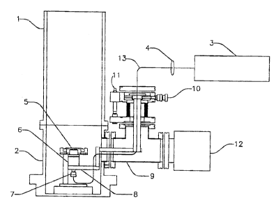
FIG. I depicts one embodiment of a system for focusing light to a
-1~-
Final_App.doc
CA 02430750 2003-05-30
submicron spot size for matrix assisted laser desorption/ionization (MALDI).
This
embodiment utilizes a time-of flight mass spectrometer 1 to capture and
analyze
the ions that includes a MALDI vacuum chamber 2. The mass spectrometer 1
may be any appropriate mass analyzer. A substrate is positioned on a MALDI
sample plate 5 that is placed within the MALDI vacuum chamber 2. The sample
plate 5 holds a sample substrate that may include an analyte and an
appropriately absorbing matrix. The matrix molecules must absorb a sufficient
amount of energy at the wavelength of the laser used for desorption/ionization
to
rapidly expand (along with the analyte molecule- a protein in our case) into a
gas
phase and transfer a charge to the analyte while either in the solid or gas
phase.
Examples of an appropriately absorbing matrix for a nitrogen laser emitting a
337nm or Nd:YAG Laser emitting at 355 nm would be ferrulic acid, sinnipinic
acid, alphacyano-cinniminic acid, dihidroxybenzoic acid, and 3-
hydroxypicolinic
acid. For an infrared laser emitting at 2.94 micron, the appropriately
absorbing
matrices could be glycerol, water or other compounds with an "O-H stretch"
that
absorb at the wavelength used.
A microscopic objective 6 may be positioned below the sample plate 5 to
create a desorption/ionization laser source of <500 nm spatial resolution at
the
surface of the sample. Utilizing a laser source with a spatial beam profile
which
can be described by a Gaussian function, the spot size at the focus of an
optic in
the beam path is suitably described as twice the beam radius (w o) at the
-12-
Final_App.doc
CA 02430750 2003-05-30
Gaussian beam waist (zo) where,
wo = zA/w(z)rr as z ->C
To guarantee a well-characterized Gaussian laser source in our preferred
configuration, we have selected to use a Nd:YAG laser 3 (Coherent Infinity 40-
100) operating at 355 nm. This laser source uses a laser diode as a pump
source with the oscillator built as a ring cavity. Amplification occurs using
a
process of Stimulated Brillouin Scattering (SBS). The fundamental laser beam
is
reflected through the amplification Nd:YAG rods using a mirror induced by SBS
in a cell filled with the compound CFC 113 This "phase conjugation" or "time
reversal" mirror reflects the laser beam, so that it perfectly re-traces its
wave front.
as it is amplified additional times in the Nd:YAG rods - something not
possible
with conventional optics. The Coherent Infinity 40-100 is recognized as
producing nearly perfect TEMoo single mode Gaussian spatial pulses of 3 ns
temporal width.
The Rayleigh criterion for spatial resolution is conveniently written as,
d = Diameter of spot size = 0.61A/N.A.
Where N.A. is the numeric aperture of the optic. In our case, the objective
has
an N.A. = 0.75. Thus, the diffraction limit for our spot size in the preferred
-13-
Final_App.doc
CA 02430750 2003-05-30
configuration is seen to be,
d = 289 nm
The present inventions spot size has been measured to 414 nm. By coupling the
355 nm output into our fiber optic 13, and then to our Carl Zeiss "Fluar"
confocal
microscopic objective 6, we have been able to measure the produced near-
diffraction limited laser spot. We have measured this laser spot, 2 w (z), as
it
diverges from the objective at various distances, z, from the beam waist, zo.
In
this way we have been able to calculate the effective average beam radius and
spot size at the focus of our objective in our preferred configuration.
An electrically insulating microscopic objective holder 8 holds the objective
6 and insulates the microscopic objective 6 from the electrical fields of
MALDI-
MS. A turbo molecular pump 12 pumps the vacuum chamber. A "T" shape
adapter 9 holds the objective positioner 8 and fiber optic 7. The X,Y
positioner/micrometer 10 moves the objective 6 in the X and Y co-ordinates.
The
Z positioner/ micrometer 11 moves the objective in the Z co-ordinate. A
mirror/collimating coupler from fiber optic 7 collimates the laser beam to the
aperture of the fiber optic. In operation, it focuses the laser beam to the
aperture
of the fiber optic cable.
FIG. 2 depicts prior art utilizing a nitrogen laser 3 that generates a
coherent light source and is positioned at an acute angle above the MALDI
-14-
Final_App.doc
CA 02430750 2003-05-30
sample 5. The slide resides inside a MALDI vacuum 2. A time of flight mass
spectrometer I is attached to MALDI vacuum chamber 2 to capture and analyze
the ions. A turbo pump 12 is used to pump the MALDI vacuum chamber 2. The
prior art can only reach a certain spot size because the laser interferes with
the
escaping ions. This preferred embodiment overcomes this limitation by using
confocal microscopy to introduce ionizing light and mounting it on the reverse
side of a quartz MALDI plate to create a desorption/ionization laser source of
<500 nm spatial resolution.
FIG. 3 depicts a further embodiment of the present invention as a system
for creating a correlated visual image of the ion desorption region of a
sample
substrate. This embodiment employs a ND: YAG laser 3 to generate coherent
ultra-violet light. The light is projected through a collimating fiber optic
couplers 4
and is reflected off a mirror 14 and through a second collimating fiber optic
coupler 4 which directs the ultra-violet light into a first end of a fiber
optic cable
13 and exits out a second end of fiber optic cable 13. After exiting the
second
end of the fiber optic cable, the ultra-violet light is directed through a
mirror/collimating coupler 7 that focuses the laser beam to the aperture of
the
fiber optic. A microscopic objective 6 is placed below the sample plate 5 to
create a desorption/ionization coherent light source of <500 nm spatial
resolution
at the surface sample. An electrically insulating microscopic objective holder
8
holds the objective 6 and insulates the microscopic objective 6 from the
electrical
fields of MALDI-Mass Spectrometer. A substrate is positioned on a MALDI
-15-
Final_App.doc
CA 02430750 2003-05-30
sample plate 5 that is placed within the MALDI vacuum chamber 2. A sample
illuminator 16 illuminates the sample substrate to create an optical image.
The
optical image is transported back through fiber optic cable 13. The image is
projected through collimating fiber optic couplers 4 and toward mirror 14.
Mirror
14 reflects laser light and transmits an optical image of sample to camera 15.
A
time of flight mass spectrometer I is utilized to capture and analyze the
ions.
A turbo pump 12 pumps the vacuum chamber (prior art). A "T" shape
adapter 9 holds the microscopic objective positioner 8 and fiber optic 7. The
X,Y
positioner/micrometer 10 moves the microscopic objective 6 in the X and Y
co-ordinates. The Z positioner/micrometer 10 moves the microscopic objective 6
in the Z co-ordinate. A mirror/collimating coupler from fiber optic 7
collimates the
laser beam to the aperture of the fiber optic.
While this embodiment described herein uses a time of flight mass
spectrometer for capturing, detecting and analyzing the ions, it is to be
understood that the capture, detecting and analysis of the ions may be
accomplished using any one of a number of well known analytical devices. It is
also contemplated that light in wave lengths other than ultra-violet (e.g.
infrared)
are within the scope of the invention and that the light may be transported by
other well-known methods of transferring coherent light sources.
EXAMPLE 1
The present invention was tested using Angiotensin I (mw 1296.9) that was
purchased from Sigma (St. Louis, MO) in the highest purity available. The UV-
-16-
Final_App.doc
CA 02430750 2003-05-30
MALDI matrices, a-cyano hydroxycinniminic acid (ACHA) and sinnipinic acid (SA)
were also purchased from Sigma in the highest purity available. Protein
samples
were prepared for MALDI analysis by allowing 0.5 pl of protein standard (200
ng/pl) to dry followed by addition of 0.5 pl of matrix solution (10 mg/ml
matrix in a
70:30 mixture of 0.1 % TFA:acetonitrile. The matrix solution is allowed to dry
for
minutes after which the sample plate is loaded into the instrument.
Instrumentation
All MALDI mass spectra were collected on a Perseptive Biosystems
Voyager SR. Spectra collected in linear mode used an accelerating voltage of
25
kV with a 95% grid voltage and 0.3% guide wire voltage. The m/z range was
limited to 11,352.
The sample stage was altered in several ways, with relative locations of
each piece described from the perspective facing the instrument front panel.
First, two of the PEEK supports for the sample stage towards the rear (nearest
the source region turbo pump) of the can were removed. A further modification
to
the plate holder was made to allow the objective to move directly underneath
the
sample plate. The majority of the metal on the bottom of the sample stage was
machined to leave clear access to the objective with the pin for connection of
the
extraction potential moved to the front of the stage. The majority of the
bottom
"skid" of the MALDI plate was removed with only 5 mm portions of "skids"
(portion of the plate away from the magnetic base) remaining. The magnetic
portion of the base was left attached as well. The bottom of the plate
containing
-17-
Final_App.doc
CA 02430750 2003-05-30
the sample wells was machined on both sides with a final thickness of 400 pm.
The top, or well side, of this plate had a 2.54 cm diameter portion machined
to a
depth of 200 pm where a quartz coverslip could be pressed into position
covering
a 3X3 hole pattern from wells 45 to 47 and 65 to 67. Each of those nine holes
was drilled through with a diameter of 1.5 mm.
A Carl Zeiss "Fluar"-type confocal objective lens with >85% transmission
at 355 nm was utilized. Quartz fiber optic cable with a low hydroxyl count was
purchased from Ocean Optics with a diameter of 300 microns. The purity of this
type fiber allowed for a high duty cycle at extreme laser intensities.
Alternatively,
standard UV-Vis fiber optic cable of 1,000 pm was also used.
The apparatus for positioning was mounted on a PEEK arm threaded for
the microscopic objective. The laser used for UV-MALDI was a Coherent Infinity
10-400 Nd:YAG laser. The third harmonic of 355 nm used for ionization. The
beam was focused on to the fiber optic for entrance into the confocal
objective.
The results of this experiment are shown in FIG. 4.
FIG. 4 illustrates the positive ion spectra for angiotensin I (M+H+ average
mass 1297.5) collected using a preferred embodiment of the invention and 64
laser shots. The Nd:YAG laser was operated at 20 Hz using a wavelength of 355
nm that was further focused on to the sample using the Carl Zeiss objective.
The
desorption/ioinization was performed by passing the focused beam through the
quartz coverslip. The spectra was obtained in reflectron mode using a 25 kV
accelerating voltage with the grid operated at 93%, and guide wire at 0.25%.
-18-
Final_App.doc
CA 02430750 2003-05-30
The matrix ions for a-cyano hydroxy cinniminic acid molecular ion (M+H 190)
and
common dehydration product are labeled as well as the angiotensin I peak and
its sodium adduct.
The sample spot size was estimated to be 2 mm in diameter containing
200 ng of peptide. This amount of material for angiotensin equates to 154
femtomoles within the 0.0314 cm2 (or 3.8 X 10 6 pmt) droplet area, or40
zeptomole/pmt. This is well above the minimum detectable concentration range
for substance P reported previously at 0.0083 zeptomole /pmt by Keller and Li
(Keller, B. 0. and L. Li. J. Am. Soc. Mass. Spectrom., 2001, 12, p. 1055-1063)
thus substantiating a particular advantage of the claimed invention in its
ability to
detect proteins in extremely small samples. Assuming equal sample distribution
across the spot (40 zeptomoles/pmt) the 414 nm diameter spot (0.134 pm2)
would contain 5.4 zeptomoles of angiotensin.
While the invention has been described in connection with specific
embodiments thereof, it will be understood that it is capable of further
modifications and this application is intended to cover any variations, uses,
or
adaptations of the invention following, in general, the principles of the
invention
and including such departures from the present disclosure as come within known
or customary practice within the art to which the invention pertains and as
may
be applied to the essential features herein before set forth and as follows in
scope of the appended claims.
-19-
Final_App.doc