Note: Descriptions are shown in the official language in which they were submitted.
CA 02433797 2003-07-04
WO 02/054948 PCT/US02/01138
Assessment of Tooth Structure Using Laser Based Ultrasonics
CROSS REFERENCE TO RELATED APPLICATIONS
[0001] This application is related to and claims the benefit of U.S.
Provisional Patent
Application Serial No. 60/261,090, filed January 11, 2001 entitled "Assessment
of Tooth
Structure Using Laser Based Ultrasonics".
FIELD OF THE INVENTION
[0002] The present invention is an apparatus and a method related to using
laser based
ultrasonics to aid in the assessment of tooth structure.
BACKGROUND
[0003] The benefits of ultrasonics to examining soft tissue structures,
particularly the abdominal
region, brain, and eyes have long been known. In these applications, typically
one or more
acoustic contact transducers is used to generate and detect acoustic waves in
the structure.
These procedures are simplified, at least for examination of teeth, with the
relatively large
dimensions being examined, slower acoustic wave velocity (allowing lower
frequency acoustic
waves to be used for equivalent acoustic wavelengths), and readily available
acoustic coupling
material for the transducer to the soft tissue. (Soft tissue, unlike hard
tooth enamel and dentin,
is largely composed of water, making water a very efficient coupling
material.)
[0004] Attempts to adapt conventional ultrasonic techniques to examination of
internal structure
of a tooth have met with little success. One major obstacle is identifying a
suitable couplant for
the transducer to the tooth for in-vivo measurements. Without proper coupling,
transfering
acoustic energy into the tooth is difficult. Early investigators attempted
using water, as with soft
tissue structures, but results were not convincing.
[0005] The coupling problem was overcome by replacing water with mercury.
Although
providing superior coupling efficiency, mercury is not suitable for clinical
applications due to its
toxicity.
[0006] Another solution to overcome the coupling difficulty was using a small
aluminum buffer
rod to transfer the acoustic energy from the contact transducer to the tooth.
An estimated
transmission efficiency of almost 87% was achieved using this technique,
compared to only 5%
-1-
CA 02433797 2003-07-04
WO 02/054948 PCT/US02/01138
using water. However, a significant limitation of this system was coupling the
aluminum buffer
rod with the tooth surface. To ensure proper coupling of the acoustic energy
to the tooth, a flat
spot had to be ground on the tooth surface, making this technique unsuitable
for clinical
applications. In addition, the relatively large contact area (3.2 mm diameter)
limited the spatial
resolution of the probe: For assessing anomalies in a tooth, such as poor
bonding or voids
between the restorative material and the dentin, a detection footprint smaller
than the anomaly
itself is required.
[0007] One method of increasing spatial resolution of a contact transducer is
to use a spherical
transducer that focuses abeam onto a sample (tooth) surface. This method forms
the basis of the
acoustic microscope, the acoustic equivalent of an optical microscope. This
technique was used
to study unblemished and demineralized enamel from extracted human teeth,
using water as a
couplant. The inspection depths were thus limited to approximately 0.5 to 1.5
mm.
[0008] More recently, the increased spatial resolution of the acoustic
microscope was used to
detect small caries lesions in sections of human enamel. However, as with
previous work,
special polishing of the tooth samples was required, making the technique ill-
suited fox clinical
applications.
[0009] What is needed is a tooth structure assessment system achieveable in-
vivo operation that
combines superior coupling efficiency, a small detection footprint size, and
no special tooth
surface preparation.
SUMMARY
[0010] To help overcome previous difficulties in coupling efficiency,
detection footprint size,
and special surface preparation, the present invention utilizes laser-
generated ultrasound
techniques. Laser-generated ultrasound uses a shoat-pulse laser, in place of a
contact firansducer,
to generate high frequency (broad-band) ultrasound in a material. Due to the
absorption of
pulse energy at or near the surface of the specimen, temperature gradients are
established within
the material, producing a rapidly changing strain field. This strain field, in
turn, radiates energy
as elastic (ultrasonic) waves. At low pulse energies, this is an entirely
thermo-elastic process
resulting in no damage to the material under test. An advantage of this
technique over the
previous methods is that no special surface preparation of the tooth is
required. In addition, by
focusing the laser beam onto the surface of the tooth, a very small contact
(generation) area can
-2-
CA 02433797 2003-07-04
WO 02/054948 PCT/US02/01138
be achieved. Spot size diameters on the order of tens of microns are routinely
achieved.
[0011] Both enamel and dentin have strong absorption bands in the longwave
infrared (IR)
spectrum (9 to 11 pm). These optical properties have already led to
applications for the carbon-
dioxide (C02) laser in fusing enamel, dentin, and apatite. Fusion inhibits
subsequent lesion
progression and markedly improves bonding strength of a composite resin to
dentin. For
illustration purposes with respect to the present invention, a short pulse COZ
laser has been used
to generate acoustic waves in an extracted human incisor. In some instances,
other lasers, such
as, for instance, a pulsed Nd:YAG laser may be used.
[0012] Optical detection of the ultrasound, such as by a laser vibrometer
interferometer, provides
a complementary technique for remote sensing of ultrasonic waves. Techniques
based upon the
sensing of the optical wavefront reflection from the tooth, such as Fabry-
Perot interferometers,
Mach-Zender interferometers, Michelson interferometers, photo-refractive
interferometers,
optical feedback interferometry, and several other types of laser vibrometers,
are well suited for
diffusely reflecting surfaces. For purposes of illustration with respect to
the disclosure herein, a
laser vibrometer is described to detect acoustic wave arrivals.
[0013] One embodiment of the present invention is a method of assessing tooth
structure using
laser based ultrasonics. Ultrasonic acoustic waves are generated using a
pulsed laser. The beam
of the pulsed laser is focused onto a desired area on the surface of a tooth
thereby creating
ultrasonic acoustic waves within the bulk and along the surface of the tooth
structure. These
acoustic waves are optically detected using optical interferometric means.
Finally, detected
acoustic waveforms are processed to assess the internal or surface structure
of the tooth.
[0014] Another embodiment of the present invention is an apparatus including a
pulsed laser that
generates a beam of ultrasonic acoustic waves. The beam is focused by a lens
onto a desired
area on the surface of a tooth creating ultrasonic acoustic waves within the
bulk and along the
surface of the tooth structure. Optical interferometric detection means
optically detect the
acoustic waves generated within the tooth structure and an oscilloscope
processes the detected
acoustic waveforms to assess the internal structure of the tooth. The short
pulse laser operates
in a region of absorption for the tooth structure.
[0015] In another embodiment, a thin film or coating can be placed on the
tooth surface and the
short pulse laser then operates in a region of absorption for the thin film or
coating.
-3-
CA 02433797 2003-07-04
WO 02/054948 PCT/US02/01138
BRIEF DESCRIPTION OF THE DRAWINGS
[0016] FIGURE 1 illustrates the internal structure of tooth enamel.
[0017] FIGURE 2 illustrates the internal structure of tooth dentin.
[0018] FIGURE 3a illustrates a block diagram of components used in the present
invention.
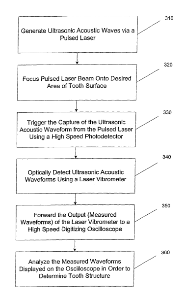
[0019] FIGURE 3b is a flowchart illustrating the steps performed in the
present invention.
[0020] FIGURE 4 illustrates a cross-section of an extracted human incisor
showing the enamel,
dentin, and pulp sections, and also an amalgam insert.
[002I] FIGURE Sa illustrates a measured temporal profile of a COZ pulse Iaser.
[0022] FIGURE Sb illustrates a measured temporal profile of an Nd:YAG pulse
laser.
[0023] FIGURE 6a illustrates an ultrasonic waveform for a tooth phantom taken
in an enamel
region.
[0024] FIGURE 6b illustrates an ultrasonic waveform for a tooth phantom taken
in an
enamel/amalgam/enamel region.
[0025] FIGURE 6c illustrates an ultrasonic wavefonn for a tooth phantom taken
in an
enamel/dentin/enamel region.
[0026] FIGURE 6d illustrates an ultrasonic wavefonm for a tooth phantom taken
in an
enamel/dentin/pulp/dentin/enamel region.
[0027] FIGURE 7 illustrates a cross-section of an extracted human iizcisor.
[0028] FIGURE 8a illustrates an ultrasonic waveform for a human incisor taken
in an enamel
region.
[0029] FIGURE 8b illustrates an ultrasonic waveform for a human incisor taken
in an
enamel/amalgam/enamel region.
[0030] FIGURE 8c illustrates an ultrasonic waveform for a human incisor taken
in an
enamel/dentin/enamel region.
[0031] FIGURE 8d illustrates an ultrasonic waveform for a human incisor taken
in an
enamel/dentin/pulp/dentin/enamel region.
DETAILED DESCRIPTION
[0032] The generation and detection of ultrasonic waves provides a method for
characterizing
the bulk and surface properties of a material by interrogating a specimen with
high frequency
acoustic waves. Up to three bulk acoustic waves can propagate in a material,
each with its own
-4-
CA 02433797 2003-07-04
WO 02/054948 PCT/US02/01138
characteristic velocity. As an ultrasonic wave propagates, the amplitude
decreases due to
geometrical spreading, attenuation from absorption, and scattering from
discontinuities.
Measurement of wave speeds, attenuation, and scattering provide the
information needed to
quantify the bulk internal and surface properties of the material. In this
analysis, we are
interested in the interaction of the acoustic waves at the interfaces between
the dental hard and
soft tissues.
[0033] The propagation of elastic plane waves in an anisotropic material is
described using
ChristofFel's equation by:
~c~;k~l;l~ -p~Zs~k~ = o.
This equation provides an analytic relation between the phase velocity, v, of
the elastic waves
and the elastic moduli, C;jkl. The direction cosines are specified by l; (with
i = 1, 2, or 3), p is the
material density, and 8;k is the Kronecker delta function.
[0034] The anisotropic nature of dental hard tissue is well-documented. Dental
enamel, the hard
protective substance covering the crown of the tooth, is the hardest biologic
tissue in the body to
resist fractures during mastication (chewing). Enamel is composed of about 96%
inorganic
mineral in the form of hydroxyapatite and 4% water and organic matter.
Hydroxyapatite is a
crystalline calcium phosphate that is also found in bone, dentin, and
cementum. As illustrated
in FIGURE 1, enamel is composed of rods 10 that extend from their origin at
the dentino-
enamel junction to the outer surface of the tooth. The rod itself resembles a
keyhole in shape,
allowing individual rods to form a strong interlocking structure. The head of
the rod measures
about 5 pm across while the tail is only about 1 pm. Each rod is filled with
crystals, whose
orientation 12 varies along the rod. At the head of the rod, these crystals
are approximately
parallel to the rod axis while near the tail of the rod, the crystals are
oriented almost
perpendicular to the rod axis. Variations in the elastic properties of enamel
are quantified by
measuring the Rayleigh velocity as a function orientation with the enamel rods
with an acoustic
microscope. It was found that the Rayleigh velocity varied by almost 5% in
these
measurements. More recently, a three-dimensional finite element model was
developed for the
prismatic nature of enamel that predicted stiffness both along and across the
rods and found that
Young's modulus varies dramatically both with direction and volumetric
fraction. Specifically,
-5-
CA 02433797 2003-07-04
WO 02/054948 PCT/US02/01138
the deviation in Young's modulus is estimated at nearly X10% parallel and X60%
perpendicular
to the orientation of crystals.
[0035] Referring to FIGURE 2, dentin 20 is the hard tissue that constitutes
the body of a tooth.
Unlike enamel, which is almost white in color, dentin appears almost
yellowish. Dentin is a
living tissue that is not normally exposed to the oral environment. Like bone,
dentin is
composed primarily of an organic matrix of collagen fibers (20%), inorganic
hydroxyapatite
crystals (70%), and about 10% water. With 20% less mineral than enamel 22,
dentin 20 is
softer and slightly elastic. Dentin 20 itself is classified as primary,
secondary, and tertiary on
the basis of the time of its development and the histoLogic (microscopic)
characteristics of the
tissue. Primary dentin is the major component of the crown and root while
secondary dentin
forms only after tooth eruption (i.e. when the teeth begin to function) and
borders the pulp.
Finally, tertiary or reparative dentin occurs in response to the presence of a
trauma to the pulp
24. The structure of dentin 20 is composed of S-shaped tubules that run from
the dentino-
enamel junction to the dentino-pulp junction. Each of these tubules is about 1-
3 ~,m in diameter
and surrounded by a matrix of needle-shaped, hydroxyapatite crystals in a
protein matrix of
composed mostly of caLlogen.
[0036] The third component of a tooth is the pulp 24, which is the soft
connective tissue located
in the central portion of each tooth. It is composed of both a crown (coronal
part) and root
(radicular part). Pulp 24 contains specialized connective tissue composed of
thin-walled blood
vessels, nerves, and nerve endings enclosed within the dentin 20.
[0037] To simplify analysis, enamel, dentin, and pulp are characterized as
elastically isotropic.
For isotropic materials, only two bulk waves need be considered, Longitudinal
and shear. It can
be shown through Christoffel's equation that the velocity of these two waves
is given by:
_ CI1 _ E _ Caa _ N
Vlong - ~ ~ and vshear - ~ -
where E is the elastic modulus and ~, is the shear modulus. The acoustic
velocities and density
for the various components of a tooth are listed in Table 1. When assessing
any multi-phase
structure, such as a tooth, both the acoustic velocity and acoustic impedance
of each layer must
be considered. The acoustic impedance, Z, is defined as:
-6-
CA 02433797 2003-07-04
WO 02/054948 PCT/US02/01138
Z = Ep = pv,o"g
[0038] When an acoustic wave travels from one medium to another (i.e. from the
enamel to the
dentin), a portion of the wave is reflected and the remaining portion is
transmitted (assuming no
other losses). The intensity of the reflected acoustic radiation, Ir, assuming
plane wave
propagation, at the interface between two different mediums with acoustic
impedances of Zl and
Z2, is given by:
Z
Ir = C z2 +z1 ) n = r1;
where r is the reflection coefficient and I; is the intensity of the incident
radiation. Due to
conservation of energy, the intensity of the transmitted acoustic radiation,
It, is:
It = I; - Ir.
[0039] Knowledge of the relative reflected and transmitted intensities at each
material interface
in a tooth structure aids in the interpretation of the final ultrasonic
waveform.
[0040] Efficient ultrasound generation depends upon the material's absorption
characteristics at
the optical wavelength of the pulsed laser. Enamel and dentin have a strong
absorption in the 9-
1 l~,m region due to the phosphate in the carbonated hydroxyapatite (CAP).
Absorption
coefficients of 5500, 8000, 1125, and 825 cm 1 at 9.3, 9.6, 10.3, and 10.6
Vim, respectively have
been determined. These correspond to absorption depths between 1.25 and 12
Vin.
[0041] Referring now to FIGURE 3a, a pulsed COz laser 30 (10.6 ~,m with a SOns
rise time) is
used to generate acoustic waves in a human tooth 32. The pulsed laser 30 is
focused to a spot
size on the order of a few tens of microns on the tooth's surface via lens 34.
The output power
of the pulsed laser 30 is controlled using polarization optics to ensure non-
destructive (thermo-
elastic) acoustic wave generation. The short pulse laser operates in a region
of absorption for
the tooth structure. In another embodiment, a thin film or coating can be
placed on the tooth
surface and the short pulse laser then operates in a region of absorption for
the thin film or
coating.
CA 02433797 2003-07-04
WO 02/054948 PCT/US02/01138
[0042] Optical detection of the acoustic wave arnvals is accomplished using a
laser vibrometer
36. The output signal from the vibrometer 36 is proportional to the surface
displacement of the
tooth 32. Ultrasound measurements can be taken in an epicentral configuration
in which the
source/laser 30 and receiver/vibrometer 36 are on opposite sides of the tooth
32. Alternatively,
ultrasound measurements can be taken in an non-epicentral configuration in
which the
source/laser 30 and receiver/vibrometer 36 are on opposite sides of the tooth
32. In addition,
ultrasound measurements can be taken wherein the source/laser 30 and
receiver/vibrometer 36
are on the same side of the tooth 32.
[0043] The output of the vibrometer 36 is passed to a high-speed digitizing
oscilloscope 37 for
recording the ultrasonic event. The capture of the ultrasonic waveform is
triggered by a high-
speed photodetector, comprised of a photo-diode 38 and an output coupler 39,
which sees a
sampling of the output pulse of the pulsed laser 30. Sampling of the output
laser pulse allows
for very accurate time-of flight measurements for the acoustic waves. In
addition, multiple
waveforms were averaged to improve the signal-to-noise ratio (SNR).
[0044] FIGURE 3b is a flowchart illustrating the steps performed in the
present invention.
Using the configuration described in FIGURE 3a, ultrasonic acoustic waves are
generated 310
via a pulsed laser. The beam of the pulsed laser is focused 320 onto the
surface of a tooth at a
desired area. The capture of the resulting acoustic wavefonns within the tooth
are triggered by a
high speed photodetector that samples the output of the pulsed laser 330. A
laser vibrometer is
used to optically detect 340 acoustic waveforms generated within the tooth
structure. The data
recorded by the laser vibrometer is then forwarded to a high speed digitizing
oscilloscope 350.
The sampling of the pulsed laser provides the oscilloscope with very accurate
time
measurements with respect to the origination of the ultrasound beam. The
detected acoustic
waveforms are then processed, analyzed and displayed by the oscilloscope 360.
[0045] Processing of the results involves analysis of the peaks and valleys of
the resulting
waveforms versus time. Certain characteristics of a tooth's structure can be
determined based
on the arrival time and amplitude of the arrival of certain wave fronts. The
processing is
typically done by a processing device (computer) that can be programmed with
the
characteristics of normal tooth structure.
[0046] An advantage of the present invention is its ability to assess the
health of the entire tooth
structure. One application is to be able to resolve the interfaces of the
various junctions that
_g_
CA 02433797 2003-07-04
WO 02/054948 PCT/US02/01138
naturally occur in a tooth. Moreover, valuable diagnostic information can be
determined by
knowing, or at least estimating, the thicknesses of the various components
that compromise a
tooth. For example, there is a need to image the margins of a restoration for
the detection of
poor bonding or voids between the restorative material and the dentin. With
conventional x-ray
techniques, it is difficult to detect cracks and to visualize interfaces
between hard media. This is
due to the x-ray providing only a two-dimensional projection of the internal
structure (i.e. a
silhouette). In addition, a high resolution imaging modality is needed to
detect tooth decay in its
early stages. If decay can be detected early enough, the process can be
monitored and
interventional procedures, such as fluoride washes and controlled diet, can be
initiated which
can help the tooth to re-mineralize itself. Currently employed x-ray imaging
is incapable of
detecting decay at a stage early enough to avoid invasive cavity preparation
followed by a
restoration with a synthetic material. Laser ultrasonics can be used to detect
early stages of
decay both in the bulk and on the surface of the tooth. Other clinical
applications include the
visualization of periodontal defects, the localization of intraosseous
lesions, and determining the
degree of osseointegration between a dental implant and the surrounding bone.
[0047] To better illustrate the present invention, results of the use of the
present invention on a
tooth phantom and an extracted human incisor are presented and discussed.
[0048] One means of better understanding the acoustic signature obtained from
an actual tooth
structure is to construct a tooth phantom made from acoustically similar
materials. A cross
section from an extracted human incisor is shown in FIGURE 4. The enamel 40,
dentin 42,
pulp 44, and an amalgam insert 46 are marked. To simplify construction, a
separate tooth
phantom was designed for four different acoustic "paths" through the tooth
section. These paths
include enamel, enamel/amalgam/enamel, enamel/dentin/enamel, and
enamel/dentin/pulp/dentin/enamel.
[0049] Materials with similar acoustic velocities and impedances to the
different components of
a real tooth are listed in Table 1. Relatively good matches were found for
enamel, pulp, and
amalgam. Only dentin proved difficult to match. As shown in Table 1, tin has
very similar
acoustic velocities but much higher acoustic impedance. This larger impedance
does not
provide the desired interface characteristics between the different components
of the tooth
phantom. Borosilicate glass, on the other hand, has much faster acoustic
velocities, but more
comparable acoustic impedance. Since the purpose of this study was to better
understand the
-9-
CA 02433797 2003-07-04
WO 02/054948 PCT/US02/01138
uiterface properties between the different dental tissues, similarities in
acoustic impedance was
viewed as more important than acoustic velocities.
TAELE 1
Layer V~o"~ Vshear Density Acoustic
[mm/ps] [mm/E~s] p [kg/m3] Impedance
Z [x 106
kg/mZS]
Enarnel 6.25 3.10 3000 18.8
Aluminum 6.30 3.10 2700 17.0
Dentin 3.80 1.90 2000 7.6
Tin 3.30 _ 1740 24.2
1.70
Borosilicate5.30 3.00 3570 18.9
glass
Pulp 1.57 0.80 1000 1.57
Teflon 1.4 2140 3.0
Amal am 4.35 2.26 7750 33.7
Copper 4.70 2.30 9670 41.6
[0050] A comparison of the reflection coefficients between the interfaces of a
real tooth and
those of the tooth phantom are listed in Table 2.
TABLE 2
Real Tooth Tooth Phantom
Components r Components r'
enamel/amalgam/enamel 0.081aluminum/copper/aluminum 0.176
enamel/dentin/enamel 0.18 aluminum/ lass/aluminum 0.026
dentin/amalgam/dentin 0.399lass/copper/glass 0.296
dentin/pulp/dentin 432 ~ glass/teflon/glass 0.369
[0051] A pulsed COZ laser is used to generate acoustic waves in the extracted
human incisor.
The measured temporal profile of a C02 laser, shown in FIGURE Sa, indicates a
pulse rise time
of SOns. A noticeable feature of this pulse is the long tail (about 1.5 ~.s).
Since only the rise of
the initial pulse is responsible for high-frequency components of the
ultrasonic waves, this tail
did not effect the ultrasonic measurements.
[0052] Ultrasound generation in the tooth phantoms is accomplished using a
pulsed Nd:YAG
laser (18 ns pulse width). The measured temporal profile of a Nd:YAG pulse is
illustrated in
FIGURE Sb. The poor absorption properties of aluminum in the tooth phantoms at
10.6 ~,m
precluded the use of the C02 laser. 111 both cases, the pulsed lasers are
focused to spot sizes on
the order of a few ten's of microns and the output power of the pulsed lasers
is controlled using
-10-
CA 02433797 2003-07-04
WO 02/054948 PCT/US02/01138
polarization optics to ensure non-destructive (thennoelastic) acoustic wave
generation.
[0053] A path-stabilized Michelson-type interferometer is used to detect the
ultrasonic wave
arrivals in the tooth phantoms. This type of interferometer is sensitive to
sub-nanometer
displacement amplitudes, typical for thermoelastically-generated ultrasound.
Michelson
interferometers are better suited to objects with specular reflections from
the surface of the
object. In each case, the front surface of the tooth phantom is polished to
allow optimal
operation of the interferometer. Since teeth do not provide a specular
reflection, a different
detection scheme is implemented. Optical detection schemes suited for
diffusely reflecting
surfaces include Fabry-Perot, Mach-Zender, photo-refractive, and optical
feedback
interferometers as well as various types of laser vibrometers. For these
measurements, a
commercially available laser vibrometer is used. As with the Michelson
interferometer, this
laser vibrometer has an output proportional to surface displacement.
TOOTH PHANTOM RESULTS
[0054] FIGURE 6a illustrates a measured thermoelastic ultrasonic waveform from
a piece of
aluminum (8.5 mm thick). This represents an ideal waveform through the enamel
of a tooth if
the enamel were truly isotropic. The first longitudinal wave (L1) and shear
wave (S1) arrivals
are marked. Scattered light from the pulsed laser denotes the beginning of the
ultrasonic
wavefonn. This initial laser pulse is visible on all of the tooth phantom
wavefonns. The
aluminum waveform also provides a baseline for the other three tooth phantom
waveforms
(FIGURES 6b-d). Each of these waveforms is distinctly different due to
reflections at the
interfaces of the different layers of the tooth phantoms. Each waveform was
averaged 100 times
to improve SNR.
[0055] The measured waveform from the second tooth phantom illustrated in
FIGURE 6b
simulates what would be found for a tooth with an amalgam restoration
(filling). This phantom
is composed of a 1.95 mm thick piece of copper (amalgam) sandwiched between
two pieces of
aluminum (enamel), 1.95 mm and 1.25 mm thick. The first longitudinal arrival
time, t~,l, occurs
at:
-11-
CA 02433797 2003-07-04
WO 02/054948 PCT/US02/01138
tLl = dlaluminum + dropper + d2aluminum
valuminum Vcopper Valuminum
1.95 mm + 1.95 mm + 1.25 mm = 0,923
~s.
6.3 ~ 4.7 ~ 6.3 ~
~s ~s ~s
[0056] The second acoustic wave arrival occurs when the longitudinal wave
traverses the thin
piece of aluminum (enamel) a second time after reflection at the enamel-
amalgam junction
(EAJ). This second arrival occurs at:
d2aluminum
tEAJI = tLl +
Valuminum
= 0.923 ~s+0.397 ~s =1.32 ~s.
[0057] The next two acoustic wave arrivals result from additional traverses of
the thicker piece
of aluminum and the copper. In each case, the acoustic wave arrivals in the
tooth phantom will
be more pronounced than what would be expected for an actual tooth due to the
larger reflection
coefficient at the almninum/copper junction (I-'=0.176 versus r=0.081). The
first shear arrival is
denoted by S1.
[0058] The ultrasonic waveform for the aluminumlglass/aluminum
(enamel/dentin/enamel) tooth
phantom is illustrated in FIGURE 6c. The first longitudinal arrival (L1) is
identified by the
initiation of the positive slope in the waveform. As with the pxevious
phantom, this arrival
corresponds to a direct acoustic path for the longitudinal wave through the
tooth phantom. In
this tooth phantom, the first section of aluminum (enamel) is 1.95 mrn thick,
the glass (dentin) is
3.3 mm thick, and the final section of enamel is 1.25 mm thick. The second
longitudinal arrival
(DEJ1) occurs after the longitudinal wave traverses the thinner section of
enamel (aluminum) a
second time after reflection at the dentino-enamel (glass/aluminum) junction
(DEJ). Subsequent
longitudinal wave arrivals are also visible due to additional traverses of the
thicker piece of
enamel (aluminum) and dentin (glass) after reflections at the DEJ. The
amplitude of each of
these acoustic wave arrivals is smaller than in the previous tooth phantom due
to the closer
acoustic impedance match of the materials and subsequent smaller reflection
coefficient
(r=0.026). The DEJ would be more visible in an actual tooth due to the larger
reflection
coefficient (r=0.18).
[0059] The final ultrasonic waveform illustrated in FIGURE 6d shows the
-12-
CA 02433797 2003-07-04
WO 02/054948 PCT/US02/01138
enamel/dentin/pulp/dentinlenamel (aluminum/glass/teflon/glass/aluminum) tooth
phantom.
This tooth phantom is composed of 1 mm thick pieces of aluminum for the
enamel, 3.S mm
thick pieces of glass for the dentin, and a 1.75 mm thick piece of teflon for
the pulp. The lower
SNR of this waveform is attributed to the increased complexity and thickness
of this tooth
phantom in comparison to the previous three. As before, the first longitudinal
wave arrival (L1)
corresponds to a single pass of the longitudinal wave through the phantom. The
next two
acoustic wave arrivals (DEJ) correspond to additional passes through the
enamel (aluminum).
As before, these reflections are very small due to the small reflection
coefficient at the junction.
The next acoustic wave arrival corresponds to a reflection at the dentin-pulp
junction (DPJ).
The amplitude of this reflection is far more pronounced due to the larger
reflection coefficient.
HUMAN INCISOR RESULTS
[0060] Laser-based ultrasonic measurements were performed on an extracted
human incisor.
Prior to the measurements, the tooth was stored in a physiological saline to
help preserve the
mechanical properties of the dental tissue. After the measurements were
completed, the incisor
was cleaved along the propagation direction of the ultrasonic waves to
determine the location of
the internal interfaces within the tooth. A cross-section of the incisor is
shown in FIGURE 7
illustrating the enamel 70, dead tracts 72, dentin 74, pulp 76, and cementum
78. The dentino-
enamel (DEJ), dentino-pulp (DPJ), and dentino-cementum (DCJ) junctions are
also visible. In
addition, a region of dentin containing dead tracts is also present. Results
from measurements
taken at four different locations through the tooth are presented here. These
measurement
locations are marked in FIGURE 7 as (1), (2), (3), and (4), respectively. In
addition, the
thickness of each dental hard and soft layer is listed in TABLE 3.
TABLE 3
MeasurementInternal
Structure
of Tooth
in mm
Location
1 Enamel
6.06
2 Enamel Dentin Enamel
0.95 4.76 1.23
3 Dentin Pulp Dentin Cementum
1.51 0.53 1.3 0.21
4 Cementum Dentin Pulp Dentin Cementum
0.32 1.13 0.47 0.86 0.48
-13-
CA 02433797 2003-07-04
WO 02/054948 PCT/US02/01138
[0061] FIGURES 8a-d illustrate the acoustic waveforms determined at the
measurement
locations shown in FIGURE 7, respectively.
[0062] The first measurement location was taken through the top portion of the
tooth and its
waveform is illustrated in FIGURE 8a. In this region, a straight path across
the tooth would
only propagate through enamel. The first longitudinal acoustic wave arrival
(L) occurs at about
1.08 ~s, which is slightly longer than the 0.98 ~,s expected assuming a
longitudinal velocity of
6.25 nn~n/~,s (see TABLE 2). However, as previously discussed, the wave speed
is known to
vary in enamel due to the anisotropic nature of the elastic properties. The
anisotropy is
especially pronounced in this region of the tooth since the ultrasonic waves
propagate both
parallel and perpendicular to the enamel rods. An interesting feature of this
waveform is the
presence of the two large acoustic wave arrivals at 1.48 ~,s and 1.98 ~,s.
These arrivals are
believed due to acoustic wave scattering from the top surface of the tooth,
which is just above
the measurement location. This phenomenon illustrates the difficulty in
interpreting bulk
measurements near interfaces. The acoustic wave arrival at 2.2 ~,s is very
close to the expected
wave arrival time of the first shear wave (S). Acoustic wave arrivals at 2.5
~s and later
correspond to reflections and scattering from the internal structure of the
tooth and do not lend
themselves to straightforward interpretation.
[0063] The second measurement location is approximately four millimeters down
from the top
of the tooth and its waveform is illustrated in FIGURE 8b. In this region, the
ultrasonic waves
propagate through two DEJs and a region of dead tracts in the dentin. The
first longitudinal
arrival (L) occurs at about 1.8 ~s. This time corresponds to a sudden negative
change in slope.
Also marked on the waveform is a wave arrival occurring at 2.03 ~s (indicated
by a positive
slope change). This is believed to be due to an additional round trip through
the enamel on the
left hand side of the dentin caused by reflections at the DEJ. This arrival is
more pronounced
than in the tooth phantom (FIGURE 6b) due to the larger reflection coefficient
at the DEJ. The
predicted arrival time for this reflection at the DEJ is 1.9 ~s. The acoustic
wave arrival at 2.25
~s is believed to be due to reflections at the interfaces of the dead tracts.
Dead tracts are
characterized by the death of odontoblasts, resulting in dentin tubules that
contain debris and
voids. It is for this reason that dead tracts appear black when teeth are
sectioned and viewed by
transmitted light. The presence of debris and open spaces in the tubules are
expected to
significantly affect the mechanical properties of the dentin, resulting in
large reflections at the
-14-
CA 02433797 2003-07-04
WO 02/054948 PCT/US02/01138
dead tract junctions (DTJ). This difference in mechanical properties
contributes to the large
acoustic wave arrival (similar to what is seen at the dentino-pulp junction).
The DTJ is probably
also responsible for the next acoustic wave arrival at 2.9 ~s. The final
marked wave arrival at
3.27 ~,s corresponds to the expected arrival time for the shear wave (S) of
3.2 ~,s. Again, there is
expected to be some variation between predicted and measured acoustic wave
arrival times,
although now the propagation direction is mostly parallel to the orientation
of the enamel rods.
In this region of the tooth, the dentin tubules run almost perpendicular to
the direction of the
wave propagation.
[0064] The third measurement location occurs much further down the tooth and
its wavefonn is
illustrated in FIGURE 8c. In this region, the acoustic wave travels through
dentin, pulp, and a
small amount of cementum. The mechanical properties (i.e. acoustic wave speeds
and density)
of cementum are not known. For analysis purposes, it is assumed that the
mechanical properties
of cementuzn are similar to those of enamel. Based upon this assumption, the
first longitudinal
wave arrival is expected at 1.1 ~.s, which is slightly longer than the
measured arrival time of
0.94 ~,s. Tlus delayed arrival time is attributed to both a degradation in the
mechanical
properties of the pulp and uncertainty in the mechanical properties of the
cementum. A second,
faint, arrival is seen at 1.27 ~s (marked by the change in slope of the
waveform). This arrival
coincides with the expected reflection at the DCJ. The next three noticeable
features occur at
1.54 ~s, 1.98 ~,s, and 2.17 ~,s. Due to the amplitude of these wave arrivals,
they are believed to
be due to reflections at the DPJ. The next wave arrival at 2.48 ~s is very
close to that expected
for the first shear wave arrival at 2.2 ~s and displays the expected sudden
change in slope as
found with the dentiupulp/dentin tooth phantom at the shear wave arrival.
[0065] The fourth measurement location is near the base of the incisor and its
waveform is
illustrated in FIGURE 8d. At this location, the acoustic wave traveled through
two layers of
cementum and dentin, as well as a single layer of pulp. The first longitudinal
arrival (L) occurs
at about 1.23 ~s, which is again slower than the expected arrival time of 0.95
~s. The next
arnval occurs at 1.52 ~,s and is attributed to a reflection at a DCJ. The next
two reflections
occur at 1.58 ~s and 1.86 ~s and are due to reflections at the DPJ. Unlike the
measurement at
the third location, there are only two wave arrivals due to reflections at the
DPJ, because the
total transit time in the pulp and dentin on the left hand side of the pulp
are identical. The final
arrival is due to the principal shear wave (S) and occurs at 2.34 ~s. As with
the longitudinal
- 15-
CA 02433797 2003-07-04
WO 02/054948 PCT/US02/01138
wave, this arrival time is slower than the predicted time of 1.89 ~s and again
attributed to
degradation of the pulp.
[0066] Sources of enor should be examined to determine the accuracy of the
final results. It has
been estimated that the error in making laser ultrasonic measurements is less
than 1% when
considering phenomena such as acoustic diffraction and timing precision from
laser alignment.
For measurements on specially prepared samples, an error of 0.08% has been
estimated in
thickness measurements. For the results illustrated herein, the thickness
measurement error is
greater since a tooth is very irregular in shape and uncertainties in the
exact acoustic path due to
possible tilt in the tooth may occur during the measurement. It is more likely
that there is a 3-
4% error in measuring the thickness of the structures within a tooth (i.e. the
enamel, dentin,
pulp, and cementum). Another consideration is the determination of the exact
arrival time of the
first longitudinal wave. As shown in FIGURES 8a-d, the arrival tune of the
first longitudinal
wave is not always clear due to noise in the laser vibrometer signal. However,
this does not
affect the determination of subsequent wave arrivals. Another source of error
is the uncertainty
of the exact wave velocities in the various dental hard and soft tissues.
Young's modulus in
enamel depends on both the orientation of the crystals as well as the
volumetric fraction and can
vary by over 10%.
[0067] Until these factors are better understood, complete characterization of
dental enamel will
be difficult. However, the problem is greatly simplified when measurements are
made in the
enamel/dentin region of a tooth. In this region, the crystals of the enamel
are predominately
oriented along the direction of the acoustic wave propagation, reducing the
uncertainty in the
Young's modulus. This allows for more accurate estimates of enamel thickness
to be made.
The s-shaped tubules in dentin also contribute to uncertainties in the Young's
modulus. Again,
in certain regions, these tubules are oriented parallel to the direction of
the acoustic wave
propagation, simplifying the analysis. Even with the uncertainties in moduli,
the measurements
presented here show that DEJ, DPJ, DCJ, and DTJ axe discernable and estimates
of the
thickness of each of these structures can be made. These estimates are not
currently possible ,
using any other known technique. In addition, uncertainties in dental
structure thickness do not
affect the ability to detect any voids within a tooth.
[0068] The application of laser ultrasonics to the in-vitro assessment of the
internal structure of
teeth has been presented herein and shown to possess significant advantages
over prior art work.
-16-
CA 02433797 2003-07-04
WO 02/054948 PCT/US02/01138
In laser ultrasonics, a short-pulse laser is used to non-destructively
generate broadband, high
frequency acoustic waves in the tooth structure. Unlike previous attempts to
characterize the
internal structure teeth using conventional contact transducers, laser
generation of ultrasound
requires no special surface preparation. Knowledge of the acoustic wave
velocities in and
reflection coefficients between the different dental structures allows for the
internal structure of
the tooth to be reconstructed. Optical detection of the acoustic waves
provides a complementary
non-contact technique requiring no special surface preparation. Another
advantage of optical
detection is that the detection footprint can easily be reduced to a few tens
of microns, providing
high spatial sensitivity in dental characterization.
[0069] In the present invention, the dentinoenamel, dentin/pulp, and
cementum/dentin interfaces
were resolved. The measured acoustic wave arnval times have been shown to
generally agree
with expected arrival times. The largest source of error in this analysis is
likely due to the large
variations in the mechanical properties of dental hard tissues. The
anisotropic nature of enamel
has been well documented. These variations will always make exact deten
ruination of the
internal structure of a tooth somewhat difficult. However, good estimates of
spatial variations
in the thicknesses of dental tissues have been shown herein and these
measurements have shown
the technique of the present invention to be very sensitive to the presence of
anomalies in a
tooth, such as dead tracts. Moreover, the dentino-enamel, dentino-pulp, and
dentino-cementum
interfaces as well as dead tracts in the dentin were able to be resolved.
[0070] In the following claims, any means-plus-function clauses are intended
to cover the
structures described herein as performing the recited function and not only
structural equivalents
but also equivalent structures. Therefore, it is to be understood that the
foregoing is illustrative
of the present invention and is not to be construed as limited to the specific
embodiments
disclosed, and that modifications to the disclosed embodiments, as well as
other embodiments,
are intended to be included within the scope of the appended claims. The
invention is defined
by the following claims, with equivalents of the claims to be included
therein.
-17-