Note: Descriptions are shown in the official language in which they were submitted.
CA 02434463 2003-07-09
WO 02/058563 PCT/USO1/49632
IMPLANTABLE SLING
BACKGROUND
(DUB ~ ~ Millions of men and women of all ages suffer from urinary
incontinence. The social implications for an
incontinent patient include loss of self esteem, embarrassment, restriction of
social and sexual activities,
isolation, depression and, in some instances, dependence on caregivers.
Incontinence is the most common
reason for institutionalization of the elderly.
(OUU2~The urinary system consists of the kidneys, ureters, bladder and
urethra. The bladder is a hollow,
muscular, balloon-shaped sac that serves as a storage container for urine. The
bladder is located behind the
pubic bone and is protected by the pelvis. Ligaments hold the bladder in place
and connect it to the pelvis and
other tissue. Figure 2 schematically illustrates female anatomy. The urethra
16 is the tube that passes urine from
the bladder 14 out of the body. The narrow, internal opening of the urethra 16
within the bladder 14 is the
bladder neck 18. In this region, the bladder's bmdled muscular fibers
transition into a sphincteric striated
muscle called the internal sphincter. Figure 3 schematically illustrates male
anatomy. The urethra 16 extends
from the bladder neck 18 to the end of the penis 22. The male urethra 16 is
composed of three portions: the
prostatic, bulbar and pendulus portions. The prostatic portion is the widest
part of the tube, which passes
through the prostate gland 24. _ '
(~~~3~ Incontinence may occur when the muscles of the urinary system
malfunction or are weakened. Other
factors, such as trauma to the urethral area, neurological injury, hormonal
imbalance or medication side-effects,
may also cause or contribute to incontinence. There are five basic types of
incontinence: stress incontinence,
urge incontinence, mixed incontinence, overflow incontinence and functional
incontinence. Stress urinary
incontinence (SUI) is the involuntary loss of urine that occurs due to sudden
increases in infra-abdominal
pressure resulting from activities such as coughing, sneezing, lifting,
straining, exercise and, in severe cases,
even simply changing body position. Urge incontinence, also termed
"hyperactive bladder" "frequency/urgency
syndrome" or "irritable bladder," occurs when an individual experiences the
immediate need to urinate and loses
bladder control before reaclung the toilet. Mixed incontinence is the most
common form of urinary
incontinence. Inappropriate bladder contractions and weakened sphincter
muscles usually cause this type of
incontinence. Mixed incontinence is a combination of the symptoms for both
stress and urge incontinence.
Overflow incontinence is a constant dripping or leakage of urine caused by an
overfilled bladder. Functional
incontinence results when a person has difficulty moving from one place to
another. It is generally caused by
factors outside the lower urinary tract, such as deficits in physical function
and/or cognitive function.
CA 02434463 2003-07-09
WO 02/058563 PCT/USO1/49632
~~~04~A variety of treatment options are currently available to treat
incontinence. Some of these treatment
options include external devices, behavioral therapy (such as biofeedback,
electrical stimulation, or Kegal
exercises), injectable materials, prosthetic devices andlor surgery. Depending
on age, medical condition, and
personal preference, surgical procedures can be used to completely restore
continence. One type of procedure,
found to be an especially successful treatment option for SUI in both men and
women, is a sling procedure.
~~~~S~A sling procedure is a surgical method involving the placement of a
sling to stabilize or support the
bladder neck or urethra. There are a variety of different sling procedures.
Slings used for pubovaginal
procedures differ in the type of material and anchoring methods. In some
cases, the sling is placed under the
bladder neck and secured via suspension sutures to a point of attachment (e.g.
bone) through an abdominal
and/or vaginal incision. Examples of sling procedures are disclosed in U.S.
Pat. Nos. 5,112,344; 5,611,515;
5,842,478; 5,860,425; 5,899,909; 6,039,686, 6,042,534 and 6,110,101.
~~~~6~Although serious complications associated with sling procedures are
infrequent, they do occur.
Complications include urethral obstruction, development of de novo urge
incontinence, hemorrhage, prolonged
urinary retention, infection, and damage to surrounding tissue and sling
erosion.
~~~~7~ The TVT Tension-free Vaginal Tape procedure utilizes a ProleneTM
nonabsorbable, polypropylene
mesh. The mesh is a substantially flat, rectangular knitted article. The mesh
includes a plurality of holes that are
sized to allow tissue ingrowth to help avoid infection. A plastic sheath
surrounds the mesh and is used to insert
the mesh. During the sling procedure, incisions are made in the abdominal
(i.e. suprapubic) area and in the
vaginal wall. Two curved, needle-like elements are each connected to an end of
the vaginal sling mesh. A sling-
free end of one of the needle-like elements is initially pushed through the
vaginal incision and into the
paraurethral space. Using a handle attached to the needle, the needle is
angulated laterally (for example, to the
right) to perforate the endopelvic fascia, guided through the retropubic space
and passed through the abdominal
incision. The handle is disconnected and the needle is then withdrawn through
the abdominal wall, thereby
threading a portion of the sling through the tissue of the patient. The handle
is then connected to the other needle
and the technique is repeated on the contralateral side, so that the mesh is
looped beneath the bladder neck or
urethra. The sling is positioned to provide appropriate support to the bladder
neck or urethra. Typically a Mayo
scissors or blunt clamp is placed between the urethra and the sling to ensure
ample looseness of the sling. When
the TVT mesh is properly positioned, the cross section of the mesh should be
substantially flat. In this condition,
the edges of the mesh do not significantly damage tissue. The sling ends are
then cut at the abdominal wall, the
sheath is removed and all incisions are closed.
(~~08~ Complications associated with the TVT procedure and other known sling
procedures include injury to
blood vessels of the pelvic sidewall and abdominal wall, hematomas, urinary
retention, and bladder and bowel
-2-
CA 02434463 2003-07-09
WO 02/058563 PCT/USO1/49632
injury due to passage of large needles. Further, a separate cystoscopy
procedure is usually required in order to
confirm bladder integrity or recognize a bladder perforation after each
insertion of the needle-like element. One
serious disadvantage of the TVT procedure, particularly for surgeons
unfamiliar with the surgical method, is the
lack of information concerning the precise location of the needle tip relative
to adjacent pelvic anatomy. If the
needle tip is allowed to accidentally pass across the surface of any blood
vessel, lymphatic duct, nerve, nerve
bundle or organ, serious complications can arise. These shortcomings, attempts
to address these shortcomings
and other problems associated with the TVT procedure are disclosed in PCT
publication nos. PCT WO 00/74613
and PCT WO 00/74594.
~~0~9~ Additional problems are associated with the TVT and other sling
procedures. Due to the tough fibrous
nature of fascia and muscle tissues, forceps or similar instruments are needed
to withdraw the needles through
the abdominal wall. However, the smooth surface of the needles, which
facilitates insertion through the tissues,
prevents secure attachment of the forceps onto the needles, causing slippage
or detachment of the forceps during
the withdrawal procedure. Removal and reuse of the handle of the TVT product
is also a cumbersome, time
consuming process, requiring the surgeon to manually rotate the handle until
the handle is unscrewed from the
needle. Reusing the handle presents a contamination risk, particularly if the
handle and screw threads are not
properly cleaned and sterilized after use on one side of the patient.
~~~'~ U~ The problems associated with improper placement of the TVT mesh are
particularly troublesome. If the
mesh is too loosely associated with its intended physiological environment,
the mesh may be ineffective in
supporting the urethra and treating incontinence. Several complications can
arise from a mesh that is too tightly
placed including retention, sling erosion and other damage to surrounding
tissue such as the urethra and vagina.
~~~1l~Once the sheath is removed from the mesh of the TVT product, friction
between the mesh and tissue
keeps the mesh in position and it becomes very difficult to subsequently
adjust the position of the mesh relative
to tissue. Because the tension of the sling is an important part of the sling
procedure, surgeons will nonetheless
attempt to adjust the tension of a sling even after the sheath is removed. TVT
mesh is elongate, substantially flat
and elastic. When pulled on longitudinally, the TVT mesh deflects elastically.
If insufficient adjustment force is
applied, the sling will simply exhibit a memory property and return to its
original, unacceptable position. As a
result, surgeons are tempted to use a great deal of force in order to loosen a
sling that is perceived to be too
tightly associated with its intended physiological environment. If excessive
force is applied, the mesh will
plastically deform and the cross section of the mesh will become arcuate.
Under excessive deformation, the
holes of the TVT mesh become significantly smaller, and risk deterring tissue
ingrowth. Without tissue
ingrowth, the potential for infection is believed to increase. In the
excessively deformed state, the edges of the
mesh tend to curl up and present a relatively sharp, frayed surface. In this
curled or deformed state, the edges of
-3-
CA 02434463 2003-07-09
WO 02/058563 PCT/USO1/49632
the TVT mesh present sharp surfaces that can readily abrade or otherwise
damage adjacent issue such as the
urethra, bladder or vagina.
[0012] Attempts to reposition the TVT sling are likely to fail in two modes.
First, the surgeon may apply
insufficient elongation force to the mesh (e.g. with forceps), resulting in
temporary elastic deformation of the
mesh followed by a rehun by the mesh to its original, unacceptable position
after the force is removed. Second,
the surgeon may apply excessive force to the mesh resulting in the curling
deformation described above with the
attendant risk of tissue damage. Additionally, an axially deformed sling necks
down (i.e. decreases in width) and
provides less cross sectional area to support the urethra. Thus, even if the
edges do not curl, excessive
deformation of the TVT sling risks adversely affecting sling performance. In
the case of an improperly
positioned sling, some surgeons will cut the TVT mesh and attempt to remove
the mesh as reported in the
literature.
[00131 There is a desire to obtain a minimally invasive yet highly effective
device that can be used with
minimal to no side effects. Such a device should reduce the complexity of a
sling procedure, be biocompatible,
adjustable, and non-toxic. The treatment methods using the device should
reduce pain, operative risks,
infections and post operative hospital stays. Further, the method of treatment
should also improve the quality of
life for patients.
~0014~ Brief Summary
[0015] The present invention comprises an implantable article suitable for
treating urological disorders in
patients. In one aspect, the present invention comprises a sling for treating
urinary incontinence in a patient.
The sling has first and second major surfaces, a pair of end portions, and a
support portion for placement in a
therapeutically effective position relative to a physiological environment
intended to be supported. The support
portion preferably has an axially elongate mesh, and a pair of ends. The
present invention includes repositioning
means, associated with the sling, for transferring either sling tightening or
sling loosening forces along the sling
to afford effective, permanent repositioning of the sling without adversely
affecting the therapeutic effect of the
sling. The repositioning means transfers sling tightening or sling loosening
forces along the sling while avoiding
permanent deformation of the sling. Preferably, the repositioning means is
constructed to afford transfer at least
some of a repositioning force applied to the sling to an end of the support
portion.
[0016] Preferably the sling is sized and shaped for treating female
incontinence with a surgical procedure that
includes a vaginal incision, and the repositioning means is free of any
structure that extends through the vaginal
incision. Preferably, the sling includes a removable sheath and the
repositioning means affords permanent
tightening of the sling when the sling is partially implanted and the sheath
is removed, by pulling on the sling
and repositioning means at a suprapubic location.
-4-
CA 02434463 2003-07-09
WO 02/058563 PCT/USO1/49632
[0017] The sling is preferably initially placed via an access incision such as
a vaginal incision, and the
repositioning means affords post operative loosening of the sling after the
vaginal incision is closed without any
subsequent vaginal incision and without any structure extending through the
original vaginal incision.
The repositioning means preferably comprises at least one, unitary, non-
separated filament threaded
along the mesh and is attached to the mesh at the ends of the support portion.
Alternatively, the repositioning
means may comprise at least one filament integrally woven in the mesh, or it
can comprise a plurality of
filaments that are each threaded along substantially the entire length of the
mesh. The filament may optionally
extend from an edge of the support portion to another edge of the support
portion.
The mesh may be woven, braided or knitted or other structures. It may be
constructed from synthetic
or non-synthetic materials, bioresorbable, permanent or non-permanent
materials. In a preferred embodiment,
when the mesh of the support portion is woven, the repositioning means
comprises a portion of the support
portion that is more tightly woven than another portion of the support
portion.
Preferably, instead of a separate elongate member, the repositioning means may
comprises a portion
of the mesh that is more tightly woven than another portion of the sling.
[0021 ~ In a preferred embodiment, the repositioning member is woven in a
weave pattern along the mesh so
that the weave pattern affords an indication of proper sling orientation after
implantation. For example, a
majority of the repositioning member may protrude above the second major side
of the support portion of the
sling.
[0022] Optionally, a sheath may be included in the assembly. The sheath can
include sheath indicia means
for assisting the surgeon in properly orienting the sling relative to the
urethra or separation means selected from
the group consisting of tear scores, perforations or holes. Preferably, the
sheath comprises first and second
sections that overlap adjacent the support portion of the sling.
[0023 In a preferred embodiment, the repositioning means comprises a one
piece, elongate member threaded
in the mesh and extending axially along substantially the entire length of the
sling, and wherein the one piece,
elongate member is attached to the sling at the ends of the support portion.
Optionally, the sling may include a
means for locating and detaching the one piece, elongate member, such as loops
in the elongate, one piece
member at the ends of the support portion.
[0024] Other optional features may be provided for the article of the present
invention. For example, the
repositioning means may include a handle situated in the support portion.
-5-
CA 02434463 2003-07-09
WO 02/058563 PCT/USO1/49632
In another aspect, the present invention comprises a method of treating
urinary incontinence in a
patient comprising the steps of establishing a pathway in tissue on both sides
of a patient's tissue intended to be
supported, the pathways extending between an abdominal wall of the patient and
a pubic space of the patient;
atraumatically dilating the pathways after the establishment of the pathways;
introducing a sling material into the
pathways while the pathways are being atraumatically dilated; positioning the
sling material in the pathways in a
therapeutic relationship relative to the tissue of the patient that is
intended to be supported (e.g. the urethra), and
so that the sling extends upward toward the abdominal wall; and repositioning
the sling to support the urethra of
the patient.
[0026] The method affords both tightening and loosening of the sling during
the surgical procedure.
Preferably, the step of establishing a pathway comprises making an original
vaginal incision and the step of
repositioning the sling occurs after the vaginal incision is closed and is
accomplished without any structure
passing through the original vaginal incision. The step of repositioning the
sling may occur post or
perioperatively.
BRIEF DESCRIPTION OF THE DRAWINGS
[0027] Other features and advantages of the present invention will be seen as
the following description of
particular embodiments progresses in conjunction with the drawings, in which:
~0028~ Fig. 1 is a side view of a sling according to one aspect of the present
invention;
[0029] Fig. 1A is a top view of a sling according to another aspect of the
present invention;
~0030~ Figure 2 is a schematic view of the female urinary system;
(0031 ~ Figure 3 is a schematic view of the male urinary system;
[0032] Figure 4 is a perspective view of one embodiment of the sling delivery
system of the present invention,
showing the sling delivery system disassembled;
[0033] Figure 5 is a perspective view of one embodiment of a sling assembly of
the present invention;
[0034] Figure 6 is an end view showing a vaginal incision and a sling properly
located according to an aspect
of the present invention;
[0035] Figure 7 is a side perspective view of one embodiment of the implanted
sling of the present invention;
-6-
CA 02434463 2003-07-09
WO 02/058563 PCT/USO1/49632
[0036] Figure 8A is a top view of sling showing a side of the sling that is
preferably placed facing the urethra;
[0037] Figure 8B is a top view of the sling of Figure 8A, showing the side of
the sling opposite the side of the
sling ShOWn 11 Figure 8A, which side is preferably positioned opposite the
urethra;
~0~38~ Figure 9A is a perspective view of an embodiment of sheath according to
the present invention;
~0039~Figure 9B is a bottom view of a sheath and sling assembly according to
the present invention after
slight removal of the sheath;
~0~4~~ Figure 10A is a perspective view of a dilator according to an aspect of
the present invention;
[0041 ~ Figure 1 OB is a top view of the dilator of Figure 10A;
~~~42~ Figure l OC is a side view of the dilator of Figure 10A;
[0043] Figure l OD is a sectional view of the dilator of Figure 10A;
[0044] Figure 10E is a side view showing a dilator assembled to either a
sheath or sling according to aspects of
the present invention;
[0045] Figure 11 is a side view of an embodiment of needle, handle and
slidable handle according to an aspect
of the present invention;
[0046] Figure 12A is a perspective view of another embodiment of the dilator
of the present invention and
portions of a sling assembly or sling in a disassembled condition;
[0047] Figure 12B is a perspective view showing the dilator of Figure 12A and
an insertion needle in a
disassembled condition;
~OO48~ Figure 13 is a side view of another embodiment of the dilator of the
present invention and portions of a
sling or sling assembly, showing the dilator in an unassembled condition;
[0049] Figure 14A is a perspective view of another embodiment of a
dilator/cystoscopy aid of the present
invention;
~~~5~~ Figure 14B is a sectional view of the dilator/cystoscopic aid of Figure
14A;
CA 02434463 2003-07-09
WO 02/058563 PCT/USO1/49632
[0051]Fig,zre 14C is a side view of a cystoscopic aid/dilator attached to a
sling assembly according to the
present invention;
~0052~Figure 15A is a side view of another embodiment of dilator according to
another aspect of the present
invention;
[0053] Figure 15B is a perspective view of the dilator of Figure 15A showing
the dilator attached to a sling or
sling assembly;
[0054 Figure 16A is a side view of a needle of the present invention;
[0055] Figure 16B is a side view of a portion of an embodiment of needle
according to the present invention;
[0056] Figure 16C is a sectional view of a needle according to the present
invention; taken approximately
along the lines of 16C-16C in Figure 16B;
[0057] Figure 16D is a perspective view of an end portion of a needle
according to an aspect of the present
invention;
t0058~ Figure 16E is an end view of a needle in an unseated position;
[0059] Figure 16F is an end view of a needle in a seated position;
~~060~ Figure 17A is a perspective view of another embodiment of the needle of
the present invention;
[0061 ~ Figure 17B is a perspective view of another embodiment of needle
according to the present invention;
[0062] Figures 18A-18E illustrate one embodiment of the handle of the present
invention, wherein:
[0063 Figure 18A is a perspective view of the handle;
[0064] Figure 18B is a sectional view of the handle, showing elements in a
disassembled condition;
[0065] Figure 18C is a sectional view of the handle of Figure 18A;
(0066 Figure 18D is a sectional view of the handle of Figure 18A showing
elements in a locked position;
[0067] Figure 18E is a perspective view of the handle of Figure 18A showing
elements in an unlocked
position;
_g_
CA 02434463 2003-07-09
WO 02/058563 PCT/USO1/49632
(U~68~Figure 19A is a perspective view of another embodiment of the handle of
the present invention,
showing two handles and portions of mating needles,
(0069] Figure 19B is a perspective view of another embodiment of handle
according to the present invention;
(~~7~~ Figure 19C is a perspective view of another embodiment of handle
according to the present invention;
[0071 ~ Figure 20A is a perspective view of another handle according to the
present invention;
(0072] Figure 20B is a sectional view of the handle of Figure 20A;
(0073 Figure 20C is an end view of the handle of Figure 20A;
(0074] Figure 21A is a side view of another embodiment of the handle of the
present invention;
(0075] Figure 21B is another side view of another embodiment of handle
according to the present invention;
[0076] Figure 22A is a side schematic illustration of one embodiment of a
slidable handle and locking
mechanism of the present invention;
(0077] Figure 22B is a schematic illustration of the slidable handle of Figure
22A;
(~~78~Figure 23A is a schematic perspective view of another embodiment of
slidable handle and locking
mechanism of the present invention;
(0079] Figure 23B is a schematic view of portions of the slidable handle and
locking mechanism of Figure
23A;
(OOHO~ Figure 23C is a perspective view of a portion of the handle of Figure
23A;
(~~$'~~Figure 24A is a perspective view of another embodiment of a slidable
handle and locking mechanism
of the present invention;
(~~82~ Figure 24B is a schematic perspective view of portions of the handle
introduced in Figure 24A;
(0083 Figure 24C is a sectional view of elements of another handle according
to the present invention;
(OO84.~ Figure 24D is a sectional view of elements of another handle according
to the present invention;
-9-
CA 02434463 2003-07-09
WO 02/058563 PCT/USO1/49632
~U 1 UU~ Figure 24E is a sectional view of elements of another handle
according to the present invention;
[0101 ~ Figure 25 is a schematic perspective view of elements of another
handle according to the present
invention;
~0~85~ Figure 26 is a sectional view of another embodiment of a slidable
handle and locking mechanism of the
present invention;
~~~86~ Figure 27 is a perspective view of another embodiment of a locking
mechanism of a slidable handle of
the present invention;
~~~87~ Figure 28 is a perspective view of elements of another embodiment of
locking mechanism of a slidable
handle of the present invention;
~~~$$~Figures 29A through 29D are perspective views sequentially showing the
insertion of a needle
suprapubically according to one aspect of the present invention, wherein:
~U~89~ Figure 29A shows the needle just passing an abdominal incision;
~~~9~~Figure 29B illustrates the needle as the surgeon seeks to identify the
tactile feel of the resistance
provided in part by the posterior portion of the pubic bone;
[0091 ~ Figure 29C shows the needle as it passes along the posterior surface
of the pubic bone which may be
used as an anatomical guide for a surgeon as the needle approaches a vaginal
incision;
[0092] Figure 29D illustrates the needle as it passes out of a vaginal
incision;
[0093 Figure 30A is a schematic end view generally illustrating regions to
avoid and preferred regions for
needle passage in a patient according to an aspect of one embodiment of the
present invention;
[0094] Figure 30B is a schematic end view showing two needles placed in a
patient and ready to receive a sling
assembly according to another aspect of the present invention;
[0095] Figure 30C is a perspective view of a sling system attached to two
needles according to a preferred
embodiment of the present invention;
-10-
CA 02434463 2003-07-09
WO 02/058563 PCT/USO1/49632
(~~96~ Figure 31A is a perspective view of the sling placed in proximity to
the urethra of a patient that shows
one method of changing the position of the sling during the surgical
procedure, which method is a method of
loosening the tension of the s1W g;
[0097] Figure 31B is a perspective view of another method of adjusting the
tension of the sling during the
surgical procedure according to the present invention, showing a method of
tightening the tension of the sling;
(OO98~Figure 31C is a perspective view the sling according to the present
invention after the dilators have been
separated from the rest of the assembly, but prior to final trimming;
[0099] Figure 32 is a perspective view of the sling according to the present
invention after the sheath has been
removed and the sling has been trimmed;
Figure 33 a schematic perspective view of another embodiment of the method of
use of the sling
delivery system of the present invention with respect to the male anatomy;
[00101 ~ Figure 34 is a perspective view of another embodiment of surgical
procedure according to the
pxesent invention showing a needle being initially inserted into the body
transvaginally as opposed to
suprapubically;
[00102] Figure 35 is an end view of two surgical needles after being inserted
in the body transvaginally as
shown in Figure 34, showing handles of the needles on one end of the needles
with dashed lines and using an
arrow and solid lines to show that the handles are removed and reattached to
the needles on the other ends of the
needles,
[00103] Figure 36 is a perspective view of the needles of Figure 35 after a
sling assembly has been
attached;
[00104] Figure 37 is a perspective view of another method of adjusting the
tension of the sling, showing a
method of loosening the tension of the sling either during or even after the
surgical procedure;
[00105] Figure 38 is a schematic view of a cadaver;
[00106] Figure 39 is a perspective view of the cadaver of Figure 38 showing
proper placement of a prior
art needle that was initially inserted transvaginally (on the left) and
showing proper placement of a needle
according to the present invention that was initially inserted suprapubically
(on the right);
-11-
CA 02434463 2003-07-09
WO 02/058563 PCT/USO1/49632
[00107] Figure 40 is a perspective view of a cadaver showing undesirable
lateral deviation of the prior art
needle that was initially inserted transvaginally (on the left) and showing
undesirable lateral deviation of the
needle according to the present invention that was initially inserted
suprapubically (on the right); and
Figure 41 is a top view of an alternative sling embodiment according to the
present invention.
~00109~ Detailed Description
The following description is meant to be illustrative ouy and not limiting.
Other embodiments of
this invention will be apparent to those of ordinary skill in the art in view
of this description.
Referring to Figure 4, an embodiment of assembly 40 in accordance with the
present invention
includes a sling assembly 46 that includes a sling 42 for treating
incontinence. The present invention is
particularly suitable for treating stress urinary incontinence (SUI) diagnosed
with urethral hypermobility ox
intrinsic sphincter deficiency in both men and women. Although the invention
as disclosed herein generally
refers to SUI, treatment of other urological disorders, such as urge
incontinence, mixed incontinence, overflow
incontinence, functional incontinence, prolapse (e.g. vaginal), enteroceles
(e.g.. of the uterus), rectoceles and
other non-urological disorders, are also included within the scope of the
present invention. It is contemplated
that the present invention may also be utilized in conjunction with other
procedures, such as, but not limited to,
procedures for addressing cystocele prolapse, vaginal prolapse and anatomic
hypermobility.
[00112] The sling assembly 46 preferably includes an implantable member (e.g.
a hammock, sling or strip)
42 within a protective sheath 44. The sheath 44 is used during insertion of
the strip 42. After the sling 42 is
implanted, the sheath 44 is removed and discarded.
[0102] Each of the two ends 48, 50 of the elongate sling assembly 46 attaches
to a first end 52 of a dilator 54
or needle-sling connector. The dilator 54 dilates a needle track for ease of
sling introduction and positioning
within the patient. A second end 56 of each dilator 54 is sized and shaped to
quickly and securely connect to a
first end 58 of a slim, arc-shaped needle 60. An adjustable handle 64 is
preferably removably and repositionably
attached to a second end 62 of the needle 60. Each end 58, 62 of the needle 60
is preferably keyed to allow for
conveuent, secure attachment of the needle 60 relative to the handle 64 and
dilator 54. In a preferred
embodiment, the key feature prevents rotation of the dilator 54 relative to
the needle 60. Alternatively, the
handle 64 may be rigidly affixed to the needle 60.
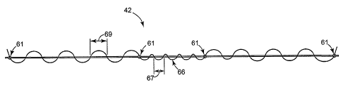
[0103] Referring to Figures 1 and 1A, the sling 42 preferably comprises first
and second major surfaces, a pair
of end portions I, and a support portion II for placement in a therapeutically
effective position relative to a
physiological environment intended to be supported (e.g. near the urethra). In
one aspect of the present
-12-
CA 02434463 2003-07-09
WO 02/058563 PCT/USO1/49632
invention, the sling 42 preferably has a tension adjustment or control member
66 associated with the sling 42, for
transferring sling adjustment forces from one portion of the sling 42 to other
portions of the sling 42 such as the
ends 61 of a support portion II of the sling (see Figures 1 and 1A). The
member 66 affords effecfiive
repositioning of the sling 42 while avoiding undesirable permanent deformation
of the sling 42. In the
embodiment of the present invention depicted in Figures 1 and 1A, the tension
adjustment member is a
filamentary member. The tension adjustment member 66 is preferably threaded
along the length of sling 42.
More preferably, the tension adjustment member 66 is connected at some points.
For example, if the sling 42
comprises a synthetic mesh material, then the filament may be affixed at the
junctures 61 between the support
portion II and the end portions I.
[0104] The sling 42 is preferably at least substantially surrounded by the
protective sheath 44, as shown in
Figures 4 and 5. The sling 42, tension control element 66 and sheath 44 are
made of biocompatible materials
having sufficient strength and structural integrity to withstand the various
forces exerted upon these components
during an implant procedure and/or following implantation within a patient.
Preferably, the protective sheath 44
is constructed of a material that affords visual examination of the
implantable sling material 42 and that affords
convenient passage of the assembly 46 through tissue of the patient.
[0105] Preferably, the overall dimensions of the sling assembly 46, including
individual sheath 44, sling 42 and
tension control member 66, are sufficient to extend from an abdominal
incision, to an undersurface of the urethra
and back to another abdominal incision with additional size to account for the
imprecision associated with the
range of human anatomy sizes. In a preferred embodiment, the sheath length L
of the device 40 of the present
invention is approximately within the range of 52.0 cm to 58.5 cm (20.5 inches
to 23.0 inches), sheath width W
is approximately within the range of 1.0 cm to 1.63 cm (0.482 inch to 0.642
inch) and sheath material thiclaiess
is approximately within the range of 0.127 mm to 0.203 mm (0.005 inch to 0.008
inch), respectively. The
associated sling 42 has a length X, width Y and thickness approximately within
the range of 49 cm to 51 cm
(19.3 inches to 20.1 inches), 1.0 cm to 1.2 cm (0.394 inch to 0.472 inch) and
0.508 mm to 0.711 mm (0.020 inch
to 0.028 inch), respectively. In addition, the length of the tension control
element 66 should be approximately
equivalent to or slightly longer than the length of the sling 42 to tighten or
loosen the sling 42 after it is placed in
the body. Alternative lengths, widths and thicknesses can also be used.
[0106]As used herein, the term "sling" is used generally to include a wide
variety of shapes and sizes,
materials and treatments. While the sling 42 is preferably rectangular for
treating SUI in females, other shapes
are also contemplated. Depending on the treatment addressed (e.g. to provide
hammock support for the bladder
or bladder neck, or to address a rectocele, enterocele or prolapse) the sling
may be any of a wide variety of
shapes. As an example, the sling may be of the general shape of the slings
described and shown in Moir et al.,
Tlae Gauze-Harramock Operation, Journal of Obstetrics and Gynaecology of the
British Commonwealth, Volume
-13-
CA 02434463 2003-07-09
WO 02/058563 PCT/USO1/49632
75, No. I, Pps. I-9 (I968). Figure 41 illustrates another example of a shape
of a sling 42G according to the
present invention. This sling shape is believed to be useful for providing a
hammock support for an anatomical
structure such as the bladder or the juncture between the bladdex and bladder
neck.
[0107] In one embodiment, the sling 42 is made of a mesh material. The mesh
material comprises one or more
woven or inter-linked filaments or fibers that form multiple fiber junctions
throughout the mesh. The fiber
junctions may be formed via weaving, bonding, ulhasonic welding or other
junction forming techniques,
including combinations thereof. In addition, the size of the resultant openngs
or pores of the mesh should be
sufficient to allow tissue in-growth and fixation within surrounding tissue.
As an example, not intended to be
Limiting, the holes may comprise polygonal shaped holes with diagonals of
0.132 inches and 0.076 inches. The
quantity and type of fiber junctions, fiber weave, pattern, and material type
influence various sling properties or
characteristics. Non-mesh sling configurations are also included within the
scope of the invention. As another
example, not intended to be limiting, the mesh may be woven polypropylene
monofllament, knitted with a warp
tricot. The stitch count may be 27.5 courses/inch (+ or - 2 courses ) and 13
wales/inch (+ or - 2 Wales). The
thickness of this example is 0.024 inches.
~~ 1 U8~ In a preferred embodiment, the mesh material of the sling 42
comprises a flexible, polypropylene
monofilament that resists weakening or degradation when implanted within a
patient. One such material is
MarlexTM material. Other mesh and non-mesh materials including, but not
limited to, synthetic biomaterials,
allografts, homografts, heterografts, autologous tissues, materials disclosed
in U.S. Provisional Applications S/N
60/263,472, S/N 60/281,350 and S/N 60/295,068, whose contents are fully
incorporated herein by reference,
synthetic materials (such as metallics, polymerics, and plastics) and any
combination of such materials may also
be used with the device of the present invention. Specific examples of
synthetic sling materials include, but are
not limited to polypropylene, polyethylene, nylon, PLLA and PGA. Preferably,
the sling material should cause
minimal to no reaction with body tissues and fluids and indefinitely retain
its particular material
characteristics/properties. Further, portions or all of the sling 42 may be
configured or fabricated from a material
to either promote or prevent tissue in-growth, or are resorbable to accomplish
the desired purpose.
[0109] In another embodiment of the invention, the sling 42, sling assembly 46
or portions thereof, may have
one or more substances associated therewith through a process such coating.
Examples of appropriate
substances include, without limitation, drugs, hormones, antibiotics,
antimicrobial substances, dyes, silicone
elastomers, polyurethanes, radiopaque filaments or substances, anti-bacterial
substances, chemicals or agents,
including any combinations thereof. The substances may be used to enhance
treatment effects, reduce potential
sling rejection by the body, enhance visualization, indicate proper sling
orientation, resist infection or other
effects. For example, a dye may be coated on one surface of the sling 42. The
dye provides the
practitioner/surgeon with a visual indicator to aid in properly orienting the
sling 42 at the target site within the
-14-
CA 02434463 2003-07-09
WO 02/058563 PCT/USO1/49632
patient and to avoid undesirable twists along the length of the sling 42. As
another example, the sling may be
coated by the process described in U.S. Pat. Nos. 5,624,704; S,7S6,14S;
S,8S3,74S; 5,902,283 and 6,162,487 (the
entire contents of which are hereby incorporated by reference).
~O ~ ~ O~ The sling 42 of the present invention need not have additional
sutures or other anchoring devices. Upon
implantation, a portion of the sling 42 is passed and/or woven through various
layers of abdominal/pelvic tissue.
The frictional forces created between the sling 42 and patient tissue prevents
movement and loss of tension once
the sling 42 is properly located at the target site within the lower abdominal
area of the patient. As a result, the
sling 42 remains securely in place, even when subj ected to various increased
abdominal pressures.
[01 'i 1 ~ The sling 42 is designed to remain within the body of a patient as
an implant for a predetermined
therapeutically effective amount of time. The sling may be non-absorbable,
absorbable or resorbable, including
any combinations of these material properties, depending on the desired
treatment. For example, portions of the
sling 42 or sling assembly 46 may be constructed of a bioabsorbable material
designed to last for a
predetermined period of time within the patient, that should be sufficiently
long to afford treatment of the
patient's need. The general characteristics of the sling material and design
should be such as to withstand the
various forces exerted upon it during implantation (for example, frictional
forces associated with tissue
resistance) and after implantation (for example, increased abdominal or
bladder pressure caused by coughing,
laughing, sneezing, or lifting). Preferably, the sling 42 is configured to
exploit the healing process and provides
adequate support to correct incontinence.
~O'~'~ 2~ The sling assembly 46 preferably has a feature that assists the
surgeon in placing the sling 42 in a
therapeutically effective anatomical position. The precise, final location of
the sling 42 will depend on a variety
of factors including the particular surgical procedures) performed, and any
preconditions of the patient such as
scar tissue or previous surgeries. For example, it may be preferred to place
the sling 42 in close proximity to, but
not in contact with, a mid portion of the urethra 16 to treat incontinence. In
a male patient, the sling 42 may be
placed proximate, but not in contact with the bulbar urethra.
[0113] Several different embodiments of tension adjustment member are within
the scope of the present
invention. Referring to the embodiment shown in Figure 7, a mesh sling 42 is
shown. A tension adjustment
member 66 is woven into the sling and attached to the sling 42 via two
attachment points 78 located near the
midsection 80 of the sling 42 and also corresponding to locations near each
side of the urethra 16.
[0114] Other attachment configurations for member 66 are also included within
the scope of the claimed
invention. The tension adjustment member 66 may be a separate element (e.g.
threaded along the length of the
sling 42) or it may be an integral part of the sling matrix. The tension
adjustment means may comprise one
filament threaded along the mesh. Alternatively, more than one filament may be
used. The tension adjustment
-1S
CA 02434463 2003-07-09
WO 02/058563 PCT/USO1/49632
member 66 shown in Figures 1 and 1A is attached to the mesh at the ends of the
middle portion II.
Alternatively, the tension adjustment means may comprise at least one filament
that is integrally woven in the
mesh and that has extension properties that are different than the other
filaments that form the mesh.
[0115] The tension adjustment means may be threaded axially along the sling
mesh, through the middle of the
sling or adjacent its ends. Preferably, this is done at the time of
manufacture to provide an assembly that is
conveniently used during a surgical procedure, without requiring the surgeon
to assemble the sling and tension
adjustment means during a surgical procedure. In one embodiment, the tension
adjustment means 66 may
comprise a plurality of elements woven axially along the sling. The plurality
of elements may be parallel or
non-parallel. For example, the elements may cross in the support portion II.
As another example, the tension
adjustment means may comprise a portion of the support portion that is more
tightly woven than another portion
of the support portion.
[0116] Preferably, the tension adjustment member is a continuous,
uninterrupted member, as opposed to a
member in separate pieces. A continuous, uninterrupted member allows the sling
to be tightened and loosened
and provides a plurality of locations that can be grasped along the sling 42
to modify the tension of the sling.
Also preferably, the member extends the entire length of the sling, from one
end to the other. A continuous,
uninterrupted member allows the entire sling to be repositioned as opposed to
merely isolated portions of the
sling.
[0117] The tension adjustment member 66 may comprise a monofilament element or
a braided member. The
tension adjustment member 66 may be constructed from a biodegradable material
or a non-biodegradable
material or combinations thereof. The monofilament may be round, flat or other
shapes to aid in fixation or
identification.
[0118] The position adjustment member 66 enables surgeons to easily tighten or
loosen the sling tension during
the surgical procedure, even after the surgeon removes the sheath 44. To
reduce the tension of the sling 42 using
the position adjustment member 66, the surgeon contacts the sling 42 and
position adjustment member 66
adjacent the urethra and pulls away from the urethra. The tension of the sling
may be increased by grasping the
sling 42 and position adjustment member 66 above the abdominal incision and
pulling upward. One or both ends
of the sling 42 and position adjustment member 66 may be grasped to increase
the tension of the sling 42.
Affording adjustment of the sling 42 position after removal of the sheath 44
facilitates proper sling placement
and helps avoid complications such as retention and sling erosion arising out
of improper sling placement.
[0119]The various configurations, properties or characteristics of the
position adjushnent member 66 may
vary or remain constant along the length of the position adjustment member 66.
For example, the position
adjustment member 66 may be made of a variety of materials including, but not
limited to, ProleneTM, nylon,
-16
CA 02434463 2003-07-09
WO 02/058563 PCT/USO1/49632
polypropylene, DekleneTM, poly-L-lactide (PLLA), polyethylene glycol (PGA),
polyester and any combination
of materials. Depending on the desired treatment, the member 66 or portions
thereof, may be absorbable, non-
absorbable and/or resorbable. If the member 66 is constructed from an
absorbable, bioabsorbable or
bioresorbable material or the like, then the member 66 may be optionally Ieft
in the sling 42 after the surgical
procedure. This offers the advantage of affording the use of the tension
adjustment member 66 in a minimally or
non-invasive near term, post operation sling tension adjustment procedure.
[0120] Figure 37 illustrates an example of a post operative sling tension
adjustment procedure. For example,
the patient may be experiencing slight retention shortly after the surgical
procedure and the surgeon may wish to
slightly loosen the sling 42. While the surgeon may make a slight dissection
in the vagina 20 to reach the
member 66, the surgeon may also have the option of placing a blunt instrument
382 into the urethra 16 and
slightly deflecting the urethra to thereby loosen the tension of the sling in
a lasting fashion. In contrast, if this
step were attempted with prior art s1W gs, the elastic nature of such slings
would likely result in temporary, elastic
deformation of the sling without a lasting change in the position of the
sling. The prior art procedure also risks
loss of sling functionality as previously described.
[0121 ~ The individual fibers or filaments comprising the tension adjustment
member 66 may be extruded,
woven, braided, spun, knitted, non-woven or have other similar configurations.
Member 66 properties, such as
tensile strength, elongation at break point, stiffness, surface finish, etc.,
may be similar to or different from those
of the slilig 42 and may vary along the length of the member 66.
j0122] In one embodiment, the tension adjustment member 66 may be secured to
the assembly 40 by attaching
one or more ends of the tension adjustment member 66 to the sheath 44. In
another embodiment, the tension
adjustment member 66 is secured to the device 40 simply by interlacing or
weaving the tension adjustment
member 66 at predetermined points along the length of the sling 42. In yet
another embodiment, the tension
adjustment member 66 may include one or more points of attachment along the
length of the sling 42. The
tension adjustment member 66 may be attached to the sling assembly 46 via
knotting, weaving, bonding,
ultrasonic welding or other attaclnnent techniques, including combinations
thereof, to prevent tension adjustment
member 66 detachment during and/or following sling implantation.
[0123] Preferably, the tension adjustment member 66 is knotted at preselected
locations along the length of the
sling 42 without any additional elements added to the assembly to comiect the
member 66 to the sling 42.
Knotting allows attachment of the member 66 to the sling 42 without additional
securement structure. This
embodiment avoids contact between such additional retaining structure and
tissue and any attendant
complications. The knot may comprise a single throw, half hitch knot, square
knot; single overhand knot, a
slipknot or a heat formed knot. Optionally, a loop or other shape may be
formed in the member 66 adjacent the
-17-
CA 02434463 2003-07-09
WO 02/058563 PCT/USO1/49632
end 61 of the support portion II to afford convenience in identifying the end
61 of the support portion II. Such a
loop or other shape may be conveniently located and cut should it be desired
to remove the portion of the
member 66 associated with the support portion II.
[0124] It is noted that, in an embodiment with a continuous length position
adjustment member 66 that is
anchored at a plurality of locations 61 (as shown in Figures 1 and 1A), when a
user grasps a mid portion II of the
sling 42 and member 66 and pulls, some of the pulling force is distributed or
transmitted from the grasped
location to a plurality of attachment points 61. This is believed to assist in
providing a sling that is more
effectively repositioned in a permanent fashion.
[0125] The means 66 for adjusting the tension or anatomical location of the
sling 42 may optionally comprise a
means for indicating proper orientation of the sling 42. Referring to Figure
1, the tension adjustment element 66
is woven along the length of the sling 42. In the support portion II of the
sling 42, the tension adjustment
element 66 is woven more frequently 67 than the less frequent weave 69 of the
element 66 in the end portions I
of the sling 42. Additionally, as shown in Figures 1 and 1A, a majority of the
element 66 is woven above one
major side surface of the sling 42 in the support portion. As shown in Figure
6, the major side of the sling with
the majority of protruding tension adjustment means 66A is located opposite
the urethra. If the material of the
element 66A is constructed of a different color, shape or size relative to the
material of the sling 42A, the
surgeon may more readily visualize proper placement of the sling 42A.
[0126] Referring to the embodiment of the invention shown in Figures 8A and
8B, the tension adjustment
member 66 is woven approximately along the centerline or axial length of the
sling 42. In one embodiment, the
weave pattern of the tension adjustment member 66 is used as an indicator of
proper sling orientation after
implantation. For example, the weave pattern on a first major side surface 82
of the sling 42, shown in Figure
8A, has small segments or loops of exposed member 66. The second major side
surface 84 (i.e. opposite side 82
or reverse side) of the sling 42, shown in Figure 8B, has larger segments or
loops of exposed tension adjustment
member 66. Upon implantation of the sling 42, the first surface 82 of the
sling 42, having minimal lengths of
filament segments or loops protruding above the material of the sling 42, is
preferably positioned to face the
urethra 16 of the patient. It is preferred that this first surface 82 of the
sling 42 face the urethra 16 to minimize
filament 66-urethra contact, particularly during adjustment of the sling 42,
and to assist the surgeon in
identifying the location of the member 66.
[0127] In another embodiment of the invention, one or more substances may be
associated with the member 66
by, for example, a coating process. The coatings may be selected from the same
group mentioned above with
respect to coatings for the sling 42. The substances may be used to enhance
treatment effects, indicate proper
sling orientation, enhance tension adjustment member visibility, and resist
infection or other effects. For
-18-
CA 02434463 2003-07-09
WO 02/058563 PCT/USO1/49632
example, the tension adjustment member 66 may be dyed a contrasting color
(e.g. blue) with respect to the sling
color (e.g. white). The contrasting color of the tension adjustment member 66
provides the surgeon with a visual
indicator that can be used to confirm proper sling orientation. In addition to
coating substances, other
components including, without limitation, tags, labels or iiidicia may also be
used to indicate proper sling
orientation or enhance tension adjustment member 66 visibility/identification.
[0128] Figure 6 illustrates a sling 42A in a proper position. The surgeon may
look through the vaginal incision
and view substantially all of the position adjustment member 66A protruding
above a support II (see Figure 1A)
or middle portion of the sling 42A when the sling 42A is properly placed. If
only a minor portion of the position
adjustment member 66A is visible protruding above a major surface of the sling
42A, then the sling is misplaced
and corrective action should be taken. Once the sling 42A is located in its
final position, the portion of the
position adjustment member 66A in the support portion II of the sling (see
Figure 1A) may optionally be cut or
released at the ends 61 of the support portion II and removed prior to closing
the vaginal incision. Optionally,
the sling 42A may include a means for conveniently locating and cutting the
tension member 66A at this point to
assist in removal of that portion of the tension member 66. As described
above, that means may comprise a loop
or other shape in the tension member 66. Alternatively, but not preferably, a
structure attached to the position
adjustment member 66 my be used to facilitate visualization, maneuverability
and cutting of the position
adjustment member 66.
[0129] Also optionally, the sling 42A may include a means for grasping the
sling 42A and/or the tension
member 66A in the support portion II of the sling. For example, the means may
comprise a small handle 15
attached to the tension member 66 in the support portion II of the sling 42A.
[0130] Referring to Figures 4 and 5, the sling 42 and tension adjustment
member 66 may be at least partially
housed within a sheath 44. Preferably, the sheath 44 is made of a relatively
transparent and flexible material
having a smooth outer surface. The transparency of the sheath 44 enables a
manufacturer or user of the device
40 to view the sling and tension adjustment member 66 encased within the
sheath 44 and visually determine
whether the sling 42 assembly contains any defects, such as a twisted sling,
detached tension adjustment member
66, torn sling fibers or other related flaws, as well as orientation within
the sheath. In addition, the sheath
provides a protective covering for the sling 42 and tension adjustment member
66 which also resists bacterial
and viral contamination of these components.
[0131 ~ In a preferred embodiment, the sheath 44 is made of polyethylene.
Other materials including, without
limitation, polypropylene, nylon, polyester or Teflon may also be used to
fabricate the sheath 44. The sheath
material should be flexible and provide sufficient structural integrity to
withstand the various forces exerted on
the sheath 44 throughout the sling delivery procedure. In general, the sheath
44 is configured to have sufficient
-19-
CA 02434463 2003-07-09
WO 02/058563 PCT/USO1/49632
flexibility to facilitate user manipulation and adequate structural strength
to withstand the various forces applied
to the sheath 44 during delivery and/or positioning of the sling assembly 46.
It should also conveniently separate
from the sling material 42 after the sling 42 is implanted without materially
changing the position of the sling 42.
~0132~ As shown in Figure 9A, the sheath 44 preferably comprises two elongate
sections 86, portions of which
detachably and telescopically overlap near the middle portion 80 of the sling
(not shown). In a preferred
embodiment, the length S of the overlapping section is approximately 3.8 cm
(1.5 inch). However, alternative
lengtlis may also be used. The length is preferably sufficient to resist
exposure of most of the sling 42 and
tension adjustment member 66 prior to sheath 44 removal. In addition to
resisting sling exposure, the
overlapping section may also be used as a visual indicator for the
practitioner or user of the device. In particular,
positioning the overlapping portion of the sheath 44 under the bladder neck or
urethra 16 ensures proper sling
placement (e.g. symmetrical sling placement) and tension within the patient.
Additionally, orientation indicia
(not shown) may be placed on the overlapping portion to indicate proper
orientation of the sling relative to the
urethra 16.
[0133] Alternatively, other configurations of the sheath 44 are within the
scope of the present invention. In
particular, the sheath may be unitary as opposed to telescoping with
perforations, holes, scores or tear lines
designed to allow separation and removal of the sheath 44.
(~~ 34~ During sheath removal, the first section 86 and the second section 86
of the sheath 44 are slid off the
sling 42 by pulling each end of the sheath 44 away from the middle portion 80
of the sling assembly 46 (as
shown by reference directional arrows in Figure 9B). Removal of the sheath 44
causes separation of the
overlapping sheath sections, thereby exposing the sling 42 and tension
adjustment member 66. In addition, the
smooth outer surface of the sheath 44 provides a relatively frictionless
surface to facilitate passage of the sheath
44 through the various tissues. The relatively frictionless motion also avoids
disturbing the position of the sling
42 relative to the anatomy of the patient.
(0135] In another embodiment of the invention, the sheath 44, or a portion
thereof, is associated with one or
more substances includW g those substances identified with respect to the
member 66 and sling 42. The
substances may be used to enhance sheath removal, identify twists along the
sheath 44 (and thereby indicate
proper sling orientation), indicate cutting/separation points, indicate center-
point, resist infection or provide other
desirable effects. For example, a first surface of the sheath 44 may include a
colored stripe that should lie
opposite the urethra 16 or bladder neck to ensure proper sling orientation.
Thus, the stripe provides the
practitioner/surgeon with a visual indicator to aid in properly orienting the
sling assembly 46, and ultimately the
sling 42, within the patient.
-20-
CA 02434463 2003-07-09
WO 02/058563 PCT/USO1/49632
[0136] The ends of the sheath are preferably connected to a dilator.
Alternatively, the sheath may be connected
to the sling, and the sling can be associated with the dilator. The number of
dilators will depend on factors such
as the shape of the sling. For example, the sling 42P shown in Figure 41
includes four dilators 54P.
[0137] At least two dilators are preferred. The sling 42 shown in Figure 4
includes two dilators. The first end
48 and second end 50 of the sheath 44 are preferably configured for attachment
to a dilator 54.
~~'~ 38~ The dilator 54 is a component that atraumatically creates and/or
expands the passageway through the
tissues for sling assembly delivery. The dilator 54 includes a means for
associating with a needle 60. The
dilator 60 is preferably short relative to a needle 60 for ease of passage of
the assembly and to reduce the overall
amount of tissue that is deflected at one time. Preferably, the dilator is
less than 2.5 inches in length, and more
preferably, it is less than one inch in length. The maximum radius of a
dilator 54 is preferably less than 10 mm,
more preferably less than 7.5 mm, even more preferably less than 5 mm. The tip
of the dilator 54 is preferably
blunt, as, in preferred embodiments, the leading tip of the dilator 54 will
pass through tissue that lias already
been pierced by a needle 60.
[0139] Tlie dilator 54 may be made from a variety of biocompatible and
sterilizable materials including,
without limitation, acetal, Delrin~, Acrylonitrile-Butadiene-Styrene (ABS),
polyethylene, nylon and any
combination of materials. Alternatively, the sheath 44 may be additionally or
solely connected to an end portion
of the sling 42.
[0140] The dilator 54 preferably includes means for associating with a
surgical needle 60. In a preferred
embodiment, the association means affords a permanent affixation between the
dilator 54 and the needle 60. By
"permanent affixation", it is meant that it would be very difficult to
manually separate the dilator from the needle
after they have become permanently affixed. After implantation of the sling
42, to separate the sling 42 from the
dilator 54/needle 60, the surgeon cuts an end of the sling 42 as described
more fully below. The association
means preferably affords quick and convenient attachment of the dilator 54 to
the needle 60 to avoid wasting
time in the midst of a surgical procedure. The attachment should also be
secure to avoid separation of the needle
60 and dilator 54 while the combination is passed through tissue.
[0141 ~ The dilator 54 also includes a means for association with the sling 42
and/or the sheath 44. For
example, the dilator 54 may be preattached to the sling 42 and/or sheath 44,
particularly if the sling is a synthetic
material. Alternatively, the dilator may include means for conveniently
attaching to a sling material (e.g.
cadaveric or autologous sling material) just prior to sling placement.
[0142]Referring to the embodiment of Figures l0A-10E, the dilator 54 may be
approximately 3.1 cm (1.2
inches) in length. The dilator 54 preferably includes a gentle taper 88 near
its second end 56. The dilator is
-21
CA 02434463 2003-07-09
WO 02/058563 PCT/USO1/49632
sized and shaped to provide atraumatic passage through body tissue. The taper
88 and relatively smooth outer
surface of the dilator 54 facilitate atraumatic passage of the dilator 54 and
attached sling assembly 46 through the
various tissues of the patient. The presence of the dilator 54 allows a gentle
transition between the diameter of
the needle, to the shape of the dilator, and finally to the sling assembly 46,
as opposed to prior art assemblies,
where the structure of the sling assembly abruptly increases the profile of
the needle and thereby the size of the
structure that must pass through tissue.
[0143] Preferably, the first end 52 of the dilator 54 attaches to one end of
the sling 42, or sheath 44 or sling
assembly 46 (shown in Figure 10E) and the second end 56 of the dilator 54 may
be quickly attached or
assembled to a needle 60 (not shown). The sheath 44 is preferably attached to
the dilator 54 via a first opening
or through-hole 90 located near the first end of the dilator 54. In this
embodiment, the opening 90 operates as a
universal sling material or assembly attachment point which can receive a
variety of materials, such as fascia,
autologous materials, s5mthetics, biologic tissues and any other similar
tissues, including any combinations. The
edge portion 91 of one end of the sheath 44 is threaded through the opening 90
of the dilator 54 and secured to
the sheath 44, thereby forming a loop 92. The edge portion 91 may be fastened
onto the sheath 44 via ultrasonic
welding, bonding, melting, suturing, sealing or other attaclnnent techniques.
Further, as shot~m in Figures 10A
and 10B, the first end 52 of the dilator 54 includes a cut-away section 94 to
provide room to receive sling
assembly material to reduce the overall profile of the sling assembly
experienced by tissue during sling passage.
Therefore, when the sheath is attached to the cut-away section, the additional
sheath material is not apt to
signficantly increase the relative thickness, diameter or profile of the
dilator 54.
[0144] Alternatively, for dilators 54 manufactured via molding techniques, the
end of the sheath 44 may be
encased within and secured to the first end 52 of the dilator 54 during the
molding process. In yet another
embodiment, the end of the sheath 44 may be fixedly attached within a
longitudinal slot located near the first end
52 of the dilator 44 using an adhesive, ultrasonic welding or other attachment
techniques.
[0145] Referring to Figures 10A-1 OD, the second end 56 of the dilator 54
includes a second opening or
through-hole 96 that extends substantially internally along the longitudinal
axis of the dilator 54. The second
opening 96 has an internal diameter generally configured for convenient
attachment to a needle 60 or similar
sling-delivery device. In one embodiment, the internal diameter of the second
opening 96 of the dilator 54 is
approximately within the range of 0.239 cm to 0.318 cm (0.094 inch to 0.125
inch). A shoulder 98 located on
the surface 100 of the second opening 96 of the dilator 54 and a complementary
mating recess located on the
surface of the first end of the needle 60 (see Figure 4) securely and
permanently attach or lock the dilator 54 and
needle 60 together. Once the needle 60 is inserted into the dilator 54, they
are preferably not separated
thereafter. After the sling 42 is implanted, the connected needle 60 and
dilator 54 are removed from the sling by
-22-
CA 02434463 2003-07-09
WO 02/058563 PCT/USO1/49632
cutting an end of the sling as described in greater detail below. Preferable,
the needle 60 and dilator 54 are
disposed.
[0146] One or more longitudinal slots 102 located on the outer surface of the
dilator 54 and in communication
with the second openng 96 allow the wall of the dilator 54 to expand in a
radially outward direction when the
first end of the needle 60 is inserted into the second opening 96 of the
dilator 54. When the shoulder 98 of the
dilator 54 passes the recess of the needle 60, the wall of the dilator 54
collapses around the needle 60 as the
shoulder 98 seats into the recess, thereby securing the dilator 54 on the
needle 60 and blocking separation of the
dilator 54 and needle 60.
~0147~Although the invention has been described in terms of a shoulder 98 and
mating recess, alternative
dilator-needle attachment mechanisms such as bumps, grooves, slots, wedges,
tabs and other mechanisms are
also included witlun the scope of the claimed invention. The dilator 54
preferably includes one or more relief
ports 104 to facilitate convenient needle connection. The relief ports 104 may
be formed at the ends of the
longitudinal slots 102 or at various high-resistance locations along the
dilator 54. The relief ports 104 decrease
the rigidity or resistance of radially outward expansion of the dilator wall
and, reduce the amount of force
required to insert or securely attach the needle 60 to the dilator 54. In yet
another embodiment, superficial bands
or rings, arc-shaped slots, superficial grooves or other mechanisms may be
provided to provide improved
expansion or attachment characteristics. ,
~0'~ 48~ A portion of the dilator 54 includes a taper 88 having a decreasing
profile toward the second end 96 of
the dilator 54. The taper 88 preferably gently cams tissue out of the path of
the sling assembly 46 as the sling
assembly is inserted in the body. The taper 88 is also sized and shaped to
reduce the amount of friction or
resistance as the device is drawn through the tissues of the patient. The
amount of force required to manipulate
the device through the tissues is thereby reduced. This in turn provides the
user of the assembly with additional
control over device insertion and maneuverability through tissue and witlun
the patient. In addition to tapered
profiles, other dilator profiles such as conical, flared, frusto-conical,
pyramid-shaped, elliptical or other
applicable profiles may also be used. Overall, the profile of the dilator 54
is preferably configured to provide
easy dilation of the tissue to accommodate smooth passage of the sling
42/sling assembly 46 and subsequent
collapse of the surrounding tissue to securely anchor the sling 42 into the
tissue (after sheath removal).
[0149] In other embodiments of the invention shown in Figures 12A and 12B, the
dilator 54A or 54B includes
a sling fastening snap mechanism 106 on one end of the dilator. The embodiment
disclosed in Figure 12A
includes a keyed/locking mechanism on its other end. As shown in Figurel2A,
the first end of the dilator 54A
includes a slot or slot-shaped opening 110 configured for convenient insertion
of one end of a sling 42 (such as
one made from autologous tissue) or sling assembly 46 either at the surgical
site (e.g. by the operating room
-23-
CA 02434463 2003-07-09
WO 02/058563 PCT/USO1/49632
nurse or surgeon) or other location (such as manufacturing location).
Additional shapes for the dilator opening
110 include, without limitation, oval, circular, square, rectangular and other
shapes. The slot-shaped opening
110 is located along a portion of the longitudinal axis of the dilator 54A.
~~'~ 50~ A snap-like element 112' is located on an outer surface near the
first end of the dilator 54B. The snap-
like element 1 I2' includes a barb or spike 114 that fits within an opening
116 situated near the first end of the
dilator 54B. The opening 116 for the barb 114, preferably configured
perpendicular to the slot-shaped opening
110', is sized and shaped to match or mate with the barb 114 of the snap-like
element 112'. When the barb 114
is fully seated within the opening 116 of the dilator 54B, the tip 118 of the
barb 114 extends into the slot-shaped
opening 110' of the dilator 54B. A first ridge 120 and a second ridge 122
located along the length of the barb
114 further secure and/or fasten the barb 114 within the opening 116 of the
dilator 54B. Other fastening
configurations including, but not limited to, bumps, shoulders, tabs, detents,
tongue in grooves, snaps and any
combinations of fastening means may also be used with the present invention.
[0151 ~ During use, one end of the sling 42, sheath 44 or sling assembly 46 is
inserted into the slot 110' of the
dilator 54B. With the end of the sling 42/sling assembly 46 properly
positioned within the slot 110', the barb
114 of the snap-like element 112' is inserted into the opening 116 of the
dilator 54B. The barb 114 is fully
seated within the opening 116 when both ridges 120, 122 pass through the
opening 116 of the dilator 54B. This
causes the tip 118 of the barb 114 to bear down on or penetrate a portion of
the sling 42/sling assembly 46
extending withal the slot 110' of the dilator 54B, thereby securely fastening
the sling 42/sling assembly 46 to the
dilator 54B.
[0152] A keyed/locking mechanism 108 is located near the second end 56B of the
dilator 54B. As shown in
Figure 12B, a square-shaped opening 124 extends along a portion of the
longitudinal axis near the second end
56B of the dilator 54B. The shape of the dilator opening I24 matches the
square-shaped perimeter of the
keying-segment 126 located near the first end 58 of the needle 60 and allows
keyed-rotation of the dilator 54B at
ninety-degree intervals. Otlier appropriate shapes for the dilator opening 124
may also be used provided that the
shape of the opening 124 complements the corresponding keying-segment shape
located near the first end 58 of
the needle 60. When the first end 58 of the needle 60 is positioned within the
dilator 54B, the square-shaped
opening 124 of the dilator 54B together with the keying-segment 126 of the
needle 60 prevents axial rotation of
the dilator 54B relative to the needle 60 and, thus, twisting of the sling
42/sling assembly 46. This optional
feature provides the practitioner or user of the assembly with improved
control and maneuverability of the
assembly before and during the insertion procedure.
[07 53] The dilator 54B also includes a locking mechanism 128. Referring to
Figure 12B, the locking
mechanism 128 comprises one or more tension-loaded ribs located within the
longitudinal opening of the dilator
-24-
CA 02434463 2003-07-09
WO 02/058563 PCT/USO1/49632
S4B. The configuration of the ribs generally matches and corresponds to a
complementary recess 130 located
near the first end S8 of the needle 60. Thus, the first end S8 of the needle
60 is inserted through the longitudinal
opening 124 of the dilator S4B until the ribs of the dilator S4B seat within
the recess 130 of the needle 60. The
dilator S4B is securely attached or locked onto the needle 60 when the dilator
ribs are fully seated within the
needle recess I30. Although the invention has been described in terms of a rib
and complementary recess,
alternative dilator-needle attachment mechanisms, such as those previously
described, are also included herein.
[0154] Referring to Figure 13, in an alternate embodiment of the invention,
the sheath 44 (or sling 42 or
assembly 46) is attached to the dilator S4C via a locking (or compression)
collet 132 and adapter connector 134.
The compression collet 132 comprises a ring-shaped portion 136 having one or
more barbed snap tongs 138.
Tlie complementary adapter 134 comprises a cylindrical element 140 having a
first end 142 and a second end
144. The internal profile near the first end 144 of the adapter connector 134
includes a tubular lumen or channel
146, having one or more recesses, shoulders, grooves or similar indentations
148, surrounding an internal prong
150. The second end 144 of the adapter connector 134 includes one or more
barbed snap tongs 152, similar to
the tongs 138 of the compression collet 132. In addition, the first end S2 of
the dilator S4C includes a
longitudinal opening 1S4 having one or more recesses, grooves, slots or
related types of indentations 1S6
configured to engage the tongs 1 S2 of the adapter connector 134.
(~'~ 55~ In use, one end of the sling 42/sling assembly 46 of the present
invention is configured into a tubulax or
appropriate shape that enables a sufficient portion of the end of the sling
42/sling assembly 46 to be inserted
through the compression collet 132. The tongs 138 of the compression collet
132 are then inserted into the first
end 142 of the adapter comiector 134, causing the tongs 138 to snap into
engagement with the adapter connector
134. The end portion of the sling 42/sling assembly 46 is compressed between
the tongs 138 of the compression
collet 132 and the internal prong 1S0 of the adapter connector 134, thereby
securely fixing the sling 42/sling
assembly 46 to the collet/adapter assembly. In a similar fashion, the tongs
1S2 of the adapter 134 are then
inserted and snap-locked into the first end S2C of the dilator S4C, creating a
secure fixation between the
collet/adapter assembly and dilator S4C.
[0156] In another embodiment of the invention, the length of the dilator S4D
is substantially equivalent to the
length of the needle 60 used for the sling delivery procedure. For example, as
shown in Figures 14A and 14B,
the dilator S4D comprises a hollow, ataumatic trocar-shaped component
generally made of a soft, semi-flexible
material, such as high density polyethylene, polypropylene, polyvinyl chloride
(PVC), polytetrafluoroethylene
(PTFE) or other similar materials, including combinations thereof. The
material and design of the dilator S4D
allows the dilator to be positioned over or passed along the length of the
needle 60, thereby totally or partially
encasing the needle 60, similar to an Amplatz sheath/dilator. In the spirit of
convenience and brevity, this
embodiment of the dilator S4D will be hereafter referenced as the cystoscopy
aid S4D. Optionally, tongue and
-2S-
CA 02434463 2003-07-09
WO 02/058563 PCT/USO1/49632
groove structure may be supplied in the needle 60 and cystoscopic aid 54D to
guide the cystoscopic aid 54D
along the needle 60.
[0157]Alternatively, the hollow portion or internal lumen 158 of the
cystoscopy aid 54D may be sized and
shaped to accommodate passage of a dilator 54 and/or sling 42 and/or sling
assembly 46, similar to those
previously described. As such, after the cystoscopy aids 54D are positioned
over the needles 60, the dilators 54
and/or sling 42 and/or sling assembly 46 are connected onto the ends of the
needles 60. The needles 60 and
attached components are then pulled through the internal lumen 158 of the
cystoscopy aids 54D until the sling 42
is positioned adjacent the target site or urethra 16 and the needles 60
connected components are withdrawn from
the patient. With the sling 42 properly positioned in a therapeutically
effective relationship with the urethra 16,
the cystoscopy aids 54D are then removed from the patient, allowing the tissue
to gently collapse around the
sling 42. This configuration of the device allows components such as dilator
54, sling assembly and subsequent
needle maneuvering to be performed substantially within the hollow portion 158
of the cystoscopy aid 54D,
thereby reducing potential tissue trauma and infection.
~~'~ 58~ In an alternate embodiment, one or more apertures or perforations
160, that function to facilitate
verification of bladder and urethra integrity, are disposed along the length
of the cystoscopy aid 54D. For
example, during use, after the needles 60 have been inserted within the
patient, the cystoscopy aid 54D may be
pushed along the exterior surface of each needle 60. If the bladder has been
punctured during needle insertion
causing urine leakage or drainage within the patient, the urine or bladder
fluid will enter the apertures 160 of the
cystoscopy aid 54D and flow along the surface and out from the needle 60. This
allows the practitioner to
quickly and easily confirm urethra and bladder integrity.
[0159] In another embodiment of the invention, shown in Figure 14C, a first
end 162 of the cystoscopy aid 54E
is attached to an end of a sling 42, or sling assembly 46 or portions thereof.
The sling 42/sling assembly 46 may
be attached to the cystoscopy aid 54E using attachment mechanisms and
techniques similar to those previously
described throughout this disclosure. Following handle 64 removal, cystoscopy
aid 54E is pushed along the
exterior of a needle 60 to maneuver and properly position the sling 42/sling
assembly 46 in a therapeutic position
relative to anatomical structures such as the urethra or bladder.
[0160] Alternatively, the cystoscopy aid 54E or dilator 54 may include a
hollow portion configured to house
the sling assembly 46. In other words, the sling 42/sling assembly 46 would be
folded, rolled or similarly
configured for placement inside the hollow portion of the dilator or
cystoscopy aid 54E. One role of the dilator
54E or cystoscopy aid 54E in this embodiment, similar to that of the sheath
44, is to reduce friction or tissue
trauma as the sling 42 is drawn through the various tissues. It is noted that
the sheath 44 is a wholly optional
-26-
CA 02434463 2003-07-09
WO 02/058563 PCT/USO1/49632
element of the present invention. The embodiment of the present invention
shown in Figure 14C is believed to
potentially reduce sling assembly component contamination/exposure and wound
infection.
[0161 ~ In an alternate embodiment of the invention, a set of grasping jaws
164 may incorporate a dilator, as
shown iii Figures 15A and 15B. Preferably, the jaws 164 are constructed from a
bioabsorbable material. A first
end 166 of the jaws 174 attaches to the needle 60 via a snap or quick fitting
attachment. A second end 168 of the
jaws 164 attaches or clamps onto the sling 42 or sling assembly 46.
Optionally, the jaws 164 may serve to
anchor the sling 42 within tissue of a patient.
[0162] The mechanism by which the jaws 164 attach to and release the sling 42
or sling assembly 46 may be
similar to that of a bioptome. Other exemplary mechanisms such as a ball-
detent used on a ratchet wrench,
spring loaded clamps, memory alloys, and other mechanisms may also be used.
The jaws 164 may optionally
be operably connected to and controlled by the handle 64 of the device.
Manipulation of the handle 64 causes
the jaws 164 to open and close, thereby enabling the device to clamp onto
and/or release the sling 42 or sling
assembly 46. In addition, the handle 64 may be further manipulated to detach
the jaws 164 or a portion thereof
from the needle 60. As such, once the sling 42 or sling assembly 46 is
properly located within the patient
(further described below), the bioresorbable jaws 164 are detached from the
needle 60 either inside or outside the
body of the patient.
[0163] Referring to Figure 16A, the needle 60 is generally curved or arcuate.
Preferably, the needle is arc-
shaped and includes a first end 58 and a second end 62. Although a variety of
needle designs and/or
configurations may be used including, without limitation, straight, bent,
curved, arc-shaped, Stamey, Raz and
other configurations, all references hereinafter will be made to an arc-shaped
needle in the spirit of brevity and
reader convenience.
[0164] Overall, the shape of the needle 60 should facilitate and provide
controlled passage of the needle 60
through tissue, preferably from an abdominal incision to the vagina or,
alternatively, from the vagina to an
abdominal incision. The ends or tip of the needle 60 are preferably not
sharpened, but may be tapered to afford
easy passage through tissue while providing a blunt surface that avoids
cutting sensitive tissue such as the
bladder or urethra. In a preferred embodiment, the length N of the needle 60
is approximately within the range
of 16.5 cm to 24.1 cm (6.5 inches to 9.5 inches) and has a preferred external
diameter of approximately 3.175
mm (0.125 inch). It is preferred that the diameter of the needle 60 be small
relative to the prior art to reduce
tissue trauma.
[0165] The needle 60 is made of a malleable, yet durable, biocompatable
surgical instrument materials such as,
but not limited to, stainless steel, titanium, Nitinol, polymers, plastics and
other materials, including
combinations of materials. The needle 60 should have sufficient structural
integrity to withstand the various
-27
CA 02434463 2003-07-09
WO 02/058563 PCT/USO1/49632
forces (e.g. forces caused by dilator attachment, cystoscopy aid passage, and
penetration/passage of the needle
60 through the various tissues) without undergoing any significant structural
deformation. Optionally, the
needles 60 could be sufficiently malleable to allow a practitioner or user of
the device to modify the needle 60 to
a desired shape and, thereby, optimize the procedural approach.
[0166]
As shown in the embodiment of Figure 16A, the first end 58 and second end 62
of the needle 60 may include a
keying feature 170 affording secure association between the needle and handle
64 and/or dilator 54 and/or sheath
assembly 46. In one embodiment, the keying feature 170 comprises a recess 130
and/or square-shaped portion
126. As previously described, the recess 130 and square-shaped portion 126 are
designed for complementary
engagement to the appropriate end of a dilator 54 or handle 64. Another
embodiment of the invention includes a
reversible keying feature. The reversible keying feature allows the handle 64
to be interchangeably attached yet
securely affixed to either end of the needle 60. In a preferred embodiment,
the needle 60 may be substantially
symmetric about a centerpoint, that is, the radius of curvature of the needle
60 may be substantially constant and
either a handle ox a dilator may be attached to either end of the needle 60.
[0167] In an alternate embodiment, the keying feature of the needle 60B
comprises an end cap 172 and an
elongate reduced width segment 174 having a square-shaped cross sectional
profile, as shown in Figures 16B to
16D. The second end 62B of the needle 60 shown in these Figures is inserted
into the keying feature or channel
176 that extends along the longitudinal axis of the handle 64B (partially
shown in Figures 16D to 16F). When
the needle 60B is properly positioned within the handle 64B, a yoke or other
fastening component 178 receives
and secures the elongate segment 174 in the narrow portion 180 of the channel
176, as shown in Figures 16D and
16E. The complementary configuration of the channel's narrow portion 180 and
the needle's elongate segment
174 prevents the handle 64B from rotating around the axis of the needle 60B.
In addition, this configuration may
also provide additional needle/handle stability and improved tactile feedback
for a user of the device.
[0168] The present invention may optionally include structure that allows the
surgeon to change the orientation
or position of the handle relative to the needle. The handle may be rotatably
repositioned relative to the needle
or, in some embodiments, the handle may be axially slidable and repositionable
along the length of the needle.
The handle may be repositioned in any orientation as determined by the surgeon
or it may be indexed between a
plurality of predetermined orientations depending on the particular embodiment
of the present invention.
[0169] Figure 16E illustrates the needle seated in a locked position relative
to handle 64B. In order to rotate the
handle 64B, a user or practitioner manipulates a trigger or button that
actuates the fastening component 178
thereby causing the channel 176 to disengage from the elongate segment 174, as
shown in Figure 16F. In this
position, segment 174 of the needle 60B is no longer seated in the handle 64B.
With the elongate segment 174
-28
CA 02434463 2003-07-09
WO 02/058563 PCT/USO1/49632
positioned in the wider portion I82 of the channel I76, the needle 60B is free
to rotate. However, the
configuration of the needle's end cap 172 prevents the needle 60B from
becoming completely disengaged from
the handle 64B. Thus, the keying features maintain the needle 60B in proper
alignment with the handle 64B
when in the locked position and also allow a user to controllably rotate the
needle 60B to obtain a desired handle
64B orientation.
~~'~ 70~ In an alternate embodiment of the invention, the needle 60C comprises
an assembly 184 having one or
more detachable components. For example, referring to embodiments shown in
Figures 17A and 17B, the
needle assembly 184 comprises a body portion 186 and one or more segments 188.
An external thread 189
formed near the end of the body portion 186 allows needle segments 188,
dilators 54, slings 42, or sing
assembly 46 to be screwed onto the body portion 188 thereby forming the needle
assembly 184. In addition, the
devices may be easily removed by simply unscrewing them from the body portion
186 of the needle assembly
184. Other configurations or designs of the needle assembly 184 may include,
but are not limited to, hollow or
solid body portions 186, snap fit, memory alloy or latching mechanisms,
internal threading or other designs .
[0171 ~ In another embodiment, the handle 64 may be permanently attached to an
end 62 of the needle 60.
More particularly, the handle 64 may be rigidly affixed to the needle 60 so
that substantially no relative
movement may occur between the needle 60 and the handle 64.
[0172] Referring to Figure 18A, one embodiment of the adjustable handle 64G
comprises a relatively smooth,
ergonomic body made of delrin, ABS, nylon, polycarbonate, acetal,
polyetherimide, polysulfone or other
sterilizable materials. The body of the handle 64G may be hollow, solid or
semi-solid. One or more surfaces of
the handle include a plurality of ridges 190 andlor indentations 192 that
provide an enhanced gripping surface for
a user of the device. Alternatively, various portions of the surface of the
handle 64G may also include grasping
features such as bumps, grooves, ridges or other gripping means, that enable
improved manipulation of the
handle 64G. In addition, the handle 64G may include an indentation formed near
the middle 194 of the handle
64G that provides a user of the device with better control of, and an improved
grip on, the handle 64G.
[0173] A push button 198 and keyed opening 200 are located near the needle
attachment end 196 of the handle
64G shown in Figure 18A and form a keying feature of the handle 64G. As shown
in Figure 18B, the push
button assembly 198 comprises a button or knob-shaped component 202 that
attaches to a yoke 204 (attachment
locations indicated by dashed reference line). In particular, the yoke 204 is
attached to the button 202 via snap
tongs 206 that lock the button 202 and yoke 204 together. Prior to attachment,
the button 202 and yoke 204,
including a spring 208, are fitted within their respective grooves and/or
slots formed near the needle attachment
end 196 of the handle 64G, as generally shown in Figure 18C. The spring 208
provides the appropriate tension
to maintain the assembly in a locked position.
-29-
CA 02434463 2003-07-09
WO 02/058563 PCT/USO1/49632
~O 174 When the assembly is in a locked position (shown in Figure 18D), the
spring forces push the button 202
in a direction away from the longitudinal axis of the device. This in turn
causes the groove or recess 210 of the
attached yoke 204 to protrude witlun the keyed longitudinal opening 200
resulting in a non-square-shaped
opening formed along an axial portion near the needle attachment end of the
handle 64G. In the locked
configuration, the handle 64G is securely attached in a stationary position on
the needle 60. Pressing or pushing
the button 202 inwardly toward the axis of the device unlocks the device and
creates a square-shaped or keyed
opening 200 for the needle 60. Figure 18E illustrates a cross-section of the
keyed, longitudinal opening 200 in
an unlocked position.
[0175] The quick-release push button of the handle 64G enables a user of the
device to easily attach or detach
the handle 64G from the needle 60 or reposition the orientation of handle 64G
relative to the needle 60, using
one hand. While gripping the handle 64G, the user of the device simply
depresses the push button 202 with one
finger to unlock the handle. Still using a single hand to control the handle
64G, the user can then insert one end
of the needle 60 into the keyed opening 200 of the handle 64G and, upon
releasing the button 202, secure the
handle 64G to the needle 60.
[0176] As previously disclosed, the needle 60 includes a similar keying
feature configured for complementary
engagement with the keyed portion of the handle 64G. These complementary,
square-shaped keying features
allow a practitioner or user of the device to rotatably index the handle 64G
between predetermined positions
located in ninety-degree increments around the needle axis. Thus, the
practitioner may position the handle 64G
in a preferred configuration on the needle 60 that provides the greatest
comfort and ease of insertion. In
addition, via the locking mechanism, the keying features also prevent the
handle 64G from uncontrollably
rotating around the axis of the needle 60, for instance, during a sling or
needle insertion procedure. Although the
invention has been described with respect to a square-shaped keying feature,
other geometrical configurations
and keying means are also included within the scope of the present invention.
~~177~Another embodiment of needle attachment mechanism for a handle is shown
in Figures I9A to I9C.
The handle 64H includes a quick-release feature 212 comprising one or more
levers 214 and an associated
border or frame 216 that surrounds an opening 218. The opening 218 is
generally located near the needle end of
the handle 64 and along the longitudinal axis of the device. The frame 216
bordering the opening 218 may
include various indentations or ridges 220 that provide improved gripping
capabilities. In addition, the handle
may also include a square-shaped keying feature 222 similar to the previously
described keying features. A
different shaped handle 64I is shown in Figures 19B and 19C.
[0178] During use, a practitioner or user of the device simply compresses the
levers 214 of the handle 64H
together using, for example, a thumb and forefinger. Compression of the levers
214 changes the configuration of
-30-
CA 02434463 2003-07-09
WO 02/058563 PCT/USO1/49632
the frame 216 and opening 218 to allow insertion of a needle 60 therein. The
user of the device releases the
levers 214 when the needle 60 is properly positioned within the handle 64I,
causing a portion of the frame 216 to
compress against a portion of the needle 60 (e.g., a recessed portion) thereby
blocking axial movement of the
needle relative to the handle 64I and securely attaching the handle 64I onto
the needle 60. The handle 64I can be
quickly released from the needle 60 by pressing on the handles 214.
~O~ 79~Another embodiment of a quick-release feature for the handle 64K is
shown in Figures 20A-20C. For
this embodiment of the invention, the handle 64K may be made from a single
molded or machined component.
A quick-release button 224, located near the needle end of the handle 64K,
controls a keyed needle-latching
mechanism 226. As best seen in Figure 20B, the needle latching mechanism 226
generally includes a
geometrically shaped opening section 228, a locking section 230 and an end
section 232. When a practitioner or
user of the device depresses the quick-release button 224, the semi-resilient
material of the handle 64K causes
displacement of the locking section 230, thereby allowing the needle 60 (not
shown) to be inserted into the
latching mechanism 226 of the handle 64K. After the needle 60 and handle 64K
are positioned or aligned
according to user preference, the button 224 is released causing the locking
section 230 to return to its initial
configuration and, in so doing, seat within the complementary, recessed
feature of the needle 60. This not only
secures or locks the handle 64K onto the needle 60 but also prevents the
handle 64K from rotating around the
needle axis.
~0 ~ 8~~ In another embodiment of the invention, the keyed, locking portion
and/or quick release feature of the
handle 64K may be located near the middle of the handle 64K, near the end of
the handle 64K close to the needle
(figure 18A) or at any preferred location on the handle 64K. A large or small
section or length of the needle 60
may be housed within and contact the handle 64K of the device, thereby
providing enhanced user-control and
stabilization of the needle 60 relative to handle 64K. The increased surface
contact between the needle 60 and
handle 64K may also strengthen the associated gripping or frictional forces,
resulting in improved locking or
attachment capabilities of the device.
~~'~ $ ~ ~ The associated quick-release feature (such as push button 198,
button 224, levers 214, etc.) may also be
positioned at any preferred location on the handle 64 of the present
invention. For example, referring to Figures
21A and 21B, positioning the button 202 opposite to the needle insertion end
196 of the handle 64L may reduce
or prevent accidental triggering of the button 202. Further, this particular
design may provide additional
ergonomic advantages for the user of the device. For example, the bottom could
be flush or recessed with the
surface of the handle.
[0182] Various configurations of the overall size, weight and shape of the
handle 64 are also included within
the scope of the claimed invention. Still referring to Figures 21A and 21B,
another embodiment of the handle
-31-
CA 02434463 2003-07-09
WO 02/058563 PCT/USO1/49632
64L comprises a compact profile. The smaller size of the handle 64L reduces
the weight of the handle 64L,
thereby making the device 40 less heavy at the top and better balanced.
Alternatively, the handle 64L may also
be configured to be permanently, but rotatably, affixed to the needle 60 (not
shown). As such, the user or
practitioner may rotate the handle 360° around the axis of the needle
60 and lock the handle 64L in position once
the desired orientation is reached.
[0183] Figure 11 illustrates an alternate embodiment of the present invention
that includes a slidable second
handle 64' which may be used alone or in combination with handle 64. In
general, the slidable handle may
provide additional ergonomic advantages and control during the needle
insertion procedure. For example, when
used in combination with the handle 64, the slidable handle 64' is initially
positioned and optionally locked near
the first end 58 of the needle 60. During needle insertion (further described
below), the slidable handle allows
the user or practitioner to maneuver the needle 60 more accurately along the
insertion pathway. In the example
of an initial suprapubic approach, after the slidable handle 64' comes close
to or in contact with the abdomen, the
slidable handle 64' is unlocked and repositioned closer to the handle 64. The
slidable handle is then secured at
the new position and locked in place, thereby allowing further insertion of
the needle 60.
[0184] The second handle 64' may optionally be locked in a position that
blocks inadvertent lurching of the
needle 60 within the tissue. Preferably, the second handle 64' is sized and
shaped to engage the abdominal tissue
to act as a stop to prevent further penetration of the needle 60 until the
second handle 64' is unlocked and moved
to a location closer to the handle 64. This feature is believed to be useful
in resisting uncontrolled passage of the
needle 60 into the reiropubic space after the end 58 of the needle 60 bursts
through the tough rectus fascia. Once
the rectus fascia is penetrated, the second handle 64' is unlocked and moved
to a location closer to the handle 64
and the needle can be controllable passed through tissue.
~~~ $5~ Optionally, the second handle 64' may include means for affording
sliding of the handle 64' toward the
handle 64, but that resists movement of the handle 64' away from the handle
64. The means may comprise a
plurality of ribs within handle 64' that engage the needle 60 and that are
angled toward the handle 64.
[0186] Referring to another embodiment shown in figures 22A and 22B, a
slidable handle 204 comprises a
body portion 206 (partially shown in Figures 22A and 22B), latch 208, o-ring
210 and spring ring 212 contained
in a handle cavity 207. In general, the body portion 206 and latch 208 may be
made of delrin, ABS, nylon,
polycarbonate, acetal, polyetherimide, polysulfone, or other sterilizable
materials. In addition, the o-ring 210
and spring ring 212 may be made from high durometer polyurethane, teflon and
other rigid or semi-rigid
materials.
(~'~ 87J The frustro-concially shaped spring ring 212 comprises a first end
214, a second end 216 and a lumen
218. In general, the external diameter of the first end 214 of the spring ring
212 is greater than the external
-32
CA 02434463 2003-07-09
WO 02/058563 PCT/USO1/49632
diameter near the second end 216 of the spring ring 212, thereby forming an
inclined surface. The lumen 218,
situated along the axis of the spring ring 212, is configured to slidably
engage a needle 60.
~~ ~ $$~ Located adjacent to the spring ring 212 is a frusto-cylindrically
shaped o-ring 210. The o-ring 210
comprises a first end 220, a second end 222 and a lumen 224 having a first
surface 226 and a second surface 228.
The first surface 226 of the lumen 224 is located near the first end 220 of
the o-ring 210 and forms an incline
configured for complimentary engagement with the inclined surface of the
spring ring 212. In contrast, the
second surface 228 of the lumen 224 is located near the second end 222 of the
o-ring 210 and is designed to
slidably engage the needle 60.
[0189]Adjacent to the o-ring 210 is a latch 208 comprising two posts 230 and
two tabs 232, wherein similar
ends of each post 230 are attached to a tab 232. In addition, the posts 230
border the needle 60 in perpendicular
alignment with the needle axis, thereby forming, together with the tabs 232, a
frame around a portion of the
needle 60. One end 234 of each post 230 also includes a flange 236 that
triggers the locking mechanism of the
handle 204. The handle 204 is locked onto a needle 60 by depressing a tab 232
so that the flange 236 contacts a
portion of the o-ring 210 and causes the o-ring 210 to engage the spring ring
212. The force of the o-ring 210
against the spring ring 212 compresses the longitudinal length and causes
radial expansion and compression of
the spring rhig 212, thereby generating frictional forces among the spring
ring 212, needle 60 and handle cavity
207. These frictional forces prevent needle movement in the longitudinal
direction (i.e. along the needle axis).
To prevent handle 204 rotation on the needle 60, a projection 238 may be
formed on an external surface of the o-
ring 210 and configured for complimentary engagement with an indentation 240
formed on an internal surface of
the handle 204. Further, the handle 204 may be unlocked in a similar fashion
by simply depressing the other tab
208 and, thereby, releasing the compressive forces which causes the components
to disengage.
[0190] Figure 22B illustrates an embodiment of lockable handle similar to that
of Figure 22A. Elements in
Figure 22B have been given reference characters similar to those of Figure
22A, to which the suffix "B" has
been added.
[0191 ~ Referring to Figures 23A-23C, an alternate embodiment of the slidable
handle comprises a body
portion, o-ring 2I2, spring ring 210 and slider 242 contained in a handle
cavity. The o-ring 2I0 and spring ring
212 of this embodiment of the slidable handle 204 are similar to those
previously described. However, the
cylindrically shaped o-ring 210 includes at least one rod 244 extending
perpendicular to the needle axis and
partially projecting from the cylindrical surface of the o-ring 210.
~0192~ The slider 242 of the handle 204 comprises two shafts 246, .that pivot
on a rod (not shown) about a pivot
point 248, and a switch 250. In general, the shafts 246, switch 250 and rod
244 may be made from substantially
the same materials, such as delrin, ABS, nylon, polycarbonate, acetal,
polyetherimide, polysulfone or other
-33
CA 02434463 2003-07-09
WO 02/058563 PCT/USO1/49632
similar materials. The first end 252 and second end 254 of each shaft are
configured to securely engage the
switch 250 and rod 244, respectively, thereby forming the slidgr assembly. The
slider 242 in combination with ,
the o-ring 219 and spring ring 212 are the mechanisms by which the needle 60
and handle 204 may be locked
and unlocked.
(0193] For example, a user locks the handle 204 by pushing or pressing the
switch 250 in one direction. This
action causes the shafts 246 to move the o-ring 210 into complementary
engagement with the spring ring 212.
As previously described, the resulting frictional forces prevent linear
displacement of the needle 60, thereby
securely locking the handle 204 onto the needle 60. The handle 204 may be
unlocked by simply pushing the
switch 250 in the opposite direction.
(0194] In another embodiment, shown in Figures 24A-24D, the slidable handle
comprises a body portion 206,
upper block 256, lower block 258, load distributor 260 and force providing
member 262 (e.g. a cam). The body
portion 206 of the handle 204 may be made of materials similar to those
described in previous embodiments. In
addition, the lower and upper blocks 258, 256 may be made of high-density
polyurethane, whereas the load
distributor 260 and force providing member 262 may be made of a material with
a high coefficient of friction.
(0195] Referring to Figures 24A-24C, the generally square-shaped blocks 256,
258 include a channel 264
formed within a portion of each block. The channels 264 are configured to
house a needle 60 when the blocks
256,258 are properly aligned witlun the handle body 206. In addition, ridges,
bumps, or other similar gripping
features are formed on the surface of each chaimel 264 to enhance the needle
gripping capabilities of the blocks
256,258.
[0196] The handle of Figures 24A-24E locks onto a needle 60 by depressing the
force providing member 262.
The force providing member 262 forces the upper block 256 into close proximity
with the lower block 258,
subsequently compressing or sandwiching the needle 60 therebetween. The
compression forces, which are
evenly displaced via the load distributor 260, together with the gripping
surfaces of the blocks 256,258 prevent
linear displacement of the needle 60 relative to the handle when locked within
the handle body 206, as shown in
Figure 24D. Although the gripping features should sufficiently prevent the
handle body 206 from rotating about
the needle axis, additional keying features may also be added. For example,
the needle 60 and needle lumen 268
of the handle body 206 may include complementary features, such as flattened
surfaces 270 shown in Figure
24E, that provide added stability to the present invention.
(U'~ 971 Referring to Figure 25, an alternate embodiment of the locking
mechanism of the slidable handle 204
comprises an upper clamping block 272, lower block (not shown), two cams 276,
a rod 278 and two pins 280.
The needle is designed to be placed between the upper and lower blocks and
sandwiched therebetween. Rotation
of the wheel cams 276 provide balanced pressuxe on clamping block 272.
-34-
CA 02434463 2003-07-09
WO 02/058563 PCT/USO1/49632
[0198] In another embodiment of the present invention, the slidable handle 204
comprises a body portion 206
and locking mechanism 282. The body portion may be made from silicone rubber
or other elastomeric materials.
As shown in Figure 26, the body portion 206 includes a barbed inner lumen 284
that functions as the locking
mechanism for the needle 60 (not shown). As such, the orientation of the barbs
prevent the slidable handle 204
from sliding in one direction along the needle 60 (e.g. toward the end of the
needle that is placed in the tissue),
yet permit the handle 204 to slide in the opposite direction along the needle
60. Tllis allows the practitioner to
use the slidable handle 204 to control or guide the needle 60 through tissue
and also reposition the slidable
handle along the length of the needle 60.
[0199] Another embodiment of the locking mechanism is shown in Figure 27. This
mechanism is similar to
the embodiment of the locking mechanism referenced in Figures 24A-24E.
However, instead of depressing a
cam 262, a user depresses a button 286 that latches into a mating release
element 288. Yet another embodiment
of a locking mechanism, shown in Figure 28, comprises a screw-like device 290
that can be locked and unlocked
simply by twisting or rotating a portion of the device 290. Other embodiments
of locking mechanisms are also
included within the scope of the claimed invention.
~0200~ In another aspect, the present invention comprises a kit for treating a
patient (e.g. for SUI). The
kit preferably comprises at least two needles, an implantable material for
supporting structure and at least two
dilators. Two or more needles reduces the need to reuse a needle at a
different location with a patient, thereby
eliminating cross contamination issues. Additional needles, dilators and other
elements may also be included for
surgical convenience, for avoidance of contamination from one portion of the
body to another, for ease of
manufacturing or sterilization or for surgical requirements. For example, four
needles may be utilized to implant
the sling of Figure 41. The needles would pass through abdominal incisions and
through a vaginal incision.
10201 ~ Optionally, the sling 42 may includes a means for determining the
tension in the sling. The
tension determination means may comprise an element attached to the sling or
incorporated in the sling that is
capable of measuring sling tension.
(0202 The elements of the assembly of the present invention may be any color.
Preferably, the elements
are of constructed to be a color that contrasts with the intended
physiological environment and with other
elements. For example, the sling 42 is preferably white and the position
adjustment member 66 may be blue.
This helps the surgeon identify the location and discern the elements of the
assembly.
~0203~ Examples
[0204] Many methods are contemplated herein. Although the methods of use as
disclosed herein generally
relate to female incontinence conditions and treatments/procedures, male
incontinence conditions and
-35
CA 02434463 2003-07-09
WO 02/058563 PCT/USO1/49632
treatments/procedures are also included within the scope of the present
invention. Procedures that address
problems other than incontinence (e.g. cystocele, enterocele or prolapse) are
also contemplated alone or in
conjunction with the present invention. Further, the term "urethra," with
respect to sling positioning, is used for
brevity and reader convenience. It should be noted that the present invention
is particularly suitable for placing a
sling in a therapeutically effective position. The method may be utilized to
support a variety of structures at
different anatomical locations. As such, the terms "target site," "bladder",
"uretnro-vesical juncture", "vaginal
vault", "U-V juncture" and "bladder neck" are also included within the scope
of the present invention.
[0205] Referring now to figures 29A through 30C, a preferred embodiment of
surgical procedure for treating
female incontinence is disclosed according to an aspect of the present
invention. Initially, the patient is placed
under local, spinal or general anesthesia. A small transverse incision 404 is
made in the anterior vaginal wall 20
of a female patient followed by a transurethral dissection. Two small
transverse suprapubic abdominal stab
incisions 400 are also made near the back of the pubic bone (e.g. each about 1
cm from the midline, or
alternatively, one large incision may be made) to allow for needle entry.
Optionally, two paraurethral dissections
(incisions next to the urethra) lateral to the midline may be created to allow
the surgeon's forger to meet the end
58 of the needle 60 during the procedure.
[0206]A handle 64 is optionally adjusted relative to needle 60 according to
surgeon preference and securely
associated with the second end 62 of the needle 60. Optionally, the attachment
and configuration of the needle-
handle assembly may be adjusted or customized to user preference. The handle
64 may be optionally released
from the needle 60 by puslung a button or compressing levers located on the
handle 64. Once released, the
handle 64 can then be rotated or displaced along an axis of the needle 60 to a
preferred position. After the
handle 64 is properly positioned on the needle 60, the button or levers are
released, thereby causing the handle
64 to become securely attached to the needle 60.
[0207] Figure 29A shows the second end 58 of needle 60 just passing an
abdominal incision 400. Preferably,
after the second end 58 of the needle 60 passes the suprapubic abdominal
incision 400, the surgeons seeks to
encounter resistance associated with the posterior portion of the patient's
pubic bone 402 with the second end 58
of the needle 60 to controllably move the end 58 of the needle toward the
vaginal incision 404 and to help avoid
damaging structures such as the urethra and bladder of the patient. The second
end 58 of the needle 60 is used to
identify the location of the pubic bone 402. The surgeon exploits the
resistance provided by the pubic bone 402
to controllably pass the end of the needle 58. This approach is preferred as
it helps keep the needle 60 away
from major pelvic vessels, nerves and anatomical structures such as the
urethra, bowels and bladder.
~~ZUB~ Figure 29B illustrates the end of the needle as it just passes the
suprapubic incision. Figure 29C
illustrates the needle 60 as the surgeon experiences the tactile feel of the
resistance provided in part by the
-36-
CA 02434463 2003-07-09
WO 02/058563 PCT/USO1/49632
posterior portion of the pubic bone 402. Figure 29C shows the needle 60 as it
passes in proximity to the
posterior surface of the pubic bone 402 which continues to operate as an
anatomical guide for the surgeon as the
needle end 58 approaches vaginal incision 404 (see Figure 29D).
[0209] Figure 30A is a schematic end view generally illustrating regions to
avoid 390 during the surgical
procedure and preferred passage region 385. Deviation of the end 58 of the
needle 60 outside of the preferred
passage region 385 into the regions to avoid 390 is believed to increase the
potential for damaging arteries,
veins, organs, lymph tissue and other tissues that are likely to lead to
complications. Passing the needle 60 in the
preferred passage region 385 avoids contact between the end of the needle 58
and these structures.
~U2'~ ~~ Figure 29D illustrates the needle as it passes out of a vaginal
incision 404. The surgeon typically holds
the handle 64 of the needle 60 during this time by using predominantly one
hand. Optionally, with the index
finger of the opposite hand, the surgeon may meet the end 58 of the needle via
the paraurethral dissection. The
surgeon's forger may be delicately placed adjacent endopelvic fascia of the
patient and used to guide the needle
60 through the relatively tough endopelvic fascia and into the vaginal
incision 404. This helps the surgeon keep
away from structures such as the bladder, urethra and other sensitive tissue.
[0211 ~ The small diameter and curvature of the needles 60 help to provide
precise passage of the needles 60 to
the vaginal incision 404. In addition, this needle configuration creates a
minimally invasive pathway through
tissue extending between the abdominal wall and pubic space, thereby reducing
the risk of perforating the bowel
and/or blood vessels and nerves located lateral to the bladder 14.
[0212] The steps described above are repeated as needed for a second needle 60
on the other side of the
urethra 16. Figure 30B is a schematic end view showing two needles placed in a
patient and ready to receive a
sling or sling assembly. Once both needles are placed, surgeons typically
perform a cystoscopy to ensure that
the bladder is not punctured before implanting the sling. A cystoscopy
confirms the integrity of the bladder 14
and urethra 16 or recognizes a bladder perforation. The plastic cystoscopy aid
shown in Figure 14A may
optionally be used for this pwpose. The cystoscopy aid may be used separately
or in conjunction with
cystoscopy.
[0213] Figure 30C is a perspective view of a sling system associated with two
needles 60. To attach the sling
assembly, the plastic sheath 44 is oriented so that the optional center
orientation indicia (e.g. a blue mark) is
facing away from the surgical field, toward the surgeon. The dilators 54 are
then pushed onto the ends 58 of
needles 60 as shown in Figure 30C. The dilators 54 are preferably snapped
irreversibly into place for a secure
coimection. Also preferably, the dilators 54 are connected to the needle in a
fashion that prevents rotation of the
dilators 54 relative to the needles 60.
-37-
CA 02434463 2003-07-09
WO 02/058563 PCT/USO1/49632
[0214] Alternatively, in another embodiment of the invention, the dilator need
not be directly connected to the
needles 60 and, instead, a flexible dilator with a lumen (e.g. dilator 54E
shown in Figure 14C) may be pushed
along the exterior portion of the needle 60 in order to implant a sling.
Preferably, the dilator of this embodiment
is pushed in a direction from the vaginal incision 404 toward the suprapubic
incision 400, but the opposite
direction is also contemplated as within the present invention.
[0215] Returning to Figure 30C, before snapping the second dilator 54 onto the
second needle 60, the surgeon
determines that the maj ority of any optional adjusting filament 66 is facing
away from the urethra 16 (see Figure
6), and that the sling mesh is untwisted
[0216] Dilators 54, including a pre-attached sling assembly 46, are attached
to the first ends 58 of the needles
60 protruding from the vagina 20. As discussed above, after the first dilator
54 is attached to one needle 60, the
sling assembly 46 is properly oriented so that the sling assembly 46 is not
twisted prior to attaching the second
dilator 54 to the end of the other needle 60. W addition, the sling assembly
46 is oriented so that the larger
filament loops (of the position adjustment member 66) are facing outward or
away from the urethra 16. After
the dilators 54 and sling assembly 46 are properly positioned, the dilators 54
are securely attached to the needles
60 to ensure that they do not become detached as the needles 60 are preferably
pulled simultaneously through the
tissues of the patient.
[0217] Once the dilators 54 are securely attached, the needles are pulled up
through the suprapubic incisions
as shown by the arrows in Figure 30C, taking care to avoid contact with
sensitive tissue. The sling is then
clamped with surgical clamps (not shown). Preferably, the handles 64 are used
to pull the needles 60 up through
the suprapubic incisions 400. During this portion of the process, the attached
dilators 54 and sling assembly 46
are atraumatically pulled up through the needle paths, advancing the sling
assembly 46 adjacent to and looped
beneath the urethra 16 or target site. A portion of each end of the sling
assembly 46 extending beyond the
suprapubic incisions 400 is clamped and then cut to release the needles 60 and
attached dilators 54.
[0218] The sling is placed in a therapeutically effective position. The
precise anatomical position will depend
upon a variety of factors including the type and degree of anatomical damage
or insufficiency, whether the sling
procedure is combined with other procedures and other surgeon decisions.
Typically, the sling is placed
midurethra, without tension, but in position to support the midurethra.
Alternatively, the sling could be placed to
support the bladder neck and/or I1V junction.
[0219] Once the sling assembly 46 is carefully positioned under the
miduretlira or target site to provide
sufficient support to the target site, the overlapping portion of the sheath
44 located near the center of the sling
-3 8-
CA 02434463 2003-07-09
WO 02/058563 PCT/USO1/49632
assembly 46 and the axially located member 66 (i.e. tensioning filament) may
then be used to center and
properly position the sling assembly 46 under the midurethra. The sheath 44 is
then removed.
[0220] Figure 31A is a perspective view of the sling placed in proximity to
the urethra of a patient that shows
one method of permanently adjusting the position or "tension" of the sling
during the surgical procedure. Using
the position adjustment member 66 on the sling mesh to reposition the sling
42, the surgeon pulls down or away
from the urethra on the sling 42 and position adjustment member 66 using a
blunt instrument 372 to shift the
sling away from the urethra 16, thereby reducing tension. The blunt instrument
(e.g. a clamp) is used to pull
down and, thereby, displace the sling 42 as desired. The position adjusting
member 66 transfers some of the
force placed on the sling 42 by the blunt instrument 372 to another location
on the sling (e.g. the end 61 of the
support portion II of the sling 42 shown in Figure 1A). This action is
believed to be effective in permanently
reducing the tension of the sling 42 and increasing the space between the
sling 42 and the urethra 16, even after
the sheath 44 is removed.
[0221 ~ Preferably, the position adjustment member 66 is a continuous member
that extends the length of the
support portion II (Figure 1A) of the sling 42 and avoids contact with the
vaginal incision 404. This affords
convenient contact between the sling 42/member 66 and member 372 at any
location along the length of the
support portion II. In contrast, a member 66 that is separated at the mid
portion of the sling would be difficult to
engage with member 372. A sling with a continuous, non-separated position
adjustment member 66 is
particularly helpful, as the surgeon is working at a remote location in
cramped quarters. Additionally, a position
adjustment member that hung down into the vaginal incision 404 may cause
complications due to interaction
with the incision 404.
[0222] After achieving the desired sling location, the position adjustment
member 66 laterally located on both
sides of the urethra 16 may be cut (e.g. at the ends 61 of the support portion
II) and removed. Alternatively, the
position adjustment member 66 may be left in place, particularly if it
consists of a degradable material or is an
integral part of the sling 42. The sling 42 is also trimmed adjacent to the
suprapubic incisions 400, thereby
removing the excess sling material extending outside the body of the patient.
[0223] Preferably the position adjustment member 66 extends substantially
along the entire length of the sling
42 (see Figures 1 and 1A) so that the member 66 can be used to increase the
tension of the sling (e.g. position the
sling closer to the urethra). Figure 31B is a perspective view of another
method of adjusting the position or
"tension" of the sling during the surgical procedure. Sling tension may be
tightened by placing a device, such as
a clamp, across one or both ends of the sling 42, suprapubically. The entire
sling width and the associated
member 66 should also be captured within the clamp. In addition, the sling 42
may be rolled or looped around
the clamp to improve the grip. As such, the end of the sling 42 is then pulled
in an upward direction to tighten
-39-
CA 02434463 2003-07-09
WO 02/058563 PCT/USO1/49632
the sling 42 as desired. The tension adjustment member 66 transfers some of
the force provided by the clamp to
another location of the sling (e.g. the ends 61 of the support portion II) to
more effectively reposition the sling
42. If necessary, this tightening procedure can also be repeated on the other
end of the sling 42 located on the
contra lateral side. In contrast, a member 66 that does not extend
substantially along the entire length of the
sling 42 (see Figures 1 and 1A) could not be used to increase the tension of
the sling.
[0224 Generally, the surgeon grasps the mesh and tensioning filament together
adjacent the suprapubic
incision 400 and pulls to increase the tension of the mesh. Adjustment may
occur before or after the dilators 54
or sheath 44 are separated. Figure 31C shows the sling after the dilators have
been cut off, but prior to final
trimming.
(0225] The position adjustment member 66 may be cut lateral to the urethra on
both sides (e.g. at 61 in Figure
1) and it is removed prior to the end of the surgical procedure. Optionally,
it may be left in place after the
surgical procedure. The sling 42 is finally cut to size at the suprapubic
incisions 400 as shown in Figure 32.
After the procedure, the surgeon closes the suprapubic and vaginal incisions.
A Foley catheter may be used to
facilitate voiding at the surgeon's discretion.
[0226] The surgeon verifies the proper placement of the sling 42 as the sling
mesh may be difficult to move
after the plastic sheath 44 is removed. After the dilators 54 are trimmed off
as shown in Figure 31C, the plastic
sheath 44 is removed from the sling mesh 42 by pulling up on both sides of the
sheath 44, preferably one at a
time, and preferably in the direction of the arrows of Figure 31 C.
Optionally, to avoid overtightening the sling
mesh 42 while removing the sheath 44, a forceps or other blunt instrument may
be placed between the sling and
the urethra.
[0227a In an alternate embodiment, the member 66 is not cut and remains
attached to the sling 42.
Maintaining filament 66 attachment to the sling 42 affords convenient post-
operative adjustments to sling
tension. Further, with respect to the embodiment of the invention whereby the
member 66 is coated with a
radiopaque substance, retaining the member 66 allows the practitioner to track
post-operative changes to the
position of the sling 42 and/or urethra 16.
[0228] The position of the sling may be adjusted using the member 66 even
after the surgical procedure
without requiring a subsequent vaginal incision and without having any
structure passing through the original
vaginal incision 404. Figure 37 is a perspective view of another method of
permanently repositioning or
adjusting the "tension" of the sling. In this procedure, typically after the
surgical procedure and before any
optional bioresorbable portion of the filament is absorbed by the body or
rendered ineffective for the purpose of
tension adjustment, the surgeon places a blunt device in the urethra 16 and
pulls down, thereby permanently
loosening the tension of the sling 42. This may help avoid the need to
reposition the sling by dissecting the
-40
CA 02434463 2003-07-09
WO 02/058563 PCT/USO1/49632
vagina 20 and grasping the sling. Alternatively, but not preferably, the
vagina may be dissected and the member
66 or sling 42 directly accessed through another vaginal incision.
[0229] Referring to the alternate embodiment shown in Figure 33, a small
incision is made in the perineal area
406 of a male patient. As with the female patient, two small transverse
suprapubic incisions 400 are also made
to allow for needle entry. After the handle 64 is securely attached and
properly positioned on the needle 60, the
first end of the needle 60 is passed through one of the suprapubic incisions
400, down the posterior side of the
pubic bone 402, through the endopelvic fascia and into the perineal W cision
406. The user of the device utilizes
the handle 64 to guide the needle 60 through the various tissues, avoiding
major pubic vessels, the bladder 14
and prostate gland. The second needle 60 is inserted in a similar fashion on
the contra-lateral side. A
cystoscopy procedure may be performed to confirm bladder integrity. The
dilators 54 and sling assembly 46 are
then positioned under the target site, sling tension is adjusted and the
remainder of the procedure is performed
similar to that previously described for a female patient.
[0230] In another embodiment of the invention, the previously described
cystoscopy aids 54D (Figure 14B)
can be used in addition to, or optionally in place of the cystoscopy
procedure. Once both needles 60 are in place,
the cystoscopy aids 54D are passed along the length of the needles 60. If the
bladder has been punctured during
needle insertion causing urine leakage within the patient, the urine enters
the apertures 160 of the cystoscopy
aids 54D, flows along the surface and out from the needle 60. $ased on the
configuration of the cystoscopy aids
54D and desired treatment/procedure, the cystoscopy aids 54D may be removed,
the sling 42 or sling assembly
46 may be attached to the cystoscopy aids 54D or the sling 42/sling assembly
46 may be hidden within or pulled
through the cystoscopy aids 54D. Preferably, the cystoscopic aids are a
contrasting color (e.g. blue) to afford
ready identification of blood or other leakage from the bladder or other
structures.
[0231 ~ In an alternate embodiment, the slidable handle 204 is used in place
of or in combination with the
handle 64. As previously described, the slidable handle 204 is positioned in a
locked configuration near the first
end 58 of the needle 60 and handle 64 is positioned near the second end 62 the
needle 60. The repositionable
handle 204 may be used as a stop to prevent inadvertent lurching of the needle
58 into sensitive tissue. As the
needle 60 is inserted into the incision, the user or practitioner pushes the
needle 60 through the incision 400
using handle 64 and guides or maneuvers the needle 60 through the various
tissues and spaces using slidable
handle 204. When the slidable handle 204 comes in close proximity to the
incision, the user unlocks the handle
204 and slides the handle 204 along a length of the needle 60. The slidable
handle 204 is thereby repositioned
away from the incision and closer to the first end 62 of the needle 60. Once
properly located, the slidable handle
204 is then locked in place and the insertion procedure continues. The
unlocking, repositioning and locking
actions are repeated at the convenience and discretion of the surgeon until
the needle 60 is fully inserted. Thus,
this embodiment provides a system with more controlled and precise
maneuverability than prior art structures.
-41-
CA 02434463 2003-07-09
WO 02/058563 PCT/USO1/49632
[0232] In another embodiment of the invention, shown in Figures 34 through 36,
one end of the needle 60 is
initially passed through a vaginal incision 404 and toward one of the
suprapubic incisions 400. While inserting
the needles 60 initially through the vagina is not preferred, it is within the
scope of the present invention as some
surgeons may prefer this approach due to previous surgical training, custom or
personal preference. The handles
64 are used to push and precisely guide the needle 60 through the various
tissues, without perforating or
damaging the bowel and/or blood vessels. With the first needle 60 in place, a
second needle 60 may be inserted
iii the same way on a contra-lateral side. As before, a separate cystoscopy
procedure may be performed to
confirrri bladder integrity.
~0233~As shown in Figure 35, the handles 64 are detached from one end of the
needles 60 and securely
attached at the opposite ends of the needles 60 protruding from the abdominal
incision 400. In this
configuration, a user of the device can use the same handles 64 to also
withdraw the needles 60 from the patient.
Alternatively, the first pair of handles 64 can be detached from the needles
60 protruding from the vagina and
discarded. A second pair of new or different handles 64 can then be attached
to the needles 60 protruding from
the abdominal incision 400 and used for the remainder of the procedure.
[0234] Referring to Figure 36, the dilators 54 and sling assembly 46 are
attached to the ends of the needles 60
protruding from the vagina 20. The remainder of the procedure is similar to
that described in previous
embodiments of the invention.
[0235] When using the embodiment of the present invention described in
conjunction with Figures 34-36,
additional attention is directed to keeping the needles away from major pubic
vessels, nerves and organs such as
the urethra, bowel and bladder. Figure 38 is a schematic view of a cadaver.
Figure 39 is an illustration of an
opened cadaver showing, on the left, a prior art TVT needle VA that was
properly placed using an initial vaginal
approach; and on the right, a needle SPA properly placed according to a
preferred embodiment of the present
invention (using a suprapubic approach). The TVT procedure requires the
surgeon to blindly pass a large
diameter stainless steel trocar upward in a retrograde, retropubic fashion
through the retropubic space to position
a sling beneath the urethra. The upward approach lacks anatomical guides to
assist in positioning the needle in a
path that is spaced from pelvic vessels, nerves, organs and sensitive tissue
such as the urethra, bladder and
bowel.
[0236] Figure 40 is another view of the cadaver showing the TVT needle VA
laterally deviated from its proper
path and the needle SPA laterally deviated from its proper path. Because the
vaginally inserted TVT needle VA
is blindly passed upward through the retropubic space, it is believed that the
end E of the needle VA is more
prone to injure pelvic vessels and nerves 502 or even the bladder or bowel. In
contrast, even if the end 58 of the
needle SPA inserted according to a preferred embodiment of the present
invention deviates slightly laterally as
-42-
CA 02434463 2003-07-09
WO 02/058563 PCT/USO1/49632
shown in Figure 40, the surgeon may exploit the resistance provided by the
posterior portion of the pelvic bone
402 to correct the path of the needle SPA back into the preferred passage
region 385 (Figure 30A) and avoid the
pelvic vessels and nerves 504. This also helps reduce the risk of puncturing
sensitive tissue such as that of the
bladder, bowels and urethra.
[0237 As previously described, the device of the present invention can also be
used for male patients. Just as
the vaginal approach may be used for female patients, a perineal approach may
be used for male patients. One
end of a needle 60 is initially passed through a perineal incision 406 and
toward one of the suprapubic incisions
400. The insertion of the second needle 60 and the remainder of the procedure
are similar to that previously
described.
[0238] In an alternate embodiment, sheath tags, center markers or other means
may be used to aid the
practitioner in accurately centering the sheath 44 under the urethra or
bladder neck in females or bulbar urethra
in males. Thus, end and/or center markings may be used as additional aids for
separating the delivery system
from the sling 42 and centrally placing the sling 42 at the target site.
[0239] In another embodiment of method according to the present invention,
four needles may be utilized to
implant the sling shown in Figure 41. The needles may extend from four
abdominal incisions to a vaginal
incision. The sling 42P may be used as a hammock to support the bladder or for
other procedures to address a
cystocele or prolapse or a vaginal vault treatment.
~O24O~ Although the invention has been described in terms of particular
embodiments and applications, one of
ordinary skill in the art, in light of this teaching, can generate additional
embodiments and modifications without
departing from the spirit of or exceeding the scope of the claimed invention.
Accordingly, it is to be understood
that the cliawings and descriptions herein are proffered by way of example to
facilitate comprehension of the
invention and should not be construed to limit the scope thereof.
-43-