Note: Descriptions are shown in the official language in which they were submitted.
CA 02440888 2003-09-11
WO 02/072173 PCT/US02/07859
PRE-FILLED SAFETY VIAL INJECTOR
PRIORITY APPLICATIONS
The present application claims priority to U.S. Provisional
Application No. 60/275,569 filed March 13, 2001, and U.S. Provisional
Application No. 60/309,867, filed August 3, 2001. Each of the foregoing
applications is hereby incorporated herein by reference.
FIELD OF THE INVENTION
The present invention relates to medical devices and more
particularly to medical devices for injecting fluid from a pre-filled vial.
After
use, the needle is shielded to prevent inadvertent contact with the
contaminated needle.
BACKGROUND
A common method for injecting medicine into a patient utilizes a
vial of medicine and a standard syringe having a needle. The vial has a
septum, which seals an end of the vial. To prepare for an injection, the
medical professional draws back the plunger in the syringe so that there is a
quantity of air in the syringe. The medical professional then pierces the vial
septum with the needle and injects the air from the syringe into the vial to
pressurize the medicine in the vial. The medicine is then drawn into the
syringe from the vial by drawing back the plunger until the desired dose is in
the syringe. The syringe is then removed from the vial's septum.
After the syringe is removed from the vial, the needle may be
removed and replaced with a new needle either because the needle may
become dulled by the vial septum or it may be desirable to utilize a smaller
gauge needle for the injection. After the needle is replaced, air is purged
from
the syringe by inverting the syringe so that the needle faces upwardly, and
the plunger is advanced to purge the air. The injection is then given to the
patient.
CA 02440888 2003-09-11
WO 02/072173 PCT/US02/07859
Handling of such medical devices after the needle is withdrawn
from the patient can result in transmission of various pathogens, most notably
human immune virus (HIV), due to an inadvertent needle stick to medical
personnel. Accordingly, it is desirable to create an easier, safer and more
efficient manner for extracting medicine from a standard vial and injecting it
into a patient.
SUMMARY OF THE INVENTION
In light of the shortcomings of the prior art, the present invention
provides a method and apparatus for safely and easily injecting medicinal
fluid from a pre-filled container.
One aspect of the present invention provides a device for
injecting medicinal fluid from a vial, wherein the device has a transfer
chamber for receiving fluid from the vial. The fluid is injected into a
patient
through a needle. After use, the needle is shielded to prevent contact with
the contaminated needle. Preferably, but not necessarily, the needle is
automatically retracted after use to shield the contaminated needle.
Another aspect of the present.invention provides a device
cooperable with a vial having fixed forward and rearward walls. A holder is
configured to receive the vial, and a needle is provided for injecting
medicinal
fluid from the vial. A fluid path between the vial and the needle allows the
medicinal fluid to flow from the vial to the injection needle. After use, the
needle is protected to shield the contaminated needle against inadvertent
contact. Preferably, the invention also provides a biasing element for
retracting the needle and a needle retainer for releasably retaining the
needle
against the bias of the biasing element during use.
The present invention also provides a method for injecting
medicine. The method comprises providing a container having a quantity of
medicinal fluid and an injection device having a chamber and a needle. The
container is attached to the device and the medicinal fluid is transferred
from
2
CA 02440888 2003-09-11
WO 02/072173 PCT/US02/07859
the container to the chamber. The medicinal fluid is then expelled from the
chamber and the needle is retracted to shield the needle against inadvertent
contact.
A further aspect of the present invention provides a method for
injecting medicinal fluid from a container having first and second ends, in
which the first and second ends are fixed to prevent displacement of the first
end relative to the second end. An injection device having a needle is
provided and the container is attached to the device. The medicinal fluid is
expelled from the container through the needle and the needle is retracted to
shield the needle against inadvertent contact.
Yet another aspect of the present invention provides a medical
device cooperable with a needle assembly having a retractable needle and a
pre-filled container of medicinal fluid. The device includes a housing
cooperable with the needle assembly, a socket cooperable with the container
and a pressurizing element operable to provide positive fluid pressure to the
container when the container is disposed in the socket. A chamber in the
housing is provided for receiving the medicinal fluid. The housing has an
actuation surface cooperable with the needle assembly to shield the needle
after use.
DESCRIPTION OF THE DRAWINGS
The foregoing summary as well as the following detailed
description of the preferred embodiments will be best understood when read
in conjunction with the following drawings, in which:
Fig. 1 is a cross-sectional view of a safety pre-filled vial injection device
illustrating the device in a position prior to transfer of medicine from the
vial;
Fig. 2 is a cross sectional view of the device illustrated in Fig. 1
illustrating the
device after a pressurization stroke;
3
CA 02440888 2003-09-11
WO 02/072173 PCT/US02/07859
Fig. 2a is an enlarged detail of a portion of the device in Fig: 2,
illustrating
details of a control valve in the open position;
Fig. 3 is a cross-sectional view of the device illustrated in Fig. 1,
illustrating
the device after transfer of medicine from the vial;
Fig. 4 is a cross-sectional view of a portion of the device illustrated in
Fig. 1,
illustrating the device after the control valve is closed;
Fig. 4a is an enlarged detail view of a portion of the device in Fig. 4,
illustrating details of the control valve in a closed position;
Fig. 5 is a cross-sectional view of the device illustrated in Fig. 1,
illustrating
the device in a ready to inject position;
Fig. 6 is a cross-sectional view of the device illustrated in Fig. 1,
illustrating
the device at the end of an injection stroke;
Fig. 7 is a cross-sectional view of the device illustrated in Fig. 1,
illustrating
the device after retraction;
Fig. 8 is an exploded perspective view of the device illustrated in Fig. 1;
Fig. 9 is a perspective view of an alternative embodiment of a safety
prefilled
vial injection device;
Fig. 10 is an exploded perspective view of the device illustrated in Fig. 9;
Fig. 11 is a side elevational view of the device illustrated in Fig. 9;
Fig. 12 is a cross-sectional view of the device illustrated in Fig. 11, taken
along the line 12-12;
4
CA 02440888 2003-09-11
WO 02/072173 PCT/US02/07859
Fig. 13 is a cross-sectional view of the device illustrated in Fig. 12,
illustrating
the device after the vial has been pressurized;
Fig. 14 is a cross-sectional view of the device illustrated in Fig. 12,
illustrating
the device after fluid has been withdrawn from the vial;
Fig. 15 is a cross-sectional view of the device illustrated in Fig. 14,
illustrating
the device just prior to injection;
Fig. 16 is a cross-sectional view of the device illustrated in Fig. 12, taken
along the line 16-16;
Fig. 17 is a cross-sectional view of the portion of the device illustrated in
Fig.
16 designated Detail 17;
Fig. 18 is a cross-sectional view of the device illustrated in Fig. 13, taken
along the line 18-18;
Fig. 19 lesson is a cross-sectional view of the portion of the device
illustrated
in Fig. 18 designated Detail 19;
Fig. 20 is a cross-sectional view of the device illustrated in Fig. 15,
illustrating
the device after completion of an injection;
Fig. 21 is a cross-sectional view of the device illustrated in Fig. 20,
illustrating
the device after retraction of the needle;
Fig. 22 is a cross-sectional view of the device illustrated in Fig. 21, taken
along the line 22-22;
Fig. 23 is an exploded perspective view of an second alternative embodiment
of a safety prefilled vial injection device;
s
CA 02440888 2003-09-11
WO 02/072173 PCT/US02/07859
Fig. 24 is an enlarged cross-sectional view of a vial assembly of the device
illustrated in Fig. 23;
Fig. 25 is a cross-sectional view of the vial assembly illustrated in Fig. 24,
illustrating the vial assembly during transfer of fluid;
Fig. 26 is a cross-sectional view of the vial assembly illustrated in Fig. 24,
illustrating the vial assembly after completion of fluid transfer;
Fig. 27 is a cross-sectional view of the device illustrated in Fig. 23,
illustrating
the device prior to venting the vial assembly;
Fig. 28 is a cross-sectional view of the device illustrated in Fig. 27,
illustrating
the device prior to injection;
Fig. 29 is a cross-sectional view of the device illustrated in Fig. 28,
illustrating
the device after injection; and
Fig. 30 is a cross-sectional view of the device illustrated in Fig. 29,
illustrating
the device after retraction of the needle.
Detailed Description of the Preferred Embodiments
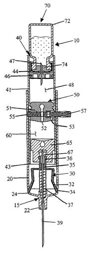
Referring now to the drawings in general and to Fig. 1
specifically, a vial injector for injecting fluid from a pre-filled vial is
designated
10. The device 10 includes an injection needle 39 having a sharpened tip for
piercing a patient. After injection, the injection needle 39 is automatically
shielded to prevent inadvertent contact with the contaminated needle.
The device 10 comprises an injector assembly 15 and a vial
holder assembly 40. The vial holder 40 holds a vial 70 of medicine, and is
attached to the injector assembly 15. The injector assembly 15 has a barrel
20 for receiving the vial holder 40, and a needle hub 35 that carries the
injection needle 39.
6
CA 02440888 2003-09-11
WO 02/072173 PCT/US02/07859
Referring to Figs. 1 and 7, the injection needle 39 is a double-ended
needle that is operable between two positions, an extended position and a
retracted position. In the extended position, the injection needle 39 projects
forwardly from the forward end of the barrel 20. In the retracted position,
the
injection needle is retracted into the barrel so that the forward sharpened
tip
of the needle is enclosed within the barrel to prevent inadvertent contact
with
the sharpened tip: When the injection needle 39 is in the extended position, a
spring 37 biases the needle rearwardly toward the retracted position. A
needle retainer 30 releasably retains the injection needle in the extended
position against the bias of the spring. During the injection stroke, the vial
holder 40 cooperates with the needle retainer 30 to allow the injection needle
to retract into the barrel 20, as shown in Fig. 7.
Some pre-filled injectors utilize a pre-filled cartridge or ampule
having a seal or plug that is displaceable to eject medicine from the
cartridge
or ampule. The present device may be utilized with such cartridges and/or
ampules. However, preferably, the present device utilizes a pre-filled vial 70
formed of a container 72 that has fixed walls and a seal 74 that is not
displaceable. During use, the medicine is withdrawn from the vial and then
expelled.
For instance, referring to Fig. 1, the device 10 comprises a vial
holder assembly 40, which has an empty transfer chamber 60 for receiving
medicine from the vial. During use, the medicine is drawn out of the vial 70
under negative pressure and into the transfer chamber. The medicine is then
ejected from the transfer chamber 60 into the patient.
More specifically, referring to Figs. 2-4, the medicine from the
vial is drawn out of the vial 70 by pushing the vial 70 forwardly to
pressurize
the fluid in the vial. The pressurized fluid is then transferred into the
transfer
chamber 60. Any air that is present in the transfer chamber 60 can then be
vented as shown in Figs. 4 and 5. The medicine can then be injected into the
patient by driving the vial holder 40 forwardly.
7
CA 02440888 2003-09-11
WO 02/072173 PCT/US02/07859
A valve 55 controls the flow of medicine between the vial and
the transfer chamber. The valve 55 is a sliding valve that is controlled by
pulling the valve. The valve 55 has a port that is slidable within a transfer
seal 50 in the vial assembly. In the open position, the valve aligns with a
fluid passage 52 in the transfer seal 50, as shown in Fig. 3, so that fluid
can
flow from the vial into the transfer chamber 60. After the fluid is
transferred
into the transfer chamber 60, the valve 55 is pulled into the closed position
so
that the fluid passage is blocked to prevent the medicine from leaking back
into the vial 70 during the injection stroke.
To inject the medicine, the vial 70 and vial holder 40 are driven
forwardly to expel the medicine from the transfer chamber 60. At the end of
the injection, the forward end of the vial assembly 40 engages the needle
retainer 30 thereby releasing the needle 39 for retraction, as shown in Fig.
6.
The spring 37 then displaces the needle rearwardly so that the contaminated
needle is shielded within the barrel 20, as shown in Figs. 7.
INJECTOR ASSEMBLY
Referring now to Fig. 1, the elements of the injector assembly
15 will be described in greater detail. The injector assembly 15 comprises a
barrel 20 and a needle retainer 30 releasably retaining the injection needle
39. A needle hub 35 attached to the needle retainer 30 has a mounting stem
36 for attaching the vial holder 40 to the injector assembly 15.
As shown in Figs. 1 and 8, the barrel 20 is generally cylindrical
and the distal end of the barrel has a tapered nose 22. The nose 22 has an
opening through which the injection needle 39 extends so that the sharpened
forward tip of the needle can be inserted into a patient. The rearward end of
the barrel 20 is open, forming a cylindrical socket adapted to receive the
vial
holder 40. Two laterally extending flanges 28 project outwardly from the
barrel 20, transverse the longitudinal axis of the barrel, forming a pair of
finger
grips for operating the device 10. The barrel 20 further includes a pair of
retaining apertures 24 and a pair of lockout windows 26 that cooperate with
s
CA 02440888 2003-09-11
WO 02/072173 PCT/US02/07859
the needle retainer 30 as described further below. In addition, the barrel 20
has a post hole 25 that extends through opposing sidewalls. The post holes
25 cooperates with the valve 55 to releasably lock the injector assembly 15
with the vial holder assembly 40, as discussed further below.
The needle hub 35 is a generally cylindrical element having a
central bore. The injection needle 39 is disposed within the central bore of
the hub 35 so that the rearward end of the needle projects rearwardly from
the hub and the forward end of the needle projects forwardly from the hub.
The needle 39 can be attached to the hub 35 in one of several ways. For
example, the needle 39 can be attached to the hub 35 by an adhesive such
as a UV curable adhesive. Alternatively, the needle 39 can be molded into
the hub 35, which is formed of plastic. The rearward end of the hub 35
includes a mounting stem 36 in the form of a barbed connector configured to
cooperate with the vial holder 40 to connect the vial holder to the needle hub
35 as discussed further below.
The needle retainer 30 is attached to the needle hub 35, and
preferably is integrally molded with the needle hub as a single piece. The
needle retainer 30 is preferably molded out of a rigid, high strength resin,
such as polycarbonate. Prior to retraction, the needle retainer 30 maintains
the needle hub 35 and attached needle 39 in a fixed axial position while the
medication is expelled from the vial holder 40. After the injection, the
needle
retainer 30 releases the needle hub 35 and the attached needle 39, which are
displaced rearwardly by a compression spring 37.
The spring 37 is a compression spring and may be formed of
stainless steel, treated carbon steel wire or other suitable non-corrosive
spring metal. The residual compression of the spring prior to disengagement
of the needle retainer is of sufficient magnitude to facilitate complete
needle
retraction and overcome the frictional resistance between sliding components
within the device 10.
Referring again to Figs. 1 and 8, the needle retainer 30 includes
a pair of retaining arms 32 that extend radially outwardly and forwardly from
9
CA 02440888 2003-09-11
WO 02/072173 PCT/US02/07859
the distal end of the needle retainer 30. During operation, the needle
retainer
30 is operable between a locked position and an unlocked position. In the
locked position, the retaining arms 32 engage the retaining apertures 24 in
the barrel wall to maintain the needle in a fixed axial position with the
forward
tip of the injection needle 39 projecting forwardly from the barrel 20. More
specifically, in the locked position, the retaining arms 32 engage the barrel
20
to hold the needle hub 35 and needle 39 against the rearward bias of the
spring 37. In the unlocked position, the retaining arms 32 are positioned so
as to allow the needle hub 35 and needle 39 to be retracted rearwardly, as
shown in Fig. 6. More specifically, in the unlocked position, the retaining
arms 32 are disengaged from the retaining apertures 24, allowing the spring
37 to propel the needle hub 35 and needle 39 rearwardly as shown in Fig. 7.
Referring to Fig. 1, as discussed above, the retaining arms 32
on the needle retainer 30 project forwardly and outwardly into engagement
with the retaining apertures 24 in the wall of the barrel 20. The terminal end
of each arm forms a retaining tab 34 that is configured to project into a
retaining aperture 24. More specifically, the retaining tabs 34 engage the lip
formed by each retaining aperture 24 in the wall of the barrel 20. In this
way,
the retaining tabs 34 operate as a pair of latches to retain the needle hub 35
and the injection needle 39 against the rearward bias of the spring.
As shown in Fig. 7, when the injection needle 39 is retracted,
the needle, needle retainer 30 and vial holder 40 are displaced rearwardly
together. Preferably, the injection device 10 includes a mechanism for
limiting rearward displacement of the retracted elements. Specifically, as
shown in Fig. 7, the injector assembly 15 preferably includes a pair of guide
arms 38 that cooperate with a pair of lockout windows 26 in the barrel 20 to
lock the retracted elements in the retracted position after use.
The guide arms 38 cooperate with a pair of alignment channels
or grooves formed in the interior wall of the barrel 20. The guide arms 38
may be molded out of a rigid, resilient high strength resin, such as
to
CA 02440888 2003-09-11
WO 02/072173 PCT/US02/07859
polycarbonate. The guide arms 38 extend forwardly from the needle hub 35
and project radially outwardly into engagement with the alignment grooves.
Each guide arm 38 includes a linear elongated rear portion
which preferably is generally parallel to the longitudinal axis of barrel 20.
The
forward portion of each guide arm 38 bends outwardly transverse the
longitudinal axis of the barrel 20 and extends into one of the alignment
grooves. When the needle retainer 30 is disposed within the barrel, the guide
arms 38 are deflected radially inwardly from their natural state. In this
position, the guide arms 38 are biased radially outwardly against the inner
wall of the barrel 20 due to the resilient properties of the guide arms.
The forward ends of guide arms 38 are contained within the
alignment grooves to substantially limit rotation of the needle and needle
retainer 30 during needle retraction. This engagement ensures that the guide
arms are aligned with the lockout windows 26 so that the guide arms snap
into the lockout windows at the end of retraction. In this way, the needle
retainer 30 is limited to axial displacement during needle retraction. During
retraction, the frictional resistance between the forward ends of the guide
arms 38 and the inside wall of the barrel 20 is overcome by the expansion
force of the spring 37.
As shown in Fig. 7, the linear elongated rear portion of each
guide arm 38 is spaced radially inwardly from the inner wall of the barrel 20
to
create a clearance space between the linear portion of the guide arms and
the barrel. Preferably, the minimum radial thickness of the clearance space is
greater than the thickness of the wall of the vial assembly housing 41. In
this
way, when the vial holder 40 is advanced forwardly to disengage the retaining
arms 32, the vial holder does not engage the guide arms 38 which could
otherwise prevent the guide arms from locking in the lockout windows 26 at
the end of retraction.
Each alignment groove is substantially parallel to the
longitudinal axis of the barrel 20. The groove may extend to the rearward
11
CA 02440888 2003-09-11
WO 02/072173 PCT/US02/07859
end of the barrel. However, it may be desirable to terminate the groove
forward of the rearward end of the barrel. The rearward portion of each
alignment groove intersects a lockout window 26 formed in the wall of the
barrel 20. The lockout windows 26 are adapted to receive the forward ends
of the guide arms 38, as shown in Fig. 7. In particular, as the front end of
each guide arm 38 aligns with the corresponding lockout window 26 during
needle retraction, the radially outward bias of the guide arm displaces the
arm
outwardly so that the forward end projects into the lockout window. The
engagement between the guide arms 38 and lockout windows 26 prevent
further axial movement of the injection needle 39. As a result, the retracted
elements are limited from further displacement in the forward or rearward
direction.
Referring now to Figs. 5-7 the automatic retraction of the
injection needle 39 shall be described. The vial holder 40 is axially advanced
to the proximal end of the barrel 20 until the medication is completely
expelled
from the transfer chamber 60 as shown in Figs. 5 and 6. As the vial holder 40
is advanced, the forward rim 43 of the housing 41 is displaced into
engagement with the retaining arms 32 of needle retainer 30.
After the rim 43 of the vial holder housing 41 engages the
retaining arms 32, continued axial advancement of the vial holder deflects the
retaining arms radially inwardly so that the retaining tabs 34 are displaced
inwardly, as shown in Fig. 6. In the inward position, the retaining tabs 34
are
disengaged from the retaining apertures 24 of the barrel 20. In this way, the
vial holder 40 operates as an actuator, such that axial advancement of the
vial holder assembly displaces the needle retainer 30 into an unlocked
position. In the unlocked position, the needle retainer 30 is no longer locked
in place against the force of the spring 37. After the needle retainer 30 is
in
the unlocked position and the user releases pressure on the vial holder 40,
the spring 37 propels the needle 39 rearwardly until the sharpened distal tip
of
the needle is enclosed within the barrel 20.
VIAL HOLDER ASSEMBLY
12
CA 02440888 2003-09-11
WO 02/072173 PCT/US02/07859
Referring to Fig. 1, the details of the vial holder assembly 40 will
be described in detail. The vial holder 40 comprises an elongated hollow
housing 41. The housing 41 comprises three seals. A front seal 65 seals the
forward end of the housing 42, and a slidable vial carrier 44 seals the
rearward end of the housing. The third seal is a transfer seal 50 disposed
between the front seal 65 and the carrier 44 which separates the housing into
two chambers: (1 ) a transfer chamber 60 between the front seal 65 and the
transfer seal; and (2) an air-pump chamber 48 between the vial carrier and
the transfer seal. The transfer seal 50 is pierceable to allow fluid transfer
between the vial 70 and the transfer chamber, as described further below. A
valve 55 in the transfer seal 50 is operable to reseal the transfer seal to
prevent leakage of fluid from the transfer chamber back into the vial 70.
The vial carrier 44 includes a double-ended transfer needle 47. The
rearward end of the transfer needle 47 projects into the socket in the vial
carrier 44 to pierce the septum 72 on the vial. Preferably, the length of the
transfer needle projecting into the carrier socket is slightly longer than the
thickness of the vial septum. In this way, the heel of the needle bevel is
spaced from the inner edge of the septum a short distance which preferably is
less than the length of the needle bevel.
. The front seal 65 cooperates with the barbed connector 36 on
the needle hub 35 to attach the vial holder assembly 40 to the injector
assembly 15. The front seal 65 is an elastomeric seal, which may be molded
in a self-sealing biocompatible elastomer such as polyisoprene. The front
seal 65 is generally cylindrical, having a plurality of axially-spaced
circumferential ribs. The ribs, frictionally and sealingly engage the interior
of
the housing 41 to provide a fluid tight seal, thereby preventing fluid from
leaking from the vial holder 40. The front seal 65 also has a wall that is
pierceable by the rearward sharpened tip of the injection needle 39. After
being pierced, the front end of the front seal 65 reseals around the needle 39
to prevent fluid from leaking from the transfer chamber 60.
13
CA 02440888 2003-09-11
WO 02/072173 PCT/US02/07859
Referring now to Fig. 1 the front seal 65 has a socket 67
configured to cooperate with the barbed connector 36 on the needle hub 35.
The socket 67 includes two radially relieved recesses that mate with the
barbed connector 36. Specifically, the barbed connector 36 matingly
engages the front seal 65 in a first position and a second position.
In the first position, the barbed connector 36 engages the first
recess, as shown in Fig. 1. In this position, the vial is attached to the hub,
but
the rearward end of the needle does not pierce the front seal 65. Displacing
the vial holder assembly forwardly relative to the needle hub 35, displaces
the
forward seal over the barb into the second position. In the second position,
the barbed connector 36 engages the second recess, as shown in Fig. 4. In
this position, the rearward end of the injection needle 39 pierces the front
seal
65.
The front seal 65 includes an elongated hollowed socket 67 in
which the rearward end of the needle projects. The rearward end of the
socket 67 is sealed by a pierceable wall. As shown in Fig. 1 prior to use, the
front seal 65 is mounted in the first position so that the barbed connector 36
engages the first recess. In this position, the injection needle 39 does not
penetrate the pierceable wall in the forward seal. As the vial holder assembly
40 is displaced forwardly, the barbed connector 36 engages the second
recess in the front seal 65, and the rearward end of the injection needle 39
pierces the wall so that the needle is in fluid communication with the
transfer
chamber 60. After the injection needle 39 penetrates the pierceable wall, the
wall reseals around the needle to form a fluid-tight seal and prevent
medication in the vial holder 40 from leaking around the needle.
The connection between the front seal 65 and the needle hub
35 is preferably a one-way engagement. In other words, when the front seal
65 is mounted on the barbed connector 36, the vial holder 40 can be
displaced forwardly relative to the barbed connector, but the front seal
cannot
be displaced rearwardly relative to the barbed connector. In this way, the
vial
holder 40 cannot be readily removed from the needle hub 35 in the barrel 20.
14
CA 02440888 2003-09-11
WO 02/072173 PCT/US02/07859
The one-way connection is facilitated by the rearward-facing
tapered shoulder of the barbed connector 36 and the square shaped forward-
facing shoulder of the recesses in the forward seal 65. In particular, the
rearward-facing shoulder of the barbed connector 36 cooperates with tapered
sides in the first and second radial recesses to permit relative displacement
of
the plug from the first recess to the second recess. Reverse displacement
from the second recess back to the first recess is resisted by the square
shaped forward-facing shoulders on barbed connector 36, which act to
impede reverse displacement.
Referring to Fig. 1, the rearward end of the vial holder housing
41 is sealed by the vial carrier 44. The vial carrier is a cylindrical
element,
having a circumferential groove around its exterior, into which a carrier seal
46 is seated. The carrier seal 46 is an elastomeric seal, such as an o-ring,
that forms a fluid-tight seal between the vial carrier 44 and the interior
wall of
the vial holder housing 41. In addition, the carrier seal 46 provides a
sliding
seal so that the vial carrier 44 can slide within the housing 41, while
maintaining an air-tight seal with the interior wall of the housing. This
sliding
seal allows the vial carrier 44 to operate as a piston to pump air into the
vial
70 as described further below.
The rearward end of the vial carrier 44 is open, forming a socket
configured to cooperate with and receive a vial 70. Specifically, the socket
is
adapted to receive the head of a vial, as shown in Fig. 1, so that preferably
there is a light interference fit between the head of the vial and the
interior of
the socket. In this way, the vial carrier 44 grips the vial to secure the vial
in
the vial carrier. Preferably, the vial carrier 44 cooperates with an annular
ridge 42 formed in the interior of the housing 41. The ridge 42 forms an
interference fit with the vial carrier to prevent axial displacement of the
carrier
44 when the vial 70 is inserted into the carrier. However, the interference of
the ridge is light enough that the carrier 44 can be axially advanced by
pressing the vial and carrier forwardly after attachment. In other words,
preferably, the frictional force of the ridge interference is only slightly
greater
is
CA 02440888 2003-09-11
WO 02/072173 PCT/US02/07859
than the force necessary to puncture the septum and insert the vial into the
carrier.
The forward end of the vial carrier forms a wall having a reduced
diameter opening. A transfer needle 47 projects through the reduced
diameter opening in the forward wall. Preferably, the transfer needle 47 is a
double-ended needle, and the rearward end of the transfer needle projects
rearwardly into the socket to pierce the septum 74 on the vial. Further,
preferably, the length of the transfer needle 47 projecting from the forward
wall into the socket is slightly longer than the thickness of the septum 74.
In
this way, the heel of the bevel of the rearward end of the transfer needle is
either aligned with the inner surface of the septum 74 or spaced from the
inner surface of the septum a short distance. The forward end of the transfer
needle 47 projects forwardly from the vial carrier 42 and is operable to
pierce
the transfer seal 50, as discussed further below.
As shown in Figs. 1 and 2a, the transfer seal 50 is disposed in
the vial holder housing 41 between the vial carrier 44 and the front seal 65,
thereby dividing the housing into the forward transfer chamber 60 and the
rearward air-pump chamber 48. The transfer seal 50 is an elastomeric seal
that forms a fluid-tight seal with the interior wall of the housing 41. A
circumferential groove around the exterior of the transfer seal 50 cooperates
with an annular flange on the interior of the housing 41 to connect the
transfer
seal to the housing. The groove and the flange cooperate to fix the axial
position of the transfer seal relative to the housing to prevent the transfer
seal
from sliding within the housing.
Referring to Fig. 2a, the transfer seal 50 comprises a fluid path
52 that extends axially through the transfer seal, and terminates at a
rearward
wall of the transfer seal. This rearward wall forms a pierceable seal that
seals
the fluid path 52 to prevent transfer of air or liquid between the air pump
chamber 48 and the transfer chamber 60. As discussed further below, during
use, the transfer needle 47 pierces the rearward wall of the transfer seal 50,
16
CA 02440888 2003-09-11
WO 02/072173 PCT/US02/07859
extending into the fluid path 52, so that fluid flows from the vial 70,
through
the transfer needle and the fluid path, and into the transfer chamber.
A valve 55 is located in the transfer seal to control the flow of
fluid between the transfer chamber and the vial 70 after the medicine is
transferred from the vial to the transfer chamber 60. More specifically, the
transfer seal 50 comprises a valve chamber 51 (see Fig. 8) disposed
transverse the fluid path 52. The valve is a sliding valve 55 that forms a
fluid-
tight seal with the valve chamber in the transfer seal 50. The valve 55 has a
hole through its side. When the valve 55 is in the open position (see Fig.
2a),
the side hole in the valve 55 aligns with the fluid path 52 in the transfer
seal
50 to allow fluid to flow from the vial into the transfer chamber. The valve
55
is closed by pulling the valve so that the valve slides within the valve
chamber
until the side hole in the valve seals against the transfer seal 50 as shown
in
Fig. 4a. In the closed position, the valve 55 prevents fluid from flowing from
the transfer chamber 60 back into the vial 70. As shown in Fig. 8, the valve
55 has two flats that register with the valve chamber, which keeps the valve
from rotating within the valve chamber so that the hole through the valve
aligns with the fluid path 52 in the transfer seal 50.
Referring again to Figs. 2a and 8, a detachable pull pin 57 is
attached to the first end of the valve 55. The pull pin 57 projects through
one
of the post holes 25 in the injector barrel 20 and through one of the locking
holes 49 in the vial holder housing 41. The second end of the valve 55
projects through the other locking hole 49 in the housing and the other post
hole 25 in the barrel 20. In this way, when the valve is disposed in the open
position, the valve 55 and pull pin 57 cooperate to releasably lock the barrel
20 and the housing 41 together to prevent axial displacement of the vial
holder assembly 40 relative to the injector assembly 15.
Referring to Figs. 4a and 8, when the valve is displaced to the closed
position, the second end of the valve 55 is drawn inwardly, out of engagement
with the post hole 25 in the barrel 20, and out of engagement with the locking
hole 49 in the housing 41. In addition, by detaching the pull pin after
closing
17
CA 02440888 2003-09-11
WO 02/072173 PCT/US02/07859
the valve, the pull pin is pulled out of engagement with the barrel 20 and the
housing 41, so that the vial holder assembly 40 is displaceable relative to
the
injector assembly 15.
The first end of the valve 55 forms an enlarged head 56 having
a socket for receiving the end of the pull pin 57 The pull pin 57 engages the
socket in the enlarged head 56 of the valve, forming an interference or snap-
fit. The frictional force between the pull pin 57 and the valve head 56 is
greater than the frictional force between the valve 55 and the valve chamber
51 in the transfer seal 50. In this way, pulling on the pull pin 57 slides the
valve 55 from the open position to the closed position. When the valve is
closed, the head 56 of the valve stops against the interior wall of the vial
holder housing 41. Continuing to pull on the pull pin 57 detaches the pull pin
from the valve 55.
Referring to Figs. 2a and 4a, the transfer seal 50 includes a vent
hole 53 for venting air from the transfer chamber 60 when fluid is transferred
from the vial 70 into the transfer chamber. The vent hole 53 is open when the
valve is open, as shown in Fig. 2a. When the valve 55 is pulled into the
closed position, the enlarged head 56 of the valve seals the vent hole 53 to
prevent medicine from leaking out the vent hole, as shown in Fig. 4a.
METHOD OF OPERATION
Before describing the details of operation, a short summary is
provided. First, the vial 70 is inserted into the vial holder 40 so that the
transfer needle 47 pierces the septum 74 on the vial (see Fig. 1 ). Air is
pumped into the vial 70 from the air pump chamber 48 to pressurize the vial
(see Fig. 2). The transfer needle 47 then pierces the transfer seal 50, and
the
pressurized medicine in the vial flows into the transfer chamber 60 (see Fig.
3). The valve 55 is then pulled closed (see Fig. 4). Air is then purged from
the transfer chamber 60 and the medicine is injected into the patient by
driving the vial holder 40 forwardly (see Figs. 5 and 6). At the end of the
injection stroke, the forward rim 43 of the vial holder assembly 40 engages
is
CA 02440888 2003-09-11
WO 02/072173 PCT/US02/07859
the needle retainer 30, displacing the needle retainer arms 32 inwardly (see
Fig. 6). The spring 37 then displaces the needle hub 35 and needle 39
rearwardly, along with the vial holder assembly 40 (see Fig. 7). At the end of
retraction, the locking arms 38 engage the lockout windows 26 to lock the
needle in the retracted position. The shielded device 10 can then be
disposed of safely.
The operation of the device 10 will now be described in detail.
Prior to use, the air pump chamber 48 and the transfer chamber 60 are
empty. Referring to Fig. 1, the vial 70 is inserted into the rearward end of
the
vial holder 40 so that the head of the vial seats in the vial carrier 44 and
the
rearward end of the transfer needle 47 pierces the septum 74 on the vial.
When the vial 70 is inserted into the needle carrier 44, the interference
between the vial carrier 44 and the ridge 42 in the housing 41 is greater than
the force required to pierce the septum, so that the ridge retains the vial
carrier in place prior to piercing the septum.
After the transfer needle 47 pierces the septum 74, the vial 70 is
displaced forwardly, which in turn displaces the vial carrier 44 forwardly
into
the air-pump chamber 48. Since the carrier seal 46 forms a fluid-tight seal
with the interior of the housing, displacing the carrier 44 forwardly pumps
the
air from the air pump chamber 48 into the vial 70, which pressurizes the vial,
as shown in Fig. 2.
At the end of the pressurizing stroke, the forward tip of the
transfer needle 47 pierces the transfer seal 50 so that the transfer needle
projects into the fluid path 52 in the transfer seal. The transfer seal
reseals
around the transfer needle 47 to prevent medicine from leaking out of the
transfer chamber around the needle. At this point, the vial is in fluid
communication with the transfer chamber. Since the device is held with the
vial down, the medicine from the pressurized vial flows downwardly into the
transfer chamber, as shown in Fig. 3. In addition, since the heel of the
rearward tip of the needle is adjacent the inner surface of the septum,
substantially all of the medicine can flow out of the vial and into the
transfer
19
CA 02440888 2003-09-11
WO 02/072173 PCT/US02/07859
chamber. As the fluid flows into the transfer chamber 60, air in the transfer
chamber vents out the vent hole 53 to prevent fluid from getting line-locked
in
the vial.
Fig. 3 illustrates the device such that at the end of the
pressurization stroke, the rearward end of the vial 70 protrudes from the
rearward end of the vial holder 40. Alternatively, and preferably, the holder
is
configured so that the length of the housing 41 rearward of the transfer seal
50 is substantially at least as long as the length of the vial carrier 44 and
the
attached vial 70. In this way, at the end of the pressurization stroke, the
rearward end of the vial is either disposed within the housing 41 or is
substantially flush with the rearward end of the housing, so that the vial
cannot be readily removed from the holder.
After the medicine is transferred into the transfer chamber, the
pull pin 57 is pulled, which in turn displaces the valve 55 sideways into the
closed position, sealing off the fluid path 52 in the transfer seal. At the
same
time, the head 56 of the valve is displaced over the vent hole 53 to seal the
vent hole. Continuing to pull on the pull pin 57 detaches the pull pin from
the
valve 55. At this point, the valve 55 and pull pin 57 are disengaged from the
post holes 25 in the barrel and the locking holes 49 in the vial holder
housing
41, so that the vial holder 40 can be displaced relative to the injector
assembly 15. In addition, at this point, the fluid is sealed in the transfer
chamber 60 between the transfer seal 50 and the front seal 65.
As shown in Fig. 4, after the valve 55 is closed, the device is
flipped vertically so that the needle is directed upwardly. The vial holder
assembly 40 is then displaced forwardly in the barrel 20 (i.e. upwardly) so
that
the front seal moves forwardly, and the barbed connector pops into the
second recess in the front seal socket 67. In doing so, the rearward tip of
the
injection needle 39 pierces the front seal 65 so that the injection needle is
in
fluid communication with the transfer chamber 60. Once the barbed
connector 36 engages the second socket in the front seal 65, the needle hub
CA 02440888 2003-09-11
WO 02/072173 PCT/US02/07859
maintains the front seal at a fixed axial position as the vial holder 40 is
displaced forwardly.
Any air in the transfer chamber is purged by displacing the vial
holder 40 forwardly over the front seal while holding the device 10 with the
needle upwardly. After the air is purged, the device is ready to inject, as
shown in Fig. 5. The forward sharpened tip of the injection needle 39 may
then be inserted into the patient, and the medicine is injected into the
patient
by driving the vial holder housing 41 forwardly
Displacing the vial holder assembly 40 forwardly relative to the
injection assembly 15 drives the housing 41 over the axially fixed front seal
65, thereby expelling the medicine from the transfer chamber. At the end of
the injection stroke, the forward rim of the housing 41 engages the arms 32 of
the needle retainer 30, displacing the arms radially inwardly, as shown in
Fig.
6. At this point, the injection needle 39 is released for retraction. As soon
as
the operator releases pressure against the rearward end of the vial holder
assembly 40, the spring 37 displaces the needle hub 35 and attached needle
rearwardly, along with the vial assembly.
As the injection needle 39 is retracted, the guide arms 38 ride in
the guide slots in the interior of the barrel, until the ends of the guide
arms
reach the lockout windows 26. At this point, the guide arms resiliently
displace outwardly into the lockout windows 26, thereby locking the needle
hub 35 and attached needle 39 in the retracted position. In this way, the
sharpened tip of the contaminated needle is automatically protected against
inadvertent contact after use, and can be safely disposed of, preferably in a
sharps container.
Referring now to Figs. 9-22, an alternate embodiment of a vial
injector for injecting fluid from a prefilled vial is designated 110. The
device
110 includes a needle 112 having a sharpened tip for piercing a patient. After
injection, the needle 112 is automatically shielded to prevent inadvertent
contact with the contaminated needle.
21
CA 02440888 2003-09-11
WO 02/072173 PCT/US02/07859
Referring to Figs. 10 and 12, the device includes a double-
ended needle 112 projecting forwardly from a generally cylindrical barrel 130.
A compression spring 126 biases the needle 112 rearwardly. A needle
retainer 120 releasably retains the needle against the bias of the spring 126.
The needle 112 is operable between two positions, an extended
position and a retracted position. In the extended position, the needle 112
projects forwardly from the forward end of the barrel 130. In the retracted
position, the needle is retracted into the barrel so that the forward
sharpened
tip of the needle is enclosed within the barrel to prevent inadvertent contact
with the sharpened tip. When the needle is in the extended position, the
spring 126 biases the needle 112 rearwardly toward the retracted position.
The needle retainer 120 releasably retains the needle into the extended
position against the bias of the spring. During the injection stroke, the vial
assembly 150 cooperates with the needle retainer 120 to allow the needle to
retract into the barrel 130, as shown in Figs. 20-22.
Some prefilled injectors utilize a prefilled vial or ampoule having
a seal or plug that is displaceable within the vial to eject medicine from the
vial. The device 110 may be configured to utilize such cartridges and/or
vials.
However, preferably, the device 110 utilizes a prefilled vial 90 that utilizes
fixed walls and a seal that is not displaceable. During use the medicine is
withdrawn from the vial and then expelled.
For instance, referring to Fig. 14, the device 110 comprises a
vial assembly 150, which has an empty transfer chamber 155 for receiving
medicine from the vial. During use, the medicine is drawn out of the vial 190
and into the transfer chamber. The medicine is then ejected from the transfer
chamber 155 into the patient.
More specifically, referring to Fig. 15, the medicine from the vial
is drawn out of the vial 190 by pulling the vial assembly 150 rearwardly to
create a vacuum that draws the fluid out of the vial and into the transfer
chamber 155. Any air that is present in the transfer chamber 155 can then be
22
CA 02440888 2003-09-11
WO 02/072173 PCT/US02/07859
vented as shown in Fig. 15. The medicine can then be injected into the
patient by driving the vial assembly 150 forwardly.
A valve 170 controls the flow of medicine from the vial into the
transfer chamber. The valve 170 is a rotary valve that is controlled by
twisting
the valve. The valve 170 has a port that is rotatable within a rearward seal
160 in the vial assembly. In a closed position, the valve seals 170 against
the
rearward seal 160 in the vial assembly, as shown in Fig. 17. When the valve
170 is closed, the medicine cannot flow from the vial 190 into the transfer
chamber 155. In the open position, the valve aligns with a fluid passage 162
in the rear seal 160, as shown in Fig. 19. Fluid can then flow from the vial
into the transfer chamber 155 by pulling the vial assembly 150 rearwardly.
After the fluid is transferred into the transfer chamber 155, the valve 170 is
twisted back into the closed position to prevent the medicine from leaking
back into the vial 190 during the injection stroke.
At the end of the injection, the forward end of the vial assembly
engages the needle retainer 120 thereby releasing the needle 112 for
retraction, as shown in Fig. 20. The spring 126 then displaces the needle
rearwardly so that the contaminated needle is shielded within the barrel 130,
as shown in Figs. 21 and 22.
NEEDLE ASSEMBLY
Referring now to Figs. 10-12, the elements of the injector device
110 will be described in greater detail. The injector 110 comprises a needle
assembly 115 and a vial assembly 150. The needle assembly 115 comprises
a barrel 130 and a needle retainer 120 releasably retaining the needle 112.
Inside the barrel is a needle hub 121 having a mounting stem 125 for
attaching the vial assembly 150 to the needle assembly 115.
The barrel 130 is generally cylindrical and the distal end of the
barrel has a tapered nose 132. The nose 132 has an opening through which
the needle 112 extends so that the sharpened forward tip of the needle can
23
CA 02440888 2003-09-11
WO 02/072173 PCT/US02/07859
be inserted into a patient. The rearward end of the barrel 130 is open,
forming a cylindrical socket adapted to receive the vial assembly 150. Two
laterally extending flanges 136 project outwardly from the barrel 130,
transverse the longitudinal axis of the barrel, forming a pair of finger grips
for
operating the device 110. The barrel 130 further includes a pair of retaining
apertures 138 and a pair of lockout windows 139 that cooperate with the
needle retainer 120 as described further below.
Needle Retainer
As shown in Fig. 12, the needle hub 121 is integrally attached to
the rearward end of the needle retainer 120. The needle hub 121 is a
generally cylindrical element having a central bore. The needle 112 is
disposed within the central bore of the hub 121 so that the rearward end of
the needle projects rearwardly from the hub and the forward end of the
needle projects forwardly from the hub. The needle 112 can be attached to
the hub 121 in one of several ways. For example, the needle 112 can be
attached to the hub 121 by an adhesive such as a UV curable adhesive.
Alternatively, the needle 112 can be molded into the hub 121, which is formed
of plastic. The rearward end of the hub 121 includes a circumferentially
barbed connector 125 configured to cooperate with the vial assembly 150 to
connect the vial assembly to the needle hub 121 as discussed further below.
The needle retainer 120 is axially displaceable within barrel 130
to facilitate needle retraction. The needle retainer 120 can be molded out of
a
rigid, high strength resin, such as polycarbonate. Prior to retraction, the
needle retainer 120 is maintained in a fixed axial position while the
medication
is expelled from the vial assembly 150. After the injection, the needle
retainer
120 and the attached needle 112 are displaced rearwardly by a compression
spring 126.
The spring 126 is a compression spring and may be formed of
stainless steel, treated carbon steel wire or other suitable non-corrosive
spring metal. The residual compression of the spring prior to disengagement
of the needle retainer is of sufficient magnitude to facilitate complete
needle
24
CA 02440888 2003-09-11
WO 02/072173 PCT/US02/07859
retraction and overcome the frictional resistance between sliding components
within the device 110.
Referring now to Figs. 10 and 12, the needle retainer 120
includes a pair of retaining arms 122 that extend radially outwardly and
forwardly from the distal end of the needle retainer 120. During operation,
the
needle retainer 120 is operable between a locked position and an unlocked
position. In the locked position, the retaining arms 122 engage the retaining
apertures 138 in the barrel wall to maintain the needle in a fixed axial
position
with the forward tip of needle 112 projecting forwardly from the barrel 130.
More specifically, in the locked position, the retaining arms 122 engage the
barrel 130 to hold the needle hub 121 and needle 112 against the rearward
bias of the spring 126. In the unlocked position, the retaining arms 122 are
positioned so as to allow the needle hub 121 and needle 112 to be retracted
rearwardly. More specifically, in the unlocked position, the retaining arms
122
are disengaged from the retaining apertures 138, allowing the spring 126 to
propel the needle hub 121 and needle 112 rearwardly.
As discussed above, the retaining arms 122 on the needle
retainer 120 project forwardly and outwardly into engagement with the
retaining apertures 138 in the wall of the barrel 130. The terminal end of
each
arm forms a retaining tab 124 that is configured to project into a retaining
aperture 138. More specifically, the retaining tabs 124 engage the lip formed
by each retaining aperture 138 in the wall of the barrel 130. In this way, the
retaining tabs 124 operate as a pair of latches to retain the needle hub 121
and needle 112 against the rearward bias of the spring.
Rearward Lock
As shown in Fig. 22, when the needle 112 is retracted, the
needle, needle retainer 120 and vial assembly 150 are displaced rearwardly
together. Preferably, the injection device 110 includes a mechanism for
limiting rearward displacement of the retracted elements. Referring now to
Figs. 10 and 22, the needle assembly 115 includes a pair of guide arms 128
2s
CA 02440888 2003-09-11
WO 02/072173 PCT/US02/07859
that cooperate with a pair of lockout windows 139 in the barrel 130 to lock
the
retracted elements in the retracted position after use.
The guide arms 138 cooperate with a pair of alignment channels
or grooves formed in the interior wall of the barrel 130. The guide arms 128
may be molded out of a rigid, resilient high strength resin, such as
polycarbonate. The guide arms 128 extend forwardly from the needle hub
121 and project radially outwardly into engagement with the alignment
grooves.
Each guide arm 128 includes a linear elongated rear portion
which preferably is generally parallel to the longitudinal axis of barrel 130.
The forward portion of each guide arm 128 bends outwardly transverse the
longitudinal axis of the barrel 130 and extends into one of the alignment
grooves. When the needle retainer 120 is disposed within the barrel, the
guide arms 128 are deflected radially inwardly from their natural state. In
this
position, the guide arms 128 are biased radially outwardly against the inner
wall of the barrel 130 due to the resilient properties of the guide arms.
The forward ends of guide arms 128 are contained within the
alignment grooves to substantially limit rotation of the needle and needle
retainer 120 during needle retraction. This engagement ensures that the
guide arms are aligned with the lockout windows 139 so that the guide arms
snap into the lockout windows at the end of retraction. In this way, the
needle
retainer 120 is limited to axial displacement during needle retraction. During
retraction, the frictional resistance between the forward ends of the guide
arms 128 and the inside wall of the barrel 130 is overcome by the expansion
force of the spring 126.
As shown in Fig. 22, the linear elongated rear portion of each
guide arm 128 is spaced radially inwardly from the inner wall of the barrel
130
to create a clearance space between the linear portion of the guide arms and
the barrel. Preferably, the minimum radial thickness of the clearance space is
greater than the thickness of the wall of the vial assembly housing 152. In
26
CA 02440888 2003-09-11
WO 02/072173 PCT/US02/07859
this way, when the vial assembly 150 is advanced forwardly to disengage the
retaining arms 122, advancement of the vial assembly is not impeded by the
guide arms 128.
Each alignment groove is substantially parallel to the
longitudinal axis of the barrel 130. The groove may extend to the rearward
end of the barrel. However, it may be desirable to terminate the groove
forward of the rearward end of the barrel. The rearward portion of each
alignment groove intersects a lockout window 139 formed in the wall of the
barrel 130. The lockout windows 139 are adapted to receive the forward
ends of the guide arms 128, as shown in Fig. 22. In particular, as the front
end of each guide arm 128 aligns with the corresponding lockout window 139
during needle retraction, the radially outward bias of the guide arm displaces
the arm outwardly so that the forward end projects into the lockout window.
The engagement between the guide arms 128 and lockout windows 139
prevent further axial movement of the retainer 122. As a result, the retracted
elements are limited from further displacement in the forward or rearward
direction.
The injection device 110 may also include a mechanism to limit
tampering or removal of the vial assembly 150 from the needle assembly 115.
Specifically, the rearward end of the barrel 130 may include an annular lip
that projects radially inwardly from the inside wall of the rearward end of
the
barrel 130. The lip is adapted to seat against a flange or beaded rim that may
be formed on the forward end of the vial assembly housing 152 so that the
vial assembly can not be easily pulled out of the rear of the barrel 130. As a
result, access to the retracted elements, and the contaminated needle in
particular, is limited.
Referring now to Figs. 20-22, the automatic retraction of the
needle 112 shall be described. The vial assembly 150 is axially advanced to
the proximal end of the barrel 130 until the medication 199 is completely
expelled from the transfer chamber 155. As the vial assembly 150 is
27
CA 02440888 2003-09-11
WO 02/072173 PCT/US02/07859
advanced, the forward rim of the housing 152 is displaced into engagement
with the retaining arms 122 of needle retainer 120
After the rim of vial assembly housing 152 engages the retaining
arms 122, continued axial advancement of the vial assembly deflects the
retaining arms radially inwardly so that the retaining tabs 124 are displaced
inwardly, as shown in Fig. 20. In the inward position, the retaining tabs 124
are disengaged from the retaining apertures 138 of the barrel 130. In this
way, the vial assembly 150 operates as an actuator, such that axial
advancement of the vial assembly displaces the needle retainer 120 into an
unlocked position. In the unlocked position, the needle retainer 120 is no
longer locked in place against the force of the spring 126. After the needle
retainer 120 is in the unlocked position and the user releases pressure on the
vial assembly 150, the spring 126 propels the needle 112 rearwardly until the
sharpened distal tip of the needle is enclosed within the barrel 130.
Lockingi Clip
Preferably, the needle assembly 115 includes a locking
mechanism for preventing the rearward end of the needle from piercing the
forward seal 156 before the medicine is drawn out of the vial 190 into the
transfer chamber 155. As shown in Figs. 9-12, the barrel 130 includes a
locking clip 145 in the barrel wall to prevent the forward seal from being
prematurely pierced. The wall of the barrel 130 includes a pair of radial
slots
134 cut through a plane that is transverse to the longitudinal axis of the
barrel.
When the locking clip 145 is inserted through the slots 134, the clip prevents
inadvertent forward displacement of the vial assembly 150 relative to the
front
seal 156, thereby preventing accidental advancement of the medicinal
components through the needle 112. The locking clip 145 is preferably
formed of a resilient high strength and high modulus resin, such as acetyl or
polycarbonate, and is configured to releasably engage the slots 134 in the
barrel 130.
Referring to Fig. 10, the locking clip 145 is preferably a flat
member having a pair of resiliently deflectable legs 147 that join to form a U-
2s
CA 02440888 2003-09-11
WO 02/072173 PCT/US02/07859
shape. The open end of the locking clip 145 has tapered edges that allow the
legs 147 to deflect outwardly as the locking clip is inserted into the
sidewall of
the barrel 130. In addition, the locking clip 145 has a plurality of teeth on
the
inside edge of the legs 147 that are adapted to engage the edges of the radial
slots 134.
As the locking clip is inserted into the sidewall of the barrel 130,
the legs 147 deflect outwardly to allow the teeth to clear the edges of radial
slots 134. Upon being deflected outwardly, the resilience of legs 147 bias the
legs radially inward toward their original position. Once the teeth are
disposed within the slots 134, the legs 147 deflect radially inwardly toward
their original position and releasably engage the outer edges of the needle
retainer 120 in barrel 130. In the inserted position, the closed end of the
locking clip 145 remains outside the barrel 130, as shown in Figs. 9 and 11.
After the medicine is transferred from the vial 90 into the transfer
chamber 155, the locking clip 145 is removed to permit the transfer chamber
to be vented and the medicine 199 to be injected into the patient, as shown in
Figs. 14 and 15. The locking clip 145 is removed from the barrel 130 by
pulling the closed end of the clip in a direction transverse to the
longitudinal
axis of the barrel. By pulling the clip in this manner, the legs are deflected
outwardly from the slots 134 to allow the teeth to clear the edges of slots
134.
After the locking clip 145 is removed from the barrel 130, the
medication 199 is injected into the patient by advancing the vial assembly
forwardly into the barrel. Initially, the rearward needle pierces the forward
seal. Then any air in the transfer chamber can be vented prior to injection.
The needle 112 is the inserted into the patient, and continued forward
displacement of the vial assembly 150 injects the medicine 199 into the
patient.
VIAL ASSEMBLY
29
CA 02440888 2003-09-11
WO 02/072173 PCT/US02/07859
Referring to Fig. 11, the details of the vial assembly will be
described in detail. The vial assembly 150 comprises an elongated hollow
housing 152 having a fluid chamber referred to as the transfer chamber. A
vial holder 180 is attached to the rearward end of the housing 152. The vial
holder 180 retains the vial 190 of medicine. A valve 170 attached to the vial
holder 180 controls the flow of fluid from the vial into the transfer chamber
155. A spring housing 155 houses a torsion spring 156 that biases the valve
toward a closed position.
The vial assembly housing 152 is an elongated generally
cylindrical hollow element. A pair of elongated ribs 154 formed on the outer
surface of the housing 152 cooperate with a pair of grooves formed in the
interior of the barrel to prevent the housing from twisting relative to the
barrel,
as shown in Figs. 9, 17 and 19. The forward end of the housing is sealed by
a forward seal 156 that cooperate with a barbed connector on the needle hub
121 to attach the vial assembly 150 to the needle assembly 115. The forward
seal 156 forms the forward end of the transfer chamber.
Front Seal
The front seal 156 is an elastomeric seal, which may be molded
in a self-sealing biocompatible elastomer such as polyisoprene. The front
seal 156 is generally cylindrical, having a plurality of axially-spaced
circumferential ribs 181. The ribs 181, which are more clearly shown in Fig.
10, frictionally and sealingly engage the interior of the container to provide
a
fluid tight seal, thereby preventing fluid from leaking from the vial 150. The
front seal 156 also has a front end that is pierceable by the rearward
sharpened tip of needle 112. After being pierced, the front end of the front
seal 156 reseals around the needle 112 to prevent fluid from leaking from the
vial 150.
The front seal 156 includes an elongated reduced diameter
neck. A substantially cylindrical sleeve 140 surrounds the neck portion of the
front seal, as shown in Fig. 12. The sleeve 140 comprises a slot through the
length of the sleeve.
CA 02440888 2003-09-11
WO 02/072173 PCT/US02/07859
Referring now to Figs. 13 and 14, the front seal 156 has a
socket 157 configured to cooperate with the barbed connector 125 on the
needle hub 121. The socket 157 includes two radially relieved recesses that
mate with the barbed connector 125. Specifically, the barbed connector 125
matingly engages the front seal 156 in a first position and a second position.
In the first position, the barbed connector 125 engages the first
recess, as shown in Fig. 12. In this position, the vial is attached to the
hub,
but the rearward end of the needle does not pierce the front seal 156.
Displacing the vial assembly forwardly relative to the hub, displaces the
forward seal over the barb into the second position. In the second position,
the barbed connector 125 engages the second recess, as shown in Fig. 15.
In this position, the rearward end of the needle 112 pierces the front seal
156.
The front seal 156 includes an elongated hollowed bore in which
the rearward end of the needle projects. The rearward end of the bore is
sealed by a pierceable wall. As shown in Fig. 12 prior to use, the vial 150 is
mounted in the first position so that the barbed connector 125 engages the
first recess. In this position, the needle 112 does not penetrate the
pierceable
wall in the forward seal. As the vial assembly is displaced forwardly, hub 121
engages the second recess in the forward seal, and the rearward end of the
needle112 pierces the wall so that the needle is in fluid communication with
the transfer cavity. After the needle 112 penetrates the pierceable wall, the
wall reseals around the needle to form a fluid-tight seal and prevent
medication in the vial assembly 150 from leaking around the needle.
The connection between the front seal 156 and the needle hub
121 is preferably a one-way engagement. In other words, when the front seal
156 is mounted on the barbed connector 125, the vial assembly 150 can be
displaced forwardly relative to the barbed connector, but the vial assembly
cannot be displaced rearwardly relative to the barbed connector. In this way,
the vial assembly 150 cannot be readily removed from the needle hub 121 in
the barrel 130, such that the vial assembly is substantially permanently
attached to the needle hub and barrel.
31
CA 02440888 2003-09-11
WO 02/072173 PCT/US02/07859
The one-way connection is facilitated by the rearward-facing
tapered shoulder of the barbed connector 125 and the square shaped
forward-facing shoulder of the recesses in the forward seal 156. In
particular,
the rearward-facing shoulder of the barbed connector 125 cooperates with
tapered sides in the first and second radial recesses to permit relative
displacement of the plug from the first recess to the second recess. Reverse
displacement from the second recess back to the first recess is resisted by
the square shaped forward-facing shoulders on barbed connector 125, which
act to impede reverse displacement.
Referring now to Fig. 12, the front seal 156 is configured to
prevent ejection of fluid when the barbed connector 125 is displaced from the
first position, in which the barbed connector 125 engages the first radial
recess, to the second position, in which the barbed connector engages the
second radial recess. Specifically, the front seal 156 includes a flared head
or circumferential flange at the forward end of the front seal. The open
forward end of the sleeve 140 surrounding the forward seal may be formed
with a beaded rim that seats against the rearward edge of the flared head.
The outside diameter of the flared head is greater than the inside diameter of
the sleeve 140, thereby impeding rearward displacement of the front seal 156
into the vial assembly is displaced forward after the locking clip 145 is
removed. In addition, the force required to overcome the frictional
engagement between the outer circumference of the front seal 156 and the
inner wall of the vial 150 is greater than the force required to displace the
plug
125 from the first recess to the second recess. Accordingly, when force is
initially applied to the vial assembly 150, the front seal 156 remains in a
fixed
position relative to the vial 150, while the barbed connector 125 is displaced
into the second position. This restriction on the front seal 156 limits the
release of fluid from the vial 150 when the needle 112 pierces the wall in the
forward seal.
The rearward end of the vial assembly housing 152 is sealed by
a rearward seal 160 that forms a fluid-tight seal with the interior of the
housing. The rearward seal forms the rearward end of the transfer chamber.
32
CA 02440888 2003-09-11
WO 02/072173 PCT/US02/07859
As shown in Figs. 17 and 19 the rearward seal 160 comprises an internal
bore forming a fluid tight seal with the valve 170. In addition, the rearward
seal 160 includes an axial channel or recess 162 intersecting the bore,
forming a fluid passage allowing fluid to flow from the vial 190 into the
transfer
chamber when the valve is open.
Referring to Figs. 12, 13, 16 and 18, the spring housing 165 is
integrally formed on the rearward end of the housing 152. The spring housing
flares out radially from the housing forming an annular space having a greater
diameter than the housing. The spring housing comprises a channel forming
a guide slot 167 that cooperates with a stop pin 169 to limit the rotation of
the
valve as discussed further below. A torsion spring in the spring housing
bears against the stop pin 169 and a post 168 to bias the valve into the
closed position. In addition, the spring housing forms a pair of radially
extending surfaces forming finger grips for use in attaching the vial 190 to
the
vial assembly 150, as discussed further below.
The vial holder 180 is pivotably displaceable within the spring
housing 165. The vial holder comprises a socket for receiving the vial 190. A
pair of radially deformable arms 185 lock the vial into the vial holder. A
piercing element 182 projects into the vial holder socket and is operable to
pierce the vial 190. The vial holder 180 forms a cap for the spring housing. A
plurality of latches formed on the spring housing 165 attach the vial holder
180 to the spring housing.
The valve 170 is integrally formed with the vial holder 180. The
valve projects forwardly from the vial holder and into engagement with the
rearward seal 160. As shown in Fig. 12, preferably the valve 170 comprises
an external barb for substantially permanently attaching the valve to the
seal.
A valve comprises a central bore having a closed forward end adjacent the
rearward seal. The piercing element is fixed attached to the valve within the
central bore of the valve, so that the valve is in fluid communication with
the
piercing element. A side port in the valve selectively engages the channel
162 through the rearward seal as discussed further below.
33
CA 02440888 2003-09-11
WO 02/072173 PCT/US02/07859
Referring again to Figs. 10 and 12, the vial 190 is a generally
cylindrical bottle that may be molded out of pharmaceutical quality glass such
as borosilicate, or a rigid inert plastic such as polyolefin or polyester. The
bottom of the vial is integral with the sides, so that it is fixed relative to
the
sidewalls. The forward end of the vial is sealed by a resealable pierceable
elastomeric septum 195. The septum 195 is fixed relative to the glass vial by
an annular cap 197 that is crimped around a circumferential flange formed on
the forward end of the vial.
The vial 190 contains a pre-filled dose of medication. The
medication is drawn out of the vial and injected into the patient as discussed
further below. Alternatively, the vial assembly can be configured to
accommodate a multi-dose vial in which a dose of medication is drawn out of
the vial and injected to the patient. After the injection, the needle is
retracted.
The vial can be removed from the device 110 and the device is safely
disposed in a sharps container. The vial can then be used with a new device
for a subsequent injection. The locking arms 185 preferably engage the
flared head of the vial to substantially permanently attach the vial to the
vial
holder. Alternatively, if the device is used in connection with a multiple
dose
vial, the arms are configured to releasably lock the vial to the vial
injector.
Use Of Device
Referring now to Figs. 12-22, the operation of the injection
device 110 will be described. Prior to use, the needle 112 is disposed in an
extended position so that the distal end of the needle projects forwardly from
the barrel 130, as shown in Fig. 12. Preferably, the device 110 is shipped
with the vial assembly 150 already mounted in barrel 130 so that the barbed
connector 125 is engaged in the first recess in the forward seal 156.
Alternatively, the vial assembly 150 may be shipped separately from the
barrel 130, so that the vial must be attached to the barrel prior to use.
As shown in Fig. 12, prior to use, the vial assembly housing 152
is partially withdrawn so that the transfer chamber 155 is an empty space
34
CA 02440888 2003-09-11
WO 02/072173 PCT/US02/07859
containing air. The vial 190 is inserted into the vial holder 180 so that the
piercing element pierces the septum 195 on the vial. The operator may grasp
the spring housing with his or her fingers and urge the vial forward with his
or
her thumb to insert the vial into the vial holder without urging the vial
assembly 150 forwardly.
The vial 180 is then pressurized as follows. The device 110 is
held vertically upright as shown in Fig. 12. The vial holder 180 is rotated
approximately 90 degrees as shown in Figs. 17 and 19 to open the valve 170.
When holding the valve open, the housing 152 is urged forwardly over the
forward seal 156. The housing is displaced forwardly until it abuts the
locking
clip 145. This is illustrated in Fig. 13, except that in Fig. 13, the housing
has
yet engaged the locking clip. In this way, the air in the transfer chamber 155
is forced into the vial 190 to pressurize the contents of the vial.
After vial 190 is pressurized, the device 110 is inverted so that
the vial is vertically oriented above the transfer chamber 155, as shown in
Fig.
14. The housing 152 is then displaced rearward over the forward seal while
the valve is maintained in the open position. This creates a vacuum in the
transfer chamber, which draws the medicine 99 out of the vial and into the
transfer chamber as shown in Fig. 14.
In the figures, the housing is elongated so that the forward seal
156 is spaced rearwardly from the rearward seal after the vial is pressurized,
as shown in Fig. 13. However, it may be desirable to shorten the housing 152
so that the forward seal abuts the rearward seal after pressurization. This
will
tend to increase the vacuum created when the housing is subsequent pulled
rearwardly to draw the medicine 199 out of the vial.
Once the medicine is drawn into the transfer chamber 155, the
medicine should be vented prior to injection to ensure that air is not
injected
into the patient. Accordingly, the device is inverted again, so that the
needle
is disposed vertically above the transfer chamber 155, as shown in Fig. 15.
The locking clip 145 is then removed so that the vial assembly 150 can be
CA 02440888 2003-09-11
WO 02/072173 PCT/US02/07859
displaced forwardly so that the rearward end of the needle pierces the
forward seal. The housing is the advanced over the forward seal to vent the
air from the transfer chamber 155. Subsequently, the forward tip of the
needle 112 is inserted into the patient and the vial assembly 150 is driven
forwardly to inject the medicine into the patient from the transfer chamber
155.
At the end of the injection stroke, the rim on the vial assembly
housing 152 engages the retaining arms 122, thereby displacing the retaining
tabs 124 radially inwardly to disengage the needle retainer 120 into the
unlocked position. Although the needle retainer 122 is in the unlocked
position, the needle 112 does not retract until the user releases pressure
from
the vial assembly. In this way, the user can retain pressure on the vial
assembly until after the needle is withdrawn from the patient. The user can
then release pressure from the vial assembly so that the needle is propelled
rearwardly by the spring 126. Alternatively, the user can release pressure
from the vial assembly while the needle 112 is still inserted in the patient.
Once the vial assembly is released, the spring 126 propels the needle 112
rearwardly so that the contaminated distal tip of the needle is enclosed
within
the barrel 130.
Referring now to Figs. 23-30, a second alternate embodiment of
a vial injector for injecting fluid from a pre-filled vial is designated 210.
The
device 210 includes an injection needle 239 having a sharpened tip for
piercing a patient. After injection, the injection needle 239 is automatically
shielded to prevent inadvertent contact with the contaminated needle.
The device 210 comprises an injector assembly 215 and a vial
holder assembly 240. The vial holder 240 holds a vial 270 of medicine, and is
attached to the injector assembly 215. The injector assembly 215 has a
barrel 220 for receiving the vial holder 240, and a needle hub 235 that
carries
the injection needle 239.
36
CA 02440888 2003-09-11
WO 02/072173 PCT/US02/07859
Referring to Figs. 23, 28 and 30, the injection needle 239 is a
double-ended needle that is operable between two positions, an extended
position and a retracted position. In the extended position, the injection
needle 239 projects forwardly from the forward end of the barrel 220. In the
retracted position, the injection needle is retracted into the barrel so that
the
forward sharpened tip of the needle is enclosed within the barrel to prevent
inadvertent contact with the sharpened tip. When the injection needle 39 is in
the extended position, a spring 237 biases the needle rearwardly toward the
retracted position. A needle retainer 230 releasably retains the injection
needle in the extended position against the bias of the spring. During the
injection stroke, the vial holder 240 cooperates with the needle retainer 230
to
allow the injection needle to retract into the barrel 220, as shown in Fig.
29.
Some pre-filled injectors utilize a pre-filled cartridge or ampule
having a seal or plug that is displaceable to eject medicine from the
cartridge
or ampule. The present device may be utilized with such cartridges and/or
ampules. However, preferably, the present device utilizes a pre-filled vial
270
formed of a container 272 that has fixed walls and a seal 274 that is not
displaceable. During use, the medicine is withdrawn from the vial and then
expelled.
For instance, referring to Fig. 24, the device 210 comprises a
vial holder assembly 240, which has an empty transfer chamber 260 for
receiving medicine from the vial. During use, the medicine is transferred out
of the vial 270 under positive pressure into the transfer chamber. The
medicine is then ejected from the transfer chamber 260 into the patient.
More specifically, referring to Figs. 24-26, the medicine from the
vial is transferred out of the vial 270 by pushing the vial 270 forwardly to
pressurize the fluid in the vial. The pressurized fluid is then transferred
into
the transfer chamber 260. Any air that is present in the transfer chamber 260
can then be vented. The medicine can then be injected into the patient by
driving the vial holder 240 forwardly.
37
CA 02440888 2003-09-11
WO 02/072173 PCT/US02/07859
To inject the medicine, the vial 270 and vial holder 240 are
driven forwardly to expel the medicine from the transfer chamber 260. At the
end of the injection, the forward end of the vial assembly 240 engages the
needle retainer 230 thereby releasing the needle 239 for retraction, as shown
in Fig. 29. The spring 237 then displaces the needle rearwardly so that the
contaminated needle is shielded within the barrel 220, as shown in Figs. 30.
INJECTOR ASSEMBLY
The details of the injector assembly 215 are substantially similar
to the injector assembly 15 of the first embodiment described above and
illustrated in Figs. 1-8. Specifically, the injector assembly 215 comprises a
barrel 220 and a needle retainer 230 releasably retaining the injection needle
239. A needle hub 235 attached to the needle retainer 230 has a mounting
stem 36 for attaching the vial holder 240 to the injector assembly 215.
The needle retainer 230 is attached to the needle hub 235, and
preferably is integrally molded with the needle hub as a single piece. Prior
to
retraction, the needle retainer 230 maintains the needle hub 235 and
attached needle 239 in a fixed axial position while the medication is expelled
from the vial holder 240. After the injection, the needle retainer 230
releases
the needle hub 235 and the attached needle 239 is displaced rearwardly by a
compression spring 237.
Referring again to Figs. 23 and 28, the needle retainer 230
includes a pair of retaining arms 232 that extend radially outwardly and
forwardly from the distal end of the needle hub 235. During operation, the
needle retainer 230 is operable between a locked position and an unlocked
position. In the locked position, the retaining arms 232 engage the retaining
apertures 224 in the barrel wall to maintain the needle in a fixed axial
position
with the forward tip of the injection needle 239 projecting forwardly from the
barrel 220. More specifically, in the locked position, the retaining arms 232
engage the barrel 220 to hold the needle hub 235 and needle 239 against the
38
CA 02440888 2003-09-11
WO 02/072173 PCT/US02/07859
rearward bias of the spring 237. In the unlocked position, the retaining arms
232 are positioned so as to allow the needle hub 235 and needle 239 to be
retracted rearwardly, as shown in Fig. 29. More specifically, in the unlocked
position, the retaining arms 232 are disengaged from the retaining apertures
224, allowing the spring 237 to propel the needle hub 235 and needle 239
rearwardly as shown in Fig. 30.
Referring to Fig. 28, as discussed above, the retaining arms 232
on the needle retainer 230 project forwardly and outwardly into engagement
with the retaining apertures 224 in the wall of the barrel 220. The terminal
end of each arm forms a retaining tab 234 that is configured to project into a
retaining aperture 224. More specifically, the retaining tabs 234 engage the
lip formed by each retaining aperture 224 in the wall of the barrel 220. In
this
way, the retaining tabs 234 operate as a pair of latches to retain the needle
hub 235 and the injection needle 239 against the rearward bias of the spring.
As shown in Fig. 30, when the injection needle 239 is retracted,
the needle, needle retainer 230 and vial holder 240 are displaced rearwardly
together. Preferably, the injection device 210 includes a mechanism for
limiting rearward displacement of the retracted elements. Specifically, as
shown in Fig. 30, the injector assembly 215 preferably includes a pair of
guide
arms 238 that cooperate with a pair of lockout windows 226 in the barrel 220
to lock the retracted elements in the retracted position after use. The guide
arms 238 cooperate with a pair of alignment channels or grooves formed in
the interior wall of the barrel 220. The guide arms 238 extend forwardly from
the needle hub 235 and project radially outwardly into engagement with the
alignment grooves.
The lockout windows 226 are adapted to receive the forward
ends of the guide arms 238, as shown in Fig. 30. In particular, as the front
end of each guide arm 238 aligns with the corresponding lockout window 226
during needle retraction, the radially outward bias of the guide arm displaces
the arm outwardly so that the forward end projects into the lockout window.
39
CA 02440888 2003-09-11
WO 02/072173 PCT/US02/07859
The engagement between the guide arms 238 and lockout windows 226
prevent further axial movement of the injection needle 239. As a result, the
retracted elements are limited from further displacement in the forward or
rearward direction.
Referring now to Figs. 28-30, the automatic retraction of the
injection needle 239 shall be described briefly. The vial holder 240 is
axially
advanced to the proximal end of the barrel 220 until the medication is
completely expelled from the transfer chamber 260 as shown in Figs. 28 and
29. As the vial holder 240 is advanced, the forward rim 243 of the housing
241 is displaced into engagement with the retaining arms 232 of needle
retainer 230.
After the rim 243 of the vial holder housing 241 engages the
retaining arms 232, continued axial advancement of the vial holder deflects
the retaining arms radially inwardly so that the retaining tabs 234 are
displaced inwardly, as shown in Fig. 29. In the inward position, the retaining
tabs 234 are disengaged from the retaining apertures 224 of the barrel 220.
In this way, the vial holder 240 operates as an actuator, such that axial
advancement of the vial holder assembly displaces the needle retainer 230
into an unlocked position. In the unlocked position, the needle retainer 230
is
no longer locked in place against the force of the spring 237. After the
needle
retainer 230 is in the unlocked position and the user releases pressure on the
vial holder 240, the spring 237 propels the needle 239 rearwardly until the
sharpened distal tip of the needle is enclosed within the barrel 220.
VIAL HOLDER ASSEMBLY
Referring to Figs. 23-26, the details of the vial holder assembly
240 will be described in detail. The vial holder 240 comprises an elongated
hollow housing 241 that is divided into two chambers by a mid-wall 245. The
forward chamber is a transfer chamber 260, the rearward chamber is an air-
pump chamber 248. A forward seal 265 seals the front of the transfer
CA 02440888 2003-09-11
WO 02/072173 PCT/US02/07859
chamber 260 and a slidable vial carrier 244 seals the rearward end of the air-
pump chamber 248.
The needle carrier 244 includes two needles. The first needle is
a pressurizing needle 249 in fluid communication with the air-pump chamber
248 and the vial 270. The second needle is a transfer needle 247 in fluid
communication with the transfer chamber 60 and the vial 270.
The rearward end of the transfer needle 247 projects into the
socket in the vial carrier 244 to pierce the septum 272 on the vial.
Preferably,
the length of the transfer needle projecting into the carrier socket is
slightly
longer than the thickness of the vial septum. In this way, the heel of the
needle bevel is spaced from the inner edge of the septum a short distance
which preferably is less than the length of the needle bevel.
The transfer needle 247 extends through a hole in the needle
carrier 244 and through a hole in the mid-wall 245, so that the rearward
sharpened tip projects rearwardly into the needle carrier socket and the
forward end projects into the transfer chamber 260. In this way, the transfer
needle spans from the vial 270 to the transfer chamber 260. The transfer
needle 247 is slidable relative to the mid-wall 245 and the needle carrier
244.
Accordingly, preferably a mid-wall seal 276 forms a fluid tight seal between
the transfer needle 247 and the mid-wall 245, and a carrier seal 277 forms a
fluid tight seal between the transfer needle and the vial carrier 244. As
shown
in Fig. 23, preferably the seals 276, 277 are o-ring seals. However, other
types of seals can be employed to provide a fluid-tight sliding fit between
the
transfer needle and the needle carrier and mid-wall.
The pressurizing needle 249 preferably projects into the socket
of the vial carrier 244 further than the transfer needle 247, so that the
pressurizing needle 249 is adapted to project further into the vial 270, as
shown in Fig. 24. In this way, preferably when the vial 270 is mounted in the
vial holder assembly 240 and held upright, the pressurizing needle 249
41
CA 02440888 2003-09-11
WO 02/072173 PCT/US02/07859
projects into the vial adjacent the bottom of the vial to pressurize the air
in the
vial.
As mentioned previously, the front seal 265 seals the forward
end of the transfer chamber 260. The front seal 265 cooperates with the
barbed connector 236 on the needle hub 235 to attach the vial holder
assembly 240 to the injector assembly 215. The front seal 265 is
substantially similar to the front seal 265 in the first device 110, and the
details of the front seal are provided previously in the description of the
first
device. The front seal 265 has a wall that is pierceable by the rearward
sharpened tip of the injection needle 239. After being pierced, the front end
of the front seal 265 reseals around the needle 239 to prevent fluid from
leaking from the transfer chamber 260.
Referring to Fig. 24, the rearward end of the vial holder housing
241 is sealed by the vial carrier 244. The vial carrier is a cylindrical
element,
having a circumferential groove around its exterior, into which a carrier seal
246 is seated. The carrier seal 246 is an elastomeric seal, such as an o-ring,
that forms a fluid-tight seal between the vial carrier 244 and the interior
wall of
the vial holder housing 241. In addition, the carrier seal 246 provides a
sliding seal so that the vial carrier 244 can slide within the housing 241,
while
maintaining an air-tight seal with the interior wall of the housing. This
sliding
seal allows the vial carrier 244 to operate as a piston to pump air into the
vial
270 as described further below.
The rearward end of the vial carrier 244 is open, forming a
socket configured to cooperate with and receive a vial. Specifically, the
socket is adapted to receive the head of a vial, as shown in Fig. 24, so that
preferably there is a light interference fit between the head of the vial and
the
interior of the socket. In this way, the vial carrier 244 grips the vial to
secure
the vial in the vial carrier. As in the first embodiment described previously,
the
vial carrier 244 may cooperate with an annular ridge formed in the interior of
the housing 241 to prevent axial displacement of the carrier 244 when the vial
42
CA 02440888 2003-09-11
WO 02/072173 PCT/US02/07859
270 is inserted into the carrier. However, in the present instance, the
housing
does not include such a ridge.
METHOD OF OPERATION
Before describing the details of operation, a short summary is
provided. First, the vial 270 is inserted into the vial holder 240 so that the
transfer needle 247 and the pressurizing needle 249 pierce the septum 274
on the vial (see Fig. 24). The vial 270 and vial carrier 244 are displaced
forwardly to pump air into the vial 270 from the air pump chamber 248 to
pressurize the vial (see Fig. 25). The pressurized medicine in the vial then
flows into the transfer chamber 260 (see Fig. 26). The rearward end of the
injection needle 239 pierces the front seal 265 (see Fig. 27), and air is
purged
from the transfer chamber 260. The medicine is then injected into the patient
by driving the vial holder 240 forwardly (see Figs. 28 and 29). At the end of
the injection stroke, the forward rim 243 of the vial holder assembly 240
engages the needle retainer 230, displacing the needle retainer arms 232
inwardly (see Fig. 29). The spring 237 then displaces the needle hub 235
and needle 239 rearwardly, along with the vial holder assembly 240 (see Fig.
30). At the end of retraction, the locking arms 238 engage the lockout
windows 226 to lock the needle in the retracted position. The shielded device
210 can then be disposed of safely.
The operation of the device 210 will now be described in detail.
Prior to use, the air pump chamber 248 and the transfer chamber 260 are
empty. Referring to Fig. 24, the vial 270 is inserted into the rearward end of
the vial holder 240 so that the head of the vial seats in the vial carrier 244
and
the rearward end of the transfer needle 247 and pressurizing needle 249
pierce the septum 274 on the vial.
After the transfer needle 247 pierces the septum 274, the vial
270 is displaced forwardly, which in turn displaces the vial carrier 244
forwardly into the air-pump chamber 248. Since the carrier seal 246 forms a
43
CA 02440888 2003-09-11
WO 02/072173 PCT/US02/07859
fluid-tight seal with the interior of the housing, displacing the carrier 244
forwardly pumps the air from the air pump chamber 248 into the vial 270,
which pressurizes the vial and transfers medicine into the transfer chamber
260 from the vial 270, as shown in Fig. 25. The vial is preferably held
upright
as shown in Fig. 25 so that gravity aids in the flow of fluid from the vial to
the
transfer chamber 260.
As the vial carrier 244 is displaced forwardly, the transfer needle
247 slides forwardly relative to the mid-wall 245. In addition, the front seal
265 displaces forwardly to increase the volume of the transfer chamber as the
pressurized medicine is transferred to the transfer chamber. At the end of the
transfer stroke, the forward tip of the transfer needle 247 engages the front
seal 265 so that the front seal seals the transfer needle to prevent medicine
from leaking from the transfer chamber 260 back into the vial 270.
As shown in Figs. 24-26, the medicine can be transferred to the
transfer chamber 260 before attaching the vial holder assembly 240 to the
needle assembly 215. However, if desired, the vial holder assembly 240 can
be attached to the needle assembly 215 before transferring the medicine into
the transfer chamber. Accordingly, if the vial holder assembly is not yet
attached to the needle assembly, it is attached after the medicine is
transferred.
After the medicine is transferred and the vial holder 240 is
attached to the injection assembly 215, the device is flipped vertically so
that
the injection needle 239 is directed upwardly, as shown in Fig. 27. The vial
holder assembly 240 is then displaced forwardly into the barrel 220 so that
the front seal moves forwardly, and the barbed connector pops into the
second recess in the front seal 265. In doing so, the rearward tip of the
injection needle 239 pierces the front seal 265 so that the injection needle
is
in fluid communication with the transfer chamber 260. Once the barbed
connector 236 engages the second socket in the front seal 265, the needle
hub maintains the front seal at a fixed axial position as the vial holder 240
is
displaced forwardly.
44
CA 02440888 2003-09-11
WO 02/072173 PCT/US02/07859
Any air in the transfer chamber is purged by displacing the vial
holder 240 forwardly over the front seal while holding the device 210 with the
needle upwardly. After the air is purged, the device is ready to inject, as
shown in Fig. 28. The forward sharpened tip of the injection needle 239 may
then be inserted into the patient, and the medicine is injected into the
patient
by driving the vial holder housing 241 forwardly
Displacing the vial holder assembly 240 forwardly relative to the
injection assembly 215 drives the housing 241 over the axially fixed front
seal
265, thereby expelling the medicine from the transfer chamber. At the same
time, the mid-wall 245 and vial carrier 244 slide relative to the transfer
needle
247, as shown in Figs. 28 and 29. At the end of the injection stroke, the
forward rim of the housing 241 engages the arms 232 of the needle retainer
230, displacing the arms radially inwardly, as shown in Fig. 29. At this
point,
the injection needle 239 is released for retraction. As soon as the operator
releases pressure against the rearward end of the vial holder assembly 240,
the spring 237 displaces the needle hub 235 and attached needle rearwardly,
along with the vial holder assembly.
As the injection needle 239 is retracted, the guide arms 238 ride
in the guide slots in the interior of the barrel, until the ends of the guide
arms
reach the lockout windows 226. At this point, the guide arms resiliently
displace outwardly into the lockout windows 226, thereby locking the needle
hub 235 and attached needle 239 in the retracted position. In this way, the
sharpened tip of the contaminated needle is automatically protected against
inadvertent contact after use, and can be safely disposed of, preferably in a
sharps container.
The terms and expressions which have been employed are
used as terms of description and not of limitation. There is no intention in
use
of such terms and expressions of excluding any equivalents of the features
shown and described or portions thereof. It is recognized, however, that
various modifications of the embodiments described herein are possible
CA 02440888 2003-09-11
WO 02/072173 PCT/US02/07859
within the scope and spirit of the invention. For instance, the different
embodiments have been described as safety devices in which the needle is
automatically shielded after use. However, in certain applications, the
present invention may be employed without the safety feature. Specifically,
the device may be modified to incorporate the automatic withdrawal and
injection of the medication from the vial without the safety features. In
addition, it is possible to modify the present invention by eliminating the
pressurization of the vial and rely on gravity or some other means to transfer
the medication from the vial. Furthermore, other changes to the structure can
be made, such as in the second embodiment 110, the torsion spring may be
eliminated, and a pair of detents can be used to releasably lock the valve in
the open and closed positions. Further still, the device can be modified to
include a step in which two separate components are mixed prior to injection.
Specifically, in the foregoing description, the device includes a transfer
chamber for receiving the medicinal fluid from the vial. The transfer chamber
has been described as an empty chamber. However, it may be desirable to
store a component, such as a powder, in the sealed transfer chamber. Then,
prior to injection, the fluid from the vial is transferred into the transfer
chamber
and mixed with the powder. The mixture can then be injected into a patient.
Furthermore, in the description above the devices are described as
incorporating a transfer chamber for receiving medicinal fluid from the vial.
In
certain instances the device need not have such a transfer chamber.
Accordingly, the present invention incorporates modifications and variations
that fall within the scope of the following claims.
46