Note: Descriptions are shown in the official language in which they were submitted.
CA 02441982 2003-09-24
WO 02/078571 PCT/US02/09455
IMPLANT INSERTED WITHOUT BONE ANCHORS
[0001 ] Background
[0002] Loss of bladder control is a condition known as urinary incontinence.
Millions
of men and women of all ages suffer from this condition, which causes
involuntary loss
of urine. Although urinary incontinence may occur at any age, it is more
common in
women and in the elderly. Women may develop incontinence during pregnancy,
childbirth or menopause. Older men may lose bladder control following prostate
surgery. In addition to the medical aspects of this condition, the social
implications for
an incontinent patient include loss of self-esteem, embarrassment, restriction
of social
and sexual activities, isolation, depression and, in some instances,
dependence on
caregivers.
[0003] Continence problems may occur when the muscles of the urinary system
malfunction or are weakened. Other factors, such as trauma to the urethral
area,
neurological injury, hormonal imbalance or medication side-effects, may also
cause or
contribute to incontinence problems.
[0004] In general, there are five basic types of incontinence: stress
incontinence, urge
incontinence, mixed incontinence, overflow incontinence and functional
incontinence.
Stress urinary incontinence (SUI) is the involuntary loss of urine that occurs
due to
sudden increases in intra-abdominal pressure resulting from activities such as
coughing,
sneezing, lifting, straining, exercise and, in severe cases, even simply
changing body
position. This condition usually occurs when the sphincter or pelvic muscles
are
weakened by, for example, childbirth or surgery.
[0005] Urge incontinence, also termed "hyperactive bladder,"
"frequency/urgency
syndrome" or "irritable bladder," occurs when an individual experiences the
immediate
need to urinate and loses bladder control. Urge incontinence is a common
problem that
increases with advancing age or results from a kidney or bladder infection.
CA 02441982 2003-09-24
WO 02/078571 PCT/US02/09455
[0006] Mixed incontinence is the most common form of urinary incontinence.
Mixed
incontinence is a combination of the symptoms for both stress and urge
incontinence.
Overflow incontinence is a constant dripping or leakage of urine caused by an
overfilled bladder. This condition often occurs in men due to the prevalence
of
obstructive prostate gland enlargement or tumor. Functional incontinence
results when
a person has difficulty moving from one place to another. It is generally
caused by
factors outside the lower urinary tract, such as deficits in physical function
and/or
cognitive function.
[0007] A variety of treatment options are currently available to treat
incontinence.
Some of these treatment options include external devices, indwelling
catheters,
behavioral therapy (such as biofeedback, electrical stimulation, or Kegal
exercises),
injectable materials, prosthetic devices and/or surgery. Surgical procedures
can be used
to'completely restore continence in some instances.
[0008] Surgical procedures include sling procedures, colposuspension
procedures, and
needle suspension procedures. Colposuspension procedures seek to place the
urethra in
high retropubic position. The Marshall-Marchetti-Krantz procedure and the
Burch
procedure are examples of colposuspension procedures. The Marshall-Marchetti-
Krantz procedure places sutures at the urethrovesical junction to the
periosteum of the
pubic bone. See Marshall et al., The Correction of Stress Incontinence By
Simple
Vesicourethral Suspension; Surg. Gynecol. Obstet. Vol. 88, Pps. 509-518
(1949).
[0009] With the Burch procedure, sutures are placed at the urethrovesical
junction to
Cooper's ligament. See Gi1j a et al., A Modified Raz Bladder Neck Suspension
Operation (Transvaginal Burch), J. of Urol. Vol. 153, Pps. 1455-1457 (May
1995). A
significant abdominal incision is associated with the Marshall-Marchetti-
Krantz
procedure. The Burch procedure has been performed abdominally, vaginally and
laparoscopically. See Burch, Urethrovaginal Fixation to Cooper's Ligament for
Correction of Stress Incontinence, Cystocele, and Prolapse, Am. J. Obst. &
Gynecology, vol. 81 (No. 2), Pps. 281-290 (Feb. 1961); and Das et al.,
Laparoscopic
Colpo-Suspension, J. of Urology, vol. 154, Pp. 1119-1121 (1995).
-2-
CA 02441982 2003-09-24
WO 02/078571 PCT/US02/09455
[0010] Needle suspension procedures elevate the urethra retropubically. They
include
Pereyra, Stamey, Raz, Gittes, Muszani and Vesica procedures. These procedures
(except the Vesica procedure) place sutures transvaginally at the
urethrovesical junction
and are sutured to the abdominal wall through two small abdominal incisions.
See
Stamey, Endoscopic Suspension of the Vesical Neck for Urinary Incontinence in
Females, Ann. Surgery, pp. 465-471, October 1980; Pereyra, A Simplified
Surgical
Procedure for the Correction of Stress Incontinence in Women, West. J. Surg.,
Obstetrics & Gynecology, pp. 243-246, July-August 1959; Holschneider et al., A
Modified Pereyra Procedure In Recurrent Stress Urinary Incontinence: A 15-Year
Review, Obstetrics & Gynecology, vol. 83, No. 4 Pps. 573-578 (1994). The
Vesica
procedure includes an abdominal incision where bone anchors are driven into
the top of
the pubic bone and sutures attached to the bone anchors are placed at the
urethrovesical
junction.
[0011 ] The first sling procedure was the Goebel-Stoeckel-Frannenheim
procedure. The
sling was autologous fascia that was placed beneath the urethra and suspended
by
sutures attached to the rectus fascia of the abdominal wall.
[0012] There are two general types of sling procedures. The first type of
sling
procedure utilizes bone screws and associated sutures to anchor a sling (e.g.
on a
posterior portion of the pubic bone). A commercial example of a bone screw
sling
procedure is a surgical procedure that utilizes the In-Fast Sling System,
available from
American Medical Systems of Minnetonka, Minnesota.
[0013] The second type of sling procedure is a minimally invasive surgical
method
involving the placement (e.g. by the use of a Stamey needle or other ligature
carrier) of
a sling to stabilize or support the bladder neck or urethra. See Horbach et
al., A
Suburethral Sling Procedure With Polytetrafluoroethylene For the Treatment of
Genuine Stress Incontinence In Patients With Low Urethral Closure Pressure, J.
Obstetrics & Gynecology, vol. 71, No. 4, Pps. 648-652 (April 1998); and Morgan
et al.,
The Marlex Sling Operation For the Treatment of Recurrent Stress Urinary
-3-
CA 02441982 2003-09-24
WO 02/078571 PCT/US02/09455
Incontinence: A 16 Year Review, Am. J. Obstet. Gynecol., vol. 151, No. 2, Pps.
224-
227, (Jan. 1985).
[0014] The slings described above differ in the type of material, sutures and
points of
anchoring based on the procedure being performed. In some cases, the sling is
placed
under the bladder neck and secured via suspension means (such as bone anchors
or
screws) through a vaginal incision. Bone anchors or screws raise the specter
of bone
infection, necrosis and other complications, although such complications are
rare.
[0015] The second type of sling procedure (pubovaginal sling procedures that
do not
include bone anchors) anchor slings in the abdominal or rectus fascia. These
types of
procedures involve puncturing the abdominal wall of the patient to pass a
needle.
Complications associated with sling procedures are rare, but they include
urethral
obstruction, infection, development of de novo urge incontinence, bladder
perforation,
hemorrhage, prolonged urinary retention, and damage to surrounding tissue
(e.g. caused
by sling erosion). The likelihood of complications due to abdominal incisions
varies
and depends on the particular surgical procedure.
[0016] The TVT Tension-free Vaginal Tape procedure is a known sling procedure
used
in the United States. During the procedure, incisions are made in the
abdominal (i.e.
suprapubic) area and in the vaginal wall. Two curved, needle-like elements are
connected at an end, to tension-free vaginal sling tape. A tape-free end of
one of the
needle-like elements is inserted through the vaginal incision and into the
paraurethral
space. Using a handle attached to the needle, the needle is angulated
laterally (for
example, to the right) to perforate the endopelvic fascia, guided through the
retropubic
space and passed through the abdominal incision. The handle is disconnected
and the
needle is then withdrawn through the abdominal wall, thereby threading a
portion of the
tape through the tissue of the patient. This technique is repeated with the
other needle
on the other side (for example, to the left), so that the tape is looped
beneath the bladder
neck or urethra. The tape is adjusted to provide appropriate support to the
bladder neck
or urethra. The tape ends are then cut at the abdominal wall leaving the ends
of the
sling anchored in the abdominal (rectus) fascia.
-4-
CA 02441982 2003-09-24
WO 02/078571 PCT/US02/09455
[0017] Complications associated with the TVT procedure include injury to blood
vessels of the pelvic sidewall and abdominal wall, hematomas, urinary
retention, and
bladder and bowel injury. One serious disadvantage of the TVT procedure,
particularly
for surgeons unfamiliar with the surgical method, is the lack of information
concerning
the precise location of the needle tip relative to adjacent pelvic anatomy. A
cadaver
study indicated that the TVT needle is placed in close proximity to sensitive
tissue such
as superficial epigastric vessels, inferior epigastric vessels, the external
iliac vessel and
the obturator. See, Walters, Mark D., Percutaneous Suburethral Slings: State
of the
Art, presented at the conference of the American Urogynecologic Society,
Chicago
(October 2001).
[0018] If the TVT needle tip is allowed to accidentally pass across the
surface of any
blood vessel, lymphatic duct, nerve, nerve bundle or organ, serious
complications can
arise. These shortcomings, attempts to address these shortcomings and other
problems
associated with the TVT procedure are disclosed in PCT publication nos. PCT WO
00/74613 and PCT WO 00/74594.
[0019] Examples of incontinence procedures are disclosed in U.S. Pat. Nos.
5,112,344;
5,611,515; 5,842,478; 5,860,425; 5,899,909; 6,039,686, 6,042,534 and
6,110,101.
[0020] Brief Summary
[0021] Fig. 1 A schematically represents the position of anatomical structures
such as the
pubic bone 12, retropubic space 11, bladder 14 and urethra 16. The retropubic
space 11 is
a highly defonnable cavity. It expands and collapses under the influence of
surrounding
tissue such as the bladder, etc. The relative positions of these structures or
regions are
shown at rest. In a healthy, continent individual, the external sphincter and
other tissues
and structures cooperate to resist flow of urine out of the bladder 14. In the
rest or "non-
stressed" condition, the distance between a midpoint of the retropubic space
and an axial
midpoint of the urethra 16 is B1. The distance between the axial midpoint of
the urethra
16 and an upper, relatively fixed structure (for example, the rectus fascia,
the top of the
pubic bone or Cooper's ligament) is Al.
-5-
CA 02441982 2003-09-24
WO 02/078571 PCT/US02/09455
[0022] Fig. 1B schematically illustrates the effect of a stress event (e.g.
coughing or
sneezing) on the anatomical structures of Fig. 1A. There can be a marked
descent of the
bladder and urethra during certain types of stress events. The retropubic
space 11 and its
midpoint descend slightly. The distance A2 (between the relatively fixed
structure and an
axial midpoint of the urethra 16) is greater than the distance Al (see Fig.
1A). The
increase from Al to A2 is more than the increase from B1 to B2. Healthy,
continent
individuals can nonetheless retain urine as their support structure can
continue to close the
urethra 16. With many types of incontinence, however, the intraurethral
pressure during
the stress event rises above the support structure's ability to close the
urethra, resulting in
leakage.
[0023] There is a debate in the medical community concerning the precise
mechanism
responsible for the success of sling procedures. Some commentators believe
that slings
correct incontinence by providing a backstop effect (i.e. preventing the
distance A2 from
expanding beyond a limit). The present invention recognizes the possibility
that
continence may be restored by providing dynamic support (i.e. a sling that is
not securely
attached to a fixed anatomical reference point). The dynamic support and
continence may
be provided without the need for invasive procedures that secure a sling to a
fixed
reference (e.g. Cooper's ligament, the pubic bone or rectus fascia). As a
result, it is
believed that the present invention is much less invasive and risks far fewer
complications
than the prior art sling procedures.
[0024] As used herein, the term "retropubic space" means that region of the
body that is
posterior to the pubic bone (i.e. the region that is posterior to the pubic
ramus and pubic
symphysis). This is an area of loose connective tissue between the bladder
with its related
fascia and the pubis. It includes endopelvic fascia. The retropubic space
extends upward
to the rectus fascia, but does not include the suprapubic area with the rectus
fascia itself.
The retropubic space does not extend beyond the sacrum. The phrases "space of
Retzius"
or "cave of Retzius" are also used to describe portions of the retropubic
space.
[0025] Conventional procedures exclude the possibility of anchoring a sling
solely in the
retropubic space. The prior art procedures suture the sling to a bone screw,
the bone itself
-6-
CA 02441982 2003-09-24
WO 02/078571 PCT/US02/09455
or tough, fixed tissue such as Cooper's ligament (which is fixed relative to
the pubic
bone). Other prior art procedures extend the sling through abdominal incisions
and anchor
the sling in rectus fascia of the suprapubic area.
[0026] Some surgeons believe that the retropubic space does not offer a
sufficiently robust
foundation for anchoring a sling. For example, for conventional sling
procedures that do
not use bone anchors, surgeons will typically extend the sling into the rectus
fascia to
firmly anchor the sling. Some procedures even suture the sling to the rectus
fascia.
[0027] The present invention recognizes that, when disturbed by an implantable
material,
the retropubic space will generate tough fibrous tissue, providing substantial
holding
power for an implant placed in that space. This body reaction can be exploited
to help
restore continence.
[0028] The present invention recognizes that an implantable article (e.g. a
dynamic sling
or hemi-sling) may be anchored to structure in the retropubic space, without
the need of
bone anchors and without the need to suture the implant to Cooper's ligament,
the pubic
bone or the tough rectus fascia. With the present invention, the implant may
be anchored
in the retropubic space (e.g. to endopelvic fascia) without the need to extend
upward into
the abdominus or rectus fascia. This avoids complications associated with
invasive
abdominal incisions.
[0029] As used herein, the phrase "endopelvic fascia" means tissue that covers
the pelvic
organs and surrounds vessels and nerves in the pelvic region (e.g. in the
subperitoneal
space). Endopelvic fascia includes collagen, elastin and smooth muscle. These
structures
surround and support the viscera in the pelvic cavity and extend from the
pelvic floor to
the rectus fascia and respiratory diaphragm. As used herein, endopelvic fascia
can include
pubocervical fascia and periurethral fascia. Endopelvic fascia is also
referred to as
visceral pelvic fascia.
[0030] Pubocervical fascia is a significant component of urethrovescial
junction support.
Pubocervical fascia is a sheet of thick fibrous tissue that is located on the
vagina
underneath the bladder. Pubocervical fascia is anterior vaginal fascia that
fuses with
-7-
CA 02441982 2003-09-24
WO 02/078571 PCT/US02/09455
vaginal tissue, providing a hammock for the urethra and bladder. Proximally,
the
pubocervical fascia attaches to the cervix; distally it extends beneath the
urethra and fuses
with the perineal membrane of the ureogenital triangle; and laterally, it is
connected to the
pelvic wall at the fascial white line (arcus tendineus fasciae pelvis).
(0031 ] The pubocervical fascia forms a horizontal platform that supports the
bladder, and
its anterior portion supports the urethra. With increased abdominal pressure,
the lower
urinary tract is forced inferiorly and compressed against the pubocervical
fascia while this
fascial layer displaces to a lesser degree because of its elastic suspensory
characteristics.
[0032] The present invention is directed to methods of placing implants, hemi-
slings,
dynamic slings or other articles for treating incontinence that do not require
abdominal
incisions, or bone anchors. The present invention recognizes that it is not
necessary to
anchor a sling or other implantable article directly in bone or in the tough
abdominal
(rectus) fascia. As a result, the present invention is less invasive than
conventional
procedures and exhibits less potential for experiencing the complications
associated with
bone anchoring procedures.
[0033] Since surgical tools for this procedure need not extend through the
abdominal
wall, the present invention reduces the risk that vulnerable tissue (such as
the bladder) will
be damaged by a surgical instrument. The implant is preferably inserted
through a vaginal
incision that is preferably as small as possible. Other surgical routes such
as transurethral
and transperineal are also within the scope of the present invention. The
present invention
is particularly suitable for use with concomitant procedures such as a sacral
colpopexy or
pelvic floor repair. The present invention also preferably does not preclude
subsequent
surgeries.
[0034] In one aspect, the present invention comprises a method of treating
incontinence
in a patient comprising the steps of i) providing an implant capable of
eliciting a foreign
body response, the implantable material being sized and shaped to be placed in
the
patient's retropubic space without extending through the patient's rectus
fascia, ii) placing
the implant in the retropubic space without securing the implant to
substantially fixed
anatomical structures such as the patient's pubic bone, periosteum of the
pubic bone,
-8-
CA 02441982 2007-03-13
51531-7
Cooper's ligament and rectus fascia; and iii) eliciting a
foreign body response with the implantable material. The
invention also provides a use of an implantable material for
treating incontinence in a patient without the implantable
material having been secured to a substantially fixed
anatomical structure in the patient, wherein the implantable
material is capable of eliciting a foreign body response in
the patient and is sized and shaped for placement in the
patient's retropubic space without extending through the
patient's rectus fascia.
[0035] Preferably, the step of placing the implant in the
retropubic space includes the step of associating the
implant with the patient's endopelvic fascia to more closely
mimic characteristics of endopelvic fascia of a continent
individual. More preferably, the step of associating the
implant with the patient's endopelvic fascia includes the
step of anchoring the implant with a mechanical fastener or
a tissue adhesive or foam. There are a variety of different
novel techniques and articles that may be used to place an
implant in the retropubic space.
[0036] In another aspect, the present invention can
comprise the steps of i) providing an implant that is sized
and shaped to be implanted in the patient's retropubic space
and that is capable of eliciting a foreign body response;
and ii) placing the implant in the retropubic space in a
therapeutically effective position relative to the patient's
urethra without extending the implant to the patient's
rectus fascia, without suturing the implant to the patient's
Cooper's ligament, and without using bone anchors.
Preferably, the step of placing the implant includes the
step of anchoring a first end of the implant with endopelvic
fascia on one side of the patient's urethra and anchoring a
-9-
CA 02441982 2009-06-22
50239-10
second end of the implant with endopelvic fascia on the
other side of the patient's urethra. A therapeutically
effective position may, for example, be mid-urethra. The
invention also provides a use of an implantable material for
treating incontinence in a patient without the implantable
material having been sutured to the patient's Cooper's
ligament or having been secured using bone anchors, wherein
the implantable material is capable of eliciting a foreign
body response in the patient, and is sized and shaped for
implantation in the patient's retropubic space in a
therapeutically effective position relative to the patient's
urethra and without extension into the patient's rectus
fascia.
[0037] A variety of surgical procedures are contemplated.
For example, the method could include the steps of passing a
deployable anchoring member with an associated suture
through endopelvic fascia; deploying the anchoring member in
endopelvic fascia, and securing the implant to the suture.
In a preferred embodiment, the method includes the step of
extending the implant from the endopelvic fascia on one side
of the patient's urethra, underneath approximately the mid-
urethra, and to the endopelvic fascia on the other side of
the patient's urethra.
[0038] A variety of different surgical approaches are
contemplated including approaches utilizing a vaginal
incision, transurethral approaches and laparoscopic
approaches.
[0038.1] According to another aspect of the present
invention, there is provided use of an implantable material
for treating incontinence in a patient, wherein the
implantable material (i) is adapted to elicit a foreign body
response in the patient, (ii) has a size and shape adapted
-9a-
CA 02441982 2009-10-30
50239-10
for placement in the patient's retropubic space, without
extending through the patient's rectus fascia, (iii) is in a
form free from attachment to a substantially fixed
anatomical structure in the patient following implantation.
[0038.2] According to another aspect of the present
invention, there is provided use of an implantable material
for treating incontinence in a patient, wherein the
implantable material (i) is adapted for eliciting a foreign
body response in the patient, (ii) has a size and shape
adapted for implantation in the patient's retropubic space
in a therapeutically effective position relative to the
patient's urethra, without extending into the patient's
rectus fascia, and (iii) is in a form free from attachment
to the patient's Cooper's ligament with sutures or bone
anchors following implantation.
[0038.3] According to still another aspect of the present
invention, there is provided use of an implantable material
for treating fecal incontinence in a patient, wherein the
implantable material (i) has a size and shape adapted for
placement in the patient's retropubic space without
extending through the patient's rectus fascia, (ii) is
adapted to be looped underneath the rectum to correct the
patient's ano-rectal angle, (iii) is adapted to elicit a
foreign body response in the patient, and (iv) is in a form
free from attachment to a substantially fixed anatomical
structure in the patient following implantation.
[0038.4] According to yet another aspect of the present
invention, there is provided use of an implantable material
for treating incontinence in a patient, wherein the
implantable material (i) is adapted to elicit a foreign body
response in the patient, (ii) has a size and shape adapted
for placement in the patient's retropubic space without
-9b-
CA 02441982 2010-07-29
50239-10
extending through the patient's rectus fascia, (iii) is
adapted to be secured in the retropubic space with a sponge-
like substance, and (iv) is in a form free from attachment
to a substantially fixed anatomical structure in the
patient.
[0038.5] According to a further aspect of the present
invention, there is provided an implant for treating
incontinence in a patient comprising: a substantially thin,
flexible sheet having a geometry, size and shape suitable
for implanting in the patient's retropubic space without
extending through the patient's rectus fascia, and for
associating with the patient's endopelvic fascia, wherein
the implant is adapted to be securely anchored in the
retropubic space by tissue ingrowth over time thereby
displacing a need for the implant to be secured to a
substantially fixed anatomical structure in the patient.
[0038.6] According to yet a further aspect of the present
invention, there is provided a kit for a surgical procedure
to treat incontinence comprising: an implant that is sized
and shaped to be implanted in a patient's retropubic space;
at least two deployable members for associating the implant
with endopelvic fascia of a retropubic space; and an
inserter that is sized and shaped to associate the
deployable members with endopelvic fascia.
-9c-
CA 02441982 2003-09-24
WO 02/078571 PCT/US02/09455
Treatments for male incontinence and fecal incontinence are also contemplated
herein,
with the attendant inclusion of a transperineal approach.
[0039] The present invention also contemplates a novel assembly of components
for a
surgical procedure designed to treat incontinence. The components are useful
in placing
an implant in a patient's retropubic space during a surgical procedure. In one
embodiment, the assembly comprises at least one deployable member for
associating the
implant with endopelvic fascia; and an inserter. A variety of inserters and
deployable
members are contemplated.
[0040] In a preferred embodiment, the inserter includes a sheath, and a
movable member
within the sheath. The movable member is operatively associated with the
deployable
member to move the deployable member between i) a retracted position with the
deployable member at least partially received within the sheath of the
inserter, and ii) an
extended position that is spaced more distally to a distal end of the sheath
than in the
retracted position. Movement of the movable member causes the deployable
member to
move from the retracted position toward the extended position. Rotational and
linear
movement embodiments are disclosed. Preferably, the inserter includes a tissue
stop to
resist penetration of the distal end of the inserter beyond a predetermined
distance.
[0041 ] In one embodiment, the deployable member is capable of assuming a
first
orientation that affords at least partial receipt of the deployable member
within the sheath
of the inserter, and a second orientation that affords association between the
deployable
member and endopelvic fascia. The deployable members can comprise disc-shaped,
conical shaped, tube-shaped, clover shaped and various other suitably shaped
members.
[0042] In another aspect, the present invention comprises an implant for
treating
incontinence in a patient. The implant comprises a substantially thin,
flexible sheet that
has a geometry, size and shape suitable for implanting in the patient's
retropubic space
without extending through the patient's rectus fascia and without requiring
the implant to
be secured to substantially fixed anatomical structures such as the patient's
pubic bone,
periosteum of the pubic bone, Cooper's ligament and rectus fascia. Preferably,
the sheet is
capable of eliciting a foreign body response. Also preferably, the sheet
comprises a
-10-
CA 02441982 2003-09-24
WO 02/078571 PCT/US02/09455
synthetic mesh material having a plurality of holes, the holes being sized and
shaped to
afford tissue ingrowth to anchor the implant in the retropubic space. For
example, woven
and/or knitted polypropylene mesh materials are believed suitable.
[0043] Some patients have significant scarring in the retropubic space due to
previous
surgeries. In some instances the scarring can be so severe as to preclude the
use of
conventional sling procedures. The present invention is believed to be
particularly
suitable for an incontinent patient with scarring in the retropubic space as
the surgeon need
not significantly invade the suprapubic region.
[0044] These and other advantages of the invention are more fully shown and
described in
the drawings and detailed description of this invention, where like reference
numerals are
used to represent similar structures. It is to be understood, however, that
the drawings and
description are for the purposes of illustration only and should not be read
in a manner that
would unduly limit the scope of this invention.
[0045] Brief Description of the Drawing
[0046] Other features and advantages of the present invention will be seen as
the
following description of particular embodiments progresses in conjunction with
the
drawings, in which:
[0047] Fig. 1 is a schematic view of selected female anatomy structures;
[0048]Fig. 1A is a schematic view of selected elements of the female anatomy
at rest;
[0049]Fig. 1B is a schematic view of selected elements of a female anatomy
during a
stress event such as a cough;
[0050] Fig. 2 is a schematic view of an implant placed in a female according
to the present
invention;
[0051]Fig. 3. is a perspective view of a surgical instrument or inserter in
accordance with
an aspect of the present invention;
-11-
CA 02441982 2003-09-24
WO 02/078571 PCT/US02/09455
[0052] Fig. 4 is a schematic illustration of the surgical instrument of Fig. 3
used to place
an implant in a female patient;
[0053] Fig. 5 is a side, partial section view of the surgical instrument of
Fig. 3;
[0054]Fig. 6 is side view of another surgical instrument or inserter according
to the
present invention, after it has passed through endopelvic fascia, but prior to
deploying an
anchor;
[0055]Fig. 7 is a side view of the surgical instrument of Figure 6 after it
has deployed an
anchor;
[0056] Fig. 8 is a perspective view of an implant that is anchored to the
endopelvic fascia
with an anchor;
[0057] Fig. 9 is a perspective view of one embodiment of a deployable member
or anchor
according to one aspect of the present invention;
[0058]Fig. 10 is a sectional view of additional embodiments of surgical
instrument and
anchor according to the present invention, showing the anchor just being
deployed relative
to endopelvic fascia;
[0059] Fig. 11 shows the anchor of Fig 10 after it is fully deployed relative
to the
endopelvic fascia;
[0060] Fig. 12 is a top view of another anchor according to the present
invention with
arrows showing general directions in which components of the anchor may be
folded;
[0061 ]Fig. 13 is a side view of the anchor of Fig. 12;
[0062]Fig. 14 is a top view of another anchor according to the present
invention with
arrows showing general directions in which components of the anchor may be
folded;
[0063]Fig. 15 is a sectional view of another embodiment of surgical instrument
according
to the present invention;
-12-
CA 02441982 2003-09-24
WO 02/078571 PCT/US02/09455
[0064]Fig. 16 is a sectional view of additional embodiments of surgical
instrument and
anchor according to the present invention, showing the anchor prior to being
deployed;
[0065] Fig. 17A is another side view of the invention shown in Figure 16, also
showing a
fully deployed anchor;
[0066]Fig. 17B shows the anchor of Fig. 17A after it is fully deployed;
[0067] Fig. 18 is a sectional view of additional embodiments of surgical
instrument and
anchor according to the present invention, showing the anchor prior to being
deployed;
[0068]Fig. 19 is a sectional view of the embodiment of Fig. 18 after the
anchor is
deployed;
[0069]Fig. 20 is a side view of additional embodiments of surgical instrument
and anchor
according to the present invention, showing a rotary deployment anchor;
[0070] Fig. 21 is a side view of additional embodiments of surgical instrument
and anchor
according to the present invention,
[0071 ]Fig. 22 is a side view of additional embodiments of surgical instrument
and anchor
according to the present invention, showing an expanding tube anchor just
prior to
deployment;
[0072]Fig. 23 is a side view of another embodiment of anchoring structure
according to
an aspect of the present invention;
[0073] Fig. 24 is a side view of another embodiment of anchor according to the
present
invention, showing the anchor in a deployed position;
[0074] Fig. 25 is a bottom view of the anchor of Fig. 24;
[0075] Fig. 26 is a side view of the anchor of Fig. 24, showing the anchor in
a pre-
deployment position;
-13-
CA 02441982 2003-09-24
WO 02/078571 PCT/US02/09455
[0076] Fig. 27 is a top view of another embodiment of anchor according to the
present
invention;
[0077]Fig. 28 is a side view of another embodiment of deployable member,
showing the
member in a pre-deployment position;
[0078] Fig. 29 is a side view of the anchor of Fig. 28 in a deployed position;
[0079]Fig. 30 is a perspective view of an assembly for using a tissue adhesive
according
to an aspect of the present invention;
[0080] Fig. 31 is a schematic view of another embodiment of an implant placed
relative to
selected female anatomical structures according to the present invention; and
[0081] Fig. 32 is a perspective view of another embodiment of implant
according to the
present invention.
[0082]Detailed Description
[0083] The following description is meant to be illustrative only and not
limiting. Other
embodiments of this invention will be apparent to those of ordinary skill in
the art in view
of this description.
[0084] Referring to Figures 1 and 2, there is shown an implant 10 for treating
incontinence in a patient. These figures schematically illustrate female
anatomical
features including the pubic bone 12, urethra 16, vagina 20, endopelvic fascia
15, a portion
of the retropubic space 11, uterus 7, bladder 14, and rectus fascia 17.
Notably, these
structures are not shown to scale. For example, the retropubic space 11 is
larger relative to
other anatomical structures than the size depicted in Fig. 1.
[0085] The implant 10 comprises a thin, flexible structure that has a
geometry, size and
shape suitable for placement in the patient's retropubic space and for
implantation in the
retropubic space without bone anchors or suturing to Cooper's ligament or
rectus fascia
17. Ina preferred embodiment, the implant 10 is rectangular with a pair of
sides and a
pair of ends 34. Preferably, the implant 10 is adapted to be placed in the
anatomical space
-14-
CA 02441982 2007-03-13
51531-7
above the endopelvic fascia 15 with minimum dissection and yet strengthen the
area while
providing at least a temporary fixation until healing has occurred.
[0086] The implant may be rectangular with a length of about less than ten
inches (more
preferably less than 5 or 4 inches) and a width of less than about 1 inch
(more preferably
between about 0.482 to 0.642 inches). While the implants are preferably
rectangular for
treating SUI in females, other shapes are also contemplated. Depending on the
treatment
addressed the implants may be any of a wide variety of shapes.
[0087] The present invention may be utilized in conjunction with a wide
variety of
implant materials. The implant may be integral, monolithic, or a composite of
different
components or segments of different components. Suitable non-synthetic
materials
include allografts, homografts, heterografts, autologous tissues, cadaveric
fascia and fascia
lata. Suitable synthetic materials for an implant include polymerics, and
plastics and any
combination of such materials. Commercial examples of such materials include
MarlexTM
(polypropylene), Prolong',' Mesh, polypropylene nonabsorbable synthetic
surgical mesh
available from Ethicon, of New Jersey, and Mersilene. Other -examples of
suitable
materials include those disclosed in U.S. Patent No. 7,025,063.
Specific examples of synthetic implant materials include, but are not limited
to
polypropylene, polyethylene, nylon, polyester (e.g. DacronTM) PLLA and PGA.
The implant
material may be resorbable, absorbable or non-absorbable. Optionally, some
portions may
be absorbable and other portions maybe non-absorbable.
[0088] Figure 32 shows a sling lOB with ends 34B. The sling 10B has end
portions 11B
constructed of a different material than mid portion 11A. For example, the mid
portion
11A may have a treatment that inhibits foreign body response to promote smooth
integration of the portion of the sling most proximate the urethra.
Alternatively, it can be
constructed of a different material or weave to reduce tissue erosion.
[0089] In a preferred aspect of the invention, the implant may comprise a mesh
material.
The mesh material comprises one or more woven, knitted or inter-linked
filaments or
fibers that form multiple fiber junctions throughout the mesh. The fiber
junctions maybe
-15-
CA 02441982 2003-09-24
WO 02/078571 PCT/US02/09455
formed via weaving, knitting, braiding, bonding, ultrasonic welding or other
junction
forming techniques, including combinations thereof. The size of the resultant
openings or
pores of the mesh are preferably sufficient to allow tissue in-growth and
fixation within
surrounding tissue.
[0090] Figure 2 illustrates an implant 10 with ends 34 projecting slightly
through
endopelvic fascia 15 and into endopelvic fascia (e.g. between 0.25 and 2
inches). The
portion of the implant 10 near ends 34 preferably is initially loosely placed
in the
retropubic space but will afford anchoring over time due to the body's foreign
body
response. These portions of the sling 10 preferably have holes that are sized
and shaped to
encourage tissue ingrowth. This response will help anchor the implant 10 in a
therapeutically effective position within the patient.
[0091 ] As an example, not intended to be limiting, the holes may comprise
polygonal
shaped holes with diagonals of 0.132 inches and 0.076 inches. The quantity and
type of
fiber junctions, fiber weave, pattern, and material type influence various
implant
properties or characteristics. As another example, not intended to be
limiting, the mesh
may be woven polypropylene monofilament, knitted with a warp tricot. The
stitch count
may be 27.5 courses/inch (+ or - 2 courses) and 13 wales/inch (+ or - 2
wales). The
thickness of this example is 0.024 inches. Non-mesh implant configurations are
also
included within the scope of the invention.
[0092] In another embodiment the implant material may have one or more
substances
associated therewith through a process such as coating or they may be
incorporated into
the raw material of the implant. Examples of appropriate substances include,
without
limitation, drugs, hormones, antibiotics, antimicrobial substances, dyes,
silicone
elastomers, polyurethanes, radiopaque filaments or substances, anti-bacterial
substances,
chemicals or agents, including any combinations thereof. The substances may be
used to
enhance treatment effects, reduce potential implant rejection by the body,
elicit or inhibit a
foreign body response, reduce the chances of tissue erosion, enhance
visualization,
indicate proper implant orientation, resist infection or other effects.
-16-
CA 02441982 2003-09-24
WO 02/078571 PCT/US02/09455
[0093] The sling 10 is preferably adapted to elicit a foreign body response.
It is believed
that an implant according to the present invention may be anchored in a
predetermined
position in the retropubic space even without external securing mechanisms
(such as bone
anchors or mechanical fasteners), particularly if sufficient time for tissue
ingrowth is
permitted. For example, the sling of Fig. 2 may be initially placed with
absorbable sutures
designed to last a predetermined amount of time (e.g. 1 to eight weeks),
thereafter tissue
reaction (e.g. ingrowth) may be relied upon to secure the sling 10 in place.
The portion of
the sling 10 projecting above the endopelvic fascia 15 is believed to be
particularly useful
in retaining the sling in position at that point.
[0094] Figure 31 shows another embodiment of sling 10A according to the
present
invention. The sling 10A includes end portions 27A near ends 34A that are
treated or
constructed to elicit a foreign body response (e.g. promote scarring, or
ingrowth) to afford
secure anchoring of the sling 10A in the retropubic space and a middle region
(designed to
be place underneath urethra 16) that is designed to reduce the body's foreign
body reaction
and to avoid tissue damage (e.g. sling erosion).
[0095] In a preferred embodiment, the present invention includes deployable
members
used to implant the implant 10 in the retropubic space 11.Referring to figures
3 through 5
and 9, there is shown deployable members 56. The deployable members 56 are
particularly suitable for associating the implant 10 with endopelvic fascia 15
of the
retropubic space 11. The deployable member 56 is preferably a nitinol wire
formed in the
shape of a cloverleaf (more preferably, four leaf). The anchor 56 can be
folded and
collapsed over itself to load it in an inserter or deployment tool (described
below). When
deployed, anchor 56 will preferably expand to 2-3 times the deployment tool
diameter
forming a rigid anchoring system.
[0096] The clover is wound to be flexible and thus able to collapse the
`leaves' of the
clover in the plane of the clover. However, when deployed and expanded into
its fall
state, it is very rigid in planes perpendicular to the `leaves.' This property
affords
deployment of the anchor 56 with a tool that is smaller than the anchor yet,
once the
anchor 56 is deployed it will not collapse or pull out of tissue.
-17-
CA 02441982 2003-09-24
WO 02/078571 PCT/US02/09455
[0097] The deployable member 56 could be made from a flexible material such a
Ni-Ti,
Co-Cr-Ni-Mo-Fe, or other superelastic alloy. Polymers and plastics that are
biocompatible
long term are also contemplated for use to construct the member 56.
[0098] In another aspect, the present invention includes an inserter 80. As
shown in Fig.
4, the inserter 80 is sized and shaped to associate the deployable members 56
with
endopelvic fascia 15. The inserter 80 includes a sheath 89 with a distal end,
and -a
movable member 87 within the sheath 89.
[0099] The movable member 87 is operatively associated with the deployable
member 56
to move the deployable member between i) a retracted position with the
deployable
member 56 at least partially received within the sheath 89 of the inserter 80
(see Figure 5),
and ii) an extended position spaced more distally to the distal end of the
sheath 89 than in
the retracted position. Button 88 affords linear movement of the movable
member 87 so
that it can push deployable member 56 out the distal end of sheath 89. Linear
movement
of the movable member 87 causes the deployable member 56 to move from the
retracted
position toward the extended position. Figure 8 shows the deployable member 56
after it
is anchored in endopelvic fascia 15.
[00100] The deployable member 56 is capable of assuming a first orientation
(Figure
5) that affords at least partial receipt' of the deployable member within the
sheath 89 of the
inserter 80, and a second orientation (Figure 8) that affords association
between the
deployable member 56 and endopelvic fascia 15. In the depicted embodiment, the
deployable member 56 comprises a substantially clover shaped top portion
substantially
situated in a first plane, and a stem substantially situated in a second
plane. The stem
includes a passage that anchors a suture 6. The suture 6 may then be used to
tie a sling 10
to the endopelvic fascia (see Fig. 8).
[00101] The deployable member has a first profile in the first orientation
(e.g.
substantially flat in Fig. 5) and a second profile (e.g. substantially T-
shaped as in Fig. 9) in
the second orientation. In the first orientation, the first plane is nearly
parallel to the
second plane (i.e. the deployable member 56 is substantially flat), and in the
second
orientation, the first plane is substantially perpendicular to the second
plane (i.e. the
-18-
CA 02441982 2003-09-24
WO 02/078571 PCT/US02/09455
deployable member has a substantially T-shaped profile). The first profile is
less than the
second profile so that the deployable member 56 can fit in a sheath 89 that is
smaller than
the second profile.
[00102] The inserter 80 includes a tissue stop 86 for blocking insertion of
the sheath
89 past preselected endopelvic fascia tissue 15. This helps prevent
overinsertion of the
sheath 89 into tissue, and the potential for damaging structures such as the
bladder.
[00103] Figures 6 and 7 discloses another embodiment of inserter 50 according
to the
present invention. The inserter 50 includes a body 55 with finger flanges,
sheath 57 with
tissue stop 51, movable member 54 and lockout 52. Figure 6 shows the
configuration of
the elements of the inserter 50 as the distal end of the sheath 57 pierces
endopelvic fascia
15. The lockout 52 blocks movement of the movable member 54 and prevents it
from
inadvertently moving forward (distally) prior to completely piercing the
fascia 15. Once
the distal end of the sheath 57 is placed in the predetermined position, the
lockout 52 may
be moved out of the path of the movable member 54 and the movable member maybe
used to eject the deployable member 56 from the distal end of the inserter 50.
[00104] The deployable members according to the present invention may take
several
different forms. Figures 10 and 11 show a deployable member 42 that has a
flexible,
resilient, substantially disc shaped top portion, and a stem with an
associated suture 6A.
Fig. 10 also shows an inserter with a movable member 46 and sheath 44 relative
to
endopelvic fascia 15 just prior to deployment of deployable member 42. The
movable
member 46 has a hollow passage to receive the suture 6A. The passage helps
manage the
suture and prevent unwanted twisting or tangling of the suture. Figure 11
shows the
deployable member 42 after it is ejected from the inserter by movable member
46. In this
position, the deployable member 42 is free to resiliently deform to a
configuration that
readily anchors suture 6A.
[00105] Figures 12 and 13 are top and side views of another embodiment of
deployable member 72 according to the present invention. The deployable member
72 is
resiliently deformable in the direction of the arrows in Figure 12 to a lower
profile position
to enable the member 72 to be received in an inserter device. Once the
deployable
-19-
CA 02441982 2003-09-24
WO 02/078571 PCT/US02/09455
member 72 passes through tissue, it can be deployed to anchor in tissue.
Suture 6" is
associated with the deployable member 72 so that a sling (e.g. 10) may be tied
to member
72.
[00106] Three rings can be folded over on one another in various ways to fit
in a
smaller tube but will spring outward once deployed, thereby increasing surface
area for
anchoring. Three rings can be constructed from a single wire making three
turns in it or
making three rings and attaching them to a separate wire. From this
perspective, the
present invention can include an embodiment where a plurality of wire like
structures are
bound together such that, when they are advanced out of an inserter (e.g. 50
or 80), they
spread out in a starburst fashion and form an anchor.
[00107] Figure 14 is a top view of another embodiment of deployable member 76
according to the present invention. The deployable member 76 is resiliently
deformable in
the direction of the arrows in Figure 14 to a lower profile position to enable
the member
76 to be received in an inserter device.
[00108] Figures 15 through 17b show another embodiment 90 of inserter 98 and
deployable member 92 according to the present invention. The deployable member
92
comprises a resilient, helical or conical spring 92. A suture 6B is associated
with the
deployable member 92 (e.g. by being attached to the tip of the spring).
[00109] The inserter 98 includes a sheath 94 and a pusher 96. Optionally, the
proximal
portion of the inserter 98 could be constructed to be reusable, and the distal
portion (e.g.
including portions of the sheath 94 and a pusher 96) may be disposable. As
shown in Fig.
17a, the deployable member 92 may be deformed to fit within sheath 94. After
the pusher
96 pushes the deployable member 92 and suture 6B out the distal end of the
sheath 94, the
helical spring resiliently deforms to a shape (see Fig. 17b) suitable for
anchoring in
endopelvic fascia. Optionally, the spring 92 can be designed so that rotation
of the spring
92 can afford adjustment of the sling tension (e.g. rotation in one direction
tightens the
sling, while rotation in the other direction loosens the sling).
-20-
CA 02441982 2003-09-24
WO 02/078571 PCT/US02/09455
[00110] Figures 18 and 19 illustrate another embodiment of deployable member
118
and inserter 110 according to another aspect of the present invention. The
deployable
member 118 comprises a soft, brush shape with soft, resiliently flexible
members or
fingers. The brush shape dramatically increases the surface area of the
deployable
member for interaction with tissue to firmly anchor suture 6E in tissue. The
suture 6E
attaches to a base portion that can include a ratchet mechanism that affords
adjustment of
sling tension even after the suture 6E is tied to sling 10 (e.g. perioperative
adjustment of
the sling tension).
[00111] The inserter 110 includes an outer sheath 112 and a pusher member 114.
The
outer sheath 112 and member 114 are linearly movable relative to each other.
Preferably,
the sheath 112 retracts to deliver the deployable member so that the brush
shaped
deployable member 118 is not required to move through tissue.
[00112] Figures 20 and 21 illustrate additional embodiments of inserter and
deployable
members 120 and 122. The inserter includes an outer sheath 124. The deployable
members 120 and 122 comprise screw-shaped anchor members. Preferably, the
distal
portion of the deployable member is constructed of a bioabsorbable material,
while the
portion of the deployable member that holds the suture in endopelvic fascia is
constructed
of a substantially permanent biocompatible material. In this embodiment, the
movable
member is rotatable in the direction of the arrow in the Figures.
[00113] Figure 22 illustrates another embodiment of inserter 130 and
deployable
member 134 according to the present invention. The inserter 130 includes a
sheath 132,
and movable member 136. A suture 6F is associated with the deployable member
134. A
rigid stem (not shown) attaches the suture 6F to the flexible deployable
member 134.
[00114] As shown, the deployable member 134 comprises an expanding tube
constructed from a biocompatible material. The expandable tube affords
movement into
tissue in one direction (e.g. deeper into endopelvic fascia), but resists
movement though
tissue in an opposite direction (e.g. out of endopelvic fascia). When the
pusher 136 pushes
on the rigid stem, the member 134 tends to take a smaller profile, thereby
allowing the
anchor to be placed deep in the endopelvic fascia 15. When the suture 6F is
placed in
-21-
CA 02441982 2003-09-24
WO 02/078571 PCT/US02/09455
axial tension (e.g. a pullout force), the tube 134 tends to expand to more
firmly anchor in
the tissue.
[00115] Figure 23 shows another embodiment of deployable member 140. The
deployable member 140 includes two major surfaces. The two major surfaces
allow
endopelvic fascia 15 and an implant 10 to be situated therebetween. In one
embodiment,
the implant and tissue may be compressed between the major surfaces of the
deployable
member 140.
[00116] Figures 24 through 26 show another embodiment of deployable member 150
according to the present invention. The deployable member 150 includes a
shaft, and a
pointed tip to assist in piercing tissue. Preferably, this portion is
constructed of a
biocompatible, bioabsorbable material. The deployable member 150 also includes
a
plurality of movable arms 152. These elements are preferably constructed from
a
substantially permanent material (e.g. Delrin, Teflon or Nylon). The arms 152
may
comprise living hinges associated with the shaft of the member 150.
[00117] Arms 152 could be in an extended position and bent down to load, thus
springing back out when deployed. Alternatively, arms 152 could be made to be
malleable, such that, upon deployment, the arms 152 are pushed out and are
held in an
outward position pursuant to plastic deformation. Arms 152 could be pinned and
hang in
a collapsed position and when deployed are pushed up and outward being held
outward in
an umbrella-like fashion.
[00118] Figures 24 and 25 show a configuration of the member 150 after it is
deployed
and suitable for use in anchoring a suture or implant in tissue such as
endopelvic fascia.
Figure 26 shows a configuration of the member 150 adapted to be partially
received in a
shaft of an inserter device.
[00119] Figure 27 shows another embodiment of deployable member 160 according
to
the present invention. The deployable member 160 does not include a pointed
tip.
Instead, it includes a plurality of members 162 capable of resiliently
expanding to form a
substantially disc shaped top portion of the member 160.
-22-
CA 02441982 2007-03-13
51531-7
[00120] Figures 28 and 29 show another embodiment of deployable member 170
according to the present invention. Again, the deployable member 170 does not
include a
pointed tip. The deployable member 170 includes spring fingers 172 adapted to
resiliently
expand after passing through endopelvic fascia. The deployable member 170 is
particularly suitable for use with an inserter that has a sheath with a distal
end suitable for
piercing tissue, as the deployable member 170 does not include a point or
sharp tip.
[00124] The deployable members of Figures 23 through 29 could be made from a
flexible material such as Ni-Ti, Co-Cr-Ni-Mo-Fe, or other superelastic alloy.
Also could
use stainless steel or plastics for fabrication.
[00122] The implant 10 according to the present invention need not be anchored
in the
retropubic space with a mechanical fastener. For example, bioabsorbable
sutures may be
utilized to selectively hold the implant 10 in place during tissue ingrowth.
The sutures
should be designed to function long enough to afford sufficient ingrowth to
anchor the
implant 10 in the retropubic space.
[00123] Figure 30 illustrates another embodiment of`.e present invention that
does not
utilize mechanical fasteners to anchor the implant 10 in the retropubic space.
In this
embodiment, the implant 10 is anchored by use of a tissue adhesive. Any
suitable tissue
adhesive maybe utilized .
[00124] Referring to Figure 30, a kit associated with this embodiment may
include an
implant 210, a syringe 160 and one or more tissue adhesive delivery needles
212 with ends
215 adapted to be associated with ends of the implant 210 (e.g. by loosely
fitting,
bioabsorbable sutures 211). The needles 212 may include a manifold 217 that is
seain.gly
engageable with complementary surfaces 219 on the end of the syringe 160.
-23-
CA 02441982 2003-09-24
WO 02/078571 PCT/US02/09455
[00125] Since some tissue adhesives may include different storage requirements
than
the delivery components and/or implant 210, one preferred kit includes the
implant 210,
syringe 160 and delivery needles 212. The components of the tissue adhesive
can be
packaged separately and incorporated in the tubes of the syringe 160 just
prior to use.
[00126] The delivery system optionally includes a means of attachment of the
sling
and transporting the sling into the retropubic space. After advancement of the
adhesive/foam dispensing needle through the endopelvic fascia, an elastic,
compressible
foam or tissue adhesive may be dispensed. The foam or adhesive preferably
spreads
evenly into the fibrous material of the retropubic space, thereby affording
sound
anchoring. The even distribution of the adhesive or foam applies to a porous
sling
substance and ensures desirable integration with surrounding tissue.
[00127] In one embodiment, the tissue or foam may have a predetermined set
time
(e.g. 5-8 minutes) before hardening or becoming excessively tacky. This
predetermined
time may be used to adjust the tension of the sling underneath urethra 16.
After
satisfactory placement, needle 212 may be retracted and the sling 10
automatically
disengages from the needle 212. The delivery tool may include release
mechanisms,
pushers or hooks to accomplish the disengagement.
[00128] The inserters and deployable members described above may be made from
a
variety of biocompatible and sterilizable materials including, without
limitation, stainless
steel, nitinol, acetal, Delrin , Acrylonitrile-Butadiene-Styrene (ABS),
polyethylene, nylon
and any combination of materials.
[00129] In another aspect, the present invention comprises a kit for treating
a patient
(e.g. for SUI). The kit preferably comprises an inserter, an implantable
material (e.g.
implant) that is sized and shaped to be placed in the patient's retropubic
space and at least
two deployable members. Additional elements may also be included for surgical
convenience, for avoidance of contamination from one portion of the body to
another, for
ease of manufacturing or sterilization, or for surgical requirements.
-24-
CA 02441982 2003-09-24
WO 02/078571 PCT/US02/09455
[00130] Examples of Methods
[001311 Several methods are contemplated herein. Although the methods of use
as
disclosed herein generally relate to female incontinence conditions and
treatments/procedures, male incontinence conditions and treatments/procedures
are also
included within the scope of the present invention. It should be noted that
the present
invention is particularly suitable for placing an implant in a therapeutically
effective
position. The method may be utilized to support a variety of structures at
different
anatomical locations. For example, the method may be used to correct mild to
moderate
fecal incontinence by correcting the patient's anal/rectal anatomical
configuration. As
such, the teens "space of Retzius," "bladder", "urethro-vesical juncture",
"vaginal vault",
"urethra", "mid-urethra", "U-V juncture" and "bladder neck" are also included
within the
scope of the present invention.
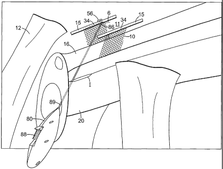
[00132] Referring now to figure 4, a preferred embodiment of surgical
procedure for
treating female incontinence is disclosed according to an aspect of the
present invention.
Initially, the patient is placed under local, spinal or general anesthesia. A
small transverse
incision I is made in the anterior vaginal wall 20 of a female patient
followed by minimal
transurethral dissection.
[00133] An implant 10 is selected that is sized and shaped be implanted in the
retropubic space. Notably, the implant 10 may be provided in a kit. The
implant 10 may
optionally be trimmed by the surgeon to address the particular needs of the
surgical
procedure (e.g. avoidance of scar tissue, or treating an individual with small
anatomic
features).
[00134] The patient is placed in a position suitable for a urological surgical
procedure.
Figure 4 simulates the position of anatomical features with a patient in the
lithotomy
position.
-25-
CA 02441982 2003-09-24
WO 02/078571 PCT/US02/09455
[00135] Figure 4 schematically illustrates one embodiment of the step of
placing the
implant 10 in the retropubic space 11 and in a therapeutically effective
position relative to
the patient's urethra 16 without extending the implant to the patient's rectus
fascia (e.g. 17
in Fig. 1), without suturing the implant 10 to the patient's Cooper's
ligament, and without
using bone anchors to anchor the implant to the pubic bone 12. In this
embodiment,
inserter 80 is used to place a deployable member (e.g. 56) in endopelvic
fascia (shown
schematically as 15) of the patient.
[00136] Figure 4 shows a preferred embodiment where the step of providing an
implant includes the step of providing an implant with first and second ends
34, and the
step of implanting the implant includes the step of anchoring the first end of
the implant
with endopelvic fascia 15 on one side of the patient's urethra 16 and
anchoring the second
end 34 of the implant 10 with endopelvic fascia 15 on the other side of the
patient's
urethra 16. Four leaf clover shaped anchors (e.g. 56) are shown, but other
fasteners could
be used to anchor the implant in the retropubic space according to the present
invention.
[00137] The implant is preferably placed mid-urethra as shown in Figure 4.
However,
it should be noted that other final locations are within the scope of the
present invention,
such as, placement of the implant 10 at the bladder neck.
[00138] Figure 4 shows an inserter 80 being used to pass a deployable
anchoring
member 56 with an associated suture 6 through endopelvic fascia 15. After the
anchoring
member 56 has substantially passed through the endopelvic fascia 15 (e.g. when
stop 86
engages endopelvic fascia 15), the button 88 may be advanced to deploy the
anchoring
member 56.
[00139] The implant 10 is secured by tying the suture 6 to the implant 10.
Figure 8
shows a suture 6' that is anchored in a step of member 56 and used to secure
one end of
the implant 10 to the anchor 56.
[00140] The steps described above are repeated as needed for a second side of
the
implant 10 on the other side of the urethra 16. As depicted, the step of
implanting the
implant 10 preferably includes the step of extending the implant 10 from the
endopelvic
-26-
CA 02441982 2003-09-24
WO 02/078571 PCT/US02/09455
fascia on one side of the patient's urethra 16, underneath approximately the
mid-urethra,
and to the endopelvic fascia 15 on the other side of the patient's urethra 16.
[00141] Other methods are also contemplated herein. For example, rather than
using a
mechanical fastener to anchor the implant 10, a tissue adhesive may be used to
place the
implant in the retropubic space. This embodiment offers the advantage that not
even the
endopelvic fascia 15 is pierced. Also, while the method preferably includes
the step of
creating a vaginal incision I, other surgical approaches are within the scope
of the present
invention including, for example, transurethral, laparoscopic and
transperineal approaches
(e.g. for treating male incontinence).
[00142] Although the invention has been described in terms of particular
embodiments and applications, one of ordinary skill in the art, in light of
this teaching, can
generate additional embodiments and modifications without departing from the
spirit of or
exceeding the scope of the invention. Accordingly, it is to be understood that
the drawings
and descriptions herein are proffered by way of example to facilitate
comprehension of the
invention and should not be construed to limit the scope thereof.
-27-