Note: Descriptions are shown in the official language in which they were submitted.
CA 02446982 2003-11-10
WO 02/090443 PCT/SE02/00860
1
TITLE
SYNTHESIS AND EVALUATION OF NEW CYANINE DYES AS MINOR GROOVE OF
[POLY(dA-dT)]Z BINDERS
DESCRIPTION
Technical field
The present invention relates to new cyanine dyes particularly suited for use
in DNA
sequencing in particular minor groove [poly(dA-dT)]Z binders.
Background of the invention
The introduction of combinatorial chemistry, the sequencing of the human
genome and
miniaturisation, e.g. lab-on a chip, nanochemistry, has enabled the creation
of vast
libraries of "new chemical entities", millions of which must be quickly tested
by high-
throughput screening to identify active sites and drugs. Drugs that bind
reversible to
DNA in the minor groove of DNA has been synthesised with the aim to generate
new
lead compounds with anticancer and antiviral properties. Formerly, radioactive
probes
have been used to study the effect of drug-DNA interactions but during the
last years
they have started to be replaced by different fluorogenic assays. Today, drug-
DNA
interactions are mainly studied with absorbance spectroscopy, fluorescence dye
displacements assays, footprinting or NMR. Since the numbers of fluorescence
markers
are limited to a few there is a challenge to discover new fluorescent dyes
that
circumvent the limitations on those that now are available. New fluorogenic
compounds
that bind in the minor groove can either work in dye displacement assays or
give insight
in how substituents may work as minor groove recognition elements.
Fluorogenic compounds can provide tremendous sensitivity due to large quantum
emission yield upon excitation. A limitation is that there are not many
fluorophores that
give a high increase in fluorescence upon hybridisation or reaction with
targets.
Asymmetric cyanine dyes have achieved much interest due to their excellent
nucleic
acid staining properties. Upon binding to nucleic acids such dyes usually
exhibit a large
enhancement in fluorescence intensity' and are widely used as fluorescent
markers for
DNA in various contexts.2"4 The interaction between double stranded DNA and
the
asymmetric cyanine dyes TO and YO (Figure 1) have been investigated
spectroscopically in several studies and were found to bind by
intercalations"' in a non-
specific fashion.8 They also bind strongly to single stranded DNA with a large
accompanying increase in fluorescence intensity.9 This makes the dyes less
useful in
CA 02446982 2003-11-10
WO 02/090443 PCT/SE02/00860
2
studies where only a signal from double stranded DNA is desirable. There are,
however,
fluorescent ligands binding in the minor groove instead of by intercalation
that bind
selectively to double and not to single stranded DNA, e.g. DAPI10 and Hoechst-
derivatives.il In contrast to most cyanine dyes these ligands have a DNA
sequence
selectivity, preferably for A/T-rich segments.12 Furthermore, compared to the
intercalating dyes they exert a smaller perturbation of the DNA-duplex upon
binding.
This is valuable in studies where its critical that the DNA is not stretched
out, for
example in certain fluorescence microscopy studies.23,1 4 Minor groove binders
do not,
however, exhibit an equally dramatic increase in fluorescence as the
asymmetric
cyanine dyes upon binding to DNA, who can display more than a thousand-fold
increase.' For BO (Figure 1) a 400-fold enhancement in fluorescence has been
reported,15 whereas Hoechst and DAPI exhibit a-95-fold16 and a-20-foid17
increase,
respectively. Another advantage of the asymmetric cyanine dyes as labels for
DNA is
their relatively long absorption maxima, which reduces problems of background
absorption from biological material. The absorption maxima of the dyes in
figure 1 when
bound to DNA varies from roughly 435 nm to 510 nm6,9,17 compared to around 350
nm
for Hoechst 33258 (Hoechst) and DAPI.17 A dye that combines the features of
the minor
groove binding ligands and the photophysical properties of the ordinary
asymmetric
cyanine dyes would thus be of great value for detection and studies of DNA.
As an initial effort towards such a dye we designed the asymmetric cyanine dye
BEBO
(Schemel). This dye has the same cyanine chromophore as the intercalating dye
BO
but the structure is extended with a benzothiazole substituent in the 6-
position. The
positioning of the benzothiazole moiety gives BEBO a crescent-shape similar to
that of
other minor groove binders, e.g. Hoechst. The short synthetic route to BEBO
starting
from the commercially available benzothiazole substituted aniline 1 motivated
the
choice of the benzothiazole group (Scheme 1). In addition, symmetrical cyanine
dyes
containing two benzothiazole moieties have been suggested to bind in the minor
groove
either as monomers18 or as dimers.19 Herein we describe the synthesis and DNA
binding
studies of BEBO and the analogous dye BO.
The fluorophores that are most frequently used today are Fluorescein, BODIPY,
DAPI,
Hoechst and asymmetric cyanine dyes such as TO, YO and TOTO.
CA 02446982 2003-11-10
WO 02/090443 PCT/SE02/00860
3
Fluorescein and BODIPY are the most common fluorescent reporter groups for
covalent
labeling of proteins whereas DAPI, Hoechst and Cyanine dyes are the most
common
fluorophores for detection of nucleic acid.
NHZ N+~ ~- OH O/N+/
HZ+N N NH2 H~N NN NH2+ N N
DAPI A HOECHST DERIVATE A CYANINE DYE
DAPI (abs. max 400 nm) and Hoechst (abs. max 350 nm) bind in the minor groove
and
are used as base-specific fluorescent probes for DNA with a 20-fold increase
in
fluorescence upon binding to DNA. In contrast, asymmetric cyanine dyes has
shown up
to a 18.000-fold increase in fluorescence upon binding to DNA. They also have
the
advantage that the absorption and emission can be easily varied by changing
the
number of double bounds between the aromatic rings. However, a major drawback
with
asymmetric cyanine dyes is that they usually bind in a non-specific fashion
towards
DNA-sequences. ( i.e. intercalate or form ion-pair complexes to DNA which may
result in
complex or weak fluorescence signal.) Therefore a cyanine dye that bind in a
more
organised way may have high fluorescence increase upon hybridisation and thus,
be a
more sensitive fluorophores.
The minor groove is a convenient site for attack since it is normally
unoccupied by
cellular compounds such as proteins. It is also a perfect complement to
concave cationic
dyes due to the negative electrostatic potential and the convex floor of the
minor
groove. Certain minor groove binders stabilise DNA duplexes and can work as
regulators
of DNA-protein function. As a consequence, the development of sequence-
specific minor
groove binders may generate new compounds with anticancer and/or antiviral
properties and thus, serve as an alternative and complementary approach to the
antisense oligonucleotide strategy. Furthermore, the minor groove binders
stabilising
effect upon DNA duplexes can be used in probes, consisting of a minor-groove
ligand-
nucleic acid conjugate, to increase the melting temperatures (Tm) of probe-DNA
duplexes. An increase of the Tm of probes will allow a more flexible assay
design since
the oligo in the probe can be shorter and still have an optimal Tm.
Sequence selective minor groove binders also has mismatch discrimination.
Nucleic acid
probes with minor groove binders as reporter group should have an increased
difference
between the Tm of match and single-base mismatch nucleic acids than the
corresponding probe with an intercalator as reporter group. Thereby increasing
the
discriminatory power of hybridisation assays.
CA 02446982 2003-11-10
WO 02/090443 PCT/SE02/00860
4
A useful feature of minor groove binders are a preference for double stranded
DNA
compared to single stranded DNA whereas intercalators usually has no
preference for
single or double stranded DNA. This feature results in that minor groove
binder probes
will have lower background fluorescence than probes with an intercalator and
as a
consequence, a greater signal-to-noise ratio upon hybridisation. Furthermore,
dyes
specific for duplex-DNA can be used for quantification of DNA in mixtures
contaminated
by RNA or single stranded DNA.
Summary of the present invention
One challenge is to develop numbers of highly sensitive fluorescent dyes with
different
well-separated emission spectra that bind in a precise way and thus allowing
multidetection of a serie of targets with high sensitivity. As mentioned,
cyanine dyes can
have up to a 18.000-fold increase in fluorescence upon hybridisation which is
almost
1000 times higher than the minor groove binders that are used today. Also the
absorption and emission are easily tuned by varying the conjugated system in
cyanine
dyes. Thus, a cyanine dye substituted so that binding in the minor groove is
govern but
with the extraordinary fluorescence properties of the known cyanine dyes
retained
seems to be a highly interesting target compound.
S NH2
~ Br2, ICSCN SI 1) Mel
~ ~ ~ NH2 -N
DMF N 2) NaOH
~ 1*
SNH
~ S Heat S ` S
/ N+ Vauum N \~ N
N I
Inspired by the concave structure of minor groove binders and the new findings
that a
benzothiazole and relatedly structured groups may govern minor groove
recognition it
has been designed and synthesised an asymmetric cyanine dye substituted with
an
extra benzothiazole group in accordance with above.
The interaction between this new dye and DNA were studied with various
spectroscopic
methods such as flow-LD and CD.
These two techniques can provide information on whether a drug is binding to
DNA by
intercalation or groove binding. Weak induction of CD is usually associated
with
CA 02446982 2003-11-10
WO 02/090443 PCT/SE02/00860
intercalating whereas asymmetric induction is due to groove binding. Groove
binding
give a strong signal in Flow-LD.
In the presence of calf thymus DNA a weak positive signai was observed in the
flow LD-
5 spectra. This can be due to heterogeneous binding with a mixture of
intercalated and
groove binding dye. On the other hand, in the presence of poly [(dA-dT)]Z a
clear
positive LD is shown providing a strong indication of minor groove binding.
For poly
[(dG-dC)]Z only a weak negative signal was observed indicating a heterogeneous
binding or a low abundance of intercalated dye.
Detailed description of the present invention
It has now turned out that the following compounds solve the above discussed
problems
and the invention is mainly characterized by new compounds according to the
following:
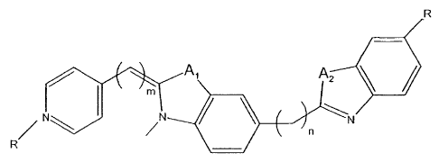
A cyanine dye binding in the groove of DNA, selected from the group of
AA
A2
or
Al
r~ \ I e~t N
s+ n
~
~ A2
N
wherein Al and A2 are each independently 0, S, or N, and R is H or a
carbohydrate that
may contain a hetero atom, and m is 0 to 5, and n is 0 to 5.
In one embodiment the cyanine dye has R being methyl, or ethyl, and m being 1
and n
being 0.
CA 02446982 2003-11-10
WO 02/090443 PCT/SE02/00860
6
In one embodiment the cyanine dye has R being methyl, or ethyl, and m being 1
and n
being 0 and Al and A2 being S.
In one embodiment the cyanine dye has R being methyl, or ethyl, and m being 1
and n
being 0 and Al and A2 being O.
In one embodiment cyanine dye has R being methyl, or ethyl, and m being 1 and
n
being 0 and Al being S and A2 being O.
In one embodiment the cyanine dye has the pyridine/quinoline ring in 2-
position.
One aspect of the present invention provides a probe for nucleic acid
hybridization
comprising a cyanine dye according to the above.
A further aspect of the present invention provides a method for carrying out a
real-time
PCR-reaction of a DNA template, wherein a fluorescent dye increasing its
fluorescent
reaction when it is looked in a minor groove position in a double stranded DNA
is used,
whereby the dye comprises at least 2 aromatic ring systems both comprising at
least
one nitrogen atom, which rings are linked by a alkine goup having up to four
carbon
atoms to form a conjugated bond, and the dye further comprises at least a
third
aromatic system linked thereto via a bond having a significant double string
character,
such as a single bond or a ethin bond, to provide a stiff conjugated system.
In one embodiment of the method the dye is an asymmetric cyanine dye.
In one embodiment of the method, one of the cyanine residues contains S and/or
O.
In one embodiment of the method the dye compound is crescent shaped.
In one embodiment the dye is a derivative according to the general formulas
given
above.
Clearly the new dye binds differently to A-T rich and G-C rich regions.
Results from CD-
measurements gave further support for groove binding of this new dye.
For poly GC almost no signal is seen which is consistent with intercalative or
external
binding, whereas for poly AT a very strong asymmetric induction is seen.
CA 02446982 2003-11-10
WO 02/090443 PCT/SE02/00860
7
It binds to the minor groove of A-T rich regions and thus it stabilises A-T
bonds more
than G-C bonds in a DNA duplex. Therefore, if a probe is designed so that an A-
T rich
region is placed under the minor groove binder it can be used in probes to
improve
mismatch discrimination.
Interestingly, our results further accentuate the preliminary reports in the
literature that
the benzothiazole group has utility as a minor groove recognition element. If
so, this is
an important finding, since its opens possibilities for design of new drugs
binding in the
minor groove.
Our first results show that it is possible to design and prepare asymmetric
cyanine dyes
that work as minor groove binders.
Further, possibilities of broadening the present scope are: Since there is a
well working
synthetic route for the substituted cyanine dye the first step is the nitrogen
in ortho
position, 2-BEBO, from the methine substituent.
g N+ S - ~ \ S NI
N N N ~ J N
BEBO 2-BEBO
/ N}
S S
N~ S_\ N~ S \N
/ N N
BETO 2-BETO
Along with the synthesis of the two quinolinium derivates, BETO and 2-BETO,
the
synthesis of the benzoxazole and benzimidazole derivates can be done.
CA 02446982 2003-11-10
WO 02/090443 PCT/SE02/00860
8
The synthesis of these new benzoxazole and benzimidazole substituted dyes will
follow a
slightly different synthetic route.
Br ~ I ~ Br \ S I N SnR3 X
NHP N / :-
/ ~i ;/
x S N S-
---- N \ I /- N
-
~ I 0
The Stille coupling of similar compounds and the synthesis of the benzooxazole-
and
benzimidazole-stannanes can be found in the literature. T he last step, the
condensation
of compound 1 with the pyridinium or quinolinium salt are routinely used in
the
synthesis of asymmetric cyanine dyes.
Synthesis
Typically asymmetric cyanine dyes are prepared by condensation of two
quaternary
heterocyclic salts with a thiomethyl group acting as leaving group on one of
the salts.
However, the use of an alternative condensation method developed by
Deligeorgiev et
al 20 furnished a synthetic route to BEBO of only four steps starting from the
commercially available 4-substituted aniiine 1(Scheme 1). Thiocyanation of the
aniline
1 with potassium thiocyanate and bromine in DMF afforded the 2-
aminobenzothiazole 2
in a 40 % yield.21,22 Methylation of 2 by iodomethane and subsequent
deprotonation
proceeded in a total 77 % yield to produce the 2-imino-3-methyl-
benzothiazoline 3. The
dye BEBO was prepared in 24 % by simply melting the benzothiazoline 3 together
with
the pyridinium salt 4 at 160 C under vacuum.20
To enable comparative DNA binding studies the presumed intercalating dye BO (1-
methyl-4-[(3-methyl-2(3H)-benzothiazolylidene)methyl]-pyridinium iodide) was
synthesised according to the classical method using a modified procedure by
Zhou et al
(Scheme 2).23 The dye was afforded in 46 % yield by condensation of the
pyridinium
salt 4 and the benzothiazolium salt 5 in dichloromethane using triethyl amine
as base.
Linear dichroism measurements
To study the effect induced by the benzothiazole substituent in BEBO on its
interaction
with DNA, binding studies of the analogous dye BO were also performed as a
comparison. Figure 2 shows the flow linear dichroism (LD) spectra of BEBO and
BO with
different DNA. LD is defined as the difference in absorption of light
polarized parallel and
CA 02446982 2003-11-10
WO 02/090443 PCT/SE02/00860
9
perpendicular to the macroscopic axis of orientation. The LD-spectra of
oriented DNA-
ligand compiexes may be analysed in terms of angles that the electronic
transition
moments of the ligands make with the DNA-helix axis to provide information
about
binding geometries.24 The orientation of the DNA complexes was achieved using
a flow
Couette cell with outer rotating cylinder. For BEBO in presence of [poly (dA-
dT)]Z (poly-
AT) a clear positive LD is shown providing a strong indication of minor groove
binding
(Figure 2). From the reduced LD, obtained through division of the LD by the
isotropic
absorption, the angle between the long wavelength transition moment of BEBO
and the
DNA-helix was calculated to be 44 . This is very similar to the angle for
known minor
groove binders, e.g., DAPI.ZS The major transition moment of BEBO can be
expected to
be polarized roughly along the line connecting the pyridine with the closest
benzothiazole ring.26 The weaker positive signal shown for BEBO in presence of
calf
thymus DNA (ctDNA) is possibly due to binding in the minor groove with an
angle close
to 54 , as suggested in earlier studies of symmetrical cyanine dyes.13
However, the
binding-angle to poly-AT of 44 in addition with CD-titration data (see below)
proposes a
more complicated binding to ctDNA with a mixture of binding modes resulting in
an
average low LD signal. Although Hoechst and DAPI have a preference for minor
groove
binding to AT-rich regions it has been suggested that they bind to GC
sequences by a
non-classical intercalation process.Z',28 This model seems to be applicable
here also,
since the reduced LD spectrum of BEBO with [poly(dG-dC)]2 (poly-GC) show a
negative
value of the same amplitude as for the DNA bases indicating intercalation
(data not
shown).
In contrast to the binding of BEBO, LD measurements indicate that BO binds by
intercalation to all three different polynucleotides studied: ctDNA (Figure
2), poly-AT
and poly-GC (Figure 3). The change in binding mode induced by the
benzothiazole
extension of the BO structure is particularly apparent in the case of poly-AT.
Circular dichroism measurements
The strong induced positive CD for BEBO in presence of poly-AT (Figure 4a)
gives
further strong support for binding in the minor groove.Z9 Figure 4a shows the
titration of
poly-AT into BEBO with binding ratios R, defined as the total number of dye
molecules
per base, varying from 0.025 to 0.1. The larger CD amplitude of BEBO at the
highest
binding ratio is rationalized by a contribution of exciton coupling
interactions between
closely bound chromophores. This is illustrated by subtracting the B spectrum
(R =
0.05) from the C spectrum (R = 0.1) in figure 4a to produce a spectrum typical
of
exciton coupling (D, Figure 4a).
CA 02446982 2003-11-10
WO 02/090443 PCT/SE02/00860
In presence of ctDNA the induced CD is smaller but still, intercalation or
external
stacking of the dye would not give rise to this large amplitude. Thus, there
must be a
significant amount of dye residing in the minor groove. The titration of ctDNA
into BEBO
with binding ratios R varying from 0.0125 to 0.10 is shown in figure 4b. As
with the
5 binding of BEBO to poly-AT, there is a feature of exciton coupling
interactions between
closely spaced ligands at higher binding ratios. At lower binding ratio the
signal is
similar to that of the corresponding poly-AT spectra, albeit with smaller
amplitude.
The binding of BEBO to poly-GC give rise to only a very small induced CD
(Figure 5a),
10 which supports an intercalative binding mode to GC-regions. This might
partly account
for the lower CD obtained upon binding to ctDNA. However, one must bear in
mind that
ctDNA is more complex than just a mixture of alternating GC- and AT-segments.
The
amplitude of the CD spectra in presence of ctDNA is about one fourth of the
poly-AT
spectra. If the binding to ctDNA is a mixture of groove binding to AT- regions
and
intercalation to GC-regions then 75 percent would be bound in an intercalative
fashion.
This does not hold since the LD should be significantly more negative in that
case.
Hence, a substantial amount of dye must be bound in a non-intercalative
fashion to
ctDNA at sites affording a lower induced CD than when bound to alternating AT.
The CD signal for BO in presence of ctDNA was only weakly negative (data not
shown)
and this further illustrates the different binding mode of BEBO compared to
BO.
Polynucleotide binding preferences
The extensive difference in amplitude of the CD signal for BEBO in presence of
poly-GC
and poly-AT allowed a simple experiment to investigate a possible AT
preference. When
poly-AT was added to a sample of BEBO in presence of poly-GC (R = 0.05) the CD
signal increased drastically showing a considerable preference for poly-AT
(Figure 5a).
These spectra were consistent with the CD spectra of BEBO in presence of poly-
AT
without poly-GC (Figure 4a) with only slightly lower amplitudes of the
signals.
A similar experiment was performed to compare the binding affinities of BEBO
to poly-
AT and ctDNA. Again poly-AT was added to a sample of BEBO now in presence of
ctDNA. There was an increase in CD signal upon addition of poly-AT but not as
large as
when the sample initially contained poly-GC (Figure 5b). Hence, there is still
a
reasonable amount of dye bound to ctDNA at these ratios showing that there
must be
other binding sites than alternating AT-regions in ctDNA that attract BEBO
significantly.
Fluorescence and absorbance measurements
CA 02446982 2003-11-10
WO 02/090443 PCT/SE02/00860
11
The absorption and fluorescence properties of BEBO with different nucleic
acids are
summarised in table 1. In analogy with other asymmetric cyanine dyes BEBO has
a
large increase in fluorescence upon binding to DNA. The clear minor groove
binding of
BEBO to poly-AT affords a 180-fold enhancement in fluorescence intensity,
whereas the
increases upon binding to ctDNA and poly-GC are somewhat larger. In buffer
solution,
the free dye has its emission peak at 542 nm compared to 492 nm for the bound
dye.
Using ethanol instead of aqueous buffer as solvent the free dye emission was
shifted to
492 nm, and the fluorescence intensity was roughly ten times lower. Recently
aggregation of TO in presence and absence of DNA was studied by absorption and
fluorescence spectroscopy and similar manifestations caused by aggregate
formation
was seen.30 Thus in buffer solution dimers or higher aggregates with longer
emission
maximum are probably formed due to the hydrophobic nature of the dye.
The shape of the absorption spectra of free BEBO in water further suggests the
presence of dimers or aggregates (Figure 6). Absorption measurements of BEBO
in
different methanol-water mixtures showed a substantial increase and a red
shift in
absorption with increasing amount of methanol (Figure 6b). The dye molecules
are
presumably present as monomers in pure methanol. The absorption spectrum of
free
BEBO in methanol and the spectrum of BEBO completely bound to DNA have a very
similar shape, which indicates that the dye is bound as monomers at low
binding ratios.
In summary, we find that the structural modifications of BO have induced a
shift in
binding mode from intercalation towards minor groove binding. Our results
further imply
the potential of the benzothiazole group as a minor groove recognition moiety.
The dye
could be synthesised in four steps only from the commercially available
aniline 1. The
binding of BEBO to poly-AT is clearly in the minor groove as deduced from the
CD- and
LD-spectra. Similarly to that of DAPI and Hoechst, the binding of BEBO to poly-
GC is
dominated by intercalation. With the random sequence ctDNA on the other hand,
BEBO
seems to interact heterogeneously. However, intercalation to GC-segments and
minor
groove binding to AT-regions cannot be the only explanation to the LD- and CD-
results
obtained with ctDNA. There must be other preferred binding sites in ctDNA for
BEBO,
which induce a lower CD than poly-AT. The relatively large amplitude of the CD
signal
show, however, that there is a significant contribution of minor groove
binding of BEBO
to ctDNA. Consistent with other minor groove binders BEBO has a distinct
preference
for poly-AT compared to poly-GC. The fluorescence increase upon binding to the
minor
groove of poly-AT is larger than for Hoechst and DAPI. The binding properties
of BEBO,
in particular its strict minor groove binding to poly-AT, give promise for the
development of a new class of asymmetric cyanine dyes with a strong preference
for
CA 02446982 2003-11-10
WO 02/090443 PCT/SE02/00860
12
minor groove binding and a large increase in fluorescence upon binding.
Synthesis and
studies of analogous dyes are underway and will be reported in due time.
Experimental
Example
Preparation according to the reaction scheme
The dye 1 was prepared in four steps starting from the commercially available
aniline 1.
Thiocyanation of the 4-substituted aniline 1 with potassium thiocyanate and
bromine in
DMF afforded the 2-aminobenzothiazole 2 in a 40 % yield. Methylation and
deprotonation of compound 2 proceeded in a total 70 % yield to produce the 2-
imino-3-
methylbenzothiazoline 3. The dye 5 was prepared in 20 % by melting compound 3
together with the pyridinium salt 4 at 160 C under vacuum.
Synthesis
2-(Tii-n-butylstannyl)-benzothiuole (1) and 2-(Tri-n-butylstannyl)-benzoxuole
(2) was
prepared by treating benzothiazole and benzoxazole, respectively with n-BuLi
at -78 C,
followed by addition of tii-n-butyltin chloride.
1) n-BuLi
:91 x 2) SnCi(n-Bu)3 X
> -78 C ~. ' N~Sn(n-Bu)s
(1),XS
(2), X = 0
Scheme : Preparation ofthe organostannanes.
6-Bromo-2-methyl-benzothiazole (5)
2,4-Dibromo-wiline was treated with acctic anhydride in pyridine to give the
acetanilide
(3). Reaction of (3) with phosphorus pentasulfide in refluxing benzene
replaced the
carbonyl oxygen by a sulphur atom to give the thioacetanilide (4). Separation
of (4)
from (3) is readily achieved by extraction with aqueous NaOH. This is possible
due to
the fact that the sulphur atom is larger and more polarizable than the oxygen
and
thereby able to form the water-soluble thioacetwilide anion (4'). This ability
to form (4')
is also utilized in the final step, in which (4) is treated with sodium
metboxide, and
elimination of the bromine in 2position leads to ring closure, giving the
product (5).
Upon removing the NMP by bulb-to-bulb distillation, it was discovered that
'(5) is easily
purified by sublimation.
CA 02446982 2003-11-10
WO 02/090443 PCT/SE02/00860
13
O s
N142 U G HNK HN K /'`. ~'`
I Nr l
=
Br-~~~' Br P2S5 Br MeONa N S
I / Pyridine BenzeneT NMP Br -Bif, 30
8r Br Br
Br
1 Br
(3) (4) (41) (6)
Scheme: Preparation of (5) from 2,4-dibromo-aniline.
S NH2
N~ Br2, KSCN ~ S~ 1) Mel
NH2 DMF N 2) NaOH
~ N+
S SNH Heat S S
~
~, N+ Vacuum N \/ N
N I
CA 02446982 2003-11-10
WO 02/090443 PCT/SE02/00860
14
6-Iodo-2-methyl-benzothiazole (9)
The synthesis of (5) and its iodo analogue (9) are very similar. However, in
this case the
dihalogenated acetanilide (7) is achieved by acylation of 4-iodo-aniline to
give (6), followed
by bromination. In this brornination step, some of the iodine in the 4-
position was
substituted by bromine. Attempts to separate the formed 2,4-dibromo-
acetanilide from (7)
were fruitless, which resulted in a product mixture of (9) and (5) in a 3:1
molar ratio. In
spite of this, the mixture was used in following Stille-coupling reactions.
O
NH2 O 0 HNK HNK HN N
j~~O) jj
' ` ` gr2 F3r p285 T ( .` I3r MeONa ` s
Pyrldine 70~1o HAc(aq) Benzene NMP
~ E I i I
(6)
Scheme: Preparation of (9) from 4-iodoaniline.
6-Bromo-2-methylthio-benzothiazole (10)
Although not being used in my subsequent reactions, it should be mentioned
that yet
another halogenated electrophile, 6-Bromo-2-methylthio-benzothiazole (10), was
prepared.
The synthesis of (10) is, as seen in the following scheme, quite
uncomplicated.
2-Methylthio-benzothiazole is simply brominated in acetic acid with FeCl3 as
catalyst.
S SMe g sMe
j Br2, FeCl3 _ Y
\ / N HAc Br ~ ~ N
(y~)
Scheme: Bromination in 6-position of 2-metylthio-benzthiazole
Stille-couplings
To study and optimise the palladium catalysed cross-coupling reaction, a
number of
experiments were carried out with different starting materials and two
different neutral
ligands on the catalyst. However, the procedure describing the synthesis of
(11) and (13)
in the experimental section was followed in all Stille-couplings. CuI is used
for its
co-catalytic effect on the coupling. Table 1 summarises the Stille-experiments
carried out
during this diploma work.
CA 02446982 2003-11-10
WO 02/090443 PCT/SE02/00860
TABLE 1
Entry Organostannane Arylhalide Catalyst Product Yield
Nz~
~. L.Qw
(1) Br \~ Pd(PPh3)4 I~. S
2 (I) ~ ~ ~ Pd(PPh3)4 Nk N \ ! 72%
3 (1) J\ f Pd(AsPh3)d CJQ-~ S 33%
4 (1) Br , / (~H Pd(PPh3)4 I / hIH 0%
- SII ~ S $II
5 (~) Br \ / N Pd(PPh3)4 ~ ~. / \ ! ~ 0%
N
(5) (11)
s
_ N
6 {I} Br \ ! N Pd(AsPh3)4 ~ .~ / N 0%
(5) ('13)
S~ ~
7 (1) 1- sN Pd(AsPh3)4 ~ N\! N 0%
\I .i
(~) (11)
~,,`
S ~
8 (1) 1 N Pd(PPh3)4 ~.- N N 98%
(s) ~~~}
s.~
.~~ 95%
0/ 9 (2) 1 Pd(PPh3)4
N
\ / N
(9) (13)
Table 1.. A summary of the Stitle-reactions performed.
CA 02446982 2003-11-10
WO 02/090443 PCT/SE02/00860
16
In entry 1, pure product could not be isolated despite flash chromatography
(chloroform
on silica). However, a small amount of product was confirmed by mass
spectrometry.
The superior performance of iodine in comparison with bromine on the
electrophiles has
been previously reported", and was therefore expected. This property is given
by
iodine's greater ability to act as a leaving group. Another reason for the
failed
experiments in entries 4-7 might be that the nitrogen in 4-position to bromine
donates
its free electron pair into the arylring, thereby deactivating the
electrophile. The reason
for trying to use brominated electrophiles anyway was their more facile
synthesis.
Although the arylhalide in entries 8-9 in reality was a 3:1 (molar-) mixture
of (9) and
(5), the yields in table 1 we calculated with respect only to the amount of
(9). This is
due to the total reluctance of the brominated clectrophiles in entries 4-7 to
react.
Using triphenylarsine as the palladium-ligand has been reported to show up to
a
1100-fold increase in reaction rates, compared to triphenylphosphine.2" '1
Surprisingly
though, triphenylarsine was less effective than triphenylphosphine in the
experiments
performed. This may, ironically enough, depend on triphenylarsine's
superiority as
ligand, which makes Pd(O) more liable to oxidize and the catalyst far more air-
sensitive
than the one with triphenylphosphirle. Hereby, a small contamination of air-
oxygen in
the reaction vessel nught substantially decrease the catalytic effect of
tripherrylarsine-coordinated palladium, whereas the catalyst with phosphine-
ligand is
less affected.
BETO & BOXTO
The two new asymmetric cyanine dyes BETO and BOXTO were prepared by the
reaction
paths shown in schemes 8 and 9.
OTs
MeOTs ccjX'
ors DcNI
(11) ~ (12) (BE74)
Scheme S. The two final steps in the synthesis of BETO.
CA 02446982 2003-11-10
WO 02/090443 PCT/SE02/00860
17
/ I \ OTs
\ " \ /
S~ + pTs Q;Iw~= \ MeOTs N
I .~ N ~ ~
~qgy (14) (77s OCM ~ /
(B4xTp)
Scheme 9. The two final steps in the synthesis of BOXTO.
By treating (11) and (13) with an excess of melted methyl tosylate, the
metylated salts
(12) and (14) were formed in 70% and 56% yields respectively. These salts were
allowed to react with 1-methyl-quinolinium tosylate in dichloromethane to
produce the
desired dyes. The yields in the last step were 27% and 30% respectively.
Column chromatography was performed using aluminium oxide (activated, neutral,
approx. 150 mesh) deactivated by addition of water to Brockman grade III.
Melting
points were determined on a Mettler FP82HT hot-stage microscope. 'H (400 MHz)
and
13C (100.6 MHz) NMR spectra were recorded at rt using a Varian UNITY-400 NMR
spectrometer. Chemical shifts are in ppm, relative to solvent peaks for DMSO
(S 2.50 for
'H and Sc 39.51 for 13C NMR); J values are given in Hz. High resolution mass
spectra
were recorded using a VG ZabSpec instrument. UV-vis spectra were measured on a
Varian Cary4 spectrophotometer. Fluorescence spectra were recorded using a
SPEX
fluorolog ti2 spectrofluorimeter. The LD and CD spectra were recorded on a
JASCO-720
spectropolarimeter. The orientation of the DNA complexes was achieved using a
flow
Couette cell with outer rotating cylinder. All spectroscopic measurements were
performed at 25 C in 25 mM sodium phosphate buffer (pH 7.0). Aqueous
solutions of
BEBO and BO were typically obtained from 2 mM stock solutions in DMSO. [Poly
(dA-
dT)]2 and [poly (dG-dC)]Z ) were purchased as solutions in buffer from
Pharmacia. Calf
thymus DNA was purchased from Fluka. Commercial reagents were purchased from
Sigma-Aldrich and used without further purification. The pyridinium salt 4 and
the
benzothiazolium salt 5 were prepared as previously reported.23
2-amino-6-(6-methyl-benzothiazol-2-yl)-benzothiazole (2)
2-(4-aminophenyl)-6-methyl-benzothiazole 1 (4.0 g, 16.6 mmol) and KSCN (2.6 g,
26.7
mmol) were dissolved in DMF (20 ml) and cooled in an ice-bath. Br2 (0.9 mi, 17
mmol)
in DMF (15 ml) was added dropwise under 3 h. The mixture was stirred for
another 20
h. Water was added and the precipitate formed was collected by filtration and
dried. The
crude product was triturated on the sinter with several portions of boiling
dichloromethane to afford 2 as a light green-yellow solid (1.97 g, 40 %). Mp
250-251
CA 02446982 2003-11-10
WO 02/090443 PCT/SE02/00860
18
C; 1H NMR (DMSO) S 2.45 (3H, s, Ar-CH3), 7.34 (1H, d, J=8.4, ArH), 7.50 (1H,
d,
J=8.4, ArH), 7.89 (1H, d, J=8.4, ArH), 7.91 (1H, s, ArH), 7.99 (1H, d, J=8.4,
ArH), 8.51
(1H, s, ArH), 8.56 (2H, s, NH2); 13C NMR (DMSO): 8 21.10, 116.7, 120.7. 121.8,
122.1,
125.6, 126.9, 128.1, 129.7, 133.3, 134.5, 135.0, 151.7, 165.9, 169.3; HR-FAB-
MS m/z
Found: 298.0521 C15H1ZN3S2 (M+H+): requires M, 298.0473.
2-amino-3-methyl-6-(6-methyl-benzothiazol-2-yl)-benzothiazolium iodide
The 2-aminobenzothiazole 2 (0.3 g, 1.0 mmol) was dissolved in DMSO (2 ml).
Methyl
iodide (0.25 ml, 2.0 mmol) was added and the mixture was stirred at 110 C for
17
hours. The mixture was cooled and poured into water. The precipitate formed
was
collected by filtration and washed with water to give the product as a yellow
solid (0.38
g, 86 %). Mp 267-269 C; 'H NMR (DMSO): S 2.47 (3H, s, Ar-CH3), 3.74 (3H, s, N-
CH3),
7.38 (1H, d, J=8.4, ArH), 7.79 (1H, d, J=8.4, ArH), 7.93 (1H, d, J=8.4, ArH),
7.95 (1H,
s, ArH), 8.22 (1H, d, J=8.4, ArH), 8.75 (1H, s, ArH), 10.19 (2H, s, NH2); 13C
NMR
(DMSO): S 21.14, 32.39, 113.9, 122.0, 122.2, 122.4, 122.7, 126.6, 128.3,
129.8,
134.7, 135.6, 140.9, 151.6, 164.7, 168.9; HR-FAB-MS m/z Found: 312.0638
C16H14N3Sa
(M+): requires M, 312.0629.
2-imino-3-methyl-6-(6-methyl-benzothiazol-2-yl)-benzothiazoline (3)
2-amino-3-methyl-6-(6-methyl-benzothiazol-2-yl)-benzothiazolium iodide (0.3 g,
0.68
mmol) was taken up in DMSO (10 ml). Water was added (20 ml) and the mixture
was
basified to pH 10 with aqueous NaOH (20%). The precipitate was collected by
filtration
and washed with water to produce 3 as a light yellow solid (0.19 g, 89%). Mp
146-148
C; iH NMR (DMSO): 2.45 (3H, s, Ar-CH3), 3.38 (3H, s, N-CH3), 7.16 (1H, d,
J=8.4,
ArH), 7.33 (1H, d, J=8.4, ArH), 7.87 (1H, s, ArH), 7.90 (1H, s; ArH), 7.93
(1H, d,
J=8.4, ArH), 8.16 (1H, s, ArH), 8.55 (1H, s, NH); 13C NMR analysis was not
possible due
to poor solubility of 4 in available deuterated solvents; HR-FAB-MS m/z Found:
312.0619 C16H14N3S2 (M+H+): requires M, 312.0629
4-[(3-methyl-6-(6-methyl-benzothiazol-2-yi)-2,3,-dihydro-(benzo-1,3-
thiazole)-2-methylidene)]-1-methyl-pyridinium iodide (BEBO)
The benzothiazoline 3 (0.1 g, 0.32 mmol) and 1,4-dimethyl-pyridinium tosylate
4 was
melted together at 160 C under vacuum for 1 hour. DMSO (5 ml) was added and
the
mixture was heated at reflux for 30 min. The mixture was added to aqueous KI
(30%)
and the precipitate formed was collected by filtration. The solid was purified
by flash
chromatography on neutral AI203 with methanol- dichloromethane (2:98) to give
BEBO
(0.04 g, 24%). Mp 280-281 C; 1H NMR (DMSO): S 2.47 (3H, s, Ar-CH3), 3.76 (3H,
s, N-
CH3), 4.02 (3H, s, N-CH3), 6.34 (1H, s, =CH-), 7.38 (1H, d, J=8.4, ArH), 7.47
(1H, d,
CA 02446982 2003-11-10
WO 02/090443 PCT/SE02/00860
19
J=6.8, PyH), 7.70 (1H, d, J=8.4, ArH) 7.93 (1H, d, J=8.4, ArH), 7.95 (1H, s,
ArH), 8.18
(1H, d, J=8.4, ArH), 8.39 (1H, d, J=6.8, PyH), 8.65 (1H, s, ArH); 13C NMR
(DMSO):
21.13, 32.99, 45.11, 90.66, 112.0, 118.8, 120.9, 121.8, 122.2, 124.6, 126.7,
127.9,
128.1, 134.5, 135.2, 142.4, 142.5, 150.1, 151.6, 156.4, 164.9; HR-FAB-MS m/z
Found:
402.1145 C23H2ON3S2 (M+): requires M, 402.1105.
2-(Tri-n-butylstannyl)-benzothiazole (1)
20 ml of freshly distilled THF was flushed for 30 min with a stream of
nitrogen after
which benzothiazole (1.0 g, 7.4 mmol) was added. After being flushed for
another 30
min, the solution was cooled to -78 C and placed under inert nitrogen
atmosphere. 0.9
equivalents of n-BuLi (2 M solution in hexane, 2.66 ml, 6.66 mmol) was added
dropwise
over a period of 30 min, during which the solution turned to deep red. The
solution was
kept at -78 C for 1 h and then tri-n-butyltin chloride (2.0 mi, 7.4 mmol) was
added
dropwise over a period of 1 h. During this addition, the solution shifted from
deep red to
brownish yellow, then to greenish blue and finally to light brown. After yet
another hour
at -78 C, the solution was allowed to reach room temperature. The THF was
removed on
a rotary evaporator and the product, a yellow oil, was isolated by
distillation in vacuo.
Yield: 2.47 g, 79%. 1H NMR (CDCI3): b 0.90 (t, J=S, 9H, Bu3Sn), 1.29 (in, 6H,
Bu3Sn),
1.35 (m, 6H, Bu3Sn), 1.63 (t, J=8, 6H, Bu3Sn), 7.37 (t, J=8, 1H, ArH), 7.46
(t, J=8, 1H,
ArH, 7.96 (d, J=8, 1H, ArH), 8.17 (D, J=8, 1H, ArH).
2-(Tri-n-butylstannyl)-benzoxazole (2)
20 ml of freshly distilled THF was flushed for 30 min with a stream of
nitrogen after
which benzoxazole (1.0 g, 8.3 mmol) was added. After being flushed for another
30
min, the solution was cooled to -78 C and placed under inert nitrogen
atmosphere. 0.9
equivalents of n-BuLi (2 M solution in hexane, 3.0 mi, 7.6 mmol) was added
dropwise
over a period of 1 h, during which the solution turned to pink. The solution
was kept at
-78 C for 30 min and then tri-n-butyltin chloride (2.3 ml, 8.3 mmol) was added
dropwise over a period of 1 h. During this addition, the solution shifted from
pink to
brown. After yet another hour at -78 C, the solution was allowed to reach room
temperature, at which it turned to deep red. The THF was removed on a rotary
evaporator and the product, an orange oil, was isolated by distillation in
vacuo. Yield:
1.37 g, 40%. 'H NMR (CDCI3): b 0.90 (t, J=7, 9H, Bu3Sn), 1.30 (m, 6H, Bu3Sn),
1.35
(m, 6H, Bu3Sn), 1.62 (t, J=7, 6H, Bu3Sn), 7.29 (t, 2H, ArH), 7.55 (d, 1H,
ArH), 7.77 (d,
1H, ArH).
CA 02446982 2003-11-10
WO 02/090443 PCT/SE02/00860
2,4-Dibromo-acetanilide (3)
A solution of 2,4-Dibromo-aniline (3.0 g, 12.0 mmol), 1.1 equivalents of
acetic
anhydride (1.35 g, 13.2 mmol) and pyridine (0.95 g, 12.0 mmol) was heated to
100 C.
After a few minutes, a precipitate had formed and pyridine (N3 ml) was added
to
5 dissolve the precipitate. The solution was kept at 100 C for two hours
after which it was
ailowed to cool to room temperature and was poured into water. The precipitate
formed
was collected by filtration and washed with water to give the product as a
white powder
in quantitative yield (3.9 g, slightly wet) 'HNMR (CDCI3): b 2.24 (a, 3H, -
CH3), 7.43 (d,
J1=8.8, 32=2, 1H, ArH) 7.69 (s, 3=2, 1H, ArH) 8.27 (d, 1=8.8, 1H, ArH).
2,4-Dibromo-thloacetanilide (4)
2,4-Dibromo-acetanilide (2.2 g, 7.51 mmol) was dissolved in 10 ml benzene and
phosphorus pentasulfide (3.34 g, 7.51 mmol) was added. The mixture was
refluxed at
80 C. After a few minutes, a gummy solid was formed at the bottom of the
flask. To
suspend the solid and make stirring possible, an additional 20 mi of benzene
was added
and the mixture was swirled vigorously. After refluxing for 5.5 h, thin layer
chromatography (TLC) on silica in chloroform suggested complete reaction and
the
heating was removed. After cooling to room temperature, the brownish slurry
was
filtered and the precipitate washed with diethyl ether. The benzene/ether
filtrate was
extracted twice with NaOH, (10%). The basic, aqueous phase was acidified to pH
N1
with cone. HCI. This gave a light brown, milky slurry. The precipitate could
not be
collected by filtration and the slurry was therefore extracted twice with
diethyl ether.
This resulted in a yellow organic phase, which was dried and evaporated to
give the
thioacetanilide as a brown, yellowish oil. Yield: 1.33 g, 57%. 'HNMR (CDCI3):
b 2.78 (S,
3H, -CH3), 7.50 (d, J1=8.8, 32=1.6, 1H, ArH) 7.90 (s, J=1.6, 1H, ArH) 8.40 (d,
3=8.8,
1H, ArH).
6-Bromo-2-methyl-benzothiazole (5)
2,4-Dibromo-thioacetanilide (1.33 g, 4.3 mmol) and 1.2 equivalents of sodium
methoxide (0.513 g, 5.2 mmol) was dissolved in 3 ml NMP After 2 h at 150 C
and
cooling to room temperature, the NMP was removed by bulb-to-bulb distillation.
The
brown remnants were purified by sublimation to give the product as white
crystals.
Yield: 707 mg, 72%. 'H NMR (CDCI3): b 2.83 (s, 3H, -CH3), 7.55 (d, J1=8.8,
JZ=1.6, 1H,
ArH) 7.80 (d, 3=8.8, 1H, ArH) 7.96 (s, 3=1.6, 1H, ArH).
4-Iodo-acetanilide (6)
4-lodo-aniline (5.0 g, 22.8 mmol) and 1.1 equivalents of acetic anhydride
(2.56 g, 25.1
mmol) was dissolved in 3 ml pyridine. After 2 h at 100 C, TLC suggested
complete
CA 02446982 2003-11-10
WO 02/090443 PCT/SE02/00860
21
reaction and the heating was removed. When the solution had reached room
temperature, it was poured into water. The precipitate formed was collected by
filtration
and washed with water to give the product as a white powder. Yield: 5.86 g,
98%. 'H
NMR (DMSO): 6 2.03 (s, 3H, -CH3) 7.41 (d, 3=8.8, 2H, ArH) 7.61 (d, 3=8.8, 2H,
ArH)
10.03 (s, 1H, NH).
2-Bromo-4-iodo-acetanilide (7)
To a solution of 4-Iodo-acetanilide (3.74 g, 14.3 mmol) in 20 ml HAcaq.,
(70%), 1.1
equivalents of bromine (2.53 g, 15.7 mmol) was added dropwise. After being
allowed to
react at 70 C for 10 min, the solution was poured into water and the formed
precipitate
was collected by filtration (3.02 g). 'H NMR showed a mixture of the desired
product
and 2,4-dibromo-acetanilide in a 9:4 molar ratio. Total yield: 3.02 g, 65%.
Yield of the
desired product: 2.18 g, 45%. 'H NMR (CDCI3,): 6 2.23 (s, 3H, -CH3), 7.60 (d,
J1=8.8,
J2=1.6, 1H, ArH) 7.85 (s, J=1.6, 1H, ArH) 8.13 (d, J=8.8, 1H, ArH). Yield of
the
by-product: 0.84 g, 20%. 'H NMR (CDCI3): Consistent with the spectrum of
2,4-dibromo-acetanilide described above.
2-Bromo-4-iodo-thloacetanilide (8)
A total amount of 2.87 g of the 2-Bromo-4-iodo-acetuilide (2.08 g, 6.1 mmol)
and 2,4-
dibromo-acetanilide (0.80 g, 2.7 mmol) mixture was dissolved in N10 ml benzene
and
phosphorus pentasulfide (3.75 g, 8.4 mmol) was added. The mixture was refluxed
at 80
C over a period of 2 h. After being allowed to cool to room temperature, the
brown
slurry was filtered and the precipitate washed with diethyl ether. The
benzene/ether
filtrate was extracted twice with NaOH, (10%) and the basic, aqueous phase was
acidified to pH N1 with conc. HCI. This gave a light brown, milky slurry,
which was
extracted twice with diethyl ether and resulted in a yellow organic phase.
This phase
was dried and evaporated to give a mixture of 2-bromo-4-iodo-thioacetanilide
and
2,4-dibromo-thioacetanilide as a brown oil. Total yield: 2,14 g, 71%. Yield of
desired
product: 1.66 g, 77% 'H NMR (CDCI3): 6 2.78 (S, 3H, -CH3), 7.68 (d, 1H, ArH)
7.96 (s,
1H, ArH) 8.29 (d, 1H, ArH). Yield of the by-product: 480 mg, 57%. 'H NMR
(CDCI3):
Consistent with the spectrum of 2,4-dibromo-thioacetanilide described above.
6-lodo-2-methyl-benzothlazole (9)
A total amount of 2.02 g of the mixture of 2-bromo-4-iodo-thioacetanilide
(1.57 g, 4.4
mmol) and 2,4-dibromo-thioacetanilide (454 mg, 1.5 mmol) was dissolved in 15
ml
NMP. Sodium methoxide (0.677 g, 6.9 mmol) was added and the mixture was
stirred at
150 C for 2.5 h. When the brown solution had cooled to room temperature, the
NMP
was removed by bulb-to-bulb distillation. The brown remnants were purified by
CA 02446982 2003-11-10
WO 02/090443 PCT/SE02/00860
22
sublimation to give a mixture of 6-iodo-2-mehyl-benzothiazole and
6-bromo-2-mehyl-benzothiazole (molar ratio 3:1) as white crystals. Total
yield: 1.33 g,
86%. Yield of desired product: 1.04 g, 86%. 'H NMR (CDCI3): b 2.82 (s, 3H, -
CH3),
7.68 (d, 3=8.4, 1H, ArH) 7.73 (d, J=8.4, 1H, ArH) 8.16 (a, 1H, ArH). Yield of
the
by-product: 289 mg, 86%. 'H NMR (CDC13) Consistent with the spectrum of 6-
bromo-2-methyl-benzothiazole described above.
6-Bromo-2-methylthio-benzothiazole (10)
To a solution of 2-methylthio-benzothiazole (1.14 g, 6.3 mmol) and bromine
(1.24 g,
7.7 mmol) in 10 nil acetic acid, a catalytic amount of FeC13 was added. After
being
refluxed at 120 C over a period of 4 h the orange reaction mixture was
allowed to cool
to room temperature and was then poured into ethyl acetate. The precipitate
formed
was collected by filtration, washed with ethyl acetate and refluxed for 1 h in
ethyl
acetate. This slurry was filtered and ethyl acetate was removed from the
filtrate by
rotary evaporation to give the product as yellow crystals (130 mg, 0.50 mmol).
The
precipitate, collected by filtration from the refluxed slurry, was Soxhlett-
extracted with
n-pentane followed by diethyl ether. Evaporation of the solvents produced
another small
amount of the desired product (70 mg, 27 mmol). Total yield: 200 mg, 0.77
mmol,
12%. 'H NMR (DMSO): 6 2.79 (s, 3H, -SCH3), 7.61 (d, J1=8.8, J2=2, 1H, ArH)
7.77 (d,
J=8.8, 1H, ArH) 8.32 (s, J=2, 1H, ArH).
2-methyl-6-(benzothiazol-2-yl)-benzothiazole (11)
216 mg of the mixture of 6-iodo-2-methyl-benzothiazole (169 mg, 0.61 mmol) and
6-bromo-2-methyl-benzothiazole (47 mg, 0.20 mmol) was dissolved in 10 ml DMF.
The
solution was flushed 30 min with nitrogen and Pd2dba3 (21 mg, 0.02 mmol)
followed by
tri-phenylphosphine (46 mg, 0.18 mmol) was added. After another 15 min of
flushing,
CuI (45 mg, 0.24 mmol) was added and the mixture was flushed for yet another
15 min
and placed under inert nitrogen atmosphere. 2-(Tri-n-butylstannyl)-
benzothiazole (500
mg, 1.18 mmol) was added and the mixture was heated to 60 C. After being kept
at 60
C for 6 h, the reaction mixture was allowed to cool to room temperature. The
DMF was
removed by bulb-to-bulb distillation to give a dark, yellow oil. This oil was
purified by
flash chromatography on silica with chloroform to produce the product as pink
crystals.
Yield: 169 mg, 74% (calculated with respect to total amount of 6-halogenated
2-methyl-benzothiazole), 98% (calculated with respect oniy to the amount of
6-iodo-2-methyl-benzothiazole). 'H NMR (CDCI3): 6 2.89 (a, 3H, -CH3), 7.41 (t,
J=7.6,
1H, ArH), 7.52 (t, J=7.6, 1H, ArH, 7.93 (d, J=7.6, 1H, ArH), 8.03 (d, J=8.4,
1H, ArH),
8.09 (d, J=8.8, 1H, ArH), 8.15 (d, 3=8.4, 1H, ArH) 8.64 (s, 1H, ArH). HR-FAB^-
MS m/z
Found: 283.038 C15H11NZS2 (M+H+): requires M, 283.036.
CA 02446982 2003-11-10
WO 02/090443 PCT/SE02/00860
23
2-methyl-3-methyl-6-(benzothiazol-2-yi)-benzothiazolium tosylate (12)
2-methyl-6-(benzothiazol-2-yl)-benzothiazole (44 mg, 0.156 mmol) was stirred
for 5 h
at 90 C in melted methyl tosylate (660 mg, 3.5 mmol). After being allowed to
cool to
room temperature, the product was precipitated by addition of acetone and
collected by
filtration. The precipitate was washed with acetone and allowed to dry over
night. This
gave the product as green crystals. Yield: 51 mg, 70%. 'H NMR (DMSO): b 2.28
(s, 3H,
-CH3), 3.20 (s, 3H, CH3), 4.24 (s, 3H, -CH3), 7.11 (d, 3=7.2, 2H, ArH), 7.46
(d, J=7.2,
2H, ArH), 7.55 (t, J=7.6, 1H, ArH), 7.62 (t, J=7.6, 1H, ArH), 8.14 (d, J=8,
1H, ArH),
8.25 (d, 3=8, 1H, ArH), 8.45 (d, 3=8.8, 1H, ArH), 8.59 (d, J=8.8, 1H, ArH),
9.24 (s, 1H,
ArH). HR-FAB-MS m/z Found: 297.067 C16H13NZSZ (M+): requires M, 297.052.
4-[(3-methyl-6-(benzothiazol-2-yi)-2,3-dihydro-(benzo-1,3-thiazole)-2-methyi
idene)]-imethyl-
quinolinium tosylate (BETO)
2-methyl-3-methyl-6-(benzothiazol-2-yl)-benzothiazolium tosylate (17 mg, 36
pmol)
and 1-methyl-quinolinium tosylate (12 mg, 36 pmol) was dissolved in 2 ml
dichloromethane. 2 equivalents of triethyl amine (10 pl, 72 pmol) was added
and the
deep red solution was allowed to react at room temperature over a period of 48
h,
during which it turned to a brownish slurry. BETO was isolated as a red solid,
by flash
chromatography on neutral A1203 with methanol- dichloromethane (2:98). Yield:
6 mg,
27%. 1H NMR (Methanol-D4): b 3.64 (s, 3H, -CH3), 3.83 (s, 3H, -CH3), 6.14 (s,
1H,
CH), 6.71 (d, J=6.8, 1H, ArH), 6.91 (t, 1=7.2, 1H, ArH), 7.02 (t, 1=7.2, 1H,
ArH), 7.29
(m, 3H, ArH), 7.50 (m, 2H, ArH), 7.67 (t, 1=7.2, 1H, ArH), 7.72 (d, J=8.8, 1H,
ArH),
7.97 (s, 1H, ArH), 8.02 (d, J=6.4, 1H, ArH), 8.09 (d, J=8.4, 1H, ArH). HR-FAB-
MS m/z
Found: 438.118 C26HaoN3S2 (M): requires M, 438.110.
2-methyl-6-(benzoxazol-2-yi)-benzothiazole (13)
216 mg of the mixture of 6-iodo-2-mehyl-benzothiazole (169 mg, 0.61 mmol) and
6-bromo-2methyl-benzothiazole (47 mg, 0.20 mmol) was dissolved in 10 mi DMF.
The
solution was flushed 30 min with nitrogen and Pd2dba3 (21 rng, 0.02 mmol), tri-
phenyl-
phosphine (46 mg, 0.18 mmol) and CuI (45 mg, 0.24 mmol) was added. The mixture
was flushed for another 15 min and placed under inert nitrogen atmosphere.
2-(Tri-n-butylstannyl)-benzoxazole (481 mg, 1.18 mmol) was added and the
mixture
was heated to 60 C. After being kept at 60 C for 7 h, the reaction mixture
was allowed
to cool to room temperature. The DMF was removed by bulb-to-bulb distillation.
The
remaining oil was purified by flash chromatography on silica with chloroform
to produce
the product as red crystals. Yield: 154 rng, 71% (calculated with respect to
total
CA 02446982 2003-11-10
WO 02/090443 PCT/SE02/00860
24
amount of 6-halogenated 2-methyl-benzothiazole), 95% (calculated with respect
only to
the amount of 6-iodo-2-methyl-benzothiazoie). 'H NMR (CDCI3): 6 2.89 (s, 3H, -
CH3),
7.37 (m, 2H, ArH), 7.59 (m, 1H, ArH), 7.78 (m, 1H, ArH), 8.06 (d, 3=8.8, 1H,
ArH),
8.40 (d, 3=8.8, 1H, ArH), 8.75 (s, 1H, ArH). HR-FAB-MS m/z Found: 267.058
C15H11N20S (M+H+): requires M, 267.059.
2-methyl-3-methyl-6. (benzoxazol-2-yl). bemothiazolium tosylate (14)
2-methyl-6-(benzoxazol-2-yl)-benzothiazole (50 mg, 0.22 mmol) was stirred for
3 h at
90 C in m excess of melted methyl tosylate (900 mg, 4.78 mmol). After being
allowed
to cool to room temperature, the product was precipitated by addition of
acetone and
collected by filtration. The precipitate was washed with acetone and allowed
to dry over
night. This gave the product as light brown crystals. Yield: 56 mg, 56%. 1H
NMR
(DMSO): 6 2.27 (s, 3H, CH3), 3.21 (s, 3H, -CH3), 4.24 (s, 3H, -CH3), 7.09 (d,
J=8, 2H,
ArH), 7.47 (M, 4H, ArH), 7.88 (t, J=8.8, 2H, ArH), 8.49 (d, J=8.8, 1H, ArH),
8.64 (d,
J=8.8, 1H, ArH), 9.32 (a, 1H, ArH). HR-FAB-MS m/z Found: 281.078 C16H13N20S
(M+)
requires M, 281.075.
4-[(3-methyl-6-(benzoxazol-2-yl)-2,3-dihydro-(benzo-1,3-th iazole)-2-
methylidene)]-1-methyl-quinolinium tosylate (BOXTO)
2-inethyl-3-methyl-6-(benzoxazole-2-yl)-benzothiazolium tosylate (20 mg, 44
pmol)
and Imethyl-quinoliniurn tosylate (14 mg, 44 pmol) was dissolved in 2 ml
dichloromethane. Triethyl amine (10 NI, 72 pmol) was added and the clear, red
solution
was allowed to react at room temperature over a weekend, during which it
turned to a
brownish red slurry. BOXTO was isolated as a red solid, by flash
chromatography on
neutral A1203 with methanol:dichloromethane (2:98). Yield: 8 mg, 30%. 'H NMR
(Methanol-D4): 6 3.74 (s, 3H, -CH3), 3.97 (s, 3H, -CH3), 6.33 (s, 1H, CH),
6.88 (d,
J=6.8, 1H, ArH), 7.01 (m, 2H, ArH), 7.11 (d, J=7.2, 1H, ArH, 7.17 (d, J=7.2,
1H, ArH),
7.42 (d, J=7.2, 1H, ArH), 7.55 (t, J=8, 1H, ArH), 7.63 (d, J=8.4, 1H, ArH),
7.70 (t,
J=7.6, 1H, ArH), 7.91 (d, J=8, 1H, ArH), 8.13 (a, 1H, ArH), 8.19 (d, 3=6.8,
1H, ArH),
8.23 (d, J=8.4, 1H, ArH). HR-FAB-MS m/z Found: 422.134 C26HZON30S (M+):
requires
M, 422.133.
Table 1. Fluorescence and absorbance properties of BEBO free in
buffer and bound to different DNA.a
Abs. Em. ~ Fbound
Peak Peak ~ / Ffree
CA 02446982 2003-11-10
WO 02/090443 PCT/SE02/00860
(nm) (nm)
Free BEBO 448 542 0.011
BEBO-
ctDNAb 467 492 0.18 245
BEBO-
polyATb 467 492 0.118 182
BEBO- '
polyGCb 471 492 0.226 264
a Measured at at 25 C in 10 mM sodium phosphate buffer (pH 7.0).
b Dye:bases ratio of 1:100.
` Fluorescence quantum yields, ¾F, were determined relative to
fluorescein in 0.1 M NaOH, assuming aoFof 0.93.
5 d Increase in fluorescence intensity at 492 nm when exciting at 467 nm.
The minor groove-binding, asymmetric cyanine dye BEBO above has been evaluated
using real-time PCR and compared with SYBR Green I. BEBO did not inhibit the
PCR at
low concentrations and the fluorescence increase upon binding to dsDNA was
sufficient
10 for reai-time measurement on the instruments used. Background fluorescence
was
caused by aggregation and it was approximately twice that of SYBR Green at
optimized
concentrations.
The fluorescence increase when binding to DNA was lower than for SYBR Green
and
15 caused a retardation of the curves and the Ct was delayed approximately 4
cycles
compared to SYBR Green.
The similar dyes BETO and BOXTO both seem to have lower background due to less
aggregation and larger fluorescence increase upon binding to DNA. Further
testing will
20 tell if these dyes are weil suited for real-time PCR.
BEBO has been used in this study in real-time PCR and compared with SYBR Green
I. A
dye binding to the minor groove of dsDNA doesn't perturb the DNA duplex like
intercalating dyes, which could be useful in for example fluorescence
microscopy
25 studies.
CA 02446982 2003-11-10
WO 02/090443 PCT/SE02/00860
26
BEBO is an asymmetric cyanine dye and is designed with a curve shape
complementary
to the convex floor of the minor groove. The cyanine chromophore of BEBO is
the same
as that of BO. The shape is similar to other minor groove binding dyes such as
Hoecht
and DAPI, but BEBO shows a higher fluorescence increase when bound to DNA and
absorbs at a higher, more convenient waveiength. Most minor groove binders and
possibly also BEBO still intercalate in GC-rich regions, while in AT-regions
it clearly binds
to the minor groove.
This study will analyze if and how the PCR-reaction is affected by the binding
of BEBO
and how it compares to the commonly used as detection reagent SYBR Green I.
Materials and Methods
BEBO was supplied in a 5.8 mM stock solution in DMSO. Two real-time PCR
instruments
were used for the investigation: LightCycler from Roche and the Rotorgene from
Corbett
Research. A previously developed and optimized PCR-system was used, amplifying
a
240 bp template from a stock of purified PCR-product. The concentrations for
reagents
used were [Mg] = 3 mM, [dNTP] = 200 M, [primers] = 0.4 M, [BSA] = 0.2 mg/ml
and
1 U of Taq polymerase. 100 M BEBO and 100X SYBR Green stock solutions were
prepared in DMSO. Absorption maximum for BEBO is 467 nm and emission at 492
nm.
The Rotorgene (Channel 1 Excit: 470, Detect: 510) and the LightCycler (Excit:
470,
Detect Ch 1: 530) both offer appropriate detection conditions. Efficiency (E)
is defined
as Pn = Po(1+E)", and is unless stated otherwise derived from a template
dilution series
as E 10"1/a - 1 where a is the slope of the corresponding standard curve. For
further
details the reader is referred to protocols and the laboratory notebook.
Results of PCR
A description of attached protocols and data-files is given in Table 1.
Dilution series of
BEBO (0.05 - 5 M) on the RotorGene (fig 9) indicated that 0.2 M is a good
balance
between background fluorescence and signal increase and this concentration was
subsequently used in template dilutions and in comparison with SYBR Green. As
an
indication of the level of PCR inhibition a template dilution series was
performed (fig 10)
and the efficiency was determined to 74 %. Figure 11-12 shows a comparison
with
SYBR Green (0.1X) and the results indicate that BEBO has higher fluorescence
background and lower fluorescence increase. Efficiencies calculated from the
dilutions
were 66 % for BEBO and 72 % for SYBR Green. This is lower than usually
observed for
this PCR-system using SYBR Green. BEBO-samples are consistently seen
approximately
4 cycles later than the SYBR Green equivalent. To test whether DMSO could
decrease
the high background fluorescence, 15 % DMSO was present in six samples (data
not
shown). Although DMSO in concentrations of up to 20 % is commonly used to
increase
CA 02446982 2003-11-10
WO 02/090443 PCT/SE02/00860
27
specificity in PCR, total inhibition was observed. A second comparison between
BEBO
and SYBR Green was performed with minor modifications to the protocol (Fig
14),
[BEBO] = 0.4 M, [SYBR Green] = 0.2X, giving 80 % and 99 % efficiency
respectively.
This study indicates that BEBO is an appropriate non-specific dsDNA-binding
dye for use
in real-time PCR. The concentration range of optimal use for real-time PCR in
the
instruments used is 0.1-0.5 M. Higher concentrations result in high unwanted
background fluorescence while lower concentration than 0.05 M does not give
enough
fluorescence increase.
BEBO does not give rise to large inhibition the polymerase chain reaction in
the lower
range of the concentration interval mentioned above. A major disturbance of
the
reaction occurs at concentrations above 1 M, where the PCR loses its
specificity and
only forms short, unspecific products, most likely primer dimers. Inhibition
is observed
at 0.4 M, while 0.2 M BEBO doesn't seem to inhibit the PCR to any great
extent.
When comparing BEBO with SYBR Green the most striking differences are the
increased
background fluorescence and the delay in Ct at the same template
concentration. The
efficiencies are higher for SYBR Green: 72 % vs 66 % for [SYBR] = 0.1X and
[BEBO] =
0.2 M, and 99 % vs 80 % for [SYBR] = 0.2X and [BEBO] = 0.4 M. The final
fluorescence reached is similar, while the background of BEBO is approximately
twice
that of SYBR Green.
The background fluorescence is caused by aggregation of BEBO, resulting in
spontaneous fluorescence. This aggregation seems to accumulate as the PCR is
running,
indicated by a linear increase in background signal seen in figures 9, 11 and
13. At high
dye concentrations, this phenomenon is also seen with SYBR Green (data not
shown).
To decrease the aggregation, which is virtually non-existent in ethanol or
methanol, 15
% DMSO was added to the reaction. The background decreased significantly, but
also
resulted in loss of specificity in the PCR. s
When using the LightCycler it was observed that much higher probe
concentrations were
needed to reach inhibition of the PCR. Up to 5 M BEBO gave specific product
using the
LightCycler while 2 M BEBO on the RotorGene gave no product. We conclude that
this
is due to significant adsorption to the glass surface of the glass capillaries
used in the
LightCycler instrument.
CA 02446982 2003-11-10
WO 02/090443 PCT/SE02/00860
28
Presently, the information about structure, binding mode, molar concentration,
etc. of
SYBR Green I is very scarce. This makes a detailed comparison with BEBO
difficult and
some applications may require information about the dye used currently only
available
for BEBO.
Curve analysis
To further analyze the amplification curves, regression of the exponential
growth phase
was calculated (fig 8). The analysis was focused on the data from the second
comparison between BEBO and SYBR Green. BEBO had an average efficiency of 0.91
and SYBR Green 0.97. This compares with the efficiency calculated from the
dilution
series: 0.80 and 0.99 respectively.
The reason for BEBO to reach threshold approximately 4 cycles later than SYBR
Green
at the same template concentration is probably the lower fluorescence increase
upon
binding to dsDNA. Lower efficiency alone cannot explain the whole delay of 4
cycles, and
this is confirmed by the efficiency derived from the curve analysis. SYBR
Green binds
effectively to the DNA during PCR and shows an early fluorescence effect.
However, this
effect seems to be too strong, as the PCR reaction is delayed during
multiplication using
SYBR Green, which is a disadvantage. Thus the SYBR Green interferes with the
DNA
molecule to an extent that may not be desirable in any way.
CA 02446982 2003-11-10
WO 02/090443 PCT/SE02/00860
29
REFERENCES AND NOTES
1. Rye, H. S.; Yue, S.; Wemmer, D. E.; Quesada, M. A.; Haugland, R. P.;
Mathies,
R. A.; Glazer, A. N. Nucleic Acids Res. 1992, 11, 2803-2812.
2. Lee, L. G.; Chen, C.-H.; Chiu, L. A. Cytometry 1986, 7, 508-517.
3. Svanvik, N.; Westman, G.; Wang, D.; Kubista, M. Anal. Biochem. 2000, 281,
26-35.
4. Gurrieri, S.; Wells, K. S.; Johnson, I. D.; Bustamante, C. Anal. Biochem.
1997,
249, 44-53.
5. Netzel, T. L.; Nafisi, K.; Zhao, M.; Lenhard, J. R.; Johnson, I. J. Phys.
Chem.
1995, 99, 17936-17947.
6. Larsson, A.; Carisson, C.; Jonsson, M.; Albinsson, B. J. Am. Chem. Soc.
1994,
116, 8459-8465.
7. Larsson, A.; Carlsson, C.; Jonsson, M. Biopolymers 1995, 36, 153-167.
8. Petty, J. T.; Bordelon, J. A.; Robertson, M. E. J. Phys. Chem. B 2000, 104,
7221-7227.
9. Nygren, J.; Svanvik, N.; Kubista, M. Biopolymers 1998, 46, 39-51.
10. Kapuscinski, J.; Skoczylas, B. Nucleic Acids Res. 1978, 5, 3775-3799.
11. Jorgenson, K. F.; Varshney, U.; van de Sande, J. H. J. Biomol. Struct.
Dyn.
1988, 5, 1005-1023.
12. Neidle, S. Biopolymers 1997, 44, 105-121.
13. Yoshinaga, N.; Akitaya, T.; Yoshikawa, K. Biochem. Biophys. Res. Comm.
2001,
286, 264-267.
14. Matsuzawa, Y.; Yoshikawa, K. Nucleosid. Nucleotid. 1994, 13, 1415-1423.
15. Isacsson, J.; Westman, G. Tetrahedron Lett. 2001, 42, 3207-3210.
16. Singer, V. L.; Jones, L. J.; Yue, S. T.; Haugland, R. P. Anal. Biochem.
1997, 249,
228-238.
17. Haugland, R. P. Handbook of Fluorescent Probes and Research Chemicals, 6th
Edition 1996, 144-152.
18. Mikheikin, A. L.; Zhuze, A. L.; Zasedatelev, A. S. J. Biomol. Struct. Dyn.
2000,
18, 59-72.
19. Seifert, J. L.; Connor, R. E.; Kushon, S. A.; Wang, M.; Armitage, B. A. J.
Am.
Chem. Soc 1999, 121, 2987-2995.
20. Deligeorgiev, T. G.; Gadjev, N. I.; Drexhage, K.-H.; Sabnis, R. W. Dyes
and
Pigments 1995, 29, 315-322.
21. Mital, R. L.; Jain, S. K. J. Chem. Soc. C 1969, 2148-2150.
22. Naim, S. S.; Singh, S. K.; Sharma, S. Ind. J. Chem. 1991, 30B, 494-498.
CA 02446982 2003-11-10
WO 02/090443 PCT/SE02/00860
23. Zhou, X. F.; Peng, Z. H.; Geise, H. J.; Peng, B. X.; Li, Z. X.; Yan, M.;
Dommisse,
R.; Carleer, R.; Claeys, M. J. Imaging Sci. Technol. 1995, 39, 244-252.
24. Norden, B.; Kubista, M.; Kurucsev, T. Quart. Rev. Biophys. 1992, 25, 51-
170.
25. Kubista, M.; Akerman, B.; Norden, B. Biochemistry 1987, 26, 4545-4553.
5 26. Carlsson, C.; Larsson, A.; Jonsson, M.; Albinsson, B.; Norden, B. J.
Phys. Chem.
1994, 98, 10313-10321.
27. Wilson, W. D.; Tanious, F. A.; Barton, H. J.; Strekowski, L.; Boykin, D.
W.;
Jones, R. L. J. Am. Chem. Soc. 1989, 111, 5008-5010.
28. Colson, P.; Bailly, C.; Houssier, C. Biophys. Chem. 1996, 58, 125-140.
10 29. Lyng, R.; Rodger, A.; Norden, B. Biopolymers 1992, 32, 1201-1214.
30. Ogul'chansky, T. Y.; Losytskyy, M. Y.; Kovalska, V. B.; Yashchuk, V. M.;
Yarmoluk, S. M. Spectrochim. Acta A 2001, 57, 1525-1532.
CA 02446982 2003-11-10
WO 02/090443 PCT/SE02/00860
31
FIGURE LEGENDS
Figurel. Intercalating asymmetric cyanine dyes
Figure 2. Flow LD spectra of BEBO complexed with: A) [poly(dA-dT)]2, B) ctDNA
and C)
BO complexed with ctDNA, normalised at the DNA base transition. Binding ratio
R,
dye:bases, were 0.05. [dye] = 11 M in all spectra.
Figure 3. Normalised LD and absorption spectra of BO in presence of: AT) [poly
(dA-
dT)]2, GC) [poly (dG-dC)]Z. [BO] = 11 M. R= 0.025.
Figure 4a-b. CD spectra of BEBO in presence of (a) [poly (dA-dT)]Z, [R = 0.025
(A),
0.05 (8), 0.10 (C), (D) = (C) -(8)] and (b) ctDNA (R values from bottom to top
are
0.1, 0.05, 0.033, 0.025 and 0.0125). [dye] = 11 M in all spectra.
Figure 5a-b. Change in CD after addition of [poly (dA-dT)]2 into samples of
BEBO in
presence of (a) [poly (dG-dC)12 and (b) ctDNA (R = 0.05 in both figures).
[poly (dA-
dT)]Z was added to give mixing ratios, dye:AT-bases of; (a) (8) 0.1 and (C)
0.05, (b)
from bottom to top: 0.1, 0.05 and 0.025. [dye] = 11 M in all spectra.
Figure 6a-b. (a): Absorption spectra of BEBO free in buffer (A) and bound to
calf
thymus DNA (8) at R value of 0.02. (b): Absorption spectra of free BEBO in
water-
methanol solutions with different compositions ranging from 0 to 100 %
methanol
(thickened lines).
Figure 7. Flow LD spectra of BEBO complexed with: calf thymus DNA (top left),
poly[dA-dT]2 (bottom left), poly [dG-dC]2 (bottom right), and BO complexed
with calf
thymus (top right). Mixing ratios (R = dye / DNA bases) were 0.05 in all cases
except
for poly [dG-dC]2 (R = 0.02).
Figure S. CD spectra of BEBO complexed with: (- --) poly[dA-dT]2, ( ) poly [dG-
dC]2, at R = 0.05 and R = 0.02, respectively.
Figure 9. BEBO dilution, raw data. Triplicates of five different
concentrations of BEBO,
positive and NTC. From top to bottom (left axis): 5 M (brown), 2 M (purple),
0.8 M
(green), 0.2 M (blue) and 0.05 M (red).
CA 02446982 2007-12-14
32
Figure 10. Template dilution, normalized data, and the corresponding standard
curve.
[BEBO] = 0.2 pM. Six 10-fold dilutions of purified PCR-product, from 109 to
104
copies/rxn. E = 0.74. The fifth sample (105 copies) was.shown to be
incorrectly diluted
and should cross threshold approximately one cycle later.
Figure 11. BEBO vs SYBR Green, raw data. Triplicates with three 100-fold
template
dilutions. This figure shows the higher background fluorescence level for BEBO
and the
total fluorescence increase. Note the linear increase in background
fluorescence for the
BEBO samples.
Figure 12. BEBO vs SYBR Green, normalized data. BEBO crosses approximately
four
cycies later than SYBR Green for the same template concentration.
Figure 13. BEBO vs SYBR Green, raw data, triplicate 4-fold dilutions. {BEBO] =
0.4 M,
[SYBR] = 0.2X. The linear increase in background fluorescence is seen for BEBO
but not
for SYBR Green.
Figure 14. BEBO vs SYBR Green. Normalized data. A 4.5-cycie shift is observed
across
the whole range of dilutions.
Figure 15. Melt curve. BEBO samples have melting peak average at 87.9 C, SYBR
Green samples melting peak average at 88.9 C.
Figure 16. Logarithmic display of a part of the exponential growth phase of
SYBR Green
(upper) and BEBO (lower) and its corresponding linear regression.
Figure 17. Different types of DNA binding modes.
Figure 18. Scheme 1 and Scheme 2.
Scheme 1. Reagents and conditions: i, Br2, KSCN, DMF, 3 h; ii, 1. MeI, DMSO,
17 h,
110 C, 2. NaOHaq, DMSO; iii, 160 C, vacuum 1 h.
Scheme 2. Reagents and conditions: Triethylamine, dichloromethane, rt 14 h.