Note: Descriptions are shown in the official language in which they were submitted.
CA 02447367 2003-11-17
WO 02/092782 PCT/US02/15814
DIAGNOSIS AND TREATMENT OF MALIGNANT NEOPLASMS
Statement as to Federally Sponsored Research
This invention was made with U.S. Government support under National Institutes
of
Health grants CA-35711, AA-02666, AA-02169, and AA11431. The government has
certain
rights in the invention.
Background of the Invention
Primary malignant central nervous system (CNS) neoplasms, particularly
glioblastomas, are highly fatal due to their aggressive and widespread
infiltration of the brain and
resistance to anti-cancer treatments. Although progress has been made in
unraveling the
pathological mechanisms underlying CNS cancers as well as other cancer types,
tumor specific
therapeutic approaches and methods of diagnosis have been largely elusive.
Summary of the Invention
The invention features a method for diagnosing or inhibiting the growth of a
malignant
neoplasm in a mammal by contacting a tissue or bodily fluid from the mammal
with an antibody
which binds to an human aspartyl (asparaginyl) beta-hydroxylase (HAAH)
polypeptide under
conditions sufficient to form an antigen-antibody complex and detecting the
antigen-antibody
complex. Malignant neoplasms detected in this manner include those derived
from endodermal
tissue, e.g., colon cancer, breast cancer, pancreatic cancer, liver cancer,
and cancer of the bile
ducts. Neoplasms of the central nervous system (CNS) such as primary malignant
CNS
neoplasms of both neuronal and glial cell origin and metastatic CNS neoplasms
are also detected.
Brain cancers include metastatic brain tumors, as well as primary brain tumors
such as glioma,
astrocytomas, and hemangiomas. Patient derived tissue samples, e.g., biopsies
of solid tumors, as
well as bodily fluids such as a CNS-derived bodily fluid, blood, serum, urine,
saliva, sputum, lung
effusion, and ascites fluid, are contacted with an HAAH-specific antibody.
The invention includes a method of eliciting an immune response or confering
an immune
response to a tumor cell, e.g., a brain tumor, in a mammal by administering to
a mammal an
antibody which binds to HAAH or a polynucleotide encoding such an antibody.
Preferably, the
antibody binds to a site in an extracellular domain (e.g., a site within
residues 1-700) of HAAH.
CA 02447367 2003-11-17
WO 02/092782 PCT/US02/15814
The antibody binds to an ectodomain of HAAH (residues 19-75 of SEQ ID NO:2).
More
preferably, the antibody binds to a catalytic domain of HAAH, e.g., amino
acids 650-700 of SEQ
ID NO:2. For example, FB50 binds to a polypeptide with the amino acid sequence
NPVEDS
(residues 286-291 of SEQ ID NO:2). Monoclonal antibody HBOH1 binds to a
polypeptide with
the amino acid sequenc QPWWTPK (residues 573-579 of SEQ ID NO:2), and
monoclonal
antibody HBOH-2 binds to a polypeptide containing the amino acid sequence
LPEDENLR
(residues 613-620 of SEQ ID NO:2). The foregoing antigenic epitopes of HAAH
are located on
the cell surface of malignant cells. Other HAAH-specific antibodies suitable
for passive
immunization include 5C7, 5E9, 19B, 48A, 74A, 78A, 86A, HA238A, HA221, HA 239,
HA241,
HA329, and HA355.
The antibody to be administered is a heterodimeric antibody, a single chain
antibody, or a
high affinity single chain antibody. By high affinity is meant that the
antigen-specific binding
affinity of the antibody has a Kd in the nanomolar range. Preferably, the
binding affinity is in the
range of 100 pM or higher affinity. For example, the antibody, antibody
fragment, or single chain
antibody has an antigen-specific binding affinity in the range of 10"10 to 10-
15 molar.
The antibody, or fragment thereof, activates complement in a patient treated
with the
antibody. Preferably, the antibody mediates antibody-dependent cytotoxicity of
tumor cells in the
patient treated with the antibody. The antibody, or fragment thereof, is
administered alone or
conjugated to a cytotoxic agent. In the latter case, binding of the antibody
to a tumor cell results
in impairment or death of the cell, thereby reducing tumor load. The antibody
is conjugated to a
radiochemical, or a chemical tag which sensitizes the cell to which it is
bound to radiation or
laser-mediated killing.
Also within the invention, are methods of inducing an HAAH-specific immune
response
to reduce tumor growth by active immunization. The method involves
administering to a
mammal an HAAH polypeptide, e.g., a polypeptide containing the amino acid
sequence of SEQ
ID NO:2. Immunogenic HAAH fragments are also administered to generate an
immune response
to a particular portion of HAAH. For example, to generate an antibody response
to HAAH on the
surface of cells, a polypeptide containing an extracellular domain of HAAH
(but lacking an
intracellular domain of HAAH) is administered. To generate antibodies, which
inhibit HAAH
activity, the individual is immunized with a polypeptide containing a
catalytic domain of HAAH
2
CA 02447367 2003-11-17
WO 02/092782 PCT/US02/15814
(e.g., amino acids 650-700 of SEQ ID NO:2). Optionally, the polypeptide
compositions contain a
clinically-acceptable adjuvant compound. Such adjuvants are generally known in
the art, and
include oil-emulsions, Freunds Complete and Incomplete adjuvant, Vitamin E,
aluminum salts or
gels, such as aluminum hydroxide, -oxide or -phosphate, saponins, polymers
based on polyacrylic
acid, such as carbopols, non-ionic block polymers, fatty acid amines, such as
avridin and DDA,
polymers based on dextran, such as dextran sulphate and DEAE dextran,
muramyldipeptides,
ISCOMs (immune stimulating complexes, e.g., as described in European Patent EP
109942),
biodegradable microcapsules, liposomes, bacterial immune stimulators, such as
MDP and LPS,
and glucans. Other adjuvant compounds are known in the art, e.g., described in
Altman and
Dixon, 1989, Advances in Veterinary Science and Comparative Medicine 33: 301-
343). Alum is
preferred for human use.
An HAAH-specific immune response is also induced by administering to a mammal
a
polynucleotide composition encoding an HAAH polypeptide, or a degenerate
variant of the
HAAH-encoding polynucleotide. For example, the polynucleotide contains the
nucleotide
sequence of SEQ ID NO:3, or a degenerate variant thereof, or a fragment
thereof encoding a
specific immunogenic domain of HAAH. Preferably, the HAAH polypeptide encoded
by the
polynucleotide (or directly administered polypeptide) is enzymatically
nonfunctional. More
preferably, the HAAH polynucleotide encodes an HAAH polypeptide that is
secreted, e.g., the
construct contains a signal sequence for transport out of the cell and into an
extracellular space.
The HAAH polypeptide lacks an essential histidine. The HAAH polypeptide is a
truncated
HAAH, which contains the first 650 amino acids of SEQ ID NO:2.
Optionally, the polynucleotide composition contains a transfection-enhancing
agent, such
as a precipitating agent or a lipid. Preferably, the encoded HAAH polyeptide
contains the amino
acid sequence of SEQ ID NO:2 (full-length HAAH) or a fragment thereof, which
contains an
extracellular domain of HAAH and lacks an intracellular domain of HAAH.
Preferably, the
polynucleotide contains a catalytic domain of HAAH. The HAAH-encoding
sequences are
operably-linked to a promoter and other regulatory sequences for expression of
the polypeptides
in target cells. The polypeptide may be directed intracellularly or marked for
extracellular
expression, or secretion. The polynucleotide directs expression in a target
cell, which expresses
appropriate accessory molecules for antigen presentation, e.g., major
histocompatibility antigens.
3
CA 02447367 2003-11-17
WO 02/092782 PCT/US02/15814
Methods for diagnosis include detecting a tumor cell in bodily fluids as well
as detecting a
tumor cell in tissue (in vivo or ex vivo). For example, a biopsied tissue is
contacted with an
HAAH-specific antibody and antibody binding measured. Whole body diagnostic
imaging may
be carried out to detect microtumors undetectable using conventional
diagnostic methods.
Accordingly, a method for diagnosing a neoplasm in a mammal is carried out by
contacting a
tissue, e.g., a lymph node, of a mammal with a detectably-labeled antibody
which binds to
HAAH. An increase in the level of antibody binding at a tissue site compared
to the level of
binding to a normal nonneoplastic tissue indicates the presence of a neoplasm
at the tissue site.
For detection purposes, the antibody (or HAAH-binding fragment thereof) is
labeled with a non-
radioactive tag, a radioactive compound, or a colorimetric agent. For example,
the antibody or
antibody fragment is tagged with 1z'I, 99Tc, Gd+++, or Fe++. Green fluorescent
protein is used as a
colorimetric tag.
The invention also includes a soluble fragment of HAAH. The soluble HAAH
polypeptide contains an extracellular domain and optionally lacks part or all
of the cytoplasmic
domain or transmembrane domain of HAAH. In one example, the fragment lacks
residues 660-
758 of SEQ ID NO:2. In another example, the fragment lacks residues 679-697
(His motif) of
SEQ ID NO:2. In yet another example, the fragment, lacks at least one residue
of SEQ ID NO:2,
the residue being selected from the group consisting of residue 661, 662, 663,
670, 671, 672, and
673. An HAAH fragment is an HAAH polypeptide, the length of which is less than
that of a full-
length HAAH protein. The full-length HAAH protein is shown in Table 1.
Diagnostic kits are also encompassed by the invention. For example, a kit for
detecting a
tumor cell contains an antibody, or fragment thereof, which binds to HAAH. The
kit optionally
contains a means for detecting binding of the antibody to the tumor cell. For
example, the kit
contains a detectable marker, e.g., a nonradioactive marker such as Gd++ or
Fe++ or a radioactive
compound. The kit may also contain instructions for use, a standard reagent
for determining
positive antibody binding, or a negative control for determining lack of
antibody binding. The
components are packaged together in a kit.
The assay format described herein is useful to generate temporal data used for
prognosis of malignant disease. A method for prognosis of a malignant neoplasm
of a mammal is
carried out by (a) contacting a bodily fluid from the mammal with an antibody
which binds to an
4
CA 02447367 2003-11-17
WO 02/092782 PCT/US02/15814
HAAH polypeptide under conditions sufficient to form an antigen-antibody
complex and
detecting the antigen-antibody complex; (b) quantitating the amount of complex
to determine the
level of HAAH in the fluid; and (c) comparing the level of HAAH in the fluid
with a normal
control level of HAAH. An increasing level of HAAH over time indicates a
progressive
worsening of the disease, and therefore, an adverse prognosis.
The invention also includes an antibody which binds to HAAH. The antibody
preferably binds to a site in the carboxyterminal catalytic domain of HAAH.
Alternatively, the
antibody binds to an epitope that is exposed on the surface of the cell. The
antibody is a
polyclonal antisera or monoclonal antibody. The invention encompasses not only
an intact
monoclonal antibody, but also an immunologically-active antibody fragment, e.
g. , a Fab or
(Fab)2 fragment; an engineered single chain Fv molecule; or a chimeric
molecule, e.g., an
antibody which contains the binding specificity of one antibody, e.g., of
murine origin, and the
remaining portions of another antibody, e.g., of human origin. Preferably the
antibody is a
monoclonal antibody such as FB50, 5C7, 5E9, 19B, 48A, 74A, 78A, 86A, HA238A,
HA221, HA
239, HA241, HA329, or HA355. Antibodies which bind to the same epitopes as
those
monoclonal antibodies are also within the invention.
An HAAH-specific intrabody is a recombinant single chain HAAH-specific
antibody
that is expressed inside a target cell, e.g., tumor cell. Such an intrabody
binds to endogenous
intracellular HAAH and inhibits HAAH enzymatic activity or prevents HAAH from
binding to an
intracellular ligand. HAAH-specific intrabodies inhibit intracellular signal
transduction, and as a
result, inhibit growth of tumors which overexpress HAAH.
A kit for diagnosis of a tumor in a mammal contains an HAAH-specific antibody.
The
diagnostic assay kit is preferentially formulated in a standard two-antibody
binding format in
which one HAAH-specific antibody captures HAAH in a patient sample and another
HAAH-
specific antibody is used to detect captured HAAH. For example, the capture
antibody is
immobilized on a solid phase, e.g., an assay plate, an assay well, a
nitrocellulose membrane, a
bead, a dipstick, or a component of an elution column. The second antibody,
i.e., the detection
antibody, is typically tagged with a detectable label such as a colorimetric
agent or radioisotope.
Also within the invention is a method of inhibiting tumor growth in a mammal,
which
is carried out by administering to the mammal a compound which inhibits
expression or
5
CA 02447367 2003-11-17
WO 02/092782 PCT/US02/15814
enzymatic activity of HAAH. Preferably, the compound is substantially pure
nucleic acid
molecule such as an HAAH antisense DNA, the sequence of which is complementary
to a coding
sequence of HAAH. Expression of HAAH is inhibited by contacting mammalian
cells, e.g.,
tumor cells, with HAAH antisense DNA or RNA, e.g., a synthetic HAAH antisense
oligonucleotide. The sequence of the antisense is complementary to a coding or
noncoding region
of a HAAH gene. For example, the sequence is complementary to a nucleotide
sequence in the 5'
untranslated region of a HAAH gene. Examples of HAAH antisense
oligonucleotides which
inhibit HAAH expression in mammalian cells include oligonucleotides containing
SEQ ID
NO:10, 11, or 12. An HAAH antisense nucleic acid is introduced into
glioblastoma cells or other
tumor cells which overexpress HAAH. Binding of the antisense nucleic acid to
an HAAH
transcript in the target cell results in a reduction in HAAH production by the
cell. By the term
"antisense nucleic acid" is meant a nucleic acid (RNA or DNA) which is
complementary to a
portion of an mRNA, and which hybridizes to and prevents translation of the
mRNA. Preferably,
the antisense DNA is complementary to the 5' regulatory sequence or the 5'
portion of the coding
sequence of HAAH mRNA (e.g., a sequence encoding a signal peptide or a
sequence within exon
1 of the HAAH gene). Standard techniques of introducing antisense DNA into the
cell may be
used, including those in which antisense DNA is a template from which an
antisense RNA is
transcribed. The method is to treat tumors in which expression of HAAH is
upregulated, e.g., as
a result of malignant transformation of the cells. The length of the
oligonucleotide is at least 10
nucleotides and may be as long as the naturally-occurring HAAH transcript.
Preferably, the
length is between 10 and 50 nucleotides, inclusive. More preferably, the
length is between 10 and
20 nucleotides, inclusive.
By "substantially pure DNA or RNA" is meant that the nucleic acid is free of
the genes
which, in the naturally-occurring genome of the organism from which the DNA of
the invention is
derived, flank a HAAH gene. The term therefore includes, for example, a
recombinant nucleic
acid which is incorporated into a vector, into an autonomously replicating
plasmid or virus, or into
the genomic DNA of a procaryote or eucaryote at a site other than its natural
site; or which exists
as a separate molecule (e.g., a cDNA or a genomic or cDNA fragment produced by
PCR or
restriction endonuclease digestion) independent of other sequences. It also
includes a
recombinant nucleic acid which is part of a hybrid gene encoding additional
polypeptide sequence
6
CA 02447367 2003-11-17
WO 02/092782 PCT/US02/15814
such as a nucleic acid encoding an chimeric polypeptide, e.g., one encoding an
antibody fragment
linked to a cytotoxic polypeptide. Alternatively, HAAH expression is inhibited
by administering
a ribozyme or a compound which inhibits binding of Fos or Jun to an HAAH
promoter sequence.
Compounds, which inhibit an enzymatic activity of HAAH, are useful to inhibit
tumor
growth in a mammal. By enzymatic activity of HAAH is meant hydroxylation of an
epidermal
growth factor (EGF)-like domain of a polypeptide. For example an EGF-like
domain has the
consensus sequence CX7CX4CX10CXCX8C (SEQ ID NO: 1). HAAH hydroxylase activity
is
inhibited intracellularly. For example, a dominant negative mutant of HAAH (or
a nucleic acid
encoding such a mutant) is administered. The dominant negative HAAH mutant
contains a
mutation which changes a ferrous iron binding site from histidine of a
naturally-occurring HAAH
sequence to a non-iron-binding amino acid, thereby abolishing the hydroxylase
activity of HAAH.
The histidine to be mutated, e.g., deleted or substituted, is located in the
carboxyterminal catalytic
domain of HAAH. For example, the mutation is located between amino acids 650-
700 (such as
the His motif, underlined sequence of SEQ ID NO:2) the native HAAH sequence.
For example,
the mutation is at residues 671, 675, 679, or 690 of SEQ ID NO:2. An HAAH-
specific intrabody
is also useful to bind to HAAH and inhibit intracellular HAAH enzymatic
activity, e.g., by
binding to an epitope in the catalytic domain of HAAH. Other compounds such as
L-mimosine or
hydroxypyridone are administered directly into a tumor site or systemically to
inhibit HAAH
hydroxylase activity.
Table 1: Amino acid sequence of HAAH
MAQRKNAKSS GNSSSSGSGS GSTSAGSSSP GARRETKHGG HKNGRKGGLS GTSFFTWFMV 61
IALLGVWTSV AVVWFDLVDY EEVLGKLGIY DADGDGDFDV DDAKVLLGLK ERSTSEPAVP 121
PEEAEPHTEP EEQVPVEAEP QNIEDEAKEQ IQSLLHEMVH AEHVEGEDLQ QEDGPTGEPQ 181
QEDDEFLMAT DVDDRFETLE PEVSHEETEH SYHVEETVSQ DCNQDMEEMM SEQENPDSSE 241
PVVEDERLHH DTDDVTYQVY EEQAVYEPLE NEGIEITEVT APPEDNPVED SQVIVEEVSI 301
FPVEEQQEVP PETNRKTDDP EQKAKVKKKK PKLLNKFDKT IKAELDAAEK LRKRGKIEEA 361
VNAFKELVRK YPQSPRARYG KAQCEDDLAE KRRSNEVLRG AIETYQEVAS LPDVPADLLK 421
LSLKRRSDRQ QFLGHMRGSL LTLQRLVQLF PNDTSLKNDL GVGYLLIGDN DNAKKVYEEV 481
LSVTPNDGFA KVHYGFILKA QNKIAESIPY LKEGIESGDP GTDDGRFYFH LGDAMQRVGN 541
KEAYKWYELG HKRGHFASVW QRSLYNVNGL KAQPWWTPKE TGYTELVKSL ERNWKLIRDE 601
GLAVMDKAKG LFLPEDENLR EKGDWSQFTL WQQGRRNENA CKGAPKTCTL LEKFPETTGC 661
RRGQIKYSIM HPGTHVWPHT GPTNCRLRMH LGLVIPKEGC KIRCANETRT WEEGKVLIFD 721
DSFEHEVWQD ASSFRLIFIV DVWHPELTPQ QRRSLPAI (SEQ ID NO:2; GENBANK Accession No.
S83325;
His motif is underlined; conserved sequences within the catalytic domain are
designated by bold type)
For example, a compound which inhibits HAAH hydroxylation is a polypeptide
that
binds a HAAH ligand but does not transduce an intracellular signal or an
polypeptide which
7
CA 02447367 2003-11-17
WO 02/092782 PCT/US02/15814
contains a mutation in the catalytic site of HAAH. Such a polypeptide contains
an amino acid
sequence that is at least 50% identical to a naturally-occurring HAAH amino
acid sequence or a
fragment thereof and which has the ability to inhibit HAAH hydroxylation of
substrates
containing an EGF-like repeat sequence. More preferably, the polypeptide
contains an amino acid
sequence that is at least 75%, more preferably at least 85%, more preferably
at least 95% identical
to SEQ ID NO:2.
A substantially pure HAAH polypeptide or HAAH-derived polypeptide such as a
mutated HAAH polypeptide is preferably obtained by expression of a recombinant
nucleic acid
encoding the polypeptide or by chemically synthesizing the protein. A
polypeptide or protein is
substantially pure when it is separated from those contaminants which
accompany it in its natural
state (proteins and other naturally-occurring organic molecules). Typically,
the polypeptide is
substantially pure when it constitutes at least 60%, by weight, of the protein
in the preparation.
Preferably, the protein in the preparation is at least 75%, more preferably at
least 90%, and most
preferably at least 99%, by weight, HAAH. Purity is measured by any
appropriate method, e.g.,
column chromatography, polyacrylamide gel electrophoresis, or HPLC analysis.
Accordingly,
substantially pure polypeptides include recombinant polypeptides derived from
a eucaryote but
produced in E. coli or another procaryote, or in a eucaryote other than that
from which the
polypeptide was originally derived.
Nucleic acid molecules which encode such HAAH or HAAH-derived polypeptides are
also within the invention.
Table 2: HAAH cDNA sequence
cggaccgtgc aatggcccag cgtaagaatg ccaagagcag cggcaacagc agcagcagcg 61
gctccggcag cggtagcacg agtgcgggca gcagcagccc cggggcccgg agagagacaa 121
agcatggagg acacaagaat gggaggaaag gcggactctc gggaacttca ttcttcacgt 181
ggtttatggt gattgcattg ctgggcgtct ggacatctgt agctgtcgtt tggtttgatc 241
ttgttgacta tgaggaagtt ctaggaaaac taggaatcta tgatgctgat ggtgatggag 301
attttgatgt ggatgatgcc aaagttttat taggacttaa agagagatct acttcagagc 361
cagcagtccc gccagaagag gctgagccac acactgagcc cgaggagcag gttcctgtgg 421
aggcagaacc ccagaatatc gaagatgaag caaaagaaca aattcagtcc cttctccatg 481
aaatggtaca cgcagaacat gttgagggag aagacttgca acaagaagat ggacccacag 541
gagaaccaca acaagaggat gatgagtttc ttatggcgac tgatgtagat gatagatttg 601
agaccctgga acctgaagta tctcatgaag aaaccgagca tagttaccac gtggaagaga 661
cagtttcaca agactgtaat caggatatgg aagagatgat gtctgagcag gaaaatccag 721
attccagtga accagtagta gaagatgaaa gattgcacca tgatacagat gatgtaacat 781
accaagtcta tgaggaacaa gcagtatatg aacctctaga aaatgaaggg atagaaatca 841
cagaagtaac tgctccccct gaggataatc ctgtagaaga ttcacaggta attgtagaag 901
aagtaagcat ttttcctgtg gaagaacagc aggaagtacc accagaaaca aatagaaaaa 961
8
CA 02447367 2003-11-17
WO 02/092782 PCT/US02/15814
cagatgatcc agaacaaaaa gcaaaagtta agaaaaagaa gcctaaactt ttaaataaat 1021
ttgataagac tattaaagct gaacttgatg ctgcagaaaa actccgtaaa aggggaaaaa 1081
ttgaggaagc agtgaatgca tttaaagaac tagtacgcaa ataccctcag agtccacgag 1141
caagatatgg gaaggcgcag tgtgaggatg atttggctga gaagaggaga agtaatgagg 1201
tgctacgtgg agccatcgag acctaccaag aggtggccag cctacctgat gtccctgcag 1261
acctgctgaa gctgagtttg aagcgtcgct cagacaggca acaatttcta ggtcatatga 1321
gaggttccct gcttaccctg cagagattag ttcaactatt tcccaatgat acttccttaa 1381
aaaatgacct tggcgtggga tacctcttga taggagataa tgacaatgca aagaaagttt 1441
atgaagaggt gctgagtgtg acacctaatg atggctttgc taaagtccat tatggcttca 1501
tcctgaaggc acagaacaaa attgctgaga gcatcccata tttaaaggaa ggaatagaat 1561
ccggagatcc tggcactgat gatgggagat tttatttcca cctgggggat gccatgcaga 1621
gggttgggaa caaagaggca tataagtggt atgagcttgg gcacaagaga ggacactttg 1681
catctgtctg gcaacgctca ctctacaatg tgaatggact gaaagcacag ccttggtgga 1741
ccccaaaaga aacgggctac acagagttag taaagtcttt agaaagaaac tggaagttaa 1801
tccgagatga aggccttgca gtgatggata aagccaaagg tctcttcctg cctgaggatg 1861
aaaacctgag ggaaaaaggg gactggagcc agttcacgct gtggcagcaa ggaagaagaa 1921
atgaaaatgc ctgcaaagga gctcctaaaa cctgtacctt actagaaaag ttccccgaga 1981
caacaggatg cagaagagga cagatcaaat attccatcat gcaccccggg actcacgtgt 2041
ggccgcacac agggcccaca aactgcaggc tccgaatgca cctgggcttg gtgattccca 2101
aggaaggctg caagattcga tgtgccaacg agaccaggac ctgggaggaa ggcaaggtgc 2161
tcatctttga tgactccttt gagcacgagg tatggcagga tgcctcatct ttccggctga 2221
tattcatcgt ggatgtgtgg catccggaac tgacaccaca gcagagacgc agccttccag 2281
caatttagca tgaattcatg caagcttggg aaactctgga gaga
(SEQ ID NO:3 ; GENBANK Accession No. S83325; codon encoding initiating
methionine is
underlined).
Methods of inhibiting tumor growth also include administering a compound which
inhibits HAAH hydroxylation of a NOTCH polypeptide. For example, the compound
inhibits
hydroxylation of an EGF-like cysteine-rich repeat sequence in a NOTCH
polypeptide, e.g., one
containing the consensus sequence CDXXXCXXKXGNGXCDXXCNNAACXXDGXDC (SEQ
ID NO:4). Polypeptides containing an EGF-like cysteine-rich repeat sequence
are administered to
block hydroxylation of endogenous NOTCH.
Growth of a tumor which overexpresses HAAH is also inhibited by administering
a
compound which inhibits signal transduction through the insulin receptor
substrate (IRS) signal
transduction pathway. Preferably the compound inhibits IRS phosphorylation.
For example, the
compound is a peptide or non-peptide compound which binds to and inhibits
phosphorylation at
residues 46, 465, 551, 612, 632, 662, 732, 941, 989, or 1012 of SEQ ID NO:5.
Compounds
include polypeptides such those which block an IRS phosphorylation site such
as a GlulTyr site.
Antibodies such as those which bind to a carboxyterminal domain of IRS
containing a
phosphorylation site block IRS phosphorylation, and as a consequence, signal
transduction along
the pathway. Inhibition of IRS phosphorylation in turn leads to inhibition of
cell proliferation.
9
CA 02447367 2003-11-17
WO 02/092782 PCT/US02/15814
Other compounds which inhibit IRS phosphorylation include vitamin D analogue
EB 1089 and
Wortmannin.
HAAH-overproducing tumor cells were shown to express HAAH both intracellularly
and on the surface of the tumor cell. Accordingly, a method of killing a tumor
cell is carried out
by contacting such a tumor cell with a cytotoxic agent linked to an HAAH-
specific antibody. The
HAAH-specific antibody (antibody fragment, or ligand which binds to
extracellular HAAH)
directs the chimeric polypeptide to the surface of the tumor cell allowing the
cytotoxic agent to
damage or kill the tumor cell to which the antibody is bound. The monoclonal
antibody binds to
an epitope of HAAH such as an epitope exposed on the surface of the cell or in
the catalytic site
of HAAH. The cytotoxic composition preferentially kills tumor cells compared
to non-tumor cell.
Screening methods to identify anti-tumor agents which inhibit the growth of
tumors
which overexpress HAAH are also within the invention. A screening method used
to determine
whether a candidate compound inhibits HAAH enzymatic activity includes the
following steps:
(a) providing a HAAH polypeptide, e.g., a polypeptide which contains the
carboxyterminal
catalytic site of HAAH; (b) providing a polypeptide comprising an EGF-like
domain; (c)
contacting the HAAH polypeptide or the EGF-like polypeptide with the candidate
compound; and
(d) determining hydroxylation of the EGF-like polypeptide of step (b). A
decrease in
hydroxylation in the presence of the candidate compound compared to that in
the absence of the
compound indicates that the compound inhibits HAAH hydroxylation of EGF-like
domains in
proteins such as NOTCH.
Anti-tumor agents which inhibit HAAH activation of NOTCH are identified by (a)
providing a cell expressing HAAH; (b) contacting the cell with a candidate
compound; and (c)
measuring translocation of activated NOTCH to the nucleus of the cell.
Translocation is
measured by using a reagent such as an antibody which binds to a 110 kDa
activation fragment of
NOTCH. A decrease in translocation in the presence of the candidate compound
compared to that
in the absence of the compound indicates that the compound inhibits HAAH
activation of
NOTCH, thereby inhibiting NOTCH-mediated signal transduction and proliferation
of HAAH-
overexpressing tumor cells.
Nucleotide and amino acid comparisons described herein were carried out using
the
Lasergene software package (DNASTAR, Inc., Madison, WI). The MegAlign module
used was
1-0
CA 02447367 2003-11-17
WO 02/092782 PCT/US02/15814
the Clustal V method (Higgins et al., 1989, CABIOS 5(2):151-153). The
parameter used were
gap penalty 10, gap length penalty 10.
Hybridization is carried out using standard techniques, such as those
described in
Ausubel et al. (Current Protocols in Molecular Biology, John Wiley & Sons,
1989). "High
stringency" refers to nucleic acid hybridization and wash conditions
characterized by high
temperature and low salt concentration, e.g., wash conditions of 65 C at a
salt concentration of
0.1 X SSC. "Low" to "moderate" stringency refers to DNA hybridization and wash
conditions
characterized by low temperature and high salt concentration, e.g., wash
conditions of less than
60 C at a salt concentration of 1.0 X SSC. For example, high stringency
conditions include
hybridization at 42 C in the presence of 50% formamide; a first wash at 65 C
in the presence of 2
X SSC and 1% SDS; followed by a second wash at 65 C in the presence of 0.1% x
SSC. Lower
stringency conditions suitable for detecting DNA sequences having about 50%
sequence identity
to an HAAH gene sequence are detected by, for example, hybridization at about
42 C in the
absence of formamide; a first wash at 42 C, 6 X SSC, and 1% SDS; and a second
wash at 50 C, 6
X SSC, and 1% SDS.
Other features and advantages of the invention will be apparent from the
following
description of the preferred embodiments thereof, and from the claims.
Brief Description of the Drawings
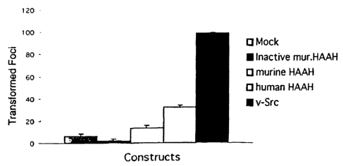
Fig. 1 is a bar graph showing colony formation induced by transient
transfection of
NIH-3T3 cells with various aspartyl (asparaginyl) beta-hydroxylase (AAH)
cDNAs. Colony
formation was induced by transient transfection with 10 g DNA. In contrast,
the mutant murine
AAH construct without enzymatic activity has no transforming activity. The
data is presented as
mean number of transformed foci + SEM.
Fig. 2 is a bar graph showing the results of a densitometric analysis of a
Western blot
assay of proteins produced by various murine AAH stably transfected cell
clones. In clones 7 and
18, there was a modest increase in HAAH gene expression, while the
overexpression was to a
lesser degree in clone 16.
Figs. 3A-B are bar graphs showing colony formation in soft agar exhibited by
HAAH
stably transfected clones compared to HAAH enzymatic activity. Fig. 3A shows a
measurement
1_i
CA 02447367 2003-11-17
WO 02/092782 PCT/US02/15814
of murine AAH enzymatic activity in clones 7, 16 and 18, and Fig. 3B shows
colony formation
exhibited by clones 7, 16 and 18. Data is presented as mean number of colonies
10 days after
plating SEM. All three clones with modest increases in HAAH enzymatic
activity, that
correlated with protein expression, exhibited anchorage independent growth.
Fig. 4 is a bar graph showing tumor formation in nude mice injected with
transfected
clones overexpressing murine AAH. Tumor growth was assessed after 30 days.
Mean tumor
weight observed in mice injected with clones 7, 16 and 18 as compared to mock
DNA transfected
clone. All animals, which were injected with clones overexpressing HAAH,
developed tumors.
Figs. 5A-D are bar graphs showing increased AAH expression in PNET2 (Fig. 5A,
5C) and SH-Sy5y (Fig. 5B) cells treated with retinoic acid (Figs. 5A, 5B) or
phorbol ester
myristate (PMA; Fig. 5C) to induce neurite outgrowth as occurs during tumor
cell invasion. The
cells were treated with 10 gM retinoic acid or 100 nM PMA for 0, 1, 2, 3, 4,
or 7 days. Cell
lysates were analyzed by Western blot analysis using an HAAH-specific
monoclonal antibody to
detect the 85 kDa AAH protein. The levels of immunoreactivity were measured by
volume
densitometry (arbitrary units). The graphs indicate the mean + S.D. of results
obtained from three
separate experiments. In Fig. 5D, PNET2 cells were treated for 24 hours with
sub-lethal
concentrations of H202 to induce neurite retraction. Viability of greater than
90% of the cells was
demonstrated by Trypan blue dye exclusion. Similar results were obtained for
SH-Sy5y cells.
Fig. 6 is a bar graph showing the effects of AAH over-expression on the levels
of
anti-apoptosis (Bcl-2), cell cycle-mitotic inhibitor (p16 and p21/Waft), and
proliferation
(proliferating cell nuclear antigen; PCNA) molecules. PNET2 neuronal cells
were stably
transfected with the full-length human cDNA encoding AAH (pHAAH) or empty
vector
(pcDNA). AAH gene expression was under control of a CMV promoter. Western blot
analysis
was performed with cell lysates prepared from cultures that were 70 to 80
percent confluent.
Protein loading was equivalent in each lane. Replicate blots were probed with
the different
antibodies. Bar graphs depict the mean S.D.'s of protein expression levels
measured in three
experiments. All differences are statistically significant by Student T-test
analysis
(P<0.01-P<0.001).
Fig. 7 is a diagram of showing the components of the IRS-1 signal transduction
pathway.
12
CA 02447367 2008-05-30
Fig. 8 is a line graph showing growth curves generated in cells expressing the
antisense HAAH compared to controls expressing GFP.
10
20 Detailed Description
HAAH is a protein belonging to the alpha-ketoglutarate dependent dioxygenase
family
of prolyl and lysyl hydroxylases which play a key role in collagen
biosynthesis. This molecule
hydroxylates aspartic acid or asparagine residues in EGF-like domains of
several proteins in the
presence of ferrous iron. These EGF-like domains contain conserved motifs,
that form repetitive
sequences in proteins such as clotting factors, extracellular matrix proteins,
LDL receptor,
NOTCH homologues, or NOTCH ligand homologues.
The alpha-ketoglutarate-dependent dioxygenase aspartyl (asparaginyl)
beta-hydroxylase (AAH) specifically hydroxylates one aspartic or asparagine
residue in EGF-like
domains of various proteins. The 4.3-kb cDNA encoding the human AspH (hAspH)
hybridizes
with 2.6 kb and 4.3 kb transcripts in transformed cells, and the deduced amino
acid sequence of
13
CA 02447367 2003-11-17
WO 02/092782 PCT/US02/15814
the larger transcript encodes an protein of about 85 kDa. Both in vitro
transcription and
translation and Western blot analysis also demonstrate a 56-kDa protein that
may result from
posttranslational cleavage of the catalytic C-terminus.
A physiological function of AAH is the post-translational beta-hydroxylation
of
aspartic acid in vitamin K-dependent coagulation proteins. However, the
abundant expression of
AAH in several malignant neoplasms, and low levels of AAH in many normal cells
indicate a role
for this enzyme in malignancy. The AAH gene is also highly expressed in
cytotrophoblasts, but
not syncytiotrophoblasts of the placenta. Cytotrophoblasts are invasive cells
that mediate
placental implantation. The increased levels of AAH expression in human
cholangiocarcinomas,
hepatocellular carcinomas, colon cancers, and breast carcinomas were primarily
associated with
invasive or metastatic lesions. Moreover, overexpression of AAH does not
strictly reflect
increased DNA synthesis and cellular proliferation since high levels of AAH
immunoreactivity
were observed in 100 percent of cholangiocarcinomas, but not in human or
experimental disease
processes associated with regeneration or nonneoplastic proliferation of bile
ducts. AAH
overexpression and attendant high levels of beta hydroxylase activity lead to
invasive growth of
transformed neoplastic cells. Detection of an increase in HAAH expression is
useful for early and
reliable diagnosis of the cancer types which have now been characterized as
overexpressing this
gene product.
Diagnosis of malignant tumors
HAAH is overexpressed in many tumors of endodermal origin and in at least 95%
of
CNS tumors compared to normal noncancerous cells. An increase in HAAH gene
product in a
patient-derived tissue sample (e.g., solid tissue or bodily fluid) is carried
out using standard
methods, e.g., by Western blot assays or a quantitative assay such as ELISA.
For example, a
standard competitive ELISA format using an HAAH-specific antibody is used to
quantify patient
HAAH levels. Alternatively, a sandwich ELISA using a first antibody as the
capture antibody
and a second HAAH-specific antibody as a detection antibody is used.
Methods of detecting HAAH include contacting a component of a bodily fluid
with an
HAAH-specific antibody bound to solid matrix, e.g., microtiter plate, bead,
dipstick. For
example, the solid matrix is dipped into a patient-derived sample of a bodily
fluid, washed, and
1-4
CA 02447367 2003-11-17
WO 02/092782 PCT/US02/15814
the solid matrix is contacted with a reagent to detect the presence of immune
complexes present
on the solid matrix.
Proteins in a test sample are immobilized on (e.g., bound to) a solid matrix.
Methods
and means for covalently or noncovalently binding proteins to solid matrices
are known in the art.
The nature of the solid surface may vary depending upon the assay format. For
assays carried out
in microtiter wells, the solid surface is the wall of the microtiter well or
cup. For assays using
beads, the solid surface is the surface of the bead. In assays using a
dipstick (i.e., a solid body
made from a porous or fibrous material such as fabric or paper) the surface is
the surface of the
material from which the dipstick is made. Examples of useful solid supports
include
nitrocellulose (e.g., in membrane or microtiter well form), polyvinyl chloride
(e.g., in sheets or
microtiter wells), polystyrene latex (e.g., in beads or microtiter plates,
polyvinylidine fluoride
(known as IMMULONTM), diazotized paper, nylon membranes, activated beads, and
Protein A
beads. The solid support containing the antibody is typically washed after
contacting it with the
test sample, and prior to detection of bound immune complexes. Incubation of
the antibody with
the test sample is followed by detection of immune complexes by a detectable
label. For
example, the label is enzymatic, fluorescent, chemiluminescent, radioactive,
or a dye. Assays
which amplify the signals from the immune complex are also known in the art,
e.g., assays which
utilize biotin and avidin.
An HAAH-detection reagent, e.g., an antibody, is packaged in the form of a
kit, which
contains one or more HAAH-specific antibodies, control formulations (positive
and/or negative),
and/or a detectable label. The assay may be in the form of a standard two-
antibody sandwich
assay format known in the art.
Production of HAAH-specific antibodies
Anti-HAAH antibodies were obtained by techniques well known in the art. Such
antibodies are polyclonal or monoclonal. Polyclonal antibodies were obtained
using standard
methods, e.g., by the methods described in Ghose et al., Methods in
Enzymology, Vol. 93, 326-
327, 1983. An HAAH polypeptide, or an antigenic fragment thereof, was used as
the immunogen
to stimulate the production of polyclonal antibodies in the antisera of
rabbits, goats, sheep, or
rodents. Antigenic polypeptides for production of both polyclonal and
monoclonal antibodies
useful as immunogens include polypeptides which contain an HAAH catalytic
domain. For
1.5
CA 02447367 2003-11-17
WO 02/092782 PCT/US02/15814
example, the immunogenic polypeptide is the full-length mature HAAH protein or
an HAAH
fragment containing the carboxyterminal catalytic domain e.g., an HAAH
polypeptide containing
the His motif of SEQ ID NO:2.
Antibodies which bind to the same epitopes as those antibodies disclosed
herein are
identified using standard methods, e.g., competitive binding assays, known in
the art.
Monoclonal antibodies were obtained by standard techniques. Ten g of purified
recombinant HAAH polypeptide was administered to mice intraperitoneally in
complete Freund's
adjuvant, followed by a single boost intravenously (into the tail vein) 3-5
months after the initial
inoculation. Antibody-producing hybridomas were made using standard methods.
To identify
those hybridomas producing antibodies that were highly specific for an HAAH
polypeptide,
hybridomas were screened using the same polypeptide immunogen used to
immunize: Those
antibodies which were identified as having HAAH-binding activity are also
screened for the
ability to inhibit HAAH catalytic activity using the enzymatic assays
described below.
Preferably, the antibody has a binding affinity of at least about 108
liters/mole and more
preferably, an affinity of at least about 109 liters/mole.
Monoclonal antibodies are humanized by methods known in the art, e.g, MAbs
with a
desired binding specificity can be commercially humanized (Scotgene, Scotland;
Oxford
Molecular, Palo Alto, CA).
HAAH-specific intrabodies are produced as follows. Following identification of
a
hybridoma producing a suitable monoclonal antibody, DNA encoding the antibody
is cloned.
DNA encoding a single chain HAAH-specific antibody in which heavy and light
chain variable
domains are separated by a flexible linker peptide is cloned into an
expression vector using known
methods (e.g., Marasco et al., 1993, Proc. Natl. Acad. Sci. USA 90:7889-7893
and Marasco et
al., 1997, Gene Therapy 4:11-15). Such constructs are introduced into cells,
e.g., using standard
gene delivery techniques for intracellular production of the antibodies.
Intracellular antibodies,
i.e., intrabodies, are used to inhibit signal transduction by HAAH.
Intrabodies which bind to a
carboxyterminal catalytic domain of HAAH inhibit the ability of HAAH to
hydroxylate EGF-like
target sequences.
16
CA 02447367 2003-11-17
WO 02/092782 PCT/US02/15814
Methods of linking HAAH-specific antibodies (or fragments thereof) which bind
to
cell surface exposed epitopes of HAAH on the surface of a tumor cell are
linked to known
cytotoxic agents, e.g, ricin or diptheria toxin, using known methods.
Deposit of Biological Materials
Under the terms of the Budapest Treaty on the International Recognition of the
Deposit of Microorganisms for the Purpose of Patent Procedure, hybridoma FB501
(which
produces monoclonal antibody FB50), hybridoma HA386A (which produces
monoclonal
antibody 86A), hybridoma HA15C7A (which produces monoclonal antibody 5C7), and
hybridoma HA219B (which produces monoclonal antibody 19B) were deposited on
May 17,
2001, with the American Type Culture Collection (ATCC) of 10801 University
Boulevard,
Manassas, Va. 20110-2209 USA..
Applicants' assignee represents that the ATCC is a depository affording
permanence of the
deposit and ready accessibility thereto by the public if a patent is granted.
All restrictions on the
availability to the public of the material so deposited will be irrevocably
removed upon the
granting of a patent. The material will be available during the pendency of
the patent application
to one determined by the Commissioner to be entitled thereto under 37 CFR 1.14
and 35 U.S.C.
122. The deposited material will be maintained with all the care necessary to
keep it viable and
uncontaminated for a period of at least five years after the most recent
request for the furnishing
of a sample of the deposited plasmid, and in any case, for a period of at
least thirty (30) years after
the date of deposit or for the enforceable life of the patent, whichever
period is longer.
Applicant's assignee acknowledges its duty to replace the deposit should the
depository be unable
to furnish a sample when requested due to the condition of the deposit.
Methods of treating malignant tumors
Patients with tumors characterized as overexpressing HAAH as such tumors of
endodermal origin or CNS tumors are treated by administering HAAH antisense
nucleic acids.
Antisense therapy is used to inhibit expression of HAAH in patients suffering
from
hepatocellular carcinomas, cholangiocarcinomas, glioblastomas and
neuroblastomas. For
example, an HAAH antisense strand (either RNA or DNA) is directly introduced
into the cells in
1-7
CA 02447367 2003-11-17
WO 02/092782 PCT/US02/15814
a form that is capable of binding to the mRNA transcripts. Alternatively, a
vector containing a
sequence which, which once within the target cells, is transcribed into the
appropriate antisense
mRNA, may be administered. Antisense nucleic acids which hybridize to target
mRNA decrease
or inhibit production of the polypeptide product encoded by a gene by
associating with the
normally single-stranded mRNA transcript, thereby interfering with translation
and thus,
expression of the protein. For example, DNA containing a promoter, e.g., a
tissue-specific or
tumor specific promoter, is operably linked to a DNA sequence (an antisense
template), which is
transcribed into an antisense RNA. By "operably linked" is meant that a coding
sequence and a
regulatory sequence(s) (i.e., a promoter) are connected in such a way as to
permit gene expression
when the appropriate molecules (e.g., transcriptional activator proteins) are
bound to the
regulatory sequence(s).
Oligonucleotides complementary to various portions of HAAH mRNA were tested in
vitro for their ability to decrease production of HAAH in tumor cells (e.g.,
using the FOCUS
hepatocellular carcinoma (HCC) cell line) according to standard methods. A
reduction in HAAH
gene product in cells contacted with the candidate antisense composition
compared to cells
cultured in the absence of the candidate composition is detected using HAAH-
specific antibodies
or other detection strategies. Sequences which decrease production of HAAH in
in vitro cell-
based or cell-free assays are then be tested in vivo in rats or mice to
confirm decreased HAAH
production in animals with malignant neoplasms.
Antisense therapy is carried out by administering to a patient an antisense
nucleic acid
by standard vectors and/or gene delivery systems. Suitable gene delivery
systems may include
liposomes, receptor-mediated delivery systems, naked DNA, and viral vectors
such as herpes
viruses, retroviruses, adenoviruses and adeno-associated viruses, among
others. A reduction in
HAAH production results in a decrease in signal transduction via the IRS
signal transduction
pathway. A therapeutic nucleic acid composition is formulated in a
pharmaceutically acceptable
carrier. The therapeutic composition may also include a gene delivery system
as described above.
Pharmaceutically acceptable carriers are biologically compatible vehicles
which are suitable for
administration to an animal: e.g., physiological saline. A therapeutically
effective amount of a
compound is an amount which is capable of producing a medically desirable
result such as
reduced production of an HAAH gene product or a reduction in tumor growth in a
treated animal.
18
CA 02447367 2003-11-17
WO 02/092782 PCT/US02/15814
Parenteral administration, such as intravenous, subcutaneous, intramuscular,
and
intraperitoneal delivery routes, may be used to deliver nucleic acids or HAAH-
inhibitory peptides
or non-peptide compounds. For treatment of CNS tumors, direct infusion into
cerebrospinal fluid
is useful. The blood-brain barrier may be compromised in cancer patients,
allowing systemically
administered drugs to pass through the barrier into the CNS. Liposome
formulations of
therapeutic compounds may also facilitate passage across the blood-brain
barrier.
Dosages for any one patient depends upon many factors, including the patient's
size,
body surface area, age, the particular nucleic acid to be administered, sex,
time and route of
administration, general health, and other drugs being administered
concurrently. Dosage for
intravenous administration of nucleic acids is from approximately 106 to 1022
copies of the nucleic
acid molecule.
Ribozyme therapy is also be used to inhibit HAAH gene expression in cancer
patients.
Ribozymes bind to specific mRNA and then cut it at a predetermined cleavage
point, thereby
destroying the transcript. These RNA molecules are used to inhibit expression
of the HAAH gene
according to methods known in the art (Sullivan et al., 1994, J. Invest. Derm.
103:85S-89S;
Czubayko et al., 1994, J. Biol. Chem. 269:21358-21363; Mahieu et al, 1994,
Blood 84:3758-65;
Kobayashi et al. 1994, Cancer Res. 54:1271-1275).
HAAH-specific antibodies inhibit tumor cell growth
HAAH-specific antibodies inhibit the proliferation of tumor cells in culture.
Two different
HAAH-specific antibodies, FB-50 and 5C7, were tested. Tumor cells (a
heptatocarcinoma cell
line, a lung carcinoma cell line, and a breast carcinoma cell line) were
seeded in a 96 well plate
and incubated with varying concentrations of antibody for 48 hours. The cells
were fixed with
acetone. Cell growth was monitored using a sulforhodamine B dye binding assay.
The data
indicated a reduction in cell viability and proliferation in the presence of
FB50 compared to in its
absence.
Passive Immunization
The HAAH-specific antibodies described herein are used to inhibit the growth
of a tumor
cell or kill the tumor cell.
Purified antibody preparations (e.g., a purified monoclonal antibody, an
antibody
fragment, or single chain antibody) is administered to an individual diagnosed
with a tumor or at
19
CA 02447367 2003-11-17
WO 02/092782 PCT/US02/15814
risk of developing a tumor. The antibody preparations are administered using
methods known in
the art of passive immunization, e.g., intravenously or intramuscularly. The
antibodies used in the
methods described herein are formulated in a physiologically-acceptable
excipient. Such
excipients, e.g., physiological saline, are known in the art.
The antibody is preferably a high-affinity antibody, e.g., an IgG-class
antibody or
fragment or single chain thereof. Alternatively, the antibody is an IgM
isotype. Antibodies are
monoclonal, e.g., a murine monoclonal antibody or fragment thereof, or a
murine monoclonal
antibody, which has been humanized. The antibody is a human monoclonal
antibody. The affinity
of a given monoclonal antibody is further increased using known methods, e.g.,
by selecting for
increasingly higher binding capacity (e.g., according to the method described
in Boder et al.,
2000, Proc. Natl. Acad. Sci. U.S.A. 97:10701-10705). Optionally, the antibody,
antibody
fragment, or high affinity single chain antibody is conjugated to a toxic
moiety prior to
administration. Toxic moities suitable for conjugation include ricin,
Psuedomonas toxin,
Diptheria toxin as well as radioisotopes and chemotherapeutic agents known in
the art. Such
antibody toxins damage or kill a tumor cell upon binding to the tumor cell or
upon internalization
into the cytoplasm of the tumor cell.
Antibody preparations or antibody-toxin preparations are administered at doses
of
approximately 0.01-2 mL/kg of body weight. Doses are readministered weekly or
monthly as
necessary to reduce tumor load in a treated individual.
Active Immunization
Active vaccination is the process of inducing an animal to respond to an
antigen. During
vaccination, cells, which recognize the antigen (B cells or cytotoxic T
cells), are clonally
expanded. In addition, the population of helper T cells specific for the
antigen also increase.
Vaccination also involves specialized antigen presenting cells, which can
process the antigen and
display it in a form which can stimulate one of the two pathways. Antigen
recognition followed
by immune cell expansion and activation leads to the production of antigen-
specific antibodies
and antigen-specific cellular immune responses. Successful immunization is
indicated by an
increase in the level of HAAH-specific antibody titer in serum of an immunized
individual
compared to the level prior to immunization. Preferably, the HAAH-specific
antibody titer is at
CA 02447367 2003-11-17
WO 02/092782 PCT/US02/15814
least 10%, more preferably at least 50%, more preferably at least 100%, and
most preferably
200% greater than the titer prior to immunization.
An individual is immunized with an AAH (e.g., HAAH) polypeptide or a
polynucleotide
encoding the peptide. For example, a human patient is immunized with full-
length 52 kDa
HAAH. Standard adjuvant formulations may be simultaneously administered to
enhance
immunogenicity of the immunizing polypeptide. Alternatively, shorter
polypeptides, e.g.,
immunogenic fragments of HAAH, are used. For example, a polypeptide contains
an
extracellular catalytic domain of HAAH (e.g., amino acids 650-700 of SEQ ID
NO:2). Other
immunogenic fragments of HAAH include a fragment contains a binding site for
alpha-
ketoglutarate, a fragment that lacks a binding site for alpha-ketoglutarate,
one which contains a
calcium binding site, and one which lacks a binding site for an EGF-like
polypeptide.
DNA vaccine
In addition to standard active vaccination using a peptide antigen, DNA
vaccination is
used to generate an immune response to HAAH, and in turn to tumor cells, which
overexpress
HAAH. Although HAAH is overexpressed on malignant cells, an effective immune
response is
not made by the patient because tumor cells lack appropriate accessory
molecules for antigen
presentation. The DNA vaccines described herein result in generation of a
humoral as well as
cellular immunity specific for HAAH (and cells expressing HAAH on their cell
surface). For
example, not only is an HAAH-specific antibody produced in the immunized
individual, HAAH-
specific cytotoxic T cells are generated. HAAH-specific cytotoxic T cells kill
tumor cells,
thereby reducing tumor load in the immunized individual.
A polynucleotide encoding an AAH polypeptide (full-length or an immunogenic
fragment
of AAH) is introduced into an individual by known methods, e.g., particle
bombardment or direct
injection via needle. Typically, the antigen (or DNA encoding the antigen) is
delivered
intramuscularly. The antigen is also directly injected into other tissues,
e.g., tumor sites. DNA is
taken up by cells at the point of injection. The cell produces proteins, and
the proteins stimulate
the immune system of the immunized individual resulting, e.g., in generation
of an HAAH-
specific antibody. Cellular immunity, e.g., cytotoxic T cells, are also
generated.
An effective DNA or mRNA dosage is generally be in the range of from about
0.05
micrograms/kg to about 50 mg/kg, usually about 0.005-5 mg/kg of body weight,
e.g., 0.5 to 5
2.1
CA 02447367 2003-11-17
WO 02/092782 PCT/US02/15814
mg/kg. The DNA to be administered is naked (in the absence of transfection-
facilitating
substances) or complexed with compounds, which enhance cellular uptake of the
polynucleotide
(e.g., charged lipids, lipid complexes or liposomes). For example, the
polynucleotide is
administered with LipofectinTM or precipitating agents such as CaPO4. The
transfected cells, e.g.,
non-proliferating muscle cells, produce the recombinant antigenic polypeptide
for at least one
month and up to several months, e.g. 3-6 months. Alternatively, transitory
expression of a
polypeptide is achieved by introducing the polynucleotide construct into a
tissue (e.g., non-
muscular tissue or tumor tissue). In the latter case, cells of the tissue
produce the polypeptide for
a shorter period of time, e.g., several days (3-5 days and up to about 20
days). The level of
protein or polypeptide expression by target cells is sufficient to induce
production of HAAH-
specific antibodies. The level of antibody production is measured using
standard methods, e.g.,
evaluation of antibody titer in patient serum, before and after immunization.
The polynucleotides are administered by standard methods, such as by injection
into the
interstitial space of tissues such as muscles or skin, introduction into the
circulation or into body
cavities or by inhalation or insufflation. Polynucleotides are injected or
otherwise delivered to the
animal with a pharmaceutically acceptable liquid carrier, e.g., a liquid
carrier, which is aqueous or
partly aqueous. The polynucleotides are associated with a liposome (e.g., a
cationic or anionic
liposome). The polynucleotide includes genetic information necessary for
expression by a target
cell, such as a promoters.
One advantage of DNA vaccination is that DNA vaccines can result in longer
lasting
production of the antigenic protein, thereby booster shots reducing or
avoiding booster
immunizations.
In addition to inducing an immune response, e.g., an HAAH-specific antibody
response,
by vaccinating with DNA encoding an HAAH polypeptide, a polynucleotide
encoding the
antibody itself is introduced. An isolated polynucleotide encoding an HAAH-
specific antibody,
e.g., variable regions of the antibody, is introduced for production of the
antibody in situ. The
antibody in turn exerts a therapeutic effect at the target site by binding a
cell surface antigen, e.g.,
extracellular HAAH, or by binding to a catalytic domain of HAAH, to inhibit
HAAH function.
In vivo diagnostic imaging
22
CA 02447367 2003-11-17
WO 02/092782 PCT/US02/15814
The antibodies (antibody fragments, and single chain antibodies) described
herein are
useful to diagnose the presence of a tumor in tissues as well as bodily
fluids. HAAH-specific
antibodies are tagged with a detectable label such as a radioisotope or
colorimeteric agent. The
labeled antibody is administered to an individual at risk of developing cancer
or an individual who
has previously been diagnosed with cancer. For example, the antibodies are
useful to diagnose
metastases of a tumor, which has been surgically excised or treated by
chemotherapeutic or
radiotherapeutic methods. The sensitivity of the method is sufficient to
detect micrometastases in
tissues such as lymph nodes. Early and sensitive diagnosis of tumors in this
manner allows
prompt therapeutic intervention.
The labeled antibody is administered to an individual using known methods,
e.g.,
intravenously, or direct injection into solid or soft tissues. The antibody is
allowed to distribute
throughout the tissue or throughout the body for a period of approximately 1
hour to 72 hours.
The whole body of the individual is then imaged using methods known in the
art. Alternatively, a
small portion of the body, e.g., a tissue site suspected of harboring a tumor,
is imaged. An
increase in antibody binding, as measured by an increase in detection of the
label, over the level
of baseline binding (to normal tissue) indicates the presence of a tumor at
the site of binding.
Activation of NOTCH signaling
NOTCH signalling is activated in cells highly expressing AAH. Fig. 14A. shows
the
presence of a 110 kDa NOTCH fragment as revealed by using Western blot.
Overexpression of
enzymatically active AAH is shown by a display of the 100 kDa cleaved, active
NOTCH-1 (Lane
1, Mock DNA transfected clone; Lane 2, clones 7; and Lane 3, clone 18). In
contrast, NOTCH-2
was not activated. There was enhanced expression of the full length Jagged
ligand in clones
expressing AAH as compared to the mock DNA transfected clone. Tubulin was used
as internal
control for protein loading.
Expression of the Hes-1, a known downstream effector gene, is activated by
NOTCH
signaling (Fig. 14B). Only AAH-expressing clones activate Notch expression as
a transcription
factor and subsequently unregulates Hes-1 gene expression as revealed by
competitive RT-PCR.
Lower panel is an RT-PCR product of GAPDII that served as internal control.
Fig. 14C shows
expression of human NOTCH-1 (hNOTCH-1) and Jagged-1 where IRS-1 signaling is
reduced by
a dominant negative mutant (DhIRS-1). Such cells demonstrate downregulation
AAH expression
23
CA 02447367 2003-11-17
WO 02/092782 PCT/US02/15814
and demonstrate a parallel decrease in NOTCH-1 and Jagged levels by Western
blot analysis.
Tubulin was used as an internal control for protein loading.
Methods of identifying compounds that inhibit HAAH enzymatic activity
Aspartyl (asparaginyl) beta-hydroxylaseydroxylase (AAH) activity is measured
in
vitro or in vivo. For example, HAAH catalyzes posttranslational modification
of P carbon of
aspartyl and asparaginyl residues of EGF-like polypeptide domains. An assay to
identify
compounds which inhibit hydroxylase activity is carried out by comparing the
level of
hydroxylation in an enzymatic reaction in which the candidate compound is
present compared to a
parallel reaction in the absence of the compound (or a predetermined control
value). Standard
hydroxylase assays carried out in a testtube are known in the art, e.g.,
Lavaissiere et al., 1996, J.
Clin. Invest. 98:1313-1323; Jia et al., 1992, J. Biol. Chem. 267:14322-14327;
Wang et al., 1991,
J. Biol. Chem. 266:14004-14010; or Gronke et al., 1990, J. Biol. Chem.
265:8558-8565.
Hydroxylase activity is also measured using carbon dioxide (14C02 capture
assay) in a 96-well
microtiter plate format (Zhang et al., 1999, Anal. Biochem. 271:137-142. These
assays are
readily automated and suitable for high throughput screening of candidate
compounds to identify
those with hydroxylase inhibitory activity.
Candidate compound which inhibit HAAH activation of NOTCH are identified by
detecting a reduction in activated NOTCH in a cell which expresses or
overexpresses HAAH,
e.g., FOCUS HCC cells. The cells are cultured in the presence of a candidate
compound. Parallel
cultures are incubated in the absence of the candidate compound. To evaluate
whether the
compound inhibits HAAH activation of NOTCH, translocation of activated NOTCH
to the
nucleus of the cell is measured. Translocation is measured by detecting a 110
kDa activation
fragment of NOTCH in the nucleus of the cell. The activation fragment is
cleaved from the large
(approximately 300 kDa) transmembrane NOTCH protein upon activation. Methods
of
measuring NOTCH translocation are known, e.g, those described by Song et al.,
1999, Proc. Natl.
Acad. Sci U.S.A. 96:6959-6963 or Capobianco et al., 1997, Mol. Cell Biol.
17:6265-6273. A
decrease in translocation in the presence of the candidate compound compared
to that in the
absence of the compound indicates that the compound inhibits HAAH activation
of NOTCH,
thereby inhibiting NOTCH-mediated signal transduction and proliferation of
HAAH-
overexpressing tumor cells.
24
CA 02447367 2003-11-17
WO 02/092782 PCT/US02/15814
Methods of screening for compounds which inhibit phosphorylation of IRS are
carried
out by incubating IRS-expressing cells in the presence and absence of a
candidate compound and
evaluating the level of IRS phosphorylation in the cells. A decrease in
phosphorylation in cells
cultured in the presence of the compound compared to in the absence of the
compound indicates
that the compound inhibits IRS-1 phosphorylation, and as a result, growth of
HAAH-
overexpressing tumors. Alternatively, such compounds are identified in an in
vitro
phosphorylation assay known in the art, e.g., one which measured
phosphorylation of a synthetic
substrate such as poly (Glu/Tyr).
Example 1: Increased expression of HAAH is associated with malignant
transformation
HAAH is a highly conserved enzyme that hydroxylates EGF-like domains in
transformation associated proteins. The HAAH gene is overexpressed in many
cancer types
including human hepatocellular carcinomas and cholangiocarcinomas. HAAH gene
expression
was found to be undetectable during bile duct proliferation in both human
disease and rat models
compared to cholangiocarcinoma. Overexpression of HAAH in NIH-3T3 cells was
associated
with generation of a malignant phenotype, and enzymatic activity was found to
be required for
cellular transformation. The data described below indicate that overexpression
of HAAH is
linked to cellular transformation of biliary epithelial cells.
To identify molecules that are specifically overexpressed in transformed
malignant
cells of human hepatocyte origin, the FOCUS hepatocellular carcinoma (HCC)
cell line was used
as an immunogen to generate monoclonal antibodies (mAb) that specifically or
preferentially
recognize proteins associated with the malignant phenotype. A lambda GT11 cDNA
expression
library derived from HepG2 HCC cells was screened, and a HAAH-specific mAb
produced
against the FOCUS cell line was found to recognize an epitope on a protein
encoded by an HAAH
cDNA. The HAAH enzyme was found to be upregulated in several different human
transformed
cell lines and tumor tissues compared to adjacent human tissue counterparts.
The overexpressed
HAAH enzyme in different human malignant tissues was found to be catalytically
active.
HAAH gene expression was examined in proliferating bile ducts and in NIH 3T3
cells.
Its role in the generation of the malignant phenotype was measured by the
formation of
transformed foci, growth in soft agar as an index of anchorage independent
growth and tumor
formation in nude mice. The role of enzymatic activity in the induction of
transformed phenotype
CA 02447367 2003-11-17
WO 02/092782 PCT/US02/15814
was measured by using a cDNA construct with a mutation in the catalytic site
that abolished
hydroxylase activity. The results indicated that an increase in expression of
HAAH gene is
associated with malignant transformation of bile ducts.
The following materials and methods were used to generate the data described
below.
Antibodies
The FB50 monoclonal antibody was generated by cellular immunization of Balb/C
mice with FOCUS HCC cells. A monoclonal anti-Dengue virus antibody was used as
a non-
relevant control. The HBOH2 monoclonal antibody was generated against a 52 kDa
recombinant
HAAH polypeptide and recognizes the catalytic domain of beta-hydroxylase from
mouse and
human proteins. Polyclonal anti-HAAH antibodies cross-react with rat
hydroxylase protein.
Control antibody anti-Erk-1 was purchased from Santa Cruz Biotechnology, Inc.,
CA. Sheep
anti-mouse and donkey anti-rabbit antisera labeled with horseradish peroxidase
were obtained
from Amersham, Arlington Heights, IL.
Constructs
The murine full length AAH construct (pNH376) and the site-directed mutation
construct (pNH376-H660) with abolished catalytic activity were cloned into the
eukaryotic
expression vector pcDNA3 (Invitrogen Corp., San Diego, CA). The full length
human AAH was
cloned into prokaryotic expression vector pBC-SK+ (Stratagene, La Jolla, CA).
The full length
human AAH (GENBANK Accession No. S83325) was subcloned into the EcoRI site of
the
pcDNA3 vector.
Animal model of bile duct proliferation
Rats were divided into 9 separate groups of 3 animals each except for group 9,
which
contained 5 rats. Group 1 was the non-surgical control group, and group 2 was
the sham-operated
surgical control. The remaining groups underwent common bile duct ligation to
induce
intrahepatic bile duct proliferation and were evaluated at 6, 12, 24, 48 hours
and 4, 8 and 16 days
as shown in Table 3. Animals were asphyxiated with C02, and liver samples were
taken from left
lateral and median lobes, fixed in 2 % paraformaldehyde and embedded in
paraffin. Liver
samples (5 m) were cut and stained with hematoxylin and eosin to evaluate
intrahepatic bile duct
proliferation. Immunohistochemistry was performed with polyclonal anti-HAAH
antibodies that
cross-react with the rat protein to determine levels of protein expression.
26
CA 02447367 2003-11-17
WO 02/092782 PCT/US02/15814
Bile duct proliferation associated with primary sclerosing cholangitis (PSC)
Liver biopsy samples were obtained from 7 individuals with PSC and associated
bile
duct proliferation. These individuals were evaluated according to standard
gastroenterohepatological protocols. Patients were 22-46 years of age and
consisted of 4 males
and 3 females. Four had associated inflammatory bowel disease (3 ulcerative
colitis and 1
Crohn's colitis). All patients underwent a radiological evaluation including
abdominal
ultrasonography and endoscopic retrograde cholangiopancreaticography to
exclude the diagnosis
of extrahepatic biliary obstruction. Tissue sections were prepared from
paraffin embedded blocks
and were evaluated by hematoxylin and eosin staining for bile duct
proliferation. Expression of
HAAH was determined by immunohistochemistry using anHAAH-specific monoclonal
antibody
such as FB50.
Immunohistochemistry
Liver tissue sections (5 m) were deparaffinized in xylene and rehydrated in
graded
alcohol. Endogenous peroxidase activity was quenched by a 30-minute treatment
with 0.6 %
H202 in 60% methanol. Endogenous biotin was masked by incubation with avidin-
biotin
blocking solutions (Vector Laboratories, Burlingame, CA). The FB50 mAb (for
PSC samples)
and polyclonal anti-HAAH-hydroxylase antibodies (for rat liver samples) were
added to slides in
a humidified chamber at 4 C overnight. Immunohistochemical staining was
performed using a
standard avidin-biotin horseradish peroxidase complex (ABC) method using
Vectastain Kits with
diaminobenzidine (DAB) as the chromogen according to manufacturer's
instructions (Vector
Laboratories, Inc., Burlingame, CA). Tissue sections were counterstained with
hematoxylin,
followed by dehydration in ethanol. Sections were examined by a light
microscopy for bile duct
proliferation and HAAH protein expression. Paraffin sections of
cholangiocarcinoma and
placenta were used as positive controls, and hepatosteatosis samples were used
as a negative
controls. To control for antibody binding specificity, adjacent sections were
immunostained in
the absence of a primary antibody, or using non-relevant antibody to Dengue
virus. As a positive
control for tissue immunoreactivity, adjacent sections of all specimens were
immunostained with
monoclonal antibody to glyceraldehyde 3-phosphate dehydrogenase.
Western blot analysis
2.7
CA 02447367 2003-11-17
WO 02/092782 PCT/US02/15814
Cell lysates were prepared in a standard radioimmunoprecipitation assay (RIPA)
buffer containing protease inhibitors. The total amount of protein in the
lysates was determined
by Bio-Rad colorimetric assay (Bio Rad, Hercules, CA) followed by 10% sodium
dodecyl
sulphate-polyacrylamide gel electrophoresis (SDS-PAGE), transferred to PVDF
membranes, and
subjected to Western blot analysis using FB50, HBOH2, anti-Erk-1 (used as an
internal control
for protein loading) as primary, sheep anti-mouse and donkey anti-rabbit
antisera labeled with
horseradish peroxidase as secondary antibodies. Antibody binding was detected
with enhanced
chemiluminescence reagents (SuperSignal, Pierce Chemical Company, Rockford,
IL) and film
autoradiography. The levels of immunoreactivity were measured by volume
densitometry using
NIH Image software.
Enzymatic activity assay
AAH activity was measured in cell lysates using the first EGF-like domain of
bovine
protein S as substrate where 14C-labeled alpha-ketogluterate hydroxylates the
domain releasing
14C containing CO2 according to standard methods, e.g., those described by Jia
et al., 1992, J.
Biol. Chem. 267:14322-14327; Wang et al., 1991, J. Biol. Chem. 266:14004-
14010; or Gronke et
al., 1990, J. Biol. Chem. 265:8558-8565. Incubations were carried out at 37 C
for 30 min in a
final volume of 40 l containing 48 g of crude cell extract protein and 75 M
EGF substrate.
Cell transfection studies
The NIH-3T3 cells were cultured in Dulbecco's modified Eagle's medium (DMEM;
Mediatech, Washington, DC) supplemented with 10 % heat-inactivated fetal calf
serum (FCS;
Sigma Chemical Co., St.Louis, MO), 1% L-glutamine, 1% non-essential amino
acids and 1%
penicillin-streptomycin (GIBCO BRL, Life Technologies, Inc., Grand Island,
NY). Subconfluent
NIH-3T3 cells (3 x 10⁵ cells/60-mm dish) were transfected with 10 g of one of
the following
plasmids: 1) non-recombinant pcDNA3 vector (Invitrogen Corp., San Diego, CA)
as a negative
control; 2) pNH376-H660, the murine AAH cDNA that was mutated in the catalytic
domain and
cloned into the pcDNA3 vector driven by a CMV promoter; 3) pNH376, the wild
type murine
AAH cDNA cloned into the pcDNA3 vector; 4) pCDHH, wild type human AAH cDNA
cloned
into the pcDNA3 vector; or 5) pLNCX-UP1, a cDNA that encodes v-Src oncogene
(positive
control). Cells were transfected using the calcium phosphate transfection kit
according to
manufacturer's instructions (5 Prime - 3 Prime, Inc., Boulder, CO). Comparison
of cellular
28
CA 02447367 2003-11-17
WO 02/092782 PCT/US02/15814
transfection efficiency was assessed with the various constructs. For this
procedure, confluent
plates obtained 48 hours after transfection were split and reseeded into 12
separate 6-cm dishes,
and 6 of them were made to grow in the presence of 400 g/ml G-418 (GIBCO BRL,
Life
Technologies, Inc., Grant Island, NY) containing medium. The number of G-418
resistant foci
was determined at 14 days after transfection and used to correct for any
variability in transfection
efficiency.
Transformation assay
The NIH-3T3 cells were transfected with the various constructs and allowed to
reach
confluence after 48 hours as described above. Each 6 cm dish was split and
seeded into 12
different 6 cm dishes. While 6 of them were made to grow in the presence of G-
418 to detect
transfection efficiency, the other six were grown in complete medium without G-
418 and with a
medium change every 4th day. The number of transformed foci were counted in
these plates
without G-418 and expressed as transformed foci per g transfected DNA.
Anchora eelpendent cell growth assay
A limiting dilution technique (0.15 cell/well of a flat bottom 96-well-plate)
was
performed on transfectants grown in G-418 in order to isolate cell clones with
different levels of
HAAH activity as measured by Western blot analysis and enzymatic assay of
hydroxylase
activity. Cloned cell lines (1.0 x 10⁴ cells) were suspended in complete
medium containing 0.4 %
low-melting agarose (SeaPlaque GTG Agarose; FMC Bioproducts, Rockland, Maine)
and laid
over a bottom agar mixture consisting of complete medium with 0.53 % low-
melting agarose.
Each clone was assayed in triplicate. The clones were seeded under these
conditions and 10 days
later the size (positive growth > 0.1 mm in diameter) and number of foci were
determined.
Tumorigenicity in nude mice
The same clones as assessed in the anchorage independent growth assay were
injected
into nude mice and observed for tumor formation. Tumorigenicity was evaluated
using 10
animals in each of 4 groups (Charles River Labs., Wilmington, MA). Group 1
received 1 x 10⁷
cells stably transfected with mock DNA, Group 2-4 received 1 x 10⁷ cells of
clones stable
transfected with pNH376 and expressing various levels of murine HAAH protein.
Nude mice
were kept under pathogen-free conditions in a standard animal facility. Thirty
days after tumor
29
CA 02447367 2003-11-17
WO 02/092782 PCT/US02/15814
cell inoculation, the animals were sacrificed using isofluorane (Aerrane,
Anaquest, NJ) containing
chambers and the tumors were carefully removed and weight determined.
Animal model of bile duct proliferation
Following ligation of the common bile duct, intrahepatic bile duct
proliferation was
evident at 48 hours. Tissue samples obtained 8 and 16 days following common
bile duct ligation
revealed extensive bile duct proliferation as shown in Table 3.
Table 3: Bile duct proliferation and HAAH expression
at different intervals after common bile duct ligation
Group Surgical Microscopy* Immunohisto-
Procedure chemistry
1 no surgery normal negative
2 sham surgery normal negative
3 6 hours post normal negative
ligation
4 12 hours post normal negative
ligation
5 24 hours post normal negative
ligation
6 48 hours post minimal bile duct negative
ligation prolif.
7 4 days post ligation moderate bile duct negative
prolif.
8 8 days post ligation extensive bile duct negative
prolif.
9 16 days post extensive bile duct negative
ligation prolif.
Investigation was performed under light microscopy following a hematoxylin and
eosin staining.
CA 02447367 2003-11-17
WO 02/092782 PCT/US02/15814
Immunohistochemical staining failed to detect presence of HAAH in
proliferating bile
ducts at any time. Analysis of HAAH expression in bile ducts derived from sham
surgical
controls was also negative, while all samples exhibited positive
immunoreactivity with control
antibodies to glyceraldehyde 3-phosphate dehydrogenase. Thus, bile duct
proliferation was not
associated with increased HAAH expression in this standard animal model
system.
HAAH expression in PSC
The liver biopsy specimens from patients with PSC exhibited bile duct
proliferation
accompanied by periductal fibrosis and a mononuclear inflammatory cell
infiltrate without
evidence of dysplasia. Adjacent sections immunostained with the an HAAH-
specific monoclonal
antibody had no detectable HAAH immunoreactivity in proliferating bile ducts.
In contrast,
sections of cholangiocarcinoma that were immunostained simultaneously using
the same antibody
and detection reagents manifested intense levels of HAAH immunoreactivity in
nearly all tumor
cells, whereas adjacent sections of the cholangiocarcinomas exhibited a
negative immunostaining
reaction with monoclonal antibody to Dengue virus. These findings indicate
that HAAH
expression was associated with malignant transformation rather than non-
cancerous cellular
proliferation of intrahepatic bile ducts.
HAAH associated transformation of NIH-3T3 cells
The transforming capability of the murine and human AAH genes, as well as the
murine AAH mutant construct without enzymatic activity were compared to mock
DNA (negative
control) and v-Src transfected NIH-3T3 cells (positive control). The
transforming capability of
murine AAH was found to be 2-3 times that of vector DNA control as shown in
Fig. 1. The
transforming capacity of the human gene was greater than that observed with
the murine AAH (32
+ 1.5 versus 13 + 2.6 transformed foci, respectively). The murine and human
AAH transfected
cells formed large foci, resembling those of v-Src transfected fibroblasts,
compared to the
occasional much smaller foci observed in cells transfected with vector DNA
that displayed the
contact inhibition of fibroblast cell lines. Parallel experiments performed
using the mutant
pNH376-H660 construct without enzymatic activity revealed no transforming
activity. This
finding indicates that the enzymatic activity of HAAH is required for the
transforming activity
exhibited by the HAAH gene.
Anchorage-independent cell growth assay
3-1
CA 02447367 2003-11-17
WO 02/092782 PCT/US02/15814
After transient transfection with the murine AAH construct, several different
transformed foci were isolated for dilutional cloning experiments to establish
stable transfected
cell clones with different levels of HAAH gene expression. Nine different
cloned cell lines were
selected for further study. The expression level of the HAAH protein was
determined by Western
blot analysis. Clones 7 and 18 had a modest increase in HAAH protein
expression, yet formed
large colonies in soft agar (Fig. 2). Protein loading was equivalent in all
lanes as shown by
immunoblotting of the same membranes with an anti-Erk-1 monoclonal antibody.
The increased
protein expression was associated with increased enzymatic activity as shown
in Fig. 3. The
capability of these clones to exhibit anchorage independent cell growth in
soft agar is presented in
Fig. 3. All 3 clones with increased HAAH gene expression demonstrated
anchorage independent
cell growth compared to the mock DNA transfected clone.
Tumor formation in nude mice
The 3 clones with increased HAAH gene expression were evaluated for the
ability to
form tumors in nude mice. Tumor size in the mouse given clone 18 was compared
to a mock
DNA transfected clone. Clones 7, 16 and 18 were highly transformed in this
assay and produced
large tumors with a mean weight of 2.5, 0.9 and 1.5 grams, respectively (Fig.
4). These data
indicate that overexpression of HAAH contributes to induction and maintenance
of the malignant
phenotype in vivo.
High level HAAH expression is indicative of malignancy
In order to determine if HAAH expression was associated with malignancy rather
than
increased cell turnover, two models of bile duct proliferation were studied.
In the animal model,
ligation of the common bile duct induced extensive intrahepatic bile duct
proliferation, yet there
was no evidence of HAAH gene expression under these experimental conditions as
shown in
Table 3. Similarly, HAAH gene expression was assessed in a human disease model
associated
with bile duct proliferation since PSC is an autoimmune liver disease
associated with destruction
as well as proliferation of the intra and extrahepatic bile ducts. PSC is
premalignant disease, and
a significant proportion of affected individuals will eventually develop
cholangiocarcinoma.
However, no evidence for increased HAAH gene expression in the presence of
extensive bile duct
proliferation.
32
CA 02447367 2003-11-17
WO 02/092782 PCT/US02/15814
Having established that HAAH protein levels were elevated in
cholangiocarcinoma
and not in normal or proliferating bile ducts, the role of HAAH in the
generation of a malignant
phenotype was studied. The HAAH gene was transfected into NTH-3T3 cells and
cellular
changes, e.g., increased formation of transformed foci, colony growth in soft
agar and tumor
formation in nude mice associated with malignant transformation, were
evaluated. The
full-length murine and human AAH genes were cloned into expression constructs
and transiently
transfected into NIH-3T3 cells. An increased number of transformed foci was
detected in cells
transfected both with the murine and human AAH genes as compared to mock DNA
transfected
controls. The increased number of transformed foci, after controlling for
transfection efficiency,
was not as high compared to v-Src gene transfected cells used as a positive
control. The
enzymatic activity of the HAAH gene was required for a malignant phenotype
because a mutant
construct which abolished the catalytic site had no transforming properties.
Several stable
transfectants and cloned NIH-3T3 cell lines with a modest increase in HAAH
protein levels and
enzymatic activity were established. Such cell lines were placed in soft agar
to examine
anchorage independent cell growth as another property of the malignant
phenotype. All cell lines
grew in soft agar compared to mock DNA transfected control, and there was a
positive correlation
between the cellular level of HAAH gene expression and the number and size of
colonies formed.
Three of these cloned cell lines formed tumors in nude mice. All three cell
lines with increased
HAAH expression were oncogenic as shown by the development of large tumors as
another
well-known characteristic of the transformed phenotype.
To determine whether cellular changes induced by overexpression of HAAH were
related to the enzymatic function, a site-directed mutation was introduced
into the gene that
changed the ferrous iron binding site from histidine to lysine at 660th
position of mouse HAAH
thereby abolishing hydroxylase activity of the murine HAAH. A corresponding
mutation in
HAAH is used as a dominant negative mutant to inhibit HAAH hydroxylase
activity. The
pNH376-H660 construct had no transformation activity indicating cellular
changes of the
malignant phenotype induced by overexpression depends on the enzymatic
activity of the protein.
33
CA 02447367 2003-11-17
WO 02/092782 PCT/US02/15814
Notch receptors and their ligands have several EGF-like domains in the N-
terminal
region that contain the putative consensus sequence for beta-hydroxylation.
Notch ligands are
important elements of the Notch signal transduction pathway and interaction of
Notch with its
ligands occurs by means of EGF-like domains of both molecules. Point mutations
affecting
aspartic acid or asparagine residues in EGF-like domains that are the targets
for
beta-hydroxylation by HAAH reduce calcium binding and protein-protein
interactions involved in
the activation of downstream signal transduction pathways. Overexpression of
HAAH and Notch
protein hydroxylation by HAAH contributes to malignancy. Tumor growth is
inhibited by
decreasing Notch protein hydroxylation by HAAH.
The data presented herein is evidence that high-level HAAH expression is
linked to
malignant transformation. An increase in expression of the HAAH cDNA in NIH-
3T3 cells
induced a transformed phenotype manifested by increased numbers of transformed
foci,
anchorage-independent growth, and tumorigenesis in nude mice. In addition,
intact
HAAH-enzyme was found to be required for HAAH-associated transformation.
Accordingly,
inhibition of as little as 20% of endogenous HAAH enzymatic activity or
expression confers a
therapeutic benefit. For example, clinical benefit is achieved by 50%-70%
inhibtion of HAAH
expression or activity after administaration of an HAAH inhibitory compound
compared to the
level associated with untreated cancer cell or a normal noncancerous cell.
HAAH is regulated at the level of transcription. Only modest increases in HAAH
expression and enzyme activity were required for cellular transformation.
These results indicate
that increased HAAH gene expression and enzyme activity contribute to the
generation or
maintenance of the transformed phenotype and that decreasing transcription of
the HAAH gene or
decreasing enzymatic activity of the HAAH gene product leads to a decrease in
malignancy.
Accordingly, HAAH transcription is inhibited by administering compounds which
decrease
binding of Fos and/or Jun (elements which regulate HAAH transcription) to HAAH
promoter
sequences.
3-4
CA 02447367 2003-11-17
WO 02/092782 PCT/US02/15814
Since HAAH is up-regulated with malignant transformation of bile duct
epithelium,
and HAAH immunoreactivity is detectable on tumor cell surface membranes, HAAH
is also a
molecule to which to target a cytotoxic agent, e.g., by linking the cytotoxic
agent to a compound
that binds to HAAH expressed on the surface of a tumor cell. Assay of HAAH
protein levels in
either biological fluids such as bile, or cells obtained by fine needle
aspiration is a diagnostic
marker of human cholangiocarcinoma.
Example 2: Expression of AAH and growth and invasiveness of malignant CNS
neoplasms
AAH is abundantly expressed in carcinomas and trophoblastic cells, but not in
most
normal cells, including those of CNS origin. High levels of AAH expression
were observed in 15
of 16 glioblastomas, 8 of 9 anaplastic oligodendrogliomas, and 12 of 12
primitive
neuroectodermal tumors (PNETs). High levels of AAH immunoreactivity were
primarily
localized at the infiltrating edges rather than in the central portions of
tumors. Double-label
immunohistochemical staining demonstrated a reciprocal relationship between
AAH and tenascin,
a substrate for AAH enzyme activity. PNET2 neuronal cell lines treated with
phorbol ester
myristate or retinoic acid to stimulate neuritic extension and invasive growth
exhibited high levels
of AAH expression, whereas H202-induced neurite retraction resulted in down-
regulation of
AAH. PNET2 neuronal cells that stably over-expressed the human AAH cDNA had
increased
levels of PCNA and Bcl-2, and reduced levels of p21/Wafl and p16, suggesting
that AAH
overexpression results in enhanced pathological cell proliferation, cell cycle
progression, and
resistance to apoptosis. In addition, the reduced levels of p16 observed in
AAH-transfectants
indicate that AAH over-expression confers enhanced invasive growth of
neoplastic cells since
deletion or down-regulation of the p16 gene correlates with more aggressive
and invasive in vivo
growth of glioblastomas. Increased AAH immunoreactivity was detected at the
infiltrating
margins of primary malignant CNS neoplasms, further indicating a role of HAAH
in tumor
invasiveness.
The following materials and methods were used to generate the data described
below.
35
CA 02447367 2003-11-17
WO 02/092782 PCT/US02/15814
Analysis of AAH Immunoreactivity in Primary Human Malignant CNS Neoplasms:
AAH immunoreactivity was examined in surgical resection specimens of
glioblastoma
(N=16), anaplastic oligodendroglioma (N=9), and primitive neuroectodermal
tumor (PNET;
supratentorial neuroblastomas (N=3) and medulloblastomas (N=9). The
histopathological
sections were reviewed to confirm the diagnoses using standard criteria.
Paraffin sections from
blocks that contained representative samples of viable solid tumor, or tumor
with adjacent intact
tissue were studied. Sections from normal adult postmortem brains (N=4) were
included as
negative controls. AAH immunoreactivity was detected using qn HAAH-specific
monoclonal
antibody. Immunoreactivity was revealed by the avidin-biotin horseradish
peroxidase complex
method (Vector ABC Elite Kit; Vector Laboratories, Burlingame, CA) using 3-3'
diaminobenzidine (DAB) as the chromogen (24) and hematoxylin as a
counterstain.
Tenascin and laminin are likely substrates for AAH due to the presence of EGF-
like
repeats within the molecules. Double-immunostaining studies were performed to
co-localize
AAH with tenascin or laminin. The AAH immunoreactivity was detected by the ABC
method
with DAB as the chromogen, and tenascin or laminin immunoreactivity was
detected by the
avidin-biotin alkaline phosphatase complex method (Vector Laboratories,
Burlingame, CA) with
BCIP/NBT as the substrate. As positive and negative controls, adjacent
sections were
immunostained with monoclonal antibody to glial fibrillary acidic protein
(GFAP) and Hepatitis
B surface antigen. All specimens were batch immunostained using the same
antibody dilutions
and immunodetection reagents.
Cell Lines and Culture Conditions
36
CA 02447367 2003-11-17
WO 02/092782 PCT/US02/15814
Studies were conducted to determine whether AAH expression was modulated with
neurite (filopodia) extension (sprouting) as occurs with invasive growth of
malignant neoplasms.
Human PNET2 CNS-derived and SH-Sy5y neuroblastoma cells were cultured and
stimulated for
0, 1, 2, 3, 5, or 7 days with 100 nM phorbol 12-ester 13-acetate or 10 M
retinoic acid to induce
sprouting. In addition, to examine the effects of neurite retraction on AAH
expression,
subconfluent cultures were treated for 24 hours with low concentrations (10-40
M) of H202. For
both studies, AAH expression was evaluated by Western blot analysis using the
an HAAH-
specific antibody.
Generation of PNET2 AAH-transfected Clones
The full-length human AAH cDNA (SEQ ID NO:3) was ligated into the pcDNA3.1
mammalian expression vector in which gene expression was under the control of
a CMV
promoter (Invitrogen Corp., San Diego, CA). PNET2 cells were transfected with
either pHAAH
or pcDNA3 (negative control) using Cellfectin reagent (Gibco BRL, Grand
Island, NY).
Neomycin-resistant clones were selected for study if the constitutive levels
of AAH protein
expression were increased by at least two-fold relative to control (pcDNA3) as
detected by
Western blot analysis. To determine how AAH overexpression altered the
expression of genes
that modulate the transformed phenotype, the levels of proliferating cell
nuclear antigen (PCNA),
p53, p21/Wafl, Bcl-2, and p16 were measured in cell lysates prepared from
subconfluent cultures
of AAH (N=5) and pcDNA3 (N=5) stably transfected clones. PCNA was used as
marker of cell
proliferation. p53, p21/Waft, and Bcl-2 levels were examined to determine
whether cells that
over-expressed AAH were more prone to cell cycle progression and more
resistant to apoptosis.
The levels of p16 were assessed to determine whether AAH over-expression has a
role in tumor
invasiveness.
Western blot analysis
37
CA 02447367 2003-11-17
WO 02/092782 PCT/US02/15814
Cells grown in 10 cm2 dishes were lysed and homogenized in a standard
radioimmunoprecipitation assay RIPA buffer containing protease and phosphatase
inhibitors. The
supernatants collected after centrifuging the samples at 12,000 x g for 10
minutes to remove
insoluble debris were used for Western blot analysis. Protein concentration
was measured using
the BCA assay (Pierce Chemical Co, Rockford, IL). Samples containing 60 g of
protein were
electrophoresed in sodium dodecyl sulfate polyacrylamide gels (SDS-PAGE) and
subjected to
Western blot analysis. Replicate blots were probed with the individual
antibodies.
Immunoreactivity was detected with horseradish peroxidase conjugated IgG
(Pierce Chemical Co,
Rockford, IL) and enhanced chemiluminescence reagents. To quantify the levels
of protein
expression, non-saturated autoradiographs were subjected to volume
densitometry using NIH
Image software, version 1.6. Statistical comparisons between pHAAH and pcDNA3
transfected
cells were made using Student T tests.
Antibodies
HAAH-specific monoclonal antibody generated against the FOCUS hepatocellular
carcinoma cells were used to detect AAH immunoreactivity. Monoclonal
antibodies to tenascin,
and glial fibrillary acidic protein, and rabbit polyclonal antibody to laminin
were purchased from
Sigma Co. (St. Louis, MO). Rabbit polyclonal antibody to human p16 was
purchased from Santa
Cruz Biotechnology Inc. (Santa Cruz, CA). The 5C3 negative control monoclonal
antibody to
Hepatitis B surface antigen was generated using recombinant protein and used
as a negative
control.
AAH immunoreactivity in primary malignant brains tumors
38
CA 02447367 2003-11-17
WO 02/092782 PCT/US02/15814
AAH immunoreactivity was detected in 15 of 16 glioblastomas, 8 of 9 anaplastic
oligodendrogliomas, and all 12 PNETs. AAH immunoreactivity was localized in
the cytoplasm,
nucleus, and cell processes. The tissue distribution of AAH immunoreactivity
was notable for the
intense labeling localized at the interfaces between tumor and intact brain,
and the conspicuously
lower levels of immunoreactivity within the central portions of the tumors.
High levels of AAH
immunoreactivity were also observed in neoplastic cells distributed in the
subpial zones,
leptomeninges, Virchow-Robin perivascular spaces, and in individual or small
clusters of
neoplastic cells that infiltrated the parenchyma. In contrast, AAH
immunoreactivity was not
detectable in normal brain. The distribution of AAH immunoreactivity appeared
not to be strictly
correlated with DNA synthesis since the density of nuclei in mitosis (1-5%)
was similar in the
central and peripheral portions of the tumors.
Relationship between AAH and tenascin immunoreactivity in glioblastomas
Tenascin is an extracellular matrix-associated antigen expressed in malignant
gliomas.
Tenascin contains EGF-like domains within the molecule, a substrate for HAAH
hydroxylation.
To localize AAH in relation to tenascin immunoreactivity in malignant brain
tumors, double-label
immunohistochemical staining was performed in which AAH was detected using a
brown
chromogen (DAB), and tenascin, a blue chromogen (BCIP/NBT). Adjacent sections
were
similarly double-labeled to co-localize AAH with laminin, another EGF domain
containing
extracellular matrix molecule expressed in the CNS. Intense levels of tenascin
immunoreactivity
were observed in perivascular connective tissue and in association with
glomeruloid proliferation
of endothelial cells. The double-labeling studies demonstrated a reciprocal
relationship between
AAH and tenascin immunoreactivity such that high levels of AAH were associated
with low or
undetectable tenascin, and low levels of AAH were associated with abundant
tenascin
immunoreactivity. Although laminins are also likely substrates for AAH enzyme
activity due to
the EGF repeats within the molecules, double labeling studies revealed only
low levels of laminin
innunoreactivity throughout the tumors and at interfaces between tumor and
intact tissue.
39
CA 02447367 2003-11-17
WO 02/092782 PCT/US02/15814
Analysis of AAH expression in neuronal cell lines treated with PMA or RA
Neuritic sprouting/filopodia extension marks invasive growth of neoplastic
neuronal
cells. PMA activates protein kinase C signal transduction pathways that are
involved in neuritic
sprouting. Retinoic acid binds to its own receptor and the ligand-receptor
complex translocates to
the nucleus where it binds to specific consensus sequences present in the
promoter/enhancer
regions of target genes involved in neuritic growth. Both PNET2 and SH-Sy5y
cells can be
induced to sprout by treatment with PMA (60-120 nM) or retinoic acid (5-10
M). Figs. 5A-D
depict data from representative Western blot autoradiographs; the bar graphs
correspond to the
mean + S.D. of results obtained from three experiments. Western blot analysis
with the FB50
antibody detected doublet bands corresponding to protein with an molecular
mass of
approximately 85 kDa. Untreated PNET2 cells had relatively low levels of AAH
immunoreactivity (Fig. 5A), whereas untreated SH-Sy5y cells had readily
detected AAH
expression (Fig. 5B). Untreated PNIET2 cells exhibited polygonal morphology
with coarse, short
radial cell processes, whereas SH-Sy5y cells were slightly elongated and
spontaneously extend
fine tapered processes. Both cell lines manifested time-dependent increases in
the levels of AAH
immunoreactivity following either RA (Figs. 5A and 5B) or PMA (Fig. 5C)
stimulation and
neurite extension. In PNET2 cells, the levels of AAH protein increased by at
least two-fold 24
hours after exposure to RA or PMA, and high levels of AAH were sustained
throughout the 7
days of study. In SH-Sy5y cells, the RA- or PMA-stimulated increases in AAH
expression
occurred more gradually and were highest after 7 days of treatment (Fig. 5B).
CA 02447367 2003-11-17
WO 02/092782 PCT/US02/15814
To examine the effect of AAH expression on neurite retraction, PNET2 and SH-
Sy5y
cells were treated with low concentrations (8-40 M) of H202. After 24 hours
exposure to up to
40 M H202, although most cells remained viable (Trypan blue dye exclusion),
they exhibited
neurite retraction and rounding. Western blot analysis using the FB50 antibody
demonstrated
H202 dose-dependent reductions in the levels of AAH protein (Fig. 5D).
Effects of AAH over-expression in PNET2 cells
To directly assess the role of AAH overexpression in relation to the malignant
phenotype, PNET2 cells were stably transfected with the human full-length cDNA
with gene
expression under control of a CMV promoter (pHAAH). Neomycin-resistant clones
that had at
least two-fold higher levels of AAH immunoreactivity relative to neomycin-
resistant pcDNA3
(mock) clones were studied. Since aggressive behavior of malignant neoplasms
is associated with
increased DNA synthesis, cell cycle progression, resistance to apoptosis, and
invasive growth, the
changes in phenotype associated with constitutive over-expression of AAH were
characterized in
relation to PCNA, p21/Waft, p53, Bcl-2, and p16. PCNA was used as an index of
DNA synthesis
and cell proliferation. p21/Wafl is a cell cycle inhibitor. Expression of the
p53 tumor-suppressor
gene increases prior to apoptosis, whereas bcl-2 inhibits apoptosis and
enhances survival of
neuronal cells. p16 is an oncosuppressor gene that is often either down-
regulated or mutated in
infiltrating malignant neoplasms.
Five pHAAH and 5 pcDNA3 clones were studied. Increased levels of AAH
expression in the pHAAH transfected clones was confirmed by Western (Fig. 6)
and Northern
blot analyses. Western blot analysis using cell lysates from cultures that
were 70 to 80 percent
confluent demonstrated that constitutively increased levels of AAH expression
(approximately 85
kDa; P<0.05) in pHAAH-transfected cells were associated with significantly
increased levels of
PCNA (approximately 35 kDa; P<0.01) and Bcl-2 (approximately 25 kDa; P<0.05),
and reduced
levels of p2l/Wafl (approximately 21 kDa; P<0.001) and p16 (approximately 16
kDa; P<0.001)
(Fig. 6). However, the pHAAH stable transfectants also exhibited higher levels
of wild-type p53
(approximately 53-55 kDa). Although AAH expression (85 kDa protein) in the
stable
transfectants was increased by only 75 to 100 percent, the levels of pl6 and
p21/Waft were
sharply reduced, and PCNA increased by nearly two-fold (Fig. 6).
4-1
CA 02447367 2003-11-17
WO 02/092782 PCT/US02/15814
Increased AAH expression is indicative of growth and invasiveness of malignant
CNS
neoplasms
The data described herein demonstrates that AAH overexpression is a diagnostic
tool
by which to identify primary malignant CNS neoplasms of both neuronal and
glial cell origin.
Immunohistochemical staining studies demonstrated that AAH overexpression was
detectable
mainly at the interfaces between solid tumor and normal tissue, and in
infiltrating neoplastic cells
distributed in the subpial zones, leptomeninges, perivascular spaces, and
parenchyma. In vitro
experiments demonstrated that AAH gene expression was modulated with neurite
(filopodium)
extension and invasiveness and down-regulated with neurite retraction. In
addition, PNET2 cells
stably transfected with the AAH cDNA exhibited increased PCNA and bcl-2, and
reduced
Wafl/p21 and p16 expression. Therefore, AAH overexpression contributes to the
transformed
phenotype of CNS cells by modulating the expression of other genes that
promote cellular
proliferation and cell cycle progression, inhibit apoptosis, or enhance tumor
cell invasiveness.
The data demonstrated readily detectable AAH mRNA transcripts (4.3 kB and 2.6
kB)
and proteins (85 kDa and 50-56 kDa) in PNET2 and SH-Sy5y cells, but not in
normal brain.
Correspondingly, high levels of AAH immunoreactivity were observed in 35 of
the 37 in
malignant primary CNS-derived neoplasms studied, whereas the 4 normal control
brains had no
detectable AAH immunoreactivity. The presence of high-level AAH
immunoreactivity at the
infiltrating margins and generally not in the central portions of the tumors
indicates that AAH
overexpression is involved in the invasive growth of CNS neoplasms.
Administration of
compounds which decrease AAH expression or enzymatic activity inhibits
proliferation of CNS
tumors which overexpress AAH, as well as metastases of CNS tumors to other
tissue types.
The AAH enzyme hydroxylates EGF domains of a number of proteins. Tenascin, an
extracellular matrix molecule that is abundantly expressed in malignant
gliomas, contains
EGF-like domains. Since tenascin promotes tumor cell invasion, its abundant
expression in
glioblastomas represents an autocrine mechanism of enhanced tumor cell growth
vis-a-vis the
frequent overexpression of EGF or EGF-like receptors in malignant glial cell
neoplasms.
Analysis of the functional domains of tenascins indicated that the mitogenic
effects of this family
of molecules are largely mediated by the fibronectin domains, and that the EGF-
like domains
42
CA 02447367 2003-11-17
WO 02/092782 PCT/US02/15814
inhibit growth, cell process elongation, and matrix invasion. Therefore,
hydroxylation of the
EGF-like domains by AAH represents an important regulatory factor in tumor
cell invasiveness.
Double-label immunohistochemical staining studies demonstrated a reciprocal
relationship between AAH and tenascin immunoreactivity such that high levels
AAH
immunoreactivity present at the margins of tumors were associated with low
levels of tenascin,
and low levels of AAH were often associated with high levels of tenascin.
These observations
indicated that AAH hydroxylation of EGF-like domains of tenascin alters the
immunoreactivity of
tenascin protein, and in so doing, facilitates the invasive growth of
malignant CNS neoplasms into
adjacent normal tissue and perivascular spaces.
AAH immunoreactivity was examined in PNET2 and SH-Sy5y neuronal cells induced
to undergo neurite extension with PMA or retinoic acid, or neurite retraction
by exposure to low
doses of H202. AAH expression was sharply increased by PMA- or retinoic acid-
induced neurite
(filopodium) extension, and inhibited by H202-induced neurite retraction and
cell rounding.
Neurite or filopodium extension and attachment to extracellular matrix are
required for tumor cell
invasion in the CNS. The EGF-like domains of tenascin inhibit neuritic and
glial cell growth into
the matrix during development.
To directly examine the role of AAH overexpression in relation to the
transformed
phenotype, genes modulated with DNA synthesis, cell cycle progression,
apoptosis, and tumor
invasiveness were examined in neuronal cell clones that stably over-expressed
the human AAH
cDNA. The findings of increased PCNA and reduced Wafl/p21 immunoreactivity
indicated that
AAH overexpression enhances cellular proliferation and cell cycle progression.
In addition, the
finding of increased Bcl-2 expression indicated that AAH overexpression
contributes to the
transformed phenotype by increasing cellular resistance to apoptosis. The
apparently
contradictory finding of higher levels of p53 in the cells that overexpressed
AAH is explained by
the observation that high levels of wildtype p53 in immature neuronal cells
were associated with
neuritic growth (invasiveness) rather than apoptosis. Levels of p16 were
reduced (compared to
normal cells) or virtually undetectable in cells that constitutively
overexpressed AAH; a deletion
mutation of the p16 gene has been correlated with invasive growth and more
rapid progression of
malignant neoplasms, including those of CNS origin. These data indicate that
p16 expression is
modulated by AAH.
43
CA 02447367 2003-11-17
WO 02/092782 PCT/US02/15814
Example 3: Increased HAAH production and IRS-mediated signal transduction
IRS-1 mediated signal transduction pathway is activated in 95% of human HCC
tumors compared to the adjacent uninvolved liver tissue. HAAH is a downstream
effector gene
involved in this signal transduction pathway. HAAH gene upregulation is
closely associated with
overexpression of IRS-1 in HCC tumors as revealed by immunohistochemical
staining and
Western blot analysis. A high level of HAAH protein is expressed in HCC and
cholangiocarcinoma compared to normal hepatocytes and bile ducts. Both of
these tumors also
exhibit high level expression of IRS-1 by immunohistochemical staining. FOCUS
HCC cell
clones stably transfected with a C-terminal truncated dominant negative mutant
of IRS-1, which
blocks insulin and IGF-1 stimulated signal transduction, was associated with a
striking reduction
in HAAH gene expression in liver. In contrast, transgenic mice overexpressing
IRS-1
demonstrate an increase in HAAH gene expression by Western blot analysis.
Insulin stimulation
of FOCUS HCC cells (20 and 40 U) in serum free medium and after 16 hr of serum
starvation
demonstrated upregulation of HAAH gene expression. These data indicate that
HAAH gene
expression is a downstream effector of the IRS-1 signal transduction pathway.
Example 4: Effects of HAAH expression levels on the characteristics of the
malignant phenotype
Overexpression of IRS-1 in NIH 3T3 cells induces transformation. The full-
length
murine HAAH construct was cloned into the pcDNA3 eukaryotic expression vector.
A second
murine construct encoded HAAH with abolished catalytic activity due to a site
directed mutation.
The full-length human HAAH cDNA was cloned into the pcDNA3 expression vector
as well as a
plasmid that encodes v-src which was used as a positive control for
transformation activity.
Standard methods were used for transfection of NIH 3T3 cells, control for
transfection efficiency,
assays of HAAH enzymatic activity, transformation by analysis of foci
formation,
anchorage-independent cell growth assays and analysis of tumorigenicity in
nude mice. The data
indicated that HAAH overexpression is associated with generation of a
malignant phenotype.
4-4
CA 02447367 2003-11-17
WO 02/092782 PCT/US02/15814
Table 4: Overexpression of enzymatically active HAAH
indicates malignancy
cDNA # of foci NIH 3T3 clone # of coloniese
+ S.D.b
pcDNA3 6.0+3.3 PcDNA 0.4+0.5
(mock) (mock)
murine 14.0+2.9 clone 18d 6.2+2.9
HAAH
mutant murine 1.6+1.0 clone 16e 4.7+6.5
HAAHa
human 32.0+5.4
HAAH
v-scr 98.0+7.1
a. enzymatically inactive HAAH
b. P<0.01 compared to mock and mutant murine HAAH
c. P<0.001 compared to mock
d. Clone 18 is a stable cloned NIH 3T3 cell line that overexpression human
HAAH by
approximately two fold.
e. Clone 16 is a stable cloned NIH 3T3 cell line that overexpresses human HAAH
by about 50%.
These data indicate that overexpression of HAAH is associated with formation
of
transformed foci. Enzymatic activity is required for cellular transformation
to occur. Cloned NIH
3T3 cell lines with increased human HAAH gene expression grew as solid tumors
in nude mice.
HAAH is a downstream effector gene of the IRS-1 signal transduction pathway.
Example 5: Inhibition of HAAH expression
The FOCUS HCC cell line from which the human HAAH gene was initially cloned
has a level of HAAH expression that is approximately 3-4 fold higher than that
found in normal
liver. To make an HAAH antisense construct, the full length human HAAH cDNA
was inserted
in the opposite orientation into a retroviral vector containing a G418
resistant gene, and antisense
RNA was produced in the cells. Shorter HAAH antisense nucleic acids, e.g.,
those corresponding
to exon 1 of the HAAH gene are also used to inhibit HAAH expression.
FOCUS cells were infected with this vector and the level of HAAH was
determined by
Western blot analysis. A reduction in HAAH gene expression was observed.
Growth rate and
morphologic appearance of cells infected with a retrovirus containing a
nonrelevant Green
CA 02447367 2003-11-17
WO 02/092782 PCT/US02/15814
Fluorescent Protein (GFP) also inserted in the opposite orientation as a
control (Fig. 8). Cells
(harboring the HAAH antisense construct) exhibited a substantial change in
morphology
characterized by an increase in the cytoplasm to nuclear ratio as well as
assuming cell shape
changes that were reminiscent of normal adult hepatocytes in culture. Cells
with reduced HAAH
levels grew at a substantially slower rate than retroviral infected cells
expressing antisense (GFP)
(control) as shown in Fig. 8. A reduction in HAAH gene expression was
associated with a more
differentiated noncancerous "hepatocyte like" phenotype. Expression of HAAH
antisense
sequences are used to inhibit tumor growth rate. Reduction of HAAH cellular
levels results in a
phenotype characterized by reduced formation of transformed foci, low level or
absent anchorage
independent growth in soft agar, morphologic features of differentiated
hepatocytes as determined
by light and phase contrast microscopy, and no tumor formation (as tested by
inoculating the cells
into nude mice).
Example 6: Inhibition of AAH expression by AAH antisense oligonucleotides
Oligonucleotides that inhibit AAH gene expression were designed and
synthesized
using standard methods. For example, antisense oligonucleotides (20 mers) were
designed to bind
to the 5' region of the AAH mRNA and overlap with the AUG initiation codon
(Table 5). The
antisense oligonucleotides were selected such that they were complementary to
sequences
beginning 1 (Location -1), 6 (Location -6), or 11 (Location -11) nucleotides
upstream (prior to)
the "A" of the AUG (methionine) codon. In addition, a sense oligonucleotide
beginning at
Location -3 was made.
Table 5: Sequence of exemplary oligonucleotide molecules
Location (-1)
5' CAT TCT TAC GCT GGG CCA TT 3' (SEQ ID NO: 10
)
Location (-6)
5' TTA CGC TGG GCC ATT GCA CG 3' (SEQ ID NO: 11)
Location (-11)
5' CTG GGC CAT TGC ACG GTC CG 3' (SEQ ID NO:12)
Sense
5' ATC ATG CAA TGG CCC AGC GTA A 3' (SEQ ID NO:13)
46
CA 02447367 2003-11-17
WO 02/092782 PCT/US02/15814
Fig. 10 shows the region of the AAH gene to which the antisense
oligonucleotides
described in Table 5 bind. All of the oligonucleotides were designed using
MacVector 6.5.3
software.
AAH antisense oligonucleotides tested were found to inhibit AAH gene
expression. Using
an in vitro cell free transcription translation assay (TNT Quick Coupled
System), the human AAH
cDNA (pHAAH) was used to synthesize AAH protein. In vitro translation was
achieved with
rabbit reticulocyte lysate included in the reaction mixture. The translated
product was labeled
with [35S] methionine in the presence of reaction buffer, RNA polymerase,
amino acid mixture,
and ribonuclease inhibitor (RNAsin). The products were analyzed by SDS-PAGE
followed by
autoradiography. A luciferase (Luc) expressing plasmid was used as a positive
control. In the
second and third lanes, synthesis of the -85 kD AAH protein is shown (AAH,
arrow) using 1 or 2
micrograms of plasmid as the template and the T7 DNA-dependent RNA polymerase
primer/promoter to generate mRNA. The addition of 100x or 1000x excess
antisense
oligonucleotide primer resulted in progressively greater degrees of inhibition
of AAH protein
synthesis, whereas the inclusion of the same amounts of sense oligonucleotide
had no effect on
AAH protein synthesis. Further studies demonstrated complete inhibition of AAH
protein
synthesis only with the antisense oligonucleotides. In addition, effective
inhibition of gene
expression was observed using all three antisense oligonucleotides tested.
Fig. 11 shows the
results of an in vitro transcription/translation analysis of AAH antisense
oligonucleotides and
shows that the antisense oligonucleotides tested block translation of the HAAH
RNA and
subsequent protein synthesis of HAAH protein.
Inhibition of AAH gene expression was also tested in cells. Fig. 11 shows the
results
of a Microititer In situ Luminescence Quantification (MILQ) Assay and
demonstrates the actual
effect of the antisense oligonucleotides inside cells. Substantial reduction
in HAAH gene
expression was detected by simply adding the antisense oligonucleotides to the
culture medium of
the cells. The MILQ assay quantifies in situ hybridization binding in cultured
cells without the
need for RNA extraction. The MILQ assay was used to study competitive
antisense binding
inhibition to illustrate that the antisense probe hybridized to the mRNA
expressed endogenously
within the Sh-SySy neuroblastoma cells. In this figure, inhibition of FITC-
labeled Location -6
antisense oligonucleotide binding using specific unlabeled antisense
oligonucleotides is shown.
4.7
CA 02447367 2003-11-17
WO 02/092782 PCT/US02/15814
Minimal inhibition of binding was observed using non-relevant
oligonucleotides. The unlabeled
specific oligonucleotide was capable of effectively competing for the binding
site designated by
the FITC-conjugated Location -6 probe, whereas the non-relevant probe
exhibited significantly
less inhibition at the same molar concentration. Bound probe (FITC-labeled)
was detected using
horseradish peroxidase conjugated antibodies to FITC, and luminescence
reagents were used to
detect the bound antibody. Luminescence units were corrected for cell density
and are arbitrary in
nature. These data indicate that cells effectively take up antisense
oligonucleotides in the
surrounding environment and that the oligonucleotides taken up effectively and
specifically
inhibit HAAH gene expression.
Inhibition of HAAH gene expression is enhanced by contacting cells with a
phosphorothioate derivative of the HAAH antisense. Phosphorothioate antisense
derivatives are
made using methods well known in the art. Fig. 13 shows inhibition of AAH gene
expression due
to antisense (Location -6) oligonucleotide gene delivery into SH-SySy
neuroblastoma cells. The
MILQ assay was used to measure gene expression resulting from antisense
oligonucleotide gene
delivery. Cells were contacted with AAH Location -6 antisense DNA, and AAH
protein
expression was measured using methods known in the art, e.g., the MICE assay
(de la Monte, et
al, 1999, Biotechniques), to determine if it was inhibited by hybridization
with the
oligonucleotide. The MICE assay is used to measure immunoreactivity in
cultured cells without
the need to extract proteins or perform gel electrophoresis. This assay is
more sensitive than
Western blot analysis. Using the MICE assay, AAH immunoreactivity was assessed
in cells
transfected with non-relevant (random) oligonucleotide sequences, specific
antisense
oligonucleotides (Location -6), and a phosphorothioate Location -6 antisense
oligonucleotide.
Phosphorothioate chemical modification of the oligonucleotide was found to
permit greater
stability of the DNA inside the cell since the sulfur group protects the DNA
from the degradation
that normally occurs with. phosphodiester bonds and cellular nucleases.
Antisense AAH
oligonucleotide (Location -6) transfection resulted in reduced levels of AAH
immunoreactivity,
and using the phosphorothioate linked Location -6 antisense oligonucleotide,
the effect of
inhibiting AAH gene expression was substantial relative to the levels observed
in cells transfected
with the random oligonucleotide. The more effective inhibition of AAH
expression using the
phosphorothioate-linked antisense oligonucleotide was likely due to the
greater stability of the
48
CA 02447367 2003-11-17
WO 02/092782 PCT/US02/15814
molecule combined, with retained effective binding to mRNA.
Example 7: Human IRS-1 mutants
Insulin/IGF-1 stimulated expression of HAAH in HCC cell lines. Dominant-
negative
IRS-1 cDNAs mutated in the plextrin and phosphotryosine (PTB) domains, and
Grb2, Syp and
P13K binding motifs located in the C-terminus of the molecule were
constructed. Human IRS-1
mutant constructs were generated to evaluate how HAAH gene expression is
upregulated by
activation of the IRS-1 growth factor signal transduction cascade. Specific
mutations in the C
terminus of the hIRS-1 molecule abolished the various domains which bind to
SH2-effector
proteins such as Grb2, Syp and P13K. The human IRS-1 protein contains the same
Grb2 and Syp
binding motifs of 897YVNI (underlined in Table 5, below and 1180YIDL
(underlined in Table 5,
below), respectively, as the rat IRS-1 protein. Mutants of hIRS-1 were
constructed by substitution
of a TAT codon (tyrosine) with a TTT codon (phenylalanine), in these motifs by
use of
oligonucleotide-directed mutagenesis suing the following primers:
(5'-GGGGGAATTTGTCAATA-3' (SEQ ID NO:8) and 5'-GAATTTGTTAATATTG-3' (SEQ ID
NO:9), respectively). The cDNAs of hIRS-1 (wild-type) and mutants (tyrosine
897-to-phenylalanine and tyrosine 1180-to-phenylalanine) were subcloned into
the pBK-CMV
expression vector and designated as hIRS-1-wt, 897F, OGrb2), 1180F, and OSyp.
49
CA 02447367 2003-11-17
WO 02/092782 PCT/US02/15814
Table 6: Human IRS-1 amino acid sequence
MASPPESDGF SDVRKVGYLR KPKSMHKRFF VLRAASEAGG PARLEYYENE KKWRHKSSAP 61
KRSIPLESCF NINKRADSKN KHLVALYTRD EHFAIAADSE AEQDSWYQAL LQLHNRAKGH 121
HDGAAALGAG GGGGSCSGSS GLGEAGEDLS YGDVPPGPAF KEVWQVILKP KGLGQTKNLI 181
GIYRLCLTSK TISFVKLNSE AAAVVLQLMN IRRCGHSENF FFIEVGRSAV TGPGEFWMQV 241
DDSVVAQNMH ETILEAMRAM SDEFRPRSKS QSSSNCSNPI SVPLRRHHLN NPPPSQVGLT 301
RRSRTESITA TSPASMVGGK PGSFRVRASS DGEGTMSRPA SVDGSPVSPS TNRTHAHRHR 361
GSARLHPPLN HSRSIPMPAS RCSPSATSPV SLSSSSTSGH GSTSDCLFPR RSSASVSGSP 421
SDGGFISSDE YGSSPCDFRS SFRSVTPDSL GHTPPARGEE ELSNYICMGG KGPSTLTAPN 481
GHYILSRGGN GHRCTPGTGL GTSPALAGDE AASAADLDNR FRKRTHSAGT SPTITHQKTP 541
SQSSVASIEE YTEMMPAYPP GGGSGGRLPG HRHSAFVPTR SYPEEGLEMH PLERRGGHHR 601
PDSSTLHTDD GYMPMSPGVA PVPSGRKGSG DYMPMSPKSV SAPQQIINPI RRHPQRVDPN 661
GYMMMSPSGG CSPDIGGGPS SSSSSSNAVP SGTSYGKLWT NGVGGHHSHV LPHPKPPVES 721
SGGKLLPCTG DYMNMSPVGD SNTSSPSDCY YGPEDPQHKP VLSYYSLPRS FKHTQRPGEP 781
EEGARHQHLR LSTSSGRLLY AATADDSSSS TSSDSLGGGY CGARLEPSLP HPHHQVLQPH 841
LPRKVDTAAQ TNSRLARPTR LSLGDPKAST LPRAREQQQQ QQPLLHPPEP KSPGEYVNIE 901
FGSDQSGYLS GPVAFHSSPS VRCPSQLQPA PREEETGTEE YMKMDLGPGR RAAWQESTGV 961
EMGRLGPAPP GAASICRPTR AVPSSRGDYM TMQMSCPRQS YVDTSPAAPV SYADMRTGIA 1021
AEEVSLPRAT MAAASSSSAA SASPTGPQGA AELAAHSSLL GGPQGPGGMS AFTRVNLSPN 1081
RNQSAKVIRA DPQGCRRRHS SETFSSTPSA TRVGNTVPFG AGAAVGGGGG SSSSSEDVKR 1141
HSSASFENVW LRPGELGGAP KEPAKLCGAA GGLENGLNYI DLDLVKDFKQ CPQECTPEPQ 1201
PPPPPPPHQP LGSGESSSTR RSSEDLSAYA SISFQKQPED RQ (SEQ ID NO:5; GENBANK Accession
No. JS0670; pleckstrin domain spans residues 11-113, inclusive; Phosphate-
binding residues
include 46, 465, 551, 612, 632, 662, 732, 941, 989, or 1012 of SEQ ID NO:5)
Table 7: Human IRS-1 cDNA
cggcggcgcg gtcggagggg gccggcgcgc agagccagac gccgccgctt gttttggttg 61
gggctctcgg caactctccg aggaggagga ggaggaggga ggaggggaga agtaactgca 121
gcggcagcgc cctcccgagg aacaggcgtc ttccccgaac ccttcccaaa cctcccccat 181
cccctctcgc ccttgtcccc tcccctcctc cccagccgcc tggagcgagg ggcagggatg 241
agtctgtccc tccggccggt ccccagctgc agtggctgcc cggtatcgtt tcgcatggaa 301
aagccacttt ctccacccgc cgagatgggc ccggatgggg ctgcagagga cgcgcccgcg 361
ggcggcggca gcagcagcag cagcagcagc agcaacagca acagccgcag cgccgcggtc 421
tctgcgactg agctggtatt tgggcggctg gtggcggctg ggacggttgg ggggtgggag 481
gaggcgaagg aggagggaga accccgtgca acgttgggac ttggcaaccc gcctccccct 541
gcccaaggat atttaatttg cctcgggaat cgctgcttcc agaggggaac tcaggaggga 601
aggcgcgcgc gcgcgcgcgc tcctggaggg gcaccgcagg gacccccgac tgtcgcctcc 661
ctgtgccgga ctccagccgg ggcgacgaga gatgcatctt cgctccttcc tggtggcggc 721
ggcggctgag aggagacttg gctctcggag gatcggggct gccctcaccc cggacgcact 781
gcctccccgc cggcgtgaag cgcccgaaaa ctccggtcgg gctctctcct gggctcagca 841
gctgcgtcct ccttcagctg cccctccccg gcgcgggggg cggcgtggat ttcagagtcg 901
gggtttctgc tgcctccagc cctgtttgca tgtgccgggc cgcggcgagg agcctccgcc 961
ccccacccgg ttgtttttcg gagcctccct ctgctcagcg ttggtggtgg cggtggcagc 1021
atggcgagcc ctccggagag cgatggcttc tcggacgtgc gcaaggtggg ctacctgcgc 1081
aaacccaaga gcatgcacaa acgcttcttc gtactgcgcg cggccagcga ggctgggggc 1141
ccggcgcgcc tcgagtacta cgagaacgag aagaagtggc ggcacaagtc gagcgccccc 1201
aaacgctcga tcccccttga gagctgcttc aacatcaaca agcgggctga ctccaagaac 1261
aagcacctgg tggctctcta cacccgggac gagcactttg ccatcgcggc ggacagcgag 1321
gccgagcaag acagctggta ccaggctctc ctacagctgc acaaccgtgc taagggccac 1381
CA 02447367 2003-11-17
WO 02/092782 PCT/US02/15814
cacgacggag ctgcggccct cggggcggga ggtggtgggg gcagctgcag cggcagctcc 1441
ggccttggtg aggctgggga ggacttgagc tacggtgacg tgcccccagg acccgcattc 1501
aaagaggtct ggcaagtgat cctgaagccc aagggcctgg gtcagacaaa gaacctgatt 1561
ggtatctacc gcctttgcct gaccagcaag accatcagct tcgtgaagct gaactcggag 1621
gcagcggccg tggtgctgca gctgatgaac atcaggcgct gtggccactc ggaaaacttc 1681
ttcttcatcg aggtgggccg ttctgccgtg acggggcccg gggagttctg gatgcaggtg 1741
gatgactctg tggtggccca gaacatgcac gagaccatcc tggaggccat gcgggccatg 1801
agtgatgagt tccgccctcg cagcaagagc cagtcctcgt ccaactgctc taaccccatc 1861
agcgtccccc tgcgccggca ccatctcaac aatcccccgc ccagccaggt ggggctgacc 1921
cgccgatcac gcactgagag catcaccgcc acctccccgg ccagcatggt gggcgggaag 1981
ccaggctcct tccgtgtccg cgcctccagt gacggcgaag gcaccatgtc ccgcccagcc 2041
tcggtggacg gcagccctgt gagtcccagc accaacagaa cccacgccca ccggcatcgg 2101
ggcagcgccc ggctgcaccc cccgctcaac cacagccgct ccatccccat gccggcttcc 2161
cgctgctcgc cttcggccac cagcccggtc agtctgtcgt ccagtagcac cagtggccat 2221
ggctccacct cggattgtct cttcccacgg cgatctagtg cttcggtgtc tggttccccc 2281
agcgatggcg gtttcatctc ctcggatgag tatggctcca gtccctgcga tttccggagt 2341
tccttccgca gtgtcactcc ggattccctg ggccacaccc caccagcccg cggtgaggag 2401
gagctaagca actatatctg catgggtggc aaggggccct ccaccctgac cgcccccaac 2461
ggtcactaca ttttgtctcg gggtggcaat ggccaccgct gcaccccagg aacaggcttg 2521
ggcacgagtc cagccttggc tggggatgaa gcagccagtg ctgcagatct ggataatcgg 2581
ttccgaaaga gaactcactc ggcaggcaca tcccctacca ttacccacca gaagaccccg 2641
tcccagtcct cagtggcttc cattgaggag tacacagaga tgatgcctgc ctacccacca 2701
ggaggtggca gtggaggccg actgccggga cacaggcact ccgccttcgt gcccacccgc 2761
tcctacccag aggagggtct ggaaatgcac cccttggagc gtcggggggg gcaccaccgc 2821
ccagacagct ccaccctcca cacggatgat ggctacatgc ccatgtcccc aggggtggcc 2881
ccagtgccca gtggccgaaa gggcagtgga gactatatgc ccatgagccc caagagcgta 2941
tctgccccac agcagatcat caatcccatc agacgccatc cccagagagt ggaccccaat 3001
ggctacatga tgatgtcccc cagcggtggc tgctctcctg acattggagg tggccccagc 3061
agcagcagca gcagcagcaa cgccgtccct tccgggacca gctatggaaa gctgtggaca 3121
aacggggtag ggggccacca ctctcatgtc ttgcctcacc ccaaaccccc agtggagagc 3181
agcggtggta agctcttacc ttgcacaggt gactacatga acatgtcacc agtgggggac 3241
tccaacacca gcagcccctc cgactgctac tacggccctg aggaccccca gcacaagcca 3301
gtcctctcct actactcatt gccaagatcc tttaagcaca cccagcgccc cggggagccg 3361
gaggagggtg cccggcatca gcacctccgc ctttccacta gctctggtcg ccttctctat 3421
gctgcaacag cagatgattc ttcctcttcc accagcagcg acagcctggg tgggggatac 3481
tgcggggcta ggctggagcc cagccttcca catccccacc atcaggttct gcagccccat 3541
ctgcctcgaa aggtggacac agctgctcag accaatagcc gcctggcccg gcccacgagg 3601
ctgtccctgg gggatcccaa ggccagcacc ttacctcggg cccgagagca gcagcagcag 3661
cagcagccct tgctgcaccc tccagagccc aagagcccgg gggaatatgt caatattgaa 3721
tttgggagtg atcagtctgg ctacttgtct ggcccggtgg ctttccacag ctcaccttct 3781
gtcaggtgtc catcccagct ccagccagct cccagagagg aagagactgg cactgaggag 3841
tacatgaaga tggacctggg gccgggccgg agggcagcct ggcaggagag cactggggtc 3901
gagatgggca gactgggccc tgcacctccc ggggctgcta gcatttgcag gcctacccgg 3961
gcagtgccca gcagccgggg tgactacatg accatgcaga tgagttgtcc ccgtcagagc 4021
tacgtggaca cctcgccagc tgcccctgta agctatgctg acatgcgaac aggcattgct 4081
gcagaggagg tgagcctgcc cagggccacc atggctgctg cctcctcatc ctcagcagcc 4141
tctgcttccc cgactgggcc tcaaggggca gcagagctgg ctgcccactc gtccctgctg 4201
gggggcccac aaggacctgg gggcatgagc gccttcaccc gggtgaacct cagtcctaac 4261
cgcaaccaga gtgccaaagt gatccgtgca gacccacaag ggtgccggcg gaggcatagc 4321
tccgagactt tctcctcaac acccagtgcc acccgggtgg gcaacacagt gccctttgga 4381
gcgggggcag cagtaggggg cggtggcggt agcagcagca gcagcgagga tgtgaaacgc 4441
cacagctctg cttcctttga gaatgtgtgg ctgaggcctg gggagcttgg gggagccccc 4501
aaggagccag ccaaactgtg tggggctgct gggggtttgg agaatggtct taactacata 4561
gacctggatt tggtcaagga cttcaaacag tgccctcagg agtgcacccc tgaaccgcag 4621
5-1
CA 02447367 2008-05-30
cctcccccac ccccaccccc tcatcaaccc ctgggcagcg gtgagagcag ctccaccccc 4681
cgctcaagtg aggatttaag cgcctatgcc agcatcagtt tccagaagca gccagaggac 4741
cgtcagtagc tcaactggac atcacagcag aatgaagacc taaatgacct cagcaaatcc 4801
tcttctaact catgggtacc cagactctaa atatttcatg attcacaact aggacctcat 4861
atcttcctca tcagtagatg gtacgatgca tccatttcag tttgtttact ttatccaatc 4921
ctcaggattt cattgactga actgcacgtt ctatattgtg ccaagcgaaa aaaaaaaatg 4981
cactgtgaca ccagaataat gagtctgcat aaacttcatc ttcaacctta aggacttagc 5041
tggccacagt gagctgatgt gcccaccacc gtgtcatgag agaatgggtt tactctcaat 5101
gcattttcaa gatacatttc atctgctgct gaaactgtgt acgacaaagc atcattgtaa 5161
attatttcat acaaaactgt tcacgttggg tggagagagt attaaatatt taacataggt 5221
tttgatttat atgtgtaatt ttttaaatga aaatgtaact tttcttacag cacatctttt 5281
ttttggatgt gggatggagg tatacaatgt tctgttgtaa agagtggagc aaatgcttaa 5341
aacaaggctt aaaagagtag aatagggtat gatccttgtt ttaagattgt aattcagaaa 5401
acataatata agaatcatag tgccatagat ggttctcaat tgtatagtta tatttgctga 5461
tactatctct tgtcatataa acctgatgtt gagctgagtt ccttataaga attaatctta 5521
attttgtatt ttttcctgta agacaatagg ccatgttaat taaattgaag aaggatatat 5581
ttggctgggt gttttcaaat gtcagcttaa aattggtaat tgaatggaag caaaattata 5641
agaagaggaa attaaagtct tccattgcat gtattgtaaa cagaaggaga tgggtgattc 5701
cttcaattca aaagctctct ttggaatgaa caatgtgggc gtttgtaaat tctggaaatg 5761
tctttctatt cataataaac tagatactgt tgatctttta aaaaaaaaaa aaaaaaaaaa 5821
aaaaaaaa (SEQ ID NO:6; GENBANK Accession No. NM 005544)
The double mutation of tyrosine 897 and 1180 was constructed by replacement of
3`-sequences coding 897F by the same region of 1180F using restriction enzymes
NheI and
EcoRl, and this construct was called 897F1 180F or AGrb2 ASyp. The expression
plasmids were
under control of a CMV promoter (hIRS-1-wt, OGrb2, OSyp, OGrb2, OSyp and pBK-
CMV
(mock) and linearized at the 3'-end of poly A signal sequences by Mlul
restriction enzymes
followed by purification. A similar approach was used to change the tyrosine
residue to
phenyalanine at positions 613 and 942 to create the double P13K mutant
construct (API3K). The
hIRS-1 mutants have a FLAG epitope (DYKDDDDK (SEQ ID NO:6) + stop codon) added
to the
C-terminus by PCR. This strategy allows to distinguish the mutant protein from
"wild type"
hIRS-1 in stable transfected cell lines. The mutants are used to define the
link between the IRS
signal transduction pathway and activation of HAAH as a downstream effector
gene and identify
compounds to inhibit transduction along the pathway to inhibit growth of
tumors characterized by
HAAH overexpression. Antibodies or other compounds which bind to
phosphorylation sites or
inhibit phosphorylation at those sites are used to inhibit signal transduction
and thus proleferation
of HAA-overexpressing tumors.
Other embodiments are within the following claims.
52
CA 02447367 2004-04-26
SEQUENCE LISTING
<110> Rhode Island Hospital, A Lifespan Partner
<120> DIAGNOSIS AND TREATMENT OF MALIGNANT NEOPLASMS
<130> 453-176
<140> 2,447,367
<141> 2002-05-17
<150> USSN 09/859,604
<151> 2001-05-17
<160> 13
<170> Patentln Ver. 2.1
<210> 1
<211> 36
<212> PRT
<213> Artificial Sequence
<220>
<223> Description of Artificial Sequence: Consensus
EGF-like domain
<220>
<221> VARIANT
<222> (2) . . (8)
<223> Wherein any Xaa may be any amino acid
<220>
<221> VARIANT
<222> (10)..(13)
<223> Wherein Xaa is any amino acid.
<220>
<221> VARIANT
<222> (15) .. (24)
<223> Wherein Xaa is anu amino acid.
<220>
<221> VARIANT
<222> (26)
<223> Wherein Xaa is any amino acid.
<220>
<221> VARIANT
<222> (28) .. (35)
<223> Wherein Xaa is any amino acid.
<400> 1
Cys Xaa Xaa Xaa Xaa Xaa Xaa Xaa Cys Xaa Xaa Xaa Xaa Cys Xaa Xaa
1 5 10 15
1
CA 02447367 2004-04-26
Xaa Xaa Xaa Xaa Xaa Xaa Xaa Xaa Cys Xaa Cys Xaa Xaa Xaa Xaa Xaa
20 25 30
Xaa Xaa Xaa Cys
<210> 2
<211> 758
<212> PRT
<213> Homo sapiens
<400> 2
Met Ala Gln Arg Lys Asn Ala Lys Ser Ser Gly Asn Ser Ser Ser Ser
1 5 10 15
Gly Ser Gly Ser Gly Ser Thr Ser Ala Gly Ser Ser Ser Pro Gly Ala
20 25 30
Arg Arg Glu Thr Lys His Gly Gly His Lys Asn Gly Arg Lys Gly Gly
35 40 45
Leu Ser Gly Thr Ser Phe Phe Thr Trp Phe Met Val Ile Ala Leu Leu
50 55 60
Gly Val Trp Thr Ser Val Ala Val Val Trp Phe Asp Leu Val Asp Tyr
65 70 75 80
Glu Glu Val Leu Gly Lys Leu Gly Ile Tyr Asp Ala Asp Gly Asp Gly
85 90 95
Asp Phe Asp Val Asp Asp Ala Lys Val Leu Leu Gly Leu Lys Glu Arg
100 105 110
Ser Thr Ser Glu Pro Ala Val Pro Pro Glu Glu Ala Glu Pro His Thr
115 120 125
Glu Pro Glu Glu Gln Val Pro Val Glu Ala Glu Pro Gln Asn Ile Glu
130 135 140
Asp Glu Ala Lys Glu Gln Ile Gln Ser Leu Leu His Glu Met Val His
145 150 155 160
Ala Glu His Val Glu Gly Glu Asp Leu Gln Gln Glu Asp Gly Pro Thr
165 170 175
Gly Glu Pro Gln Gln Glu Asp Asp Glu Phe Leu Met Ala Thr Asp Val
180 185 190
Asp Asp Arg Phe Glu Thr Leu Glu Pro Glu Val Ser His Glu Glu Thr
195 200 205
Glu His Ser Tyr His Val Glu Glu Thr Val Ser Gln Asp Cys Asn Gln
210 215 220
Asp Met Glu Glu Met Met Ser Glu Gln Glu Asn Pro Asp Ser Ser Glu
225 230 235 240
Pro Val Val Glu Asp Glu Arg Leu His His Asp Thr Asp Asp Val Thr
245 250 255
2
CA 02447367 2004-04-26
Tyr Gln Val Tyr Glu Glu Gln Ala Val Tyr Glu Pro Leu Glu Asn Glu
260 265 270
Gly Ile Glu Ile Thr Glu Val Thr Ala Pro Pro Glu Asp Asn Pro Val
275 280 285
Glu Asp Ser Gln Val Ile Val Glu Glu Val Ser Ile Phe Pro Val Glu
290 295 300
Glu Gln Gln Glu Val Pro Pro Glu Thr Asn Arg Lys Thr Asp Asp Pro
305 310 315 320
Glu Gln Lys Ala Lys Val Lys Lys Lys Lys Pro Lys Leu Leu Asn Lys
325 330 335
Phe Asp Lys Thr Ile Lys Ala Glu Leu Asp Ala Ala Glu Lys Leu Arg
340 345 350
Lys Arg Gly Lys Ile Glu Glu Ala Val Asn Ala Phe Lys Glu Leu Val
355 360 365
Arg Lys Tyr Pro Gln Ser Pro Arg Ala Arg Tyr Gly Lys Ala Gln Cys
370 375 380
Glu Asp Asp Leu Ala Glu Lys Arg Arg Ser Asn Glu Val Leu Arg Gly
385 390 395 400
Ala 11a Glu Thr Tyr Gln Glu Val Ala Ser Leu Pro Asp Val Pro Ala
405 410 415
Asp Let: Leu Lys Leu Ser Leu Lys Arg Arg Ser Asp Arg Gln Gln Phe
420 425 430
Leu Gly His Met Arg Gly Ser Leu Leu Thr Leu Gln Arg Leu Val Gln
435 440 445
Leu Phe Pro Asn Asp Thr Ser Leu Lys Asn Asp Leu Gly Val Gly Tyr
450 455 460
Leu Leu Ile Gly Asp Asn Asp Asn Ala Lys Lys Val Tyr Glu Glu Val
465 470 475 480
Leu Ser Val Thr Pro Asn Asp Gly Phe Ala Lys Val His Tyr Gly Phe
485 490 495
Ile Leu Lys Ala Gln Asn Lys Ile Ala Glu Ser Ile Pro Tyr Leu Lys
500 505 510
Glu Gly Ile Glu Ser Gly Asp Pro Gly Thr Asp Asp Gly Arg Phe Tyr
515 520 525
Phe His Leu Gly Asp Ala Met Gln Arg Val Gly Asn Lys Glu Ala Tyr
530 535 540
Lys Trp Tyr Glu Leu Gly His Lys Arg Gly His Phe Ala Ser Val Trp
545 550 555 560
Gln Arg Ser Leu Tyr Asn Val Asn Gly Leu Lys Ala Gln Pro Trp Trp
565 570 575
3
CA 02447367 2004-04-26
Thr Pro Lys Glu Thr Gly Tyr Thr Glu Leu Val Lys Ser Leu Glu Arg
580 585 590
Asn Trp Lys Leu Ile Arg Asp Glu Gly Leu Ala Val Met Asp Lys Ala
595 600 605
Lys Gly Leu Phe Leu Pro Glu Asp Glu Asn Leu Arg Glu Lys Gly Asp
610 615 620
Trp Ser Gln Phe Thr Leu Trp Gln Gln Gly Arg Arg Asn Glu Asn Ala
625 630 635 640
Cys Lys Gly Ala Pro Lys Thr Cys Thr Leu Leu Glu Lys Phe Pro Glu
645 650 655
Thr Thr Gly Cys Arg Arg Gly Gln Ile Lys Tyr Ser Ile Met His Pro
660 665 670
Gly Thr His Val Trp Pro His Thr Gly Pro Thr Asn Cys Arg Leu Arg
675 680 685
Met His Leu Gly Leu Val Ile Pro Lys Glu Gly Cys Lys Ile Arg Cys
690 695 700
Ala Asn Glu Thr Arg Thr Trp Glu Glu Gly Lys Val Leu Ile Phe Asp
705 710 715 720
Asp Ser Phe Glu His Glu Val Trp Gln Asp Ala Ser Ser Phe Arg Leu
725 730 735
Ile Phe Ile Val Asp Val Trp His Pro Glu Leu Thr Pro Gln Gln Arg
740 745 750
Arg Ser Leu Pro Ala Ile
755
<210> 3
<211> 2324
<212> DNA
<213> Homo sapiens
<400> 3
cggaccgtgc aatggcccag cgtaagaatg ccaagagcag cggcaacagc agcagcagcg 60
gctccggcag cggtagcacg agtgcgggca gcagcagccc cggggcccgg agagagacaa 120
agcatggagg acacaagaat gggaggaaag gcggactctc gggaacttca ttcttcacgt 180
ggtttatggt gattgcattg ctgggcgtct ggacatctgt agctgtcgtt tggtttgatc 240
ttgttgacta tgaggaagtt ctaggaaaac taggaatcta tgatgctgat ggtgatggag 300
attttgatgt ggatgatgcc aaagttttat taggacttaa agagagatct acttcagagc 360
cagcagtccc gccagaagag gctgagccac acactgagcc cgaggagcag gttcctgtgg 420
aggcagaacc ccagaatatc gaagatgaag caaaagaaca aattcagtcc cttctccatg 480
aaatggtaca cgcagaacat gttgagggag aagacttgca acaagaagat ggacccacag 540
gagaaccaca acaagaggat gatgagtttc ttatggcgac tgatgtagat gatagatttg 600
agaccctgga acctgaagta tctcatgaag aaaccgagca tagttaccac gtggaagaga 660
cagtttcaca agactgtaat caggatatgg aagagatgat gtctgagcag gaaaatccag 720
attccagtga accagtagta gaagatgaaa gattgcacca tgatacagat gatgtaacat 780
accaagtcta tgaggaacaa gcagtatatg aacctctaga aaatgaaggg atagaaatca 840
cagaagtaac tgctccccct gaggataatc ctgtagaaga ttcacaggta attgtagaag 900
aagtaagcat ttttcctgtg gaagaacagc aggaagtacc accagaaaca aatagaaaaa 960
cagatgatcc agaacaaaaa gcaaaagtta agaaaaagaa gcctaaactt ttaaataaat 1020
ttgataagac tattaaagct gaacttgatg ctgcagaaaa actccgtaaa aggggaaaaa 1080
4
CA 02447367 2004-04-26
ttgaggaagc agtgaatgca tttaaagaac tagtacgcaa ataccctcag agtccacgag 1140
caagatatgg gaaggcgcag tgtgaggatg atttggctga gaagaggaga agtaatgagg 1200
tgctacgtgg agccatcgag acctaccaag aggtggccag cctacctgat gtccctgcag 1260
acctgctgaa gctgagtttg aagcgtcgct cagacaggca acaatttcta ggtcatatga 1320
gaggttccct gcttaccctg cagagattag ttcaactatt tcccaatgat acttccttaa 1380
aaaatgacct tggcgtggga tacctcttga taggagataa tgacaatgca aagaaagttt 1440
atgaagaggt gctgagtgtg acacctaatg atggctttgc taaagtccat tatggcttca 1500
tcctgaaggc acagaacaaa attgctgaga gcatcccata tttaaaggaa ggaatagaat 1560
ccggagatcc tggcactgat gatgggagat tttatttcca cctgggggat gccatgcaga 1620
gggttgggaa caaagaggca tataagtggt atgagcttgg gcacaagaga ggacactttg 1680
catctgtctg gcaacgctca ctctacaatg tgaatggact gaaagcacag ccttggtgga 1740
ccccaaaaga aacgggctac acagagttag taaagtcttt agaaagaaac tggaagttaa 1800
tccgagatga aggccttgca gtgatggata aagccaaagg tctcttcctg cctgaggatg 1860
aaaacctgag ggaaaaaggg gactggagcc agttcacgct gtggcagcaa ggaagaagaa 1920
atgaaaatgc ctgcaaagga gctcctaaaa cctgtacctt actagaaaag ttccccgaga 1980
caacaggatg cagaagagga cagatcaaat attccatcat gcaccccggg actcacgtgt 2040
ggccgcacac agggcccaca aactgcaggc tccgaatgca cctgggcttg gtgattccca 2100
aggaaggctg caagattcga tgtgccaacg agaccaggac ctgggaggaa ggcaaggtgc 2160
tcatctttga tgactccttt gagcacgagg tatggcagga tgcctcatct ttccggctga 2220
tattcatcgt ggatgtgtgg catccggaac tgacaccaca gcagagacgc agccttccag 2280
caatttagca tgaattcatg caagcttggg aaactctgga gaga 2324
<210> 4
<211> 31
<212> PRT
<213> Artificial Sequence
<220>
<223> Description of Artificial Sequence: EGF-like
cysteine-rich repeat
<220>
<221> VARIANT
<222> (2) .. (5)
<223> Wherein any Xaa may be any amino acid
<220>
<221> VARIANT
<222> (7)..(8)
<223> Wherein Xaa is any amino acid.
<220>
<221> VARIANT
<222> (10)
<223> Wherein Xaa is any amino acid.
<220>
<221> VARIANT
<222> (14)
<223> Wherein Xaa is any amino acid.
<220>
<221> VARIANT
<222> (17)..(18)
<223> Wherein Xaa is any amino acid.
<220>
<221> VARIANT
<222> (25)..(26)
<223> Wherein Xaa is any amino acid.
CA 02447367 2004-04-26
<220>
<221> VARIANT
<222> (29)
<223> Wherein Xaa is any amino acid.
<400> 4
Cys Asp Xaa Xaa Xaa Cys Xaa Xaa Lys Xaa Gly Asn Gly Xaa Cys Asp
1 5 10 15
Xaa Xaa Cys Asn Asn Ala Ala Cys Xaa Xaa Asp Gly Xaa Asp Cys
20 25 30
<210> 5
<211> 1242
<212> PRT
<213> Homo sapiens
<400> 5
Met Ala Ser Pro Pro Glu Ser Asp Gly Phe Ser Asp Val Arg Lys Val
1 5 10 15
Gly Tyr Leu Arg Lys Pro Lys Ser Met His Lys Arg Phe Phe Val Leu
20 25 30
Arg Ala Ala Ser Glu Ala Gly Gly Pro Ala Arg Leu Glu Tyr Tyr Glu
35 40 45
Asn Glu Lys Lys Trp Arg His Lys Ser Ser Ala Pro Lys Arg Ser Ile
50 55 60
Pro Leu Glu Ser Cys Phe Asn Ile Asn Lys Arg Ala Asp Ser Lys Asn
65 70 75 80
Lys His Leu Val Ala Leu Tyr Thr Arg Asp Glu His Phe Ala Ile Ala
85 90 95
Ala Asp Ser Glu Ala Glu Gln Asp Ser Trp Tyr Gln Ala Leu Leu Gln
100 105 110
Leu His Asn Arg Ala Lys Gly His His Asp Gly Ala Ala Ala Leu Gly
115 120 125
Ala Gly Gly Gly Gly Gly Ser Cys Ser Gly Ser Ser Gly Leu Gly Glu
130 135 140
Ala Gly Glu Asp Leu Ser Tyr Gly Asp Val Pro Pro Gly Pro Ala Phe
145 150 155 160
Lys Glu Val Trp Gln Val Ile Leu Lys Pro Lys Gly Leu Gly Gln Thr
165 170 175
Lys Asn Leu Ile Gly Ile Tyr Arg Leu Cys Leu Thr Ser Lys Thr Ile
180 185 190
Ser Phe Val Lys Leu Asn Ser Glu Ala Ala Ala Val Val Leu Gln Leu
195 200 205
Met Asn Ile Arg Arg Cys Gly His Ser Glu Asn Phe Phe Phe Ile Glu
210 215 220
6
CA 02447367 2004-04-26
Val Gly Arg Ser Ala Val Thr Gly Pro Gly Glu Phe Trp Met Gln Val
225 230 235 240
Asp Asp Ser Val Val Ala Gln Asn Met His Glu Thr Ile Leu Glu Ala
245 250 255
Met Arg Ala Met Ser Asp Glu Phe Arg Pro Arg Ser Lys Ser Gln Ser
260 265 270
Ser Ser Asn Cys Ser Asn Pro Ile Ser Val Pro Leu Arg Arg His His
275 280 285
Leu Asn Asn Pro Pro Pro Ser Gln Val Gly Leu Thr Arg Arg Ser Arg
290 295 300
Thr Glu Ser Ile Thr Ala Thr Ser Pro Ala Ser Met Val Gly Gly Lys
305 310 315 320
Pro Gly Ser Phe Arg Val Arg Ala Ser Ser Asp Gly Glu Gly Thr Met
325 330 335
Ser Arg Pro Ala Ser Val Asp Gly Ser Pro Val Ser Pro Ser Thr Asn
340 345 350
Arg Thr His Ala His Arg His Arg Gly Ser Ala Arg Leu His Pro Pro
355 360 365
Leu Asn His Ser Arg Ser Ile Pro Met Pro Ala Ser Arg Cys Ser Pro
370 375 380
Ser Ala Thr Ser Pro Val Ser Leu Ser Ser Ser Ser Thr Ser Gly His
385 390 395 400
Gly Ser Thr Ser Asp Cys Leu Phe Pro Arg Arg Ser Ser Ala Ser Val
405 410 415
Ser Gly Ser Pro Ser Asp Gly Gly Phe Ile Ser Ser Asp Glu Tyr Gly
420 425 430
Ser Ser Pro Cys Asp Phe Arg Ser Ser Phe Arg Ser Val Thr Pro Asp
435 440 445
Ser Leu Gly His Thr Pro Pro Ala Arg Gly Glu Glu Glu Leu Ser Asn
450 455 460
Tyr Ile Cys Met Gly Gly Lys Gly Pro Ser Thr Leu Thr Ala Pro Asn
465 470 475 480
Gly His Tyr Ile Leu Ser Arg Gly Gly Asn Gly His Arg Cys Thr Pro
485 490 495
Gly Thr Gly Leu Gly Thr Ser Pro Ala Leu Ala Gly Asp Glu Ala Ala
500 505 510
Ser Ala Ala Asp Leu Asp Asn Arg Phe Arg Lys Arg Thr His Ser Ala
515 520 525
Gly Thr Ser Pro Thr Ile Thr His Gln Lys Thr Pro Ser Gln Ser Ser
530 535 540
7
CA 02447367 2004-04-26
Val Ala Ser Ile Glu Glu Tyr Thr Glu Met Met Pro Ala Tyr Pro Pro
545 550 555 560
Gly Gly Gly Ser Gly Gly Arg Leu Pro Gly His Arg His Ser Ala Phe
565 570 575
Val Pro Thr Arg Ser Tyr Pro Glu Glu Gly Leu Glu Met His Pro Leu
580 585 590
Glu Arg Arg Gly Gly His His Arg Pro Asp Ser Ser Thr Leu His Thr
595 600 605
Asp Asp Gly Tyr Met Pro Met Ser Pro Gly Val Ala Pro Val Pro Ser
610 615 620
Gly Arg Lys Gly Ser Gly Asp Tyr Met Pro Met Ser Pro Lys Ser Val
625 630 635 640
Ser Ala Pro Gln Gln Ile Ile Asn Pro Ile Arg Arg His Pro Gln Arg
645 650 655
Val Asp Pro Asn Gly Tyr Met Met Met Ser Pro Ser Gly Gly Cys Ser
660 665 670
Pro Asp Ile Gly Gly Gly Pro Ser Ser Ser Ser Ser Ser Ser Asn Ala
675 680 685
Val Pro Ser Gly Thr Ser Tyr Gly Lys Leu Trp Thr Asn Gly Val Gly
690 695 700
Gly His His Ser His Val Leu Pro His Pro Lys Pro Pro Val Glu Ser
705 710 715 720
Ser Gly Gly Lys Leu Leu Pro Cys Thr Gly Asp Tyr Met Asn Met Ser
725 730 735
Pro Val Gly Asp Ser Asn Thr Ser Ser Pro Ser Asp Cys Tyr Tyr Gly
740 745 750
Pro Glu Asp Pro Gln His Lys Pro Val Leu Ser Tyr Tyr Ser Leu Pro
755 760 765
Arg Ser Phe Lys His Thr Gln Arg Pro Gly Glu Pro Glu Glu Gly Ala
770 775 780
Arg His Gln His Leu Arg Leu Ser Thr Ser Ser Gly Arg Leu Leu Tyr
785 790 795 800
Ala Ala Thr Ala Asp Asp Ser Ser Ser Ser Thr Ser Ser Asp Ser Leu
805 810 815
Gly Gly Gly Tyr Cys Gly Ala Arg Leu Glu Pro Ser Leu Pro His Pro
820 825 830
His His Gln Val Leu Gln Pro His Leu Pro Arg Lys Val Asp Thr Ala
835 840 845
Ala Gln Thr Asn Ser Arg Leu Ala Arg Pro Thr Arg Leu Ser Leu Gly
850 855 860
Asp Pro Lys Ala Ser Thr Leu Pro Arg Ala Arg Glu Gln Gln Gln Gln
8
CA 02447367 2004-04-26
865 870 875 880
Gln Gln Pro Leu Leu His Pro Pro Glu Pro Lys Ser Pro Gly Glu Tyr
885 890 895
Val Asn Ile Glu Phe Gly Ser Asp Gln Ser Gly Tyr Leu Ser Gly Pro
900 905 910
Val Ala Phe His Ser Ser Pro Ser Val Arg Cys Pro Ser Gln Leu Gln
915 920 925
Pro Ala Pro Arg Glu Glu Glu Thr Gly Thr Glu Glu Tyr Met Lys Met
930 935 940
Asp Leu Gly Pro Gly Arg Arg Ala Ala Trp Gln Glu Ser Thr Gly Val
945 950 955 960
Glu Met Gly Arg Leu Gly Pro Ala Pro Pro Gly Ala Ala Ser Ile Cys
965 970 975
Arg Pro Thr Arg Ala Val Pro Ser Ser Arg Gly Asp Tyr Met Thr Met
980 985 990
Gln Met Ser Cys Pro Arg Gln Ser Tyr Val Asp Thr Ser Pro Ala Ala
995 1000 1005
Pro Val Ser Tyr Ala Asp Met Arg Thr Gly Ile Ala Ala Glu Glu Val
1010 1015 1020
Ser Leu Pro Arg Ala Thr Met Ala Ala Ala Ser Ser Ser Ser Ala Ala
1025 1030 1035 1040
Ser Ala Ser Pro Thr Gly Pro Gln Gly Ala Ala Glu Leu Ala Ala His
1045 1050 1055
Ser Ser Leu Leu Gly Gly Pro Gln Gly Pro Gly Gly Met Ser Ala Phe
1060 1065 1070
Thr Arg Val Asn Leu Ser Pro Asn Arg Asn Gln Ser Ala Lys Val Ile
1075 1080 1085
Arg Ala Asp Pro Gln Gly Cys Arg Arg Arg His Ser Ser Glu Thr Phe
1090 1095 1100
Ser Ser Thr Pro Ser Ala Thr Arg Val Gly Asn Thr Val Pro Phe Gly
1105 1110 1115 1120
Ala Gly Ala Ala Val Gly Gly Gly Gly Gly Ser Ser Ser Ser Ser Glu
1125 1130 1135
Asp Val Lys Arg His Ser Ser Ala Ser Phe Glu Asn Val Trp Leu Arg
1140 1145 1150
Pro Gly Glu Leu Gly Gly Ala Pro Lys Glu Pro Ala Lys Leu Cys Gly
1155 1160 1165
Ala Ala Gly Gly Leu Glu Asn Gly Leu Asn Tyr Ile Asp Leu Asp Leu
1170 1175 1180
Val Lys Asp Phe Lys Gln Cys Pro Gln Glu Cys Thr Pro Glu Pro Gln
1185 1190 1195 1200
9
CA 02447367 2004-04-26
Pro Pro Pro Pro Pro Pro Pro His Gln Pro Leu Gly Ser Gly Glu Ser
1205 1210 1215
Ser Ser Thr Arg Arg Ser Ser Glu Asp Leu Ser Ala Tyr Ala Ser Ile
1220 1225 1230
Ser Phe Gln Lys Gln Pro Glu Asp Arg Gln
1235 1240
<210> 6
<211> 5828
<212> DNA
<213> Homo sapiens
<400> 6
cggcggcgcg gtcggagggg gccggcgcgc agagccagac gccgccgctt gttttggttg 60
gggctctcgg caactctccg aggaggagga ggaggaggga ggaggggaga agtaactgca 120
gcggcagcgc cctcccgagg aacaggcgtc ttccccgaac ccttcccaaa cctcccccat 180
cccctctcgc ccttgtcccc tcccctcctc cccagccgcc tggagcgagg ggcagggatg 240
agtctgtccc tccggccggt ccccagctgc agtggctgcc cggtatcgtt tcgcatggaa 300
aagccacttt ctccacccgc cgagatgggc ccggatgggg ctgcagagga cgcgcccgcg 360
ggcggcggca gcagcagcag cagcagcagc agcaacagca acagccgcag cgccgcggtc 420
tctgcgactg agctggtatt tgggcggctg gtggcggctg ggacggttgg ggggtgggag 480
gaggcgaagg aggagggaga accccgtgca acgttgggac ttggcaaccc gcctccccct 540
gcccaatgat atttaatttg cctcgggaat cgctgcttcc agaggggaac tcaggaggga 600
aggcgcgcgc gcgcgcgcgc tcctggaggg gcaccgcagg gacccccgac tgtcgcctcc 660
ctgtgccgga ctccagccgg ggcgacgaga gatgcatctt cgctccttcc tggtggcggc 720
ggcggctgag aggagacttg gctctcggag gatcggggct gccctcaccc cggacgcact 780
gcctccccgc cggcgtgaag cgcccgaaaa ctccggtcgg gctctctcct gggctcagca 840
gctgcgtcct ccttcagctg cccctccccg gcgcgggggg cggcgtggat ttcagagtcg 900
gggtttctgc tgcctccagc cctgtttgca tgtgccgggc cgcggcgagg agcctccgcc 960
ccccacccgg ttgtttttcg gagcctccct ctgctcagcg ttggtggtgg cggtggcagc 1020
atggcgagcc ctccggagag cgatggcttc tcggacgtgc gcaaggtggg ctacctgcgc 1080
aaacccaaga gcatgcacaa acgcttcttc gtactgcgcg cggccagcga ggctgggggc 1140
ccggcgcgcc tcgagtacta cgagaacgag aagaagtggc ggcacaagtc gagcgccccc 1200
aaacgctcga tcccccttga gagctgcttc aacatcaaca agcgggctga ctccaagaac 1260
aagcacctgg tggctctcta cacccgggac gagcactttg ccatcgcggc ggacagcgag 1320
gccgagcaag acagctggta ccaggctctc ctacagctgc acaaccgtgc taagggccac 1380
cacgacggag ctgcggccct cggggcggga ggtggtgggg gcagctgcag cggcagctcc 1440
ggccttggtg aggctgggga ggacttgagc tacggtgacg tgcccccagg acccgcattc 1500
aaagaggtct ggcaagtgat cctgaagccc aagggcctgg gtcagacaaa gaacctgatt 1560
ggtatctacc gcctttgcct gaccagcaag accatcagct tcgtgaagct gaactcggag 1620
gcagcggccg tggtgctgca gctgatgaac atcaggcgct gtggccactc ggaaaacttc 1680
ttcttcatcg aggtgggccg ttctgccgtg acggggcccg gggagttctg gatgcaggtg 1740
gatgactctg tggtggccca gaacatgcac gagaccatcc tggaggccat gcgggccatg 1800
agtgatgagt tccgccctcg cagcaagagc cagtcctcgt ccaactgctc taaccccatc 1860
agcgtccccc tgcgccggca ccatctcaac aatcccccgc ccagccaggt ggggctgacc 1920
cgccgatcac gcactgagag catcaccgcc acctccccgg ccagcatggt gggcgggaag 1980
ccaggctcct tccgtgtccg cgcctccagt gacggcgaag gcaccatgtc ccgcccagcc 2040
tcggtggacg gcagccctgt gagtcccagc accaacagaa cccacgccca ccggcatcgg 2100
ggcagcgccc ggctgcaccc cccgctcaac cacagccgct ccatccccat gccggcttcc 2160
cgctgctcgc cttcggccac cagcccggtc agtctgtcgt ccagtagcac cagtggccat 2220
ggctccacct cggattgtct cttcccacgg cgatctagtg cttcggtgtc tggttccccc 2280
agcgatggcg gtttcatctc ctcggatgag tatggctcca gtccctgcga tttccggagt 2340
tccttccgca gtgtcactcc ggattccctg ggccacaccc caccagcccg cggtgaggag 2400
gagctaagca actatatctg catgggtggc aaggggccct ccaccctgac cgcccccaac 2460
ggtcactaca ttttgtctcg gggtggcaat ggccaccgct gcaccccagg aacaggcttg 2520
ggcacgagtc cagccttggc tggggatgaa gcagccagtg ctgcagatct ggataatcgg 2580
ttccgaaaga gaactcactc ggcaggcaca tcccctacca ttacccacca gaagaccccg 2640
CA 02447367 2004-04-26
tcccagtcct cagtggcttc cattgaggag tacacagaga tgatgcctgc ctacccacca 2700
ggaggtggca gtggaggccg actgccggga cacaggcact ccgccttcgt gcccacccgc 2760
tcctacccag aggagggtct ggaaatgcac cccttggagc gtcggggggg gcaccaccgc 2820
ccagacagct ccaccctcca cacggatgat ggctacatgc ccatgtcccc aggggtggcc 2880
ccagtgccca gtggccgaaa gggcagtgga gactatatgc ccatgagccc caagagcgta 2940
tctgccccac agcagatcat caatcccatc agacgccatc cccagagagt ggaccccaat 3000
ggctacatga tgatgtcccc cagcggtggc tgctctcctg acattggagg tggccccagc 3060
agcagcagca gcagcagcaa cgccgtccct tccgggacca gctatggaaa gctgtggaca 3120
aacggggtag ggggccacca ctctcatgtc ttgcctcacc ccaaaccccc agtggagagc 3180
agcggtggta agctcttacc ttgcacaggt gactacatga acatgtcacc agtgggggac 3240
tccaacacca gcagcccctc cgactgctac tacggccctg aggaccccca gcacaagcca 3300
gtcctctcct actactcatt gccaagatcc tttaagcaca cccagcgccc cggggagccg 3360
gaggagggtg cccggcatca gcacctccgc ctttccacta gctctggtcg ccttctctat 3420
gctgcaacag cagatgattc ttcctcttcc accagcagcg acagcctggg tgggggatac 3480
tgcggggcta ggctggagcc cagccttcca catccccacc atcaggttct gcagccccat 3540
ctgcctcgaa aggtggacac agctgctcag accaatagcc gcctggcccg gcccacgagg 3600
ctgtccctgg gggatcccaa ggccagcacc ttacctcggg cccgagagca gcagcagcag 3660
cagcagccct tgctgcaccc tccagagccc aagagcccgg gggaatatgt caatattgaa 3720
tttgggagtg atcagtctgg ctacttgtct ggcccggtgg ctttccacag ctcaccttct 3780
gtcaggtgtc catcccagct ccagccagct cccagagagg aagagactgg cactgaggag 3840
tacatgaaga tggacctggg gccgggccgg agggcagcct ggcaggagag cactggggtc 3900
gagatgggca gactgggccc tgcacctccc ggggctgcta gcatttgcag gcctacccgg 3960
gcagtgccca gcagccgggg tgactacatg accatgcaga tgagttgtcc ccgtcagagc 4020
tacgtggaca cctcgccagc tgcccctgta agctatgctg acatgcgaac aggcattgct 4080
gcagaggagg tgagcctgcc cagggccacc atggctgctg cctcctcatc ctcagcagcc 4140
tctgcttccc cgactgggcc tcaaggggca gcagagctgg ctgcccactc gtccctgctg 4200
gggggcccac aaggacctgg gggcatgagc gccttcaccc gggtgaacct cagtcctaac 4260
cgcaaccaga gtgccaaagt gatccgtgca gacccacaag ggtgccggcg gaggcatagc 4320
tccgagactt tctcctcaac acccagtgcc acccgggtgg gcaacacagt gccctttgga 4380
gcgggggcag cagtaggggg cggtggcggt agcagcagca gcagcgagga tgtgaaacgc 4440
cacagctctg cttcctttga gaatgtgtgg ctgaggcctg gggagcttgg gggagccccc 4500
aaggagccag ccaaactgtg tggggctgct gggggtttgg agaatggtct taactacata 4560
gacctggatt tggtcaagga cttcaaacag tgccctcagg agtgcacccc tgaaccgcag 4620
cctcccccac ccccaccccc tcatcaaccc ctgggcagcg gtgagagcag ctccacccgc 4680
cgctcaagtg aggatttaag cgcctatgcc agcatcagtt tccagaagca gccagaggac 4740
cgtcagtagc tcaactggac atcacagcag aatgaagacc taaatgacct cagcaaatcc 4800
tcttctaact catgggtacc cagactctaa atatttcatg attcacaact aggacctcat 4860
atcttcctca tcagtagatg gtacgatgca tccatttcag tttgtttact ttatccaatc 4920
ctcaggattt cattgactaa actgcacgtt ctatattgtg ccaagcgaaa aaaaaaaatg 4980
cactgtgaca ccagaataat gagtctgcat aaacttcatc ttcaacctta aggacttagc 5040
tggccacagt gagctgatgt gcccaccacc gtgtcatgag agaatgggtt tactctcaat 5100
gcattttcaa gatacatttc atctgctgct gaaactgtgt acgacaaagc atcattgtaa 5160
attatttcat acaaaactgt tcacgttggg tggagagagt attaaatatt taacataggt 5220
tttgatttat atgtgtaatt ttttaaatga aaatgtaact tttcttacag cacatctttt 5280
ttttggatgt gggatggagg tatacaatgt tctgttgtaa agagtggagc aaatgcttaa 5340
aacaaggctt aaaagagtag aatagggtat gatccttgtt ttaagattgt aattcagaaa 5400
acataatata agaatcatag tgccatagat ggttctcaat tgtatagtta tatttgctga 5460
tactatctct tgtcatataa acctgatgtt gagctgagtt ccttataaga attaatctta 5520
attttgtatt ttttcctgta agacaatagg ccatgttaat taaactgaag aaggatatat 5580
ttggctgggt gttttcaaat gtcagcttaa aattggtaat tgaatggaag caaaattata 5640
agaagaggaa attaaagtct tccattgcat gtattgtaaa cagaaggaga tgggtgattc 5700
cttcaattca aaagctctct ttggaatgaa caatgtgggc gtttgtaaat tctggaaatg 5760
tctttctatt cataataaac tagatactgt tgatctttta aaaaaaaaaa aaaaaaaaaa 5820
aaaaaaaa 5828
<210> 7
<211> 8
<212> PRT
<213> Artificial Sequence
11
CA 02447367 2004-04-26
<220>
<223> Description of Artificial Sequence: FLAG epitope
<400> 7
Asp Tyr Lys Asp Asp Asp Asp Lys
1 5
<210> 8
<211> 17
<212> DNA
<213> Artificial Sequence
<220>
<223> Description of Artificial Sequence:Mutagenesis
primer
<400> 8
gggggaattt gtcaata 17
<210> 9
<211> 16
<212> DNA
<213> Artificial Sequence
<220>
<223> Description of Artificial Sequence:Mutagenesis
primer
<400> 9
gaatttgtta atattg 16
<210> 10
<211> 20
<212> DNA
<213> Artificial Sequence
<220>
<223> Description of Artificial Sequence:Location (-1)
oligonucleotide
<400> 10
cattcttacg ctgggccatt 20
<210> 11
<211> 20
<212> DNA
<213> Artificial Sequence
<220>
<223> Description of Artificial Sequence:Location (-6)
oligonucleotide
<400> 11
ttacgctggg ccattgcacg 20
<210> 12
12
CA 02447367 2004-04-26
<211> 20
<212> DNA
<213> Artificial Sequence
<220>
<223> Description of Artificial Sequence:Locations (-11)
oligonucleotide
<400> 12
ctgggccatt gcacggtccg 20
<210> 13
<211> 22
<212> DNA
<213> Artificial Sequence
<220>
<223> Description of Artificial Sequence:Sense
Oligonucleotide
<400> 13
atcatgcaat ggcccagcgt as 22
13