Note: Descriptions are shown in the official language in which they were submitted.
CA 02457150 2004-02-18
WO 03/015651 PCT/US02/26713
TITLE OF THE INVENTION
Cryogenic Catheter with Deflectable Tip
BACKGROUND OF THE INVENTION
Field of the Invention - The present invention is in the field of cryosurgical
catheters.
Background Art - In the treatment of various medical conditions, it is
sometimes beneficial to apply an extremely cold temperature at one or more
selected,
isolated locations in or near a selected organ in the patient's body. As an
example, it
1 o can be beneficial in the treatment of cardiac arrhythmia to apply
cryosurgical
temperatures at selected locations in the patient's heart, to create localized
areas of
necrotic tissue. Similarly, it can be beneficial to apply extremely cold
temperatures at
selected locations in other organs, or in a vascular system of the patient.
The
application of extremely cold temperatures can be achieved by inserting a
flexible
cryosurgical catheter through a vascular system to the desired location. The
flexible
catheter can have a heat transfer element at or near its distal end. The heat
transfer
element can be cooled to a cryosurgical temperature and placed in contact with
a
selected area of biological tissue.
It would be desirable to facilitate the application of cold temperatures by
devising an apparatus with the ability to flex the tip of the cryosurgical
catheter in a
desired direction, to assist in guiding the catheter through a tortuous path
to the
selected location in or near a selected organ, or in a vascular system.
BRIEF SUMMARY OF THE INVENTION
According to certain embodiments of the invention, a surgical device is
provided for applying cold temperatures at locations within the human body,
via
minimally invasive techniques. More specifically, the device may comprise a
deflectable catheter, passable through the larger blood vessels and cavities
of the
heart, having a distal tip which can be deflected by remotely located means.
The
apparatus has conduits for the delivery and removal of refrigerant fluids
within the
catheter, and conductors for the monitoring of temperature and electrical
impulse. A
proximally located handle has a mechanism for activating the deflection of a
distal
CA 02457150 2004-02-18
WO 03/015651 PCT/US02/26713
2
catheter tip in a single plane. In certain embodiments, a flexible multiple
conduit
tubular vessel attached to the handle terminates in a dual channel quick
connect plug
for interfacing the catheter with a cryogenic fluid supply unit.
The catheter may have a torque transmitting tubular member extending from
the handle to a distally located flexible tubular segment which, in turn,
terminates in a
high thermal conductivity tip. A deflection mechanism in the handle may
manipulate
the curvature of the distal flexible tubular segment of the catheter, and a
braking or
locking mechanism in the handle may be used to maintain a set curvature of the
tip,
with the tip deflection being in a predefined plane. A portion of the
deflection
mechanism in the handle insures that the axial tension imposed to effect
deflection of
the catheter tip is not transferred to the catheter shaft, thereby preventing
transmission
of force to the shaft. A mechanism is also incorporated into the handle to aid
in the
straightening of the distal tip section of the catheter, once deflection is
released. A
tensioning mechanism maintains a user adjustable, relatively constant tip
deflection
force throughout the range of motion.
Another feature that may be provided in the catheter is a device for
monitoring
interior catheter pressure near the catheter tip region. The conduits for
refrigerant
fluid delivery and removal, and the conduit for pressure monitoring are
separated
from the deflection mechanism in the handle, thereby relieving the need to
hermetically seal the handle.
The novel features of this invention, as well as the invention itself, will be
best
understood from the attached drawings, taken along with the following
description, in
which similar reference characters refer to similar parts, and in which:
BRIEF DESCRIPTION OF THE SEVERAL VIEWS OF THE DRAWINGS
Figure 1 is a perspective view of the apparatus according to an embodiment of
the present invention;
Figure 2 is a partial longitudinal section view of the apparatus shown in
Figure
l;
Figure 3 is an elevation view of the proximal end of the apparatus shown in
Figure 2;
CA 02457150 2004-02-18
WO 03/015651 PCT/US02/26713
3
Figure 4 is an elevation view of a portion of the apparatus shown in Figure 2;
Figure 5 is a longitudinal section view of the portion of the apparatus shown
in
Figure 4;
Figures 6 and 7 are transverse section views of the apparatus shown in Figure
2;
Fig. 8 is an elevation view of the distal portion of the apparatus shown in
Figure 1;
Figures 9 through 15 are transverse section views of the apparatus shown in
Figure 8;
to Figure 16 is a longitudinal section view of the portion of the apparatus
shown
in Figure 8;
Figure 17 is a longitudinal section view of the distal end of the portion of
the
apparatus shown in Figure 16;
Figure 18 is a longitudinal section view of an intermediate part of the
portion
of the apparatus shown in Figure 16;
Figures 19 and 20 are longitudinal section views of an alternate embodiment
of the distal portion of the apparatus shown in Figure 1; and
Figure 21 is a partially exploded view of the apparatus of Figure 1.
DETAILED DESCRIPTION OF THE INVENTION
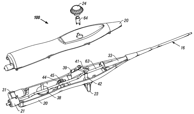
As shown in Figure 1, the apparatus 100 includes a flexible catheter 16
attached to a handle 20, which is attached by a flexible tube 25 to a
cryogenic fluid
unit (not shown). As seen in Figures 16, 17, and 18, a spring wire 4 and a
pull wire 5
are incorporated into the catheter 16, to facilitate a controlled deflection
of the distal
portion of the catheter 16.
As shown in Figures 8, 16, 17, and 18, the distal tip 1 of the catheter 16 is
a
closed end hollow tube which can be machined, formed, cast or molded from a
highly
conductive metal, preferably copper. The copper can be gold plated to insure
biocompatibility. Proximal to the catheter tip 1, there can be a tip union 3
formed
from a weldable metal, preferably stainless steel. The tip union 3 and the
catheter tip
1 can be attached and hermetically sealed together by soldering or brazing.
The tip
CA 02457150 2004-02-18
WO 03/015651 PCT/US02/26713
4
union 3 can in turn be attached to a particularly flexible segment at the
distal end of
the catheter 16.
Within the chamber 2 of the catheter tip 1, a plurality of electrical
conductors
7a,7b,7c,7d can be attached, for the transmission of electrical signals. The
electrical
conductors 7a,7b,7c,7d can be seen best in Figures 7 and 9 through 13. Two of
the
attached conductors can form a thermocouple, preferably a T type with one wire
material being copper and the second being thermocouple grade constantan. A
third
conductor, preferably formed of nickel, can be attached to the interior of the
catheter
tip 1, for monitoring of electro-physiological signals. The electrical
conductors can
1o be coated with an insulating material, such as polyimide. A capillary tube
6 can
terminate, at a distal end, in the chamber 2 of the catheter tip 1. The
capillary tube 6
preferably has inner and outer diameters of .010 inches and .016 inches,
respectively.
The distal orifice of the capillary tube 6 can be located approximately .OS to
.07
inches proximal to the distal end of the catheter tip 1. The capillary tube 6
is the
distal extension of a high-pressure refrigerant fluid line 29 which extends
proximally
through the catheter 16, the handle 20, and the flexible tubular connection 25
to the
cryogenic unit. The distal portion of the capillary tube 6 and its distal
orifice
comprise a Joule Thomson expansion element.
Welded to the interior surface of the tubular tip union 3 are two metal
components, a spring wire component 4 and a pull wire component 5, both
preferably
stainless steel, which are located diametrically opposed to each other. The
spring
wire component 4 is composed of multiple flat wires, each of which is
essentially
rectangular in cross section, with each rectangular wire having one cross-
sectional
dimension significantly greater than the cross-sectional dimension
perpendicular
thereto. This spring wire component 4 extends proximally from the tip union 3
through, and just proximal to, the flexible segment of the catheter 16.
The flat wires are stacked and attached to each other in the spring wire
component 4 to essentially form a leaf spring. More specifically, the spring
wire
component 4 consists of a base flat wire with a length slightly longer than
the length
of the distal flexible segment of the catheter 16. Near the proximal end of
this base
flat wire are stacked additional flat wires of progressively shorter lengths,
with each
having a proximal end terminating preferably a short distance distal to the
proximal
CA 02457150 2004-02-18
WO 03/015651 PCT/US02/26713
end of the base wire. In the preferred embodiment, there are at least three of
these
additional flat wires, with at least some of these having progressively
shorter lengths
than the base flat wire. All of the stacked flat wires preferably have similar
rectangular cross-sectional dimensions.
5 The distal end of the base wire of the spring wire component 4 is firmly
bonded or welded to the tip union 3 distal to the flexible catheter segment,
and the
proximal end of the base wire is firmly bonded or welded to a shaft union 15
proximal
to the flexible catheter segment. The essentially rectangular leaf spring 4
functions as
a spine through the flexible segment of the catheter 16, with the smaller
cross-
sectional dimension of the spine 4 defining a direction in which deflection of
the
flexible segment of the catheter 16 will occur. The spine 4 also resists
deflection of
the flexible catheter segment in a direction perpendicular to the defined
direction of
deflection.
The second metal component attached to the tip union 3 is a pull or tendon
wire component 5 which, when axially tensioned, imposes a bending moment on
the
flexible segment of the catheter 16, with a resulting deflection in the
direction defined
by the spine component 4. The tendon wire 5 extends proximally from the tip
union 3
to a deflection mechanism in the handle 20.
Located proximally from the catheter tip 1 is a multi-lumen core tube 9, which
extends proximally, from a point approximately two catheter diameters proximal
to
the catheter tip 1, through the flexible segment of the catheter 16. The core
tube 9 can
be extruded from a polymer material having a balance between its structural
properties and its elastomeric properties. A preferred material for the core
tube
extrusion 9 is Pebax. The core tube 9 may consist of a continuous segment, or
several
axially arranged segments of Pebax. For a continuous core tube 9, the hardness
and
the elastic modulus are constant throughout its length. For the multiple
segment
embodiment, each segment of core tube 9 can have a hardness and an elastic
modulus
less than the hardness and elastic modulus of the adjacent segment,
progressing
proximally to distally. This results in a core tube 9 which is softer and more
flexible
near its distal end than near its proximal end.
As shown in Figure 14, the core tube 9 has multiple lumens, which can be
geometrically shaped and positioned to give the flexible segment of the
catheter 16 a
CA 02457150 2004-02-18
WO 03/015651 PCT/US02/26713
6
mass moment of inertia lower in the defined direction of deflection than in
the
direction perpendicular to the direction of deflection. The preferred
embodiment of
the core tube 9 contains five lumens 1 Oa, l Ob, l Oc, l Od,10e. The core tube
9 has a
central lumen lOd for passage of the tendon wire S, and a rectangular lumen
10e
positioned outwardly from the central lumen 10d. The rectangular lumen 10e is
for
passage of the spine wire 4. Diametrically opposite the rectangular lumen 10e,
on the
other side of the central lumen 10d, is located a half annular shaped lumen
10a,
through which the capillary tube 6 passes. This half annular lumen 10a also
provides
a return path for low pressure refrigerant gas. Two additional lumens 1 Ob, l
Oc located
l0 outwardly from the central lumen lOd carry the aforementioned electrical
conductors
7a,7b,7c,7d.
Located at the distal and proximal ends of the core tube 9 are two rigid multi-
lumen coupler elements 8,11, preferably fabricated from a metal such as
stainless
steel. As shown in Figures 17 and 18, each coupler 8,11 is a multi-lumen
tubular
structure with an outer diameter equivalent in size to the outer diameter of
the core
tube 9. The preferred embodiment of the coupler 8,11 is a tubular structure
with at
least three lumens 12a,12b,12c, as shown in Figure 15. These are a center
circular
lumen 12c, an essentially oval lumen 12b located outwardly from the center
lumen
12c, and a partial annular lumen 12a that essentially encircles about 3/4 of
the
circumference of the center lumen 12c. In the catheter assembly, the center
lumen
12c of each coupler 8,11, through which the tendon wire 5 passes, axially
aligns with
the center lumen lOd of the core tube 9. The oval lumen 12b of each coupler
8,11,
through which the spine wire 4 passes, aligns axially with the rectangular
lumen 10e
of the core tube 9.
The distal coupler 8 is encased in the tip union 3, and the proximal coupler
11
is encased or captured in the shaft union 15, which is also a stainless steel
tube. In the
preferred embodiment, the shaft union I S is thin-walled, preferably having a
wall
thickness less than about .003 inch, and it has a length at least five times
longer than
the proximal coupler 11. The proximal coupler 11 is rigidly held within the
shaft
union 15 by mechanical means, such as a swage or bezel, or by soldering means,
brazing means, welding means, or a combination of the cited means.
CA 02457150 2004-02-18
WO 03/015651 PCT/US02/26713
7
In another embodiment shown in Figures 19 and 20, instead of the core tube 9,
a tubular compression spring 62 extends proximally through the flexible
segment of
the catheter 16. The tubular spring 62 is located proximally from the tip
union 3 and
firmly attached thereto, by being bonded, welded, soldered, or brazed. The
tubular
spring 62 is composed of a flat wire having a rectangular cross section, with
the
smaller of the rectangular dimensions directed radially from the center of the
tubular
shape, and with the greater of the rectangular dimensions directed
substantially
axially along the tubular shape. The pitch between coils of the tubular spring
62 is
designed to enable bending of the tubular spring 62 perpendicular to the axis
of the
catheter 16. The pitch may be fixed or variable. In the preferred embodiment,
the
proximal portion of the tubular spring 62 has a smaller gap between coils than
the
distal portion of the tubular spring 62, causing the tubular spring 62 to be
more
flexible near its distal end. The tubular spring embodiment also has a multi-
lumen
proximal coupler 11 and a shaft union 15.
Inserted into, and rigidly fixed to, the center lumen 12c of the proximal
coupler 11 is a sheath union 17. The sheath union 17 is a single lumen formed
metal
tube. In the preferred embodiment, the sheath union 17 is firmly held to the
proximal
coupler 11 by mechanical means, or by being soldered, brazed or welded to the
center
lumen 12c of the proximal coupler 11. Inserted into, and rigidly fixed to, the
center
lumen 12c of the distal coupler 8 is a distal coupler union 19. The distal
coupler
union 19 is a single lumen formed metal tube with a flared distal end. In the
preferred
embodiment, the distal coupler union 19 is firmly held to the distal coupler 8
by
mechanical means, or by being soldered, brazed, or welded to the center lumen
12c of
the distal coupler 8.
The pull or tendon wire S passes from the tip union 3 through the distal
coupler union 19, then through the center lumen 1 Od of the core tube 9 or
through the
spring tube 62, then into and through the sheath union 17. The essentially
rectangular
spine 4 passes through the oval lumens 12b of the couplers 8,11 and into the
catheter
shaft union 15. The spine 4 may be firmly attached to the shaft union 15 by
welding
means. The sensor wires 7a,7b,7c,7d passing through the core tube 9 or the
spring
tube 62 freely pass unobstructed through the partial annular lumens 12a of the
couplers 8,11. Also passing through the partial annular lumens 12a of the
couplers
CA 02457150 2004-02-18
WO 03/015651 PCT/US02/26713
8
8,11 is the capillary tube 6 on the distal end of the high pressure fluid line
29. The
portions of the lumens 12a,12b,12c of the couplers 8, I 1 not taken up by
wires and
tubes make up the low pressure refrigerant gas return.
A flexible jacket 14 covers all of the catheter elements from the shaft union
15
to the tip union 3, encasing the core tube 9 or the spring tube 62, and all
other internal
elements. The flexible jacket 14 is a tube extruded from an elastomeric
polymer with
a hardness and modulus of elasticity less than or equal to the material of the
core tube
9. The jacket 14 has sufficient wall thickness to maintain circularity without
buckling, during the bending of the jacket 14 around a one inch radius,
through a 180
1o degree angle. In the preferred embodiment, the jacket 14 has a length of
about 5
centimeters, a diameter of about .130 inch and wall thickness of about .020
inch. The
flexible tubular jacket 14 can be firmly attached to the distal portion of the
outer
diameter of the shaft union 15 and to the proximal portion of the outer
diameter of the
tip union 3, by a combination of adhesive bonding and thermal fusion. The
jacket
tube 14 can also be thermally fused to the core tube 9 or the spring tube 62.
In the
embodiment using the spring tube 62, the spring tube 62 can impart additional
hoop
strength to the jacket tube 14, thereby preventing buckling during bending.
The
adhesive bonding and thermal fusing of the jacket tube 14 to the tip union 3
and the
shaft union 15 creates a hermetically sealed cavity extending from the
catheter tip 1 to
the shaft union 15.
Two millimeters proximal to the catheter tip 1, a sensor band 13, preferably
formed from platinum, is swaged, fitted or bonded around the flexible jacket
tube 14.
Conductively attached to the platinum sensor band 13 is a nickel wire, which
is
passed through the wall of the jacket tube 14, and either into and through one
of the
conductor lumens I Ob, l Oc of the core tube 9 or between the inner diameter
of the
jacket tube 14 and the outer diameter of the spring tube 62, passing
proximally past
the shaft union 15. The sensor band 13 and the nickel wire comprise a means
for
sensing ECG electrical impulses.
A tightly wound wire coil sheath 18 encases the pull or tendon wire 5. The
3o sheath 18 terminates on its distal end within the proximal portion of the
sheath union
17 and is attached thereto. The sheath 18 extends proximally through the
catheter 16
into the handle 20. The sheath I 8 preferably has an outer diameter of about
.021 inch,
CA 02457150 2004-02-18
WO 03/015651 PCT/US02/26713
9
and is fabricated of tightly wound .003 inch diameter wire. During deflection
of the
tip, axial displacement and tensile force are imposed upon the pull or tendon
wire 5.
The sheath 18 prevents axial compression of the catheter body 16. While
preventing
axial compression of the catheter body 16, the coils of the sheath 18 pack
together,
and the sheath 18 behaves as an incompressible body, thereby allowing
efficient
transmission of tensile force and axial displacement to the flexible portion
of the
catheter 16, which results in the deflection of the flexible portion of the
catheter 16.
Connected, bonded and thermally fused to the shaft union 15 and the flexible
jacket tube 14 is the main catheter shaft 63. The catheter shaft 63 is a
tubular element
with an outer diameter comparable in size to the outer diameter of the
flexible jacket
14, and with an inner diameter comparable to the outer diameter of the shaft
union 15.
The catheter shaft 63 is a composite structure designed to transmit torque to
the
catheter tip 1 and the flexible portion of the catheter 16 during manipulation
of the
catheter 16.
In one embodiment, the catheter shaft 63 includes a relatively stiff thin
walled
inner tube of thermoplastic, such as polyimide. A stainless steel wire braid
is placed
over the polyimide tube, and a more flexible polymer covers the wire braid. In
this
embodiment, the inner polyimide tube has a thickness of about .0015 to about
.002
inch, the braid is woven from .001 inch wire, and the outer layer is a
flexible polymer
such as Pebax. The flexible outer layer thickness is significantly greater
than the
inner polyimide tube, preferably about .010 to about .015 inch. The catheter
shaft 63
terminates on its distal end at the shaft union 15 and the flexible segment of
the
catheter 16. The catheter shaft 63 extends proximally through the handle 20,
terminating proximal to the handle 20.
In another embodiment, the catheter shaft 63 is comprised of a thermoplastic
extrusion with an embedded stainless steel braid. The hardness and elastic
properties
of the extrusion, and the pitch and number of wires in the braid are chosen to
give the
desired torque transfer properties to the catheter shaft 63, as is well known
in the art.
The sensor conductors 7a,7b,7c,7d, the sheath-encased pull wire 5, and the
3o capillary tube 6 exit the proximal coupler 11, enter into and pass through
the catheter
shaft 63, and exit the catheter shaft 63 within the interior of the handle 20.
An
additional small diameter tube, the gauge tube 22, is contained within the
catheter
CA 02457150 2004-02-18
WO 03/015651 PCT/US02/26713
shaft 63 for monitoring of the return fluid pressure. The gauge tube 22 has a
preferable outer diameter of about .029 inches and inner diameter of about
.024
inches. The gauge tube 22 terminates on its distal end adjacent to the
proximal
coupler 11 and extends proximally through the catheter shaft 63, exiting the
catheter
5 shaft 63 within the interior of the handle 20.
As shown in Figure 7, a sheath tube 34 is employed about the sheath 18. The
sheath tube 34 has a preferable inner diameter of about .024 inch, thereby
allowing
free movement of the sheath 18 within the sheath tube 34. During catheter
usage, the
pressure at the distal end of the sheath tube 34 is below atmospheric. The
sheath tube
l0 34 terminates proximally within the interior of the handle 20, where
pressure is
essentially atmospheric. The length and dimensions of the sheath tube 34 are
designed to provide a high resistance to fluid movement between the interior
of the
catheter 16 and the interior of the handle 20. With the sheath 18 and the
tendon 5
passing through the sheath tube 34, the available space for fluid movement
between
the sheath tube 34 and the sheath 18, and between the sheath 18 and the tendon
S, is
minimal. Utilization of a sheath tube 34 thusly configured allows the sheath
18 and
the tendon 5 components of the deflection apparatus to exit the fluid filled
interior of
the catheter 16 with no subsequent leakage of fluid, thereby eliminating the
need to
hermetically seal the handle 20.
The high pressure capillary tube 6 extends from the catheter tip 1 to a point
about 10 inches proximal to the catheter tip l, where it transitions into a
larger high
pressure tube 29. The transition site is hermetically sealed and can withstand
pressures in excess of 1000 psi, without compromise. The high pressure tube 29
then
extends proximally through the catheter shaft 63 and exits the catheter shaft
63 within
the interior of the handle 20.
As shown in Figure 2, the handle 20 incorporates a means for securing the
catheter shaft 63, the articulation mechanism, an electrical connector or
receptacle 31,
and a pathway for the catheter shaft 63, the high pressure tube 29, and the
gauge tube
22 to pass through. As the catheter shaft 63 enters the handle 20, it is
firmly captured
and bonded into the catheter support 33. The catheter support 33 is a hollow
tubular
structure with features on its proximal end that allow for securing to slots
within the
handle 20.
CA 02457150 2004-02-18
WO 03/015651 PCT/US02/26713
11
The catheter shaft 63 enters the handle 20 on the distal end of the handle 20,
passes through the handle 20, and exits the handle 20 through an exit port on
the
proximal end of the handle 20. Four exit site holes are made in the wall of
the
catheter shaft 63 within the handle 20. The exit site holes are drilled or cut
preferably
at an angle of about 10 to 15 degrees off the axis of the catheter shaft 63,
thereby
allowing tubes within the catheter shaft lumen to exit without deformation or
buckling. One exit site hole (not shown) is provided to allow the high
pressure tube
29 to exit the catheter shaft 63. Another exit site hole (not shown) is
provided to
allow the gauge tube 22 to exit the catheter shaft 63. A third exit site hole
46 is
l0 provided to allow the sensor wires 7a,7b,7c,7d to exit the catheter shaft
63. A fourth
exit site hole is provided to allow the sheath tube 34, the sheath 18 and the
tendon
wire 5 to exit the catheter shaft 63.
In the preferred embodiment, the high pressure tube 29 exits the catheter
shaft
63 within the handle 20 at the most proximal location, extends essentially
parallel to
the catheter shaft 63, and exits the handle 20 through the exit port on the
proximal end
of the handle 20. A hermetic seal is placed about the juncture where the high
pressure
tube 29 exits the catheter shaft 63. Just distal to the high pressure tube
exit site hole,
is the gauge tube exit site hole. In the preferred embodiment, the gauge tube
22 exits
the catheter shaft 63 within the handle 20, extends essentially parallel to
the catheter
shaft 63, and exits the handle 20 through the exit port on the proximal end of
the
handle 20. A hermetic seal is placed about the juncture where the gauge tube
22 exits
the catheter shaft 63. At a site slightly distal to the gauge tube exit site
hole, the
sensor wires 7a,7b,7c,7d exit the shaft 63, pass across the handle 20 and are
conductively connected, soldered, or crimped to an electrical receptacle 31.
Hermetic
seals are placed about the connection of the wires to the receptacle 31 and
about the
wire exit site hole 46 on the shaft 63.
At a site just proximal to the point where the catheter shaft 63 enters the
handle 20, the sheath tube 34, the sheath 18 and the tendon wire 5 exit the
catheter
shaft 63. A hermetic seal is place about the sheath tube 34 exiting the
catheter shaft
63. The tightly wound coil spring which makes up the sheath 18 exits the
sheath tube
34, is looped slightly, and then transitions into a larger tightly wound coil
spring, the
sheath extension 35,36. The loop 37 in the sheath 18 as it exits the catheter
shaft 63 is
CA 02457150 2004-02-18
WO 03/015651 PCT/US02/26713
12
a service loop which allows the sheath 18 to move independently of the
catheter shaft
63, thereby preventing the imposition of tensile or compressive forces on the
catheter
shaft 63.
The sheath extension 35,36 passes through, and is firmly bonded, welded,
soldered, or brazed to an adjustment screw 44 with an attached adjustment nut
45.
The adjustment screw 44 and nut 45 are securely positioned within the handle
20.
Rotation of the adjustment nut 45 on the screw 44 moves the screw 44 and the
attached sheath extension 35,36 distally or proximally, depending on the
direction of
rotation of the nut 45. Use of the adjustment screw 44 and nut 45 allows for
fine
l0 adjustment of the service loop 37 of the sheath 18. The adjustment screw 44
also
divides the sheath extension 35,36 into a compressive segment 35 distal to the
screw
44, and a tensile segment 36 proximal to the screw 44. The purpose of this
division
will become apparent later.
The sheath extension 35,36 and the enclosed tendon wire 5 exit the proximal
side of the adjustment screw 44 and pass around a pulley 38, to a point where
they are
both firmly connected, preferably swaged or crimped, to a swivel connector 39.
In
this connector 39, the proximal end of the tightly wound coil spring of the
sheath
extension 36 and the proximal end of the tendon wire 5 are joined together.
The
swivel connector 39 is fastened to a lever arm 41 and allowed to swivel about
the
connection point. The lever arm 41, an axle 42, and an activation lever 23
make up
the deflection lever mechanism
Movement of the activation lever 23 in one direction rotates the axle 42,
which in turn moves the lever arm 41 to pull on the tendon wire 5 and the
sheath
extension 36. Movement of the lever arm 41 in this direction imparts a
proximally
directed displacement to both the tendon wire 5 and the proximal portion of
the sheath
extension 35. This proximal displacement is transmitted down the tendon wire S
to
the distal end of the distal flexible segment of the catheter 16. The initial
portion of
the proximal displacement works to compress the tightly wound coil of the
sheath 18.
The sheath 18 stiffens and prevents any further proximal displacement, and
prevents
compressive force from being transmitted to the catheter shaft 63, thereby
allowing all
remaining displacement to be used to effect a bending of the distal bendable
segment
of the catheter 18. During activation of tip deflection, the sheath 18 and the
sheath
CA 02457150 2004-02-18
WO 03/015651 PCT/US02/26713
13
extension 35 extending from the shaft union 15 in the catheter 16 to the
adjustment
screw 44 in the handle 20 are under compression. The sheath extension 36
extending
from the proximal side of the adjustment screw 44 to the lever arm 41 is under
tension.
Release of the activation lever 23 will cause the portion of the sheath
extension 36 which is extending from the proximal side of the adjustment screw
44 to
recoil and bring the lever mechanism back to its initial position. This forces
the
tendon wire 5 toward the catheter tip l, and along with the assistance of the
spine
wire 4 and the elastic properties of the distal jacket tube 14, this results
in a
to straightening of the distal deflection segment of the catheter 16. During
activation of
the deflection mechanism, the service loop 37 in the sheath 18 inside the
handle 20
allows the catheter 16 to be bent without affecting tip deflection.
A locking or braking mechanism is employed on the deflection lever
mechanism to allow the user to set a desired level of tension in the
articulation
t5 mechanism to restrain the recoil action of the sheath extension 36, and
this level of
tension will then be held throughout the articulation of the tip. Also, by
tightening the
brake knob 24, the tension level can even be set high enough to lock the
movement of
the articulation mechanism to hold deflection of the distal portion of the
catheter 16 in
any desired position from 0 to 270 degrees. Tightening of the brake knob 24
imparts
20 an axial force to the tension shaft 64 by means of a metal threaded insert
(not shown)
that is pressed into the brake knob 24. The two tabs of the tension shaft 64
in turn
apply compression to drag washers (not shown). Reactive force generated by the
drag
washers forces the lever shaft 42 against the side of the handle 20, resisting
rotation of
the lever shaft 42.
25 Extending proximally from the handle 20 is a larger flexible tube 25, the
flex
line, which houses the proximal portion of the catheter shaft 63, the high
pressure
fluid line 29, and the gauge line 22, as shown in Figures 2 and 6. In the
preferred
embodiment, the flex line 25 is a corrugated tube constructed from a polymer
such as
polyethylene. The distal end of the flex line 25 is connected to the handle
20, and its
3o proximal end is connected to a gas line connector 27. Running essentially
parallel
within the flex line 25 are the high pressure fluid line 29, the gauge line
22, and a
continuation of the catheter shaft 63, which is the low pressure fluid line
47. The
CA 02457150 2004-02-18
WO 03/015651 PCT/US02/26713
14
gauge line 22 exits the flex line 25 just distal to the gas line connector 27
and
terminates in a standard luer fitting 30.
As shown in Figures 3, 4, and 5, the high pressure fluid line 29 and the low
pressure fluid line 47 enter into and pass through the gas line connector 27,
with the
low pressure line 47 terminating at the distal portion of a dual gas line
fitting 28, and
with the high pressure fluid line 29 passing all the way through the dual gas
line
fitting 28. The tubes of the low and high pressure fluid lines 47,29 are
potted to the
gas line connector 27 to prevent fluid leakage. Where the low pressure fluid
line 47
terminates, there are orifices 51 for the passage of fluid into a mating
receptacle (not
to shown). Just distal to these low pressure orifices 51 is a quad o-ring 49
which
prevents low pressure fluid leakage when the dual gas line fitting 28 is
inserted into a
mating receptacle (not shown). The high pressure fluid line 29 passes through
the
cavity of the gas line connector 27 and through the dual gas line fitting 28.
At the
proximal extremity is a check valve actuator 53 which is actually a proximal
extension of the high pressure fluid line 29. High pressure orifices 52 are
provided in
the proximal extension of the high pressure fluid line 29, to allow for the
passage of
high pressure fluid into the high pressure fluid line 29. A second quad o-ring
50 is
located about the dual gas line fitting 28 just distal to the high pressure
orifices 52, to
prevent leakage of high pressure fluid when the dual gas line fitting 28 is
inserted into
the mating receptacle (not shown).
The dual gas line fitting 28 has a mating and locking means 48 which allows
the dual gas line fitting 28 to be securely connected to the mating receptacle
(not
shown). The check valve actuator 53 located most proximally on the dual gas
line
fitting 28 acts to open a check valve in the precooler assembly (not shown)
when the
dual gas line fitting 28 is connected to the mating receptacle (not shown).
Conversely, disconnecting the dual gas line fitting 28 from the mating
receptacle (not
shown) breaks contact between the check valve actuator 53 and the check valve
(not
shown), thus closing the check valve, minimizing gas escape from, or pressure
change
within, the cryo refrigerant system.
The invention herein disclosed is fully capable of obtaining the objects
stated,
but this disclosure is merely illustrative of the preferred embodiments of the
invention, and no limitations are intended other than as recited in the
claims.