Note: Descriptions are shown in the official language in which they were submitted.
CA 02460379 2004-03-25
I
CORTICAL AND CANCELLOUS ALLOGRAFT CERVICAL FUSION BLOCK
RELATED APPLICATION
There are no related applications.
FIELD OF INVENTION
The present invention is generally directed toward a surgical implant product
and
more specifically is a shaped allograft cortical cancellous bone block implant
for the fusion of
vertebral bones which is introduced between two vertebral bones to be fused.
BACKGROUND OF THE INVENTION
The use of substitute bone tissue dates back around 1800. Since that time
research
efforts have been undertaken toward the use of materials which are close to
bone in composition
to facilitate integration of bone grafts. Developments have taken place in the
use of grafts to use
materials such as corals, hydroxyapatites, ceramics or synthetic materials
such as biodegradable
polymer materials. Surgical implants should be designed to be biocompatible in
order to successfully
perform their intended function. Biocompatibility may be defineci as the
characteristic of an implant
acting in such a way as to allow its therapeutic function to be manifested
without secondary adverse
affects such as toxicity, foreign body reaction or cellular disruption.
Human allograft tissue is widely used in orthopaedic, neuro-, maxillofacial,
podiatric
and dental surgery. The tissue is valuable because it is biocompatible,
strong, biointegrates in time
with the recipient patient's tissue and can be shaped either by the surgeon to
fit the specific surgical
defect or shaped commercially in a manufacturing environmeint. Contrasted to
most synthetic
absorbable or nonabsorbable polymers or metals, allograft tissue integrates
with the surrounding
tissues.
Allograft bone is a logical substitute for autologous bone. It is readily
available and
precludes the surgical complications and patient morbidity associated with
obtaining autologous
bone as noted above. Allograft bone is essentially a collagen fiber reinforced
hydroxyapatite matrix
containing active bone morphogenic proteins (BMP) and can be provided in a
sterile form. The
demineralized form of allograft bone is naturally both osteoindiuctive and
osteoconductive. The
demineralized allograft bone tissue is fully incorporated in the pa.tient's
tissue by a well established
biological mechanism. It has been used for many years in bone surgery to fill
the osseous defects
previously discussed.
CA 02460379 2004-03-25
,~.
2
Allograft bone occurs in two basic forms; cancellous and cortical. The
cancellous
bone includes void areas with the collagen fiber component conitributing in
part to torsional and
tensile strength. The less dense cancellous bone provides an excellent matrix
for rapid bone
regeneration and repair.
Many devices ofvarying shapes and forms are fabricated from allograft cortical
tissue
by machining. Surgical implants such as pins, rods, screws, anchors, plates,
intervertebral spacers
and the like have been made and used successfully in human surgery. 'These pre-
engineered shapes
are used by the surgeon in surgery to restore defects in bone to the bone's
original anatomical shape.
Injury or disease processes to the head, neck, or shoulders can cause abnormal
forces
to be applied on the cervical vertebra. Arthritis, motion induced "whiplash",
or other trauma create
this malfunction. This situation is often treated_surgically by a pr=ocedure
intended to fuse the two
adjacent cervical or spinal vertebrae to each other. Such fusion relieves the
pressure the partially
displaced vertebrae place on the adjacent spinal nerves.
Many surgical devices have been developed and used successfully to immobilize
and
fuse the misaligned vertebrae. Metal plates screwed into the adjacent
vertebrae work well, but after
some time post-operatively, the stress rise occurring at the screw position
causes erosion of the bone
and resultant slipping. This has been irnproved by placing load-bearing
spacers between the two (or
more) misaligned vertebrae. The spacer is both load-bearing and of a material
which will induce,
or at least support, fusion between the vertebrae.
Removal of damaged or diseased discs, restoration of disc space height and
fusion
of adjacent vertebrae to treat chronic back pain and other ailmerits are known
medical techniques.
Implants such as intervertebral spacers are often iinplanted in the disc space
engaging the vertebrae
to maintain or reestablish disc space height after removal of all oi- a
portion of the disc. The spacers
are fonned of a variety of both resorbable and non-resorbable imaterials,
including, for example,
titanium, surgical steel, polymers, composites and bone. It is currently
considered desirable to
promote fusion between the vertebral bodies that are adjacent to the damaged
or diseased discs.
Typically, an osteogenic material is combined with a spacer and inserted in
the disc space to
facilitate and promote bone growth. While the selection of the implant
configuration and
composition can depend upon a variety of considerations, it is often desirable
to select a resorbable
material that does not shield the bone ingrowth. Bone and bone-derived
components can provide
suitable material to prepare the implants. However, bone material and in
particular cortical bone
acceptable for use in implants is a scarce resource, being derived from
limited number human tissue
donor resources.
CA 02460379 2004-03-25
' 4 c =
3
Suitable bone or bone-derived material for use in implants, in general, is
almost
exclusively obtained from allograft and xenograft sources, both of which
comefrom a limited
supply. Since intervertebral spacers must withstand the compressive loads
exerted by the spine, these
implants are often cortical bone which has the mechanical strength suitable
for use in any region of
the spine. Cortical spacers are often shaped from cortical long bones, which
are primarily found in
the lower limbs and include, for example, femur, fibula, and the tibia bones.
However, these long
bones make up only a fraction of the available bone source. Cancellous bone,
because of its superior
osteoinductive properties, would be desirable to sue in the spinal implant.
However, the lower
mechanical strength ofcancellous bone prohibits its use in many surgical
applications. Thus, sources
of bone suitable for structural intervertebral spacers are extremely limited.
The scarcity of desired
donor bone makes it difficult to provide implants having the desired size and
configuration for
implantation between vertebrae, which can require relatively large implants.
It is further anticipated
that as the population ages there will be an increased need for correction for
spinal deformities and
a concomitant increase in the demand for bone-derived components. Therefore,
these structural
bone portions must be conserved and used efficiently to provide implants. The
scarcity of suitable
bone material has also hindered efforts to design and manufacture varying
configurations of suitable
implants for arthodesis of the spine. Further, various implant configurations
have not been
physiologically possible to obtain given the structural and geometrical
constraints ofavailable donor
bone.
One known treatment for fusing two vertebrae is the insertion of a suitably
shaped
dowel into a prepared cylindrical cavity which reaches the two vertebrae to be
fused. The dowel
used is preshaped bone or allogra.ft bone.
A number of allograft bone spacers have been used in surgery as spacers. They
are
commonly called the ACF spacer constructed as a cortical bone cross section,
shaped like a washer
with teeth to discourage graft explusion and an axial center hole; a VG3
cervical spacer constructed
with two rarnp shaped cortical plates held together with cortical pins, the
top and bottom surfaces
being ridged to discourage graft expulsion; an ICW spacer constructed with an
elongated C spaced
cortical portion with a cancellous inside to allow rapid ingrowth (slice of
iliac crest) and a SBS
spacer constructed with a single piece cortical member with serrated top and
bottom surfaces and
an axial center hole.
The ICW (iliac crest wedge) has been used for a long time for cervical spine
fusion
and has a total load bearing force around 4500 Newtons. Testing has noted that
cervical vertebrae
fail in compression at about 2000 Newtons. The ICW spacer suffers from high
unit variability
CA 02460379 2004-03-25
t =
4
because of its natural, anatomic variations.
United States Patent No. 5,972,368 issued on October 26, 1999 discloses the
use
of cortical constructs (e.g. a cortical dowel for. spinal fusion) wluch are
cleaned to remove all of
the cellular material, fat, free collagen and non-collagenous protein leaving
structural or bound
collagen which is associated with bone mineral to form the trabecular struts
of bone. The shaped
bone is processed to remove associated non-collagenous bone proteins while
maintaining native
bound collagen materials and naturally associated bone minerals. The surface
ofa machined cortical
bone is characterized by a wide variety of openings resulting from exposure by
the machining
process of the Haversian canals present throughout cortical bone. These canals
serve to transport
fluids throughout the bone to facilitate the biochemical processes that occur
at variable angles and
depths within the bone.
An attempt to solve the increasing bone supply problems using a combined
cortical
and cancellous bone block is shown in United States Patent Nurnber 4,950,296
issued August 21,
1990 which uses a cubically configured cortical shell defming a through going
internal cavity and
a cancellous plug fitted into the cavity so that the end surfaces of the
cancellous plug are exposed.
Another reference, WIPO Patent Publication Number WO 02/24122 A2, published
March 28, 2002
owned by SDGI Holdings Inc. show various intervertebral spacers formed
ofcortical and cancellous
bone composites such as sandwiches, with intersecting ribs and rods.
U.S. Patent Number 6,294,187 issued September 25, 2001 is directed toward an
shaped osteimplant of compressed bone particles. The shaped implant is disc
sha.ped and has a
number of holes drilled therein for macroporosity and the holes can be filled
with an osteogenic
putty material.
Conversely, WIPO Patent Publication Number WO 02/07654 A2, published January
31, 2002 discloses intervertebral spacers formed of dense cancellous human or
animal bone. In one
embodiment, a cortical rod or cortical rods are placed in bores cut through a
cancellous bone block
to provide load bearing strength with the ends of the rods being exposed on
both sides of the
cancellous bone block. Another embodiment shows a C shapeci cortical block
with a cancellous
plug inserted into the recess of the C to form a rectangular spacer. A pin is
inserted through a bore
cut through the legs of the C block and through the cancellous plug to keep
the cancellous plug
positioned with the recess of the cortical component. U.S. Patent Number
6,379,385 issued April
30, 2002 also discloses the use of a spongy block having a plurality of
cortical rods mounted in
through going bores cut through the bone block. In another embodiment, a X-
shaped cortical
support member is mounted therein to provide structured strength to the
composite implant.
CA 02460379 2004-03-25
It is also known to mate various bone components together to form a single
implant.
In this regard, see, Albee, Bone Graft Surgery in Disease, Iniw.y and
Deformity, (1940), pp. 30,
which uses a tongue in groove and dove tail to hold separate pieces of bone
together for implant
use, and U. S. Publication No. US2002/0029084 Al, published M[arch 7, 2002,
which shows a three
component implant with a center core surrounded by two outer semicircular
portions. The outer
portions have alternative dove tail joints on adjacent bone portions to secure
the outer portions
together forming a dowel shaped bone implant.
Consequently, there is a need for an implant which should have with a load
bearing
compressive strength of 1000 to 5000 Newtons with a compressive load to be a
minimum of 3000
Newtons as a safety factor. There is also a need to liave a portion of
cancellous bone inunediately
adjacent to the load bearing cortical zone to permit rapid ingrowtlh ofa
patient's own new bone with
the cancellous bone forming the major part of the implant.
SUMMARY OF THE INVENTION
The composite allograft cervical fusion block is directed toward a two piece,
mated
bone fusion block or spacer constructed with one component member of load
bearing material
preferably cortical bone and the other component member made of cancellous
bone for use in
orthopedic surgical procedures. The cortical bone member defines a dove tail
shaped projection
extending from its outer surface with the cancellous component member having a
dove tail recess
cut therein to receive the dove tail projection of the cortical meimber. A
plurality of bores are cut
through the cortical bone member and into the cancellous member to hold pins
which are angularly
, inserted into the bores along opposite sides of the dove tail projection and
recess and through the
head of the cortical member to limit axial and lateral movementõ
Additional embodiments include the use of a single throughgoing pin, a single
transverse throughgoing pin, multiple dove tails, a bulbous projection
substituted for the dove tail
and opposing load bearing members.
It is an object of the invention to use a bone block geometry to provide a
composite
bone block ofcancellous and cortical bone components having perfonnance
characteristics that meet
or exceed conventional spinal fusion requirements.
It is another object of the invention to utilize a shaped cortical cancellous
bone
implant block which provides the mechanical stretigth characteristics that can
withstand compression
forces and provide overall strength and durability to the structure.
CA 02460379 2007-02-13
6
It is still another object of the invention to provide a spinal fusion implant
which
uses load bearing component member to take up the high forces which can arise
between
two vertebral bodies and a relatively porous cancellous component member to
accelerate
the healing process.
It is yet another object of the invention to provide a pre-machined shaped
allograft
bone structure which can effectively promote new bone growth and accelerated
healing.
It is also an object of the invention to create a sterile bone fusion implant,
which is
sterile and which can be easily handled by the physician during surgery which
eliminates or
significantly reduces the physician from having to carve or modify the
respective blocks.
In a broad aspect, the present invention relates to a sterile composite graft
comprising: a cancellous bone component member with angled sides and a load
bearing
component member mounted to said cancellous bone component, said load bearing
component member has an outside exposed surface area ranging from about 15% to
about
40% of the outside exposed surface area of the cancellous member, said
cancellous bone
component member defining a planar engagement surface with a shaped recess cut
into
said planar engagement surface of said cancellous bone component member with
at least a
portion of said shaped recess being larger than an opening leading to the
shaped recess,
said load bearing component member defining an outer base portion with an
inner planar
engagement surface, locking member extending outward from said engagement
surface
correspondingly shaped to fit into said shaped recess holding said cancellous
and load
bearing component members together in a mated relationship and pin means
mounted in
said cancellous bone component member and said load bearing component member.
In another broad aspect the present invention relates to a sterile composite
graft
comprising: a cancellous bone component member and a load bearing component
member
mounted to said cancellous bone component member, said load bearing component
member having an outside exposed surface area ranging from about 15% to about
40% of
the outside exposed surface area of the cancellous component member, said
cancellous
bone component member having a planar outer surface and defining a shaped
recess cut
CA 02460379 2007-02-13
6a
into said planar outer surface into the interior of the cancellous bone
component member,
said load bearing component member having a T' shaped configuration with a
flat inner
surface on the base of the crosspiece and a stem extending outward from said
crosspiece
flat surface, said stem being configured with a distal portion being larger
than its proximal
portion to fit into said shaped recess holding said component members together
in a mated
relationship, said load bearing and cancellous bone component members defining
bores
which are axially aligned when the component members are mated together and
pins
mounted in said axially aligned bores, said pins extending across an
intersection between
said cancellous bone component member and said load bearing component member.
In another broad aspect the present invention relates to a sterile composite
graft
comprising: a cancellous bone component member and cortical bone component
member,
said cancellous bone component defining a U shaped configuration when viewed
from a
top planar view with the legs of the U shaped configuration being provided
with ends
defining a flat surface and the interior of the U shaped configuration forming
a shaped
recess, said cortical bone component member having an outside exposed surface
area
ranging from about 15% to about 40% of the outside exposed surface area
defining a flat
inner surface which seats on said cancellous bone component member legs flat
end
surfaces and a mating member extending from said flat inner surface, said
mating member
being configured to fit into said shaped recess holding a first and second
bone components
together said component members each defining bores which are axially aligned
when the
component members are mated together and pins mounted in said axially aligned
bores,
said pins extending across an intersection between said cancellous bone
component
member and said cortical bone component member.
In another broad aspect, the present invention relates to a sterile composite
bone
graft for use in implants comprising: a load bearing member constructed of
allograft
cortical bone defining a planar surface and a dove tail mating shaped member
extending
from said planar surface, an allograft cancellous member defining tapered side
walls at an
CA 02460379 2007-02-13
6b
angle ranging from about 100 to about 110 from a line drawn through the
longitudinal
center axis, a flat proximal end surface and a flat distal end surface and a
dove tail shaped
recess cut into said flat proximal end surface with the narrowest portion of
said recess
exiting said flat proximal end surface, said load bearing member and
cancellous member
being mated together, said load bearing member having an outside exposed
surface area
ranging from about 15% to about 40% of the outside exposed surface area of
said load
bearing member.
In another broad aspect, the present invention relates to a sterile composite
graft
comprising: a cancellous bone component member with tapered side walls and two
end
cortical bone component members mounted to said cancellous bone component
member,
said cancellous bone component defining a flat surface on each end with at
least one dove
tail shaped recess formed in each end surface, each cortical bone component
member
defming a flat inner surface which seats on said cancellous bone component
member end
flat surface so that said cortical component members are opposite each other
and at least
one dove tail shaped mating member extending from said flat inner surface,
each dovetail
mating member being configured to fit into a corresponding dove tail shaped
recess
holding said bone components together, said cortical bone component members
having an
outside exposed surface area ranging from about 30% to about 40% of the
outside exposed
surface area of the cancellous bone component member, said cortical and
cancellous bone
component members defining at least one bore which is axially aligned when the
component members are mated together and at least one pin mounted in said
axially
aligned bores extending across an intersection between said cancellous bone
component
member and said load bearing component members.
These and other objects, advantages, and novel features of the present
invention
will become apparent when considered with the teachings contained in the
detailed
disclosure. This disclosure, along with the accompanying drawings and
description,
CA 02460379 2007-02-13
6c
constitutes a part of this specification and illustrates embodiments of the
invention which
serve to explain the principles of the invention.
BRIEF DESCRIPTION OF THE DRAWINGS
Figure 1 is a perspective view of the inventive composite cortical and
cancellous
component bone implant;
Figure 2 is a top plan view of the implant of Figure 1;
Figure 3 is a perspective view in phantom of the cancellous component of
Figure 1;
Figure 4 is an enlarged top plan view of the cancellous component of Figure 3
showing the bores in phantom;
Figure 5 is an enlarged front right side elevational view of the cancellous
component of Figure 3;
Figure 6 is a right side elevational view of the cancellous component of
Figure 3;
Figure 7 is a top plan of the cortical component of Figure 1;
Figure 8 is a front elevational view of the cortical component of Figure 7
showing
the dove tail in phantom;
Figure 9 is a side elevational view of the cortical component of Figure 7;
Figure 10 is a perspective view of an alternative embodiment of the composite
cortical and cancellous bone implant with a single throughgoing bore running
along the
centre axis ofthe dove tail;
Figure 11 is a top plan view of the implant of Figure 10 showing the bore in
phantom;
CA 02460379 2004-03-25
7
Figure 12 is a perspective view of an alternative embodiment of the composite
cortical and cancellous bone implant with a single throughgoing bore running
transverse the axis of
the dove tail;
Figure 13 is a top plan view of the implant of Figure 12 showing the bore in
phantom;
Figure 14 is a perspective view of an alternate einbodiment of the composite
cortical
and cancellous composite bone implant with multiple dove tails;
Figure 15 is a top plan view of the implant of Figure 14;
Figure 16 is a perspective view of an alternative enibodiment of the composite
cortical and cancellous composite bone implant with a truncated ellipsoid
interconnection;
Figure 17 is a top plan view of the implant of Figure 16;
Figure 18 is a perspective view of the embodiment of Figure 16 with two angled
bores;
Figure 19 is a top plan view of the implant of Figure 18;
Figure 20 is a perspective view ofan alternate er.nbodiment ofthe composite
cortical
and cancellous composite bore implant with a wider dove tail engagement
structure;
Figure 21 is a top plan view of the implant of Figure 20;
Figure 22 is a perspective view of the irnplant of Figure 20 with a single
bore parallel
to the center axis of the implant;
Figure 23 is a top plan view of the implant of Figure 22 with the bore shown
in
phantom;
Figure 24 is a perspective view of an alternate ernbodiment of the composite
cortical
and cancellous composite bone implant with load bearing menibers positioned at
both ends;
Figure 25 is a top plan view of the implant of F'igure 20;
Figure 26 is a perspective view of the implant of Figure 24 showing two
throughgoing bores;
Figure 27 is a top plan view of the implant of Figure 26 with the bore shown
in
phantom;
Figure 28 is a perspective view of the implant of Figure 24 showing a single
throughgoing bore; and
Figure 29 is a top plan view of the irnplant of Figure 28 showing the bore in
phantom.
DETAILED DESCRIPTION OF THE INVENTION
CA 02460379 2004-03-25
8
The preferred embodiment and best mode of the present invention is shown in
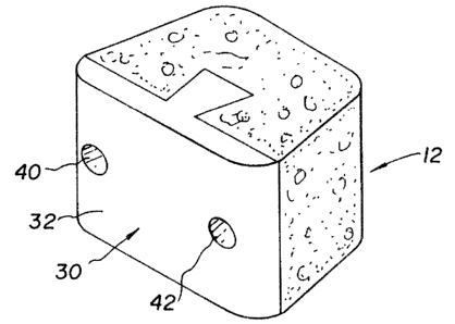
Figures 1 through 9. The composite bone implant block 10 is shown in Figure 1
in accordance with
the present invention.
The composite cortical cancellous bone block body or intervertebral spacer 10
is
preferably constructed with a first component member 12 of denser cancellous
bone taken from
donors age 45 or less cut into a truncated triangle shape. This component
accounts for a large
portion of the graft and provides a large area for bone fusion to occur. The
component member
body has a flat planar front end surface 14 and is provided with a dove tail
shaped recess 16 cut
therein into the interior of the cancellous component body. The dove tail
shaped recess 16 extends
from the access port or opening 15 to the base wall 17 forming the rear of the
recess. The access
entrance opening 15 is preferably about twice as wide as the base 17 of the
recess and the side walls
19 of the recess are angled from 76 to 95 outward from the enltrance opening
15. The cancellous
bone is harvested from a'bone such as a tibia, humerus, patella, calcaneus or
femur. The side walls
18 of the cancellous member 12 are tapered or angled from 100 to 110 ,
preferably at 101 with
a tapered distal side section 20 running into a planar rear wall surface 22.
The cancellous member
12 when implanted in the patient's body encourages tissue, vascularation and
deposition of new
bone.
The cortical cancellous bone block 10 has a T shaped cortical component member
30 with a cross piece 31 having planar outer surface 32 and two tapered or
curved side sections 33
which lead to an inner flat planar surface 34. A dove tai:l shaped projection
36 which has
approximately the same dimensions as dove tail recess 16 cut into the
cancellous member extends
outward from the planar surface 34. The projection or mating r.nember 36 has
angled side walls 37
extending outward at an angle ranging from 70 to 75 to mate with the recess
16. The end 38 of
the dove tail projection 36 is planar. The cortical member 30 has superior
wall strength for support
between load bearing body structures such as vertebrae. While it is noted that
wall surfaces 14 and
34 are flat, these surfaces can be provided with any kind of complementary
construction.
When the composite assembly is lyophilized, the pieces shrink with the
cortical bone
shrinking about 3% and the cancellous bone shrinking a greater amount ranging
from 4% to 8%.
Thus, the dove tail projection 36 will loosely fit into the dove tail recess
1.6 to hold the two
components together. The cortical member 30 has superior wall strength for
support between load
bearing body structures such as vertebrae and has a compressive load ranging
from 2000 to 5000
Newtons, preferably in excess of 3000 Newtons. The composite bone block body
10 height can
CA 02460379 2004-03-25
9
range from 8-12 mm preferably 10 mm depending upon patient needs with a
corresponding length
ranging from 12 to 20 mm, preferably 16 mm with a width ranging from 10 mm to
14 mm
preferably 12 mm, again depending upon surgeon preference atid the size of the
fusion block which
will be used on the individual patient.
Preferably, the load bearing member accounts for about 15% to 40% of the
outside
exposed area of the implant, preferably around 20%, with a volumetric area of
about 10% to about
40% of the implant, preferably around 10% to 20%.
If desired, pins 40 and 42 can be inserted in a through going bores 44 and 46
cut
through both component members 12 and 30 to increase stability to the graft.
The pins 40 and 42
are preferably constructed of cortical bone but can be constructed from any
bio-compatible material
having the necessary strength requirements including metals, plastics
compositions and the like and
are friction fit in the respective bores 44 and 46. The cortical front is
mated to the cancellous
component with the crosspiece inner planar surface being adjacent the
cancellous component. The
cortical or load bearing component bears not only a compressive load but also
serves as an
impaction surface. Thus, the surgeon can tap on the anterior cortical surface
while impacting the
graft without damaging the more brittle cancellous portion of the graft.
In an alternate embodiment of the invention, a single bore 60 is fornled
through the
center of the dove tail and the base of the U as is seen in Figures 10 and 11.
A pin 62 is inserted
through the axially aligned bores ofthe cortical load bearing me!mber 30 and
the cancellous member
12.
In Figures 12 and 13, a bore 70 is cut transverse to the axis of the dove tail
shaped
stem and across the legs of the cancellous member 12 to receive a pin 72 which
provides additional
security to the composite implant.
In Figures 14 and 15, the load bearing member 30 is formed with two dove tail
shaped projections 80 and 82 which fit into correspondingly formed recesses 81
and 83, formed in
cancellous member 12.
In Figures 16 and 17, the load bearing member 30 is formed with a bulbous or
truncated ellipsoid projection 90 which fits. into a similarly configured
recess 92 of the cancellous
member 12. In Figures 18 and 19, the construction of Figure 16 is shown with
two angled bores
94 and 96, cut through the load bearing member 30 and into the cancellous
member 12 holding pins
95 and 97.
Another modification of the invention is shown in Figures 20 and 21 in which a
CA 02460379 2004-03-25
widened dove tail mating member 100 extends from the load bearing member 30.
This widened
dove tail member is at least double the size of the originally shown dove tail
member in Figure 1 and
fits into a similarly sized recess 102 in cancellous member 12 as shown in
Figure 21. An added
feature to the Figure 20 embodiment discloses a bore 110 is cut through the
load bearing member
30 and centered on the widened dove tail 102. The throughgoing bore 110 holds
pin 114.
A double sided load bearing implant is shown in Figures 24 through 29. In this
embodiment, the load bearing members 30 and 130 are mounted on opposite sides
of the cancellous
member 12 which has corresponding recesses 132 and 134 to hold dove tail
projections 133 and
135. The cancellous member 112 is substantially I shaped. Additional component
security can be
accomplished by providing two throughgoing bores 140 and 142 to hold
respective pins 141 and
143, as is shown in Figures 26 and 27. A pin variation is shown in Figures 28
and 29. This variation
uses a single bore 150 running through the center midpoint of the load bearing
members 30 and 130
and the central stem of the I shaped member 112. A pin 152 is inserted into
the aligned bore 150.
It should be noted that all of the embodiments shown in Figures 1 through 29,
that the sidewalls of
the cancellous member are tapered from 100 to 110 and when two facing load
bearing members
30 and 130 are utilized that member 130 has a smaller innet flat surface 136
than the flat surface 138
of load bearing member 30 with the respective member 130 having a smaller area
size than the load
bearing member 30.
While the embodiments shown in Figures 1 through 23 have a volumetric ratio in
which the load bearing member accounts from 10% to 40% of the mass volume of
the composite,
the double load bearing embodiment shown in Figures 24 through 29 has a higher
volumetric mass
in that the load bearing surfaces account for about 30% to about 45% of the
total volume of the
component.
While this operation has been discussed in tertns of using the preferred
embodiment
namely, allograft cortical and cancellous component members ofthe bone blocks,
alternative sources
ofthe components ofthe components of the bone blocks may be substituted such
as xenograft bone
or synthetic graft materials. With any of these alternatives, the bone blocks
may be shaped as
described above. The devices provide the surgeon with a graft that has the
combined and best
characteristics of both cortical and cancellous bone nia.terials.
The cancellous component can be of partially demineralized or mineralized bone
and the load bearing component can be formed of partially surface
demineralized or mineralized
bone.
CA 02460379 2004-03-25
11
The spacers of the present invention were prepared by machining cancellous
bone
from donors, preferably under 45 years of age which have a denser cancellous
structure. Suitable
bones which can be used are calcaneus patella, femoral head, long bone
condyles and talus. Cortical
bone was prepared by machining and was taken from any acceptable donor age.
Suitable bones are
the radius, ulna, femur, tibia, humerus and the talus.
The unique features of allograft bone that make it desirable as a surgical
material are,
its ability to slowly resorb and be integrated into the space it occupies
while allowing the bodies own
healing mechanism to restore the repairing bone to its natural shape and
function by a mechanism
known in the art as creeping substitution.
It is well known that bone contains osteoinductive elements known as bone
morphogenetic proteins (BMP). These BMP's are present within the compound
structure ofcortical
bone and are present at a very low concentrations, e.g. 0.003%. The BMP's are
present in higher
concentrations in cancellous bone. BMP's direct the differentiation of
pluripotential mesenchymal
cells into osteoprogenitor cells which form osteoblasts. The ability of freeze
dried demineralized
bone to facilitate this bone induction principle using BMP present in the bone
is well known in the
art. However, the amount of BMP varies in the bone dependirig on the age of
the bone donor and
the botie processing. Based upon the work of Marshall Urist as shown in United
States Patent
Number 4,294,753, issued October 13, 1981 the proper demineralization of
cortical bone will
expose the BMP and present these osteoinductive factors to the surface of the
demineralized
material rendering it significantly more osteoinductive. The removal of the
bone mineral leaves
exposed portions of collagen fibers allowing the addition of BMP's and other
desirable additives to
be introduced to the demineralized outer treated surface of the bone structure
and thereby enhances
the healing rate of the cortical bone in surgical procedures. In cancellous
bone the structure is not
as dense as cortical bone exposing the naturally occurring BMP's rendering the
entire structure with
biological properties similar to full demineralized bone (DBM).
It is also possible to add one or more rhBMP's to the bone by soaking and
being
able to use a significantly lower concentration of the rare and expensive
recombinant human BMP
to achieve the same acceleration of biointegration. The addition of other
useful treatment agents
such as vitamins, hormones, antibiotics, antiviral and other therapeutic
agents could also be added
to the bone.
Any number of medically useful substances can be incorporated in the
cancellous
component member or load bearing member by adding the medically useful
substances to the same.
Such substances include collagen and insoluble collagen derivatives,
hydroxyapatite and soluble
CA 02460379 2004-03-25
12
solids and/or liquids dissolved therein. Also included are antiviricides such
as those effective against
HIV and hepatitis; antimicrobial and/or antibiotics such as erythromycin,
bacitracin, neomycin,
penicillin, polymyxin B, tetracycline, viomycin, chloromycetin and
streptomycin, cefazolin,
ampicillin, azactam, tobramycin, clindamycin, gentamycin and silver salts. It
is also envisioned that
amino acids, peptides, vitamins, co-factors for protein synthesis; hormones;
endocrine tissue or
tissue fragments; synthesizers; enzymes such as collagenase, peptidases,
oxidases; polymer cellpl
scaffolds with parenchymal cells; angiogenic drugs and polynleric carriers
containing such drugs;
collagen lattices; biocompatible surface active agents, antigenic agents;
cytoskeletal agents; cartilage
fragments, living cells and cell elements such red blood cells, white blood
cells, platelets, blood
plasnla., pluripotential cells, chondrocytes, bone marrow cells, mesenchymal
stem cells, osteoblasts,
osteoctasts and fibroblasts, epithelial cells and endothelial cells present as
a concentration of l05 and
106 per cc of a carrier, natural extracts, tissue transplants, bioadhesives,
transforming growth factor
(TGF-beta), insulin-like growth factor (IGF-1); platlet derived growth factor
(PDGF), fibroblast
growth factor (FGF) (numbers 1-23), osteopontin, vascular endothelial growth
factor (VEGF),
growth hormones such as somatotropin, cellular attractants and attachment
agents, blood elements;
natural extracts, tissue transplants, bioadhesives, bone digestors; antitumor
agents; fibronectin;
cellular attractants and attachment agents; immuno-suppressants; permeation
enhancers, e.g. fatty
acid esters such as laureate, myristate and stearate monoesters of
polyethylene glycol, enamine
derivatives, aipha-k.eto aldehydes can be added to the composition.
While the present invention is described for use in the cervical spine, it is
also suitable
for use in the lumbar and/or thoracic spine. Th implant can be provided in a
variety of sizes, each
size configured to be inserted between a specific pair of acljacent vertebrae.
For example, the
implant can be provided in selected dimensions to maintain disc lieight,
correct lordosis,.kyphosis
or other spinal deformities.
The principles, preferred embodiments and modes of operation of the present
invention have been described in the foregoing specification. However, the
invention should not be
construed as limited to the particular embodiments which have been described
above. Instead, the
embodiments described here should be regarded. as illustrative rather than
restrictive. Variations and
changes niay be made by others without departing from the scope of the present
invention as defined
by the following claims: