Note: Descriptions are shown in the official language in which they were submitted.
CA 02460949 2006-12-12
SKIN LESION EXCISER AND SKIN-CLOSURE DEVICE THEREFOR
BACKGROUND OF THE INVENTION
1. Field of Invention.
10002) The present invention relates to the excision of skin tags, moles,
lesions and other
types of discrete patches or points on the skin (herein collectively referred
to as lesions) from
a human or animal.
2. Description of the Related Art. -
10003] In 1996, the Center for Disease Control estimated that approximately 2
million
skin lesions were excised (from humans) per year in the United States. This
estimate was
based on voluntary reporting by several centers and is most likely an
underestimate of the
actual number of skin lesions excised. In that same year, it was estimated
that approximately
8 million skin lesions were excised (again, from humans) per year in
industrialized nations
worldwide.
100041 The current medical practice model for treatment of skin cancer
involves
preliminary screening of skin lesions. This requires surgical excision of the
skin lesion
typically done in the office of a plastic surgeon. Alternative methods by
which
dermatologists can biopsy lesions in screening for cancer include shaving
small segments for
microscopic analysis, or punch biopsy. A punch biopsy involves coring out a
small sample
of the skin lesion and then leaving the skin defect open with a covering
bandage. Because it
is such a small sample, no skin closure is used.
10005] When an individual identifies a mole or skin lesion that he or she
wishes excised,
either for cosmetic purposes or screening for skin cancer, the first approach
is oflen a visit to
the family practice physician or internist. At that time, evaluation of the
lesion is performed
and if necessary, referral to the dermatologist or plastic surgeon is given.
10006) Plastic surgeons or other physicians performing surgical excision
typically prepare
and drape the area, inject the area locally with an anesthetie such as
lidocaine, and then
perform a surgical excision using a scalpel. The skin is re-approximated and
closed using
suture material, which is se,%Nn and then tied.
CA 02460949 2006-12-12
2
[0007] These methods of skin lesion excision can be awkward, time consuming
and
inconvenient. Often patients fail to follow up with screening for skin lesions
because of the
inconvenience and fear of surgical procedures even though minor. A device
and/or method of
simply and effectively excising skin lesions while the underlying skin is
simultaneously re-
approximated and closed is highly desirable. Patients would then be more
likely to follow
through with the procedures and derive greater satisfaction overall. This
would also lead to
earlier detection of skin cancer when it is more easily treated.
SUMMARY OF THE INVENTION
100081 In accordance with the present invention, devices and methods are
provided by
which skin lesions are excised safely and effectively with substantially
simultaneous closure of
the skin. The excision and closure of the excision site through use of the
present invention
could change the paradigm for screening and treatment of skin cancer in the
industrialized
world.
[0009] The inventive devices are quick and easy to manipulate, and the method
requires
only a minimum of local anesthesia or analgesia for patient comfort. The
inventive methods
could be performed in the office of the internist or family practice physician
where the patient
initially presents and often by a physician extender, such as a nurse
practitioner, under the
supervision and guidance of the physician.
[0010] Through use of the present invention, it would be unnecessary for
patients to
make a secondary appointment with another physician for examination and
potential excision of
the lesion. The usual 30-minute procedure could be reduced to 2 or 3 minutes
using the present
invention. Moreover, the excised lesion may be easily retrieved from the
inventive device and
submitted for pathologic examination.
[0011] Accordingly, in one aspect of the present invention there is provided a
skin
lesion exciser comprising:
a base assembly;
a moving blade disposed within the base assembly, wherein excision of a skin
lesion placed in proximity to said blade is effected by movement of said blade
against the skin;
an aperture defined by the base assembly and adapted to frame the skin lesion;
a moving engagement portion, wherein said blade and said engagement portion
have coincident movement; and
a skin-closure device having an open condition prior to excision of the skin
lesion, and a closed condition in which said skin-closure device holds the
skin closed after
CA 02460949 2006-12-12
3
excision of the skin lesion, said skin-closure device being in engagement with
said engagement
portion during movement of said blade, said skin-closure device being moved
from its said
open to its said closed condition in response to movement of said engagement
portion, whereby
the excision of the skin lesion and the closure of the skin are substantially
simultaneous.
[0012] The present invention also provides a method for excising a skin lesion
including: positioning a skin lesion above a moving blade; slicing the lesion
from the skin with
the blade; and substantially simultaneously with excision of the lesion,
closing the skin beneath
the moving blade.
[0013] According to another aspect of the present invention there is provided
a skin
lesion exciser comprising:
a base assembly;
relatively moving first and second members associated with the base assembly;
an aperture defined by the base assembly and adapted to frame a skin lesion;
a moving blade disposed within the base assembly and attached to at least one
of
said first and second members, wherein excision of the skin lesion placed in
proximity to said
blade is effected by movement of said blade against the skin; and
a skin-closure device having first and second halves, each said device half
being
connected to a respective said first and second member, said device having an
open condition
prior to excision of the skin lesion, movement of said first and second
members bringing said
device halves into engagement wherein said device halves have a closed
condition in which
skin surrounding the perimeter of the lesion to be excised is pinched between
said device halves
and said device holds the skin closed after excision of the skin lesion; and
wherein movement of said blade against the skin occurs in said device closed
condition.
[0014] The present invention also provides a method for excising a skin lesion
including: positioning a skin lesion above a moving blade; engaging two halves
of a skin-
closure device and pinching skin located outside the perimeter of the lesion
between the device
halves; slicing the lesion from the skin with the blade above the device; and
holding the
excision site closed with the device.
[0015] The present invention also provides a skin-closure device including a
pair of
elongate legs, the device having an open condition in which the legs are
distant and a closed
condition in which the legs are proximate. Each of the legs is provided with
at least one skin-
piercing pin which extends toward the other leg. The device, in its closed
condition, is retained
CA 02460949 2006-12-12
4
to the skin by the pins.
[0016] The present invention also provides a skin-closure device including
first and
second separate halves, these halves having means for interfitting with each
other without the
interfitting means piercing the skin. The device has a first, open condition
in which the halves
are less than fully engaged with each other, a second, closed condition in
which the halves are
fully engaged with each other and retained together. At least one of the
halves includes means
for retaining the device to the skin without interconnecting the first and
second halves.
[0017] According to another aspect of the present invention there is provided
a skin
lesion exciser comprising:
a base assembly;
a blade disposed within the base assembly, wherein excision of a skin lesion
placed in proximity to said blade is effected by movement of said blade
against the skin;
an aperture defined by the base assembly and adapted to frame the skin lesion;
a skin-closure device which holds the skin closed at the site of the excision
after
excision of the skin lesion from the skin; and
means for excising the skin lesion from the skin with said blade and closing
the
skin with said skin-closure device during movement of said blade.
[0017.1] According to still yet another aspect of the present invention there
is provided an
apparatus comprising:
a base assembly;
a cutting member disposed within the base assembly, for excising a skin lesion
upon movement of said cutting member from a first position to a second
position;
an aperture defined by the base assembly and adapted to frame the skin lesion;
a closure member for closing the skin following excision of the skin lesion,
said
closure member having an open condition and a closed condition in which the
closure member
is operable to pinch the skin underneath the skin lesion and hold the skin
together; and
a driver mechanism operatively coupled to said cutting member and to said
closure member, said driver mechanism being movable to cause movement of said
cutting
member from said first position to said second position to excise the skin
lesion and to also
cause movement of said closure member from said open condition to said closed
condition to
close the skin.
CA 02460949 2006-12-12
4a
BRIEF DESCRIPTION OF THE DRAWINGS
[0018] The above-mentioned and other features and advantages of this
invention, and
the manner of attaining them, will become more apparent and the invention
itself will be better
understood by reference to the following description of embodiments of the
invention taken in
conjunction with the accompanying drawings, wherein:
[0019] Figure 1 is an oblique view of a first embodiment of the inventive
device
positioned against the skin of the patient and in a first state, prior to
lesion excision, with the
forceps retracted;
[0020] Figure 2 shows the device of Figure 1 in a second, sequential state,
prior to
lesion excision, with the forceps extended and capturing the lesion to be
excised;
[00211 Figure 3 shows the device of Figure 1 in a third, sequential state,
prior to lesion
excision, with the forceps shown in a lesion-pulling position and the safety
pin removed;
[0022] Figure 4 shows the device of Figure 1 in a fourth, sequential state,
during lesion
excision, with the staple partially closed through the skin surrounding the
lesion;
100231 Figure 5 shows the device of Figure 1 in a fifth, sequential state,
during lesion
excision, with the staple more fully closed;
[0024] Figure 6 shows the device of Figure 1 in a sixth, sequential state,
after lesion
excision, with the staple fully closed, the forceps being withdrawn from the
device and
removing the lesion from the skin;
[00251 Figure 7 shows the device of Figure 1 in a seventh, sequential state,
after lesion
excision, the device housing being removed from the skin, the forceps holding
the excised
lesion fully removed from the device housing;
[0026] Figure 8 is an enlarged fragmentary sectional view of the exciser of
Figure 1
along line 8-8, showing the staple retention feature of the housing and the
position of a lesion to
be excised from the skin;
[0027] Figure 9 is an oblique view of a second embodiment of the inventive
device
located on the skin of the patient, assembled and in a first, open position;
[0028] Figure 10 is a view of the component parts of the device of Figure 9 in
a
disassembled state;
CA 02460949 2004-03-16
WO 03/028563 PCT/US02/31037
[0029] Figure 11 is an oblique view of the first applicator block of the
device of Figure 9,
with the male staple half inserted therein;
[0030] Figure 12 is an oblique view of the second applicator block of the
device of Figure
9, with the female staple half inserted therein;
[0031] Figure 13 is an oblique view of the blade assembly of the device of
Figure 9;
[0032] Figure 14 is an oblique view of the blade assembly of Figure 13 fitted
to the
second applicator block of Figure 12;
10033] Figure 15 is a view of the male and female staple halves of Figures 11
and 12,
respectively, shown interfitted;
[0034] Figure 16 shows the device of Figure 9 in a first state, prior to
lesion excision;
[0035] Figure 17 shows the device of Figure 9 in a second, sequential state,
prior to
lesion excision and during interfitting of the staple halves;
[0036] Figure 18 shows the device of Figure 9 in a third, sequential state,
prior to lesion
excision but after closure of the staple;
[0037] Figure 19 shows the device of Figure 9 in a forth, sequential state,
during lesion
excision;
[0038] Figure 20 shows the device of Figure 9 in a fifth, sequential state,
upon lesion
excision;
[0039] Figure 21 shows the device of Figure 9 in a sixth, sequential state,
after upon
completion of the excision and during partial release of the closed staple
from the device;
[0040] Figure 22 is an oblique view of a third embodiment of the inventive
device;
[0041] Figure 23 is a sectional view of the device of Figure 22 in a fully
opened state;
[0042] Figure 24 is a sectional view of the device of Figure 22 in a first
state, prior to
lesion excision, the integral tweezers or forceps of the device closed on the
lesion to be
excised;
[0043] Figure 25 shows the device of Figure 22 in a second, sequential state,
prior to
lesion excision and during closure of the staple halves through the skin
surrounding the
lesion;
[0044] Figure 26 shows the device of Figure 22 in a third, sequential state,
subsequent to
closure of the staple and during lesion excision;
[0045] Figure 27 shows the device of Figure 22 in a fourth, sequential state,
subsequent
to lesion excision and during removal of the excised lesion from the skin;
CA 02460949 2004-03-16
WO 03/028563 PCT/US02/31037
6
[0046] Figure 28 is a side view of a fourth embodiment of the inventive device
in a fully
opened state, with separate, known tweezers or forceps also shown;
[0047] Figure 29 shows the separate tweezers pulling the lesion away from the
skin and
the device of Figure 28 in a second, sequential state, prior to lesion
excision and during
closure of the staple halves through the skin surrounding the lesion;
[0048] Figure 30 shows the device of Figure 28 in a third, sequential state,
after closure
of the staple and during lesion excision;
[0049] Figure 31 shows the device of Figure 28 in a fourth, sequential state,
subsequent
to lesion excision and during removal of the excised lesion from the skin with
the tweezers;
[0050] Figure 32 is a disassembled view of a first embodiment of a two-piece
staple for
use with the inventive device of Figure 22 or 28, the staple pieces shown
attached thereto;
and
[0051] Figure 33 is a disassembled view of a second embodiment of a two-piece
staple
for use with the inventive device of Figure 22 or 28, the staple pieces shown
attached thereto.
[0052] Corresponding reference characters indicate corresponding parts
throughout the
several views. The exemplifications set out herein illustrate various
embodiments of the
invention and such exemplifications are not to be construed as limiting the
scope of the
invention in any manner.
DETAILED DESCRIPTION OF THE INVENTION
[0053] Figure 1 shows exciser 10, a first embodiment of the present invention
which
includes base assembly 12 and separable forceps assembly 14. It is envisioned
that exciser
may be a single use device, all or part of which may be discarded after a
lesion has been
excised therewith.
[0054] Base assembly 12 includes transparent, elongate plastic housing or
frame 16
which, as shown, has the shape of a parallelepiped. It is envisioned, however,
that housing
16 may be of any suitable shape. The lower side of housing 16, that side
which, in use, lies
against skin S of the patient, is provided with rectangular first aperture 18
which frames
lesion L to be excised. At a location directly opposite first aperture 18, the
upper side of
housing 16 is provided with circular second aperture 20 into which the end of
cylindrical
body 22 of forceps assembly 14 is inserted.
[0055] Forceps assembly 14 further includes forceps or tweezers 24 having a
pair of
elongate, separable, somewhat flexible arms which are retractable into and
extendable from
the interior of cylindrical forceps body 22, and spring 26 which acts to urge
tweezers 24 into
CA 02460949 2004-03-16
WO 03/028563 PCT/US02/31037
7
the interior body 22. Forceps assembly 14 is also provided with plunger 28
which, when
depressed with the thumb, urges tweezers 24 out of body 22 against the action
of spring 26,
the extended tweezers urged into an open position in which its arms are
spread. Release of
plunger 28 allows spring 26 to force tweezers 24 upwardly and into body 22,
closing the
tweezers. Those of ordinary skill in the art will recognize that forceps
assembly 14 may
include a mechanism similar to slender, elongate tools commonly used by
mechanics for
grasping small parts such as screws and nuts, for example, which have been
dropped into
hard to reach places. Such grasping tools typically employ spring-biased
tweezers which are
opened by depression of a plunger, as described above. Alternatively, forceps
assembly 14
may include a mechanism (not shown) by which tweezers 24 are similarly
extended from a
body and opened, or retracted into the body and closed, by turning a screw
threaded into the
body, the tip of the screw attached to the tweezers inside the body. As a
further, unshown
alternative, second aperture 20 may be enlarged, or housing 16 otherwise
adequately
fashioned to allow the lesion to be manually captured with an ordinary pair of
tweezers or
forceps.
[0056] Disposed inside housing 16, adjacent to first aperture 18, is a skin-
closure device
which may be made of a surgical stainless steel or a suitable plastic
material: Unitary staple
30, in its opened condition, is somewhat V-shaped, having a pair of distant,
splayed straight
legs, 32 and 34, each having an end integrally connected to central portion 36
which extends
between one end of the legs. The free end of legs 32 and 34 are respectively
provided with
barbs 38 and 40 which, when the legs are proximate and the staple is closed,
interlock and
hold the staple in its closed condition. Staple 30 may be lightly adhered to
the inside surface
of housing 16 to help maintain its position prior to being closed.
[0057] The interfacing, or inward sides of legs 32 and 34 are provided with a
plurality of
pointed pins 42 which extend therefrom and which, when the staple is closed,
are alternating
relative to the legs from which they extend. When the staple is closed, and
pins 42 extend
through the skin below the excision site, the pointed free end of each pin 42
abuts or is at
least proximal the inward side of the opposite leg. It is to be understood
that staple 30, and/or
any of the other skin-closure devices or staples described further herein
below, are exemplary
embodiments which may be adapted for use with the inventive excisers. It is
envisioned that
other types of skin-closure devices which serve to close or maintain closed
the skin at the
lesion excision site may also be in accordance with the present invention, and
such devices or
the use thereof fall within the scope of the present invention.
CA 02460949 2004-03-16
WO 03/028563 PCT/US02/31037
8
[0058] Housing 16 is provided with inverted U-shaped clip 43 (Figure 8) which
is
integrally molded or otherwise attached thereto at the edge of rectangular
first aperture 18
nearest blade assembly 44. Clip 43 surrounds three sides of staple central
portion 36 to
prevent its movement longitudinally of housing 16 when engaged by the blade
assembly, as
disclosed further below. Notably, the opening of clip 43 is located over first
aperture 18 such
that, upon removal of base assembly 12 from the skin of the patient after
excision of the
lesion, closed staple 30 may exit the housing with clearance between its
central portion 36
and the adjacent edge of first aperture 18. Note that excisers and skin-
closure devices of
different sizes may be provided to accommodate the excision various sized
lesions and
closure of skin at the excision site.
[0059] Also disposed within housing 16 is blade assembly 44 which includes
surgical
steel blade 46 fixed between wedges or hammers 48 and 50. Hammers 48 and 50
are staple-
engaging portions of blade assembly 44 and are provided with surfaces 52 and
54 which are
curved or flat and are oblique to the longitudinal axis 55 of housing 16. As
will be described
further hereinbelow, during actuation of exciser 10, hammers 48 and 50 and
blade 46 move
coincidentally such that surfaces 52 and 54 slidably engage legs 32 and 34 of
staple and move
legs 32 and 34 together, thereby closing the staple and the skin
simultaneously with the
excision of the lesion from the skin by blade 46. Notably, the sharp edge of
blade 46 is
located adjacent to surfaces 52 and 54, and slicing of the lesion from the
skin occurs as
opposite portions of legs 32 and 34 along axis 55 are squeezed together by
surfaces 52 and 54
to their closed distance from each other. Notably, too, above-described clip
43 is located well
beneath blade 46 so that the clip will not interfere with the blade's
movement.
[0060] Blade assembly 44 further includes block 56 to which hammers 48 and 50
and
blade 46 are attached. Block 56 is provided with post 58 which extends
vertically and hole
60 (Figures 3-7) which extends laterally. Base assembly 12 is also provided
with removable
elongate safety pin 62 which, prior to actuation of exciser 10, extends into
hole 60 and
through hole 64 in housing 16.
[0061] Compression spring 66 is provided inside housing 16, and has one end
fixed
relative to the housing; the other end abuts block 56. Spring 66 thus urges
blade assembly 44
from its cocked position along axis 55 toward staple 30. With safety pin 62
installed, blade
assembly 44 is retained in its cocked position against the force of
compression spring 66 and
may not be inadvertently actuated or triggered. With safety pin 62 installed,
blade assembly
44 thus may not be slidably moved within housing 16 along axis 55. Base
assembly 12 also
CA 02460949 2004-03-16
WO 03/028563 PCT/US02/31037
9
provided with plunger 68 which extends through the lateral wall of housing 16
and has head
70, the depression of which triggers blade assembly 44 once safety pin 62 has
been removed.
[0062] Plunger 68 is provided with J-shaped latching end or hook 72 which, in
the blade
assembly cocked position, partially surrounds post 58, the free end of hook 72
extending
laterally in a direction perpendicular to axis 55 and abutting the post.
Depression of plunger
head 70 moves plunger 68 laterally such that post 58 is no longer captured
within hook 72
and, with safety pin 62 removed, spring 66 will then immediately force blade
assembly 44 to
move along axis 55 toward the lesion and staple 30.
[0063] The operation of exciser 10 will now be described with sequential
reference to
Figures 1-7. The body 16 of base assembly 12 is placed against skin S of the
patient such
that lesion L to be excised is framed by aperture 18, and safety pin 62 is
removed. Referring
to Figure 2, plunger 28 of forceps assembly 14 is depressed against spring 26
and tweezers 24
are extended into the interior of housing 16 and expand. The free ends of
tweezers 24, which
may be serrated for enhanced gripping ability, acquire or grab the lesion and,
with reference
to Figure 3, plunger 28 is released. Under the influence of spring 26,
tweezers 24 are at least
partially retracted into cylindrical body 22 and pull the lesion upwardly
through aperture 18.
Parallel lines 74 and 76 are etched or printed onto the transparent lateral
sides of body 16, and
blade 46 lies and moves in a plane containing lines 74 and 76; these lines
thus establish the
location on the skin at which the lesion will be excised by the blade.
[0064] Because body 16 is transparent, the doctor or nurse practitioner can
establish the
desired elevated position of the lesion by first sighting lines 74 and
761aterally through the
body such that they are viewed as being superposed, and adjusting the lesion
with forceps
assembly 14, if and as necessary, such that perimeter P of lesion L, which may
be irregularly
shaped, is pulled to a position above the superposed lines, as best shown in
Figure 8. So
positioned, the lesion will, after actuation of the blade assembly, be placed
in proximity to
blade 46 which cuts the skin located outside lesion perimeter P. In adjusting
forceps
assembly 14, its body 22 may be moved relative to base assembly housing 16, or
its plunger
28 may be pulled further upward, drawing tweezers 24 further into body 22.
Alternatively, as
mentioned above, the lesion may be captured manually using an ordinary pair of
tweezers or
forceps and appropriately positioned prior to triggering blade assembly 44. As
a further
alternative, the lesion may be captured with a skin hook (not shown) and
appropriately
positioned prior to triggering the blade.
CA 02460949 2004-03-16
WO 03/028563 PCT/US02/31037
[0065] Once the lesion is in its desired position within housing 16, blade
assembly 44 is
triggered by depression of plunger head 70. In immediate response to the free
end of plunger
hook 72 sliding clear of block post 58, blade assembly 44 quickly moves along
axis 55.
Blade 46 passes below the free ends of tweezers 24 and through the skin
outside of lesion
perimeter P, slicing the lesion from the skin while staple 30 simultaneously
closes the skin at
a location below the excision site. During closure of staple 30, as surfaces
52 and 54 of
hammers 48 and 50 slidably engage and close legs 32 and 34, pins 42 pierce and
protrude
through the skin of the patent, and hold the staple in place and prevent it
from being pulled
from the re-approximated skin after closure. During the simultaneous excision
and closure,
the shorn edges of the skin on opposite sides of the excision are captured
between staple legs
32, 34, and are upwardly diverted, resulting in a desirable, elliptically-
shaped closure.
Further, the dermis of these shorn skin edges, rather than merely the
epidermis, is brought
into abutting contact, thereby allowing the stronger parts of the skin to mend
together and
speeding the excision site healing time.
[0066] Referring to Figures 5 and 6, the flat interfacing and parallel
surfaces of hammers
48 and 50 are spaced such that central portion 36 of staple 30 fits closely
therebetween and
when barbed ends 38 and 40 of the staple become interlocked, the staple will
assume a
rectangular shape which is smaller than the periphery of rectangular first
aperture 18. After
blade assembly 44 has traveled its entire distance along axis 55, the lesion
will be fully
excised from the skin and staple 30 is completely closed. Base assembly 12 may
then be
removed from the patient's skin, closed staple 30 passing through first
aperture 18. Forceps
assembly 14, still gripping the excised lesion, may then be withdrawn from
hole 20 of
housing 16. In Figure 7, forceps assembly 14 is shown having been completely
and
separably withdrawn from base assembly 12 with the excised lesion captured
between the
ends of tweezers 24. The excised lesion may then be discarded or sent to a
laboratory for
biopsy or other analysis as appropriate.
[0067] It is envisioned that after approximately four days the excision wound
will have
sufficiently healed that staple 30 may be removed. Staple 30 may be removed by
cutting it,
perhaps at its central portion 36, and peeling its legs 32, 34 away from the
skin and
withdrawing pins 42 therefrom.
[0068] Referring now to Figures 9-21, there is shown exciser 100, a second
embodiment
of the present invention which was prototyped and successfully used in animal
experiments.
CA 02460949 2004-03-16
WO 03/028563 PCT/US02/31037
11
[0069] Exciser 100 comprises first applicator block 102 and second applicator
block 104.
Disposed between the applicator blocks is blade assembly 106. Guide rods 108
are fixed
within bores 110 provided in first applicator block 102 and slidably extend
through bores 112
in second applicator block 104. First and second applicator blocks 102 and 104
may be made
of a polymeric material such as nylon, for example.
[0070] Blade assembly 106 comprises block portion 114 and blade 116. Block
portion
114 is made of a material similar to that of applicator blocks 102, 104, and
blade 116 is
surgical steel or suitable plastic material, like blade 46 of first embodiment
exciser 10. Blade
116 is attached to block portion 114 through means of fastener 120 or by any
other suitable
means. Guide rods 108 slidably extend through bores 118 provided in blade
assembly block
portion 114.
[0071] The basic components of exciser 100 and its associated skin-closure
device are
shown in Figure 10. Two-part staple 122 comprises interfitting male half 124
and female half
126. Male staple half 124 comprises a pair of parallel rod portions 128, and
female staple
half 126 comprises a pair of similarly spaced parallel tube portions 130. Rod
portions 128
each include extending portion 132 and pointed engaging portion 134. Tube
portions 130
each include extending portion 136 and engaging portion 138. As further
described
hereinbelow, each solid engaging portion 134 of the male staple half slidably
and
interferingly engages its mating hollow engaging portion 138 of female staple
half 124 during
closure of the staple. When staple halves 124 and 126 are separated or less
than fully seated,
staple 122 is in its open condition, and when staple halves 124 and 126 are
fully engaged,
staple 122 is in its closed condition. The interference fit between engaging
portions 134 and
138 ensure that staple 122 remains in its closed condition after excision of
the lesion.
[0072] Extending between and fixed to rod portions 128 of male staple half 124
is
elongate leg 140, and extending between and fixed to tube portions 130 of
female staple half
126 is elongate leg 142. When fitted into exciser 100, or when staple 122 is
closed, legs 140
and 142 are parallel and extend in directions perpendicular to the
longitudinal directions of
rod and tube portions 128, 130. Each of legs 140 and 142 is provided with a
plurality of
sharpened pins 144, which correspond to pins 42 of first embodiment exciser 10
shown in
Figures 1 through 8. Pins 144 extend in the longitudinal directions of
engaging portions 134
and 136 and, when the staple 122 is closed, the pins of the male and female
staple halves are
misaligned such that they alternate along the legs, and the pointed tips of
the pins of one
staple half are in close proximity to the leg of the opposite staple half.
Notably, when staple
CA 02460949 2004-03-16
WO 03/028563 PCT/US02/31037
12
122 is closed as shown in Figure 15, engaging portions 134 of male staple half
124 extend
beyond the engaging portion 138 of female staple half 126 and into the female
staple half s
tubular extending portions 136. The distance between parallel legs 140 and 142
when staple
122 is closed may be limited by the length of female staple half engaging
portion 138 relative
to its leg 142, i.e., the ends of engaging portions 136 abut leg 140, thereby
minimizing the
distance between the staple legs.
[0073] Referring again to Figure 9, it can be seen that prior to excision of
lesion L from
skin S, extending portions 132, 136 of respective male and female staple
halves 124, 126 are
received into holes 146, 148 in first and second applicator blocks 102, 104,
respectively.
That is, holes 146 receive extending portions 132 of male staple half 124, and
the male staple
half is slid into first applicator block 102 until the interfacing surfaces of
the first applicator
block and leg 140 abut. Similarly, extending portions 136 of female staple
half 126 are
slidably received in holes 148 provided in second applicator block 104, with
the interfacing
surfaces of the second applicator block and leg 142 abutting.
[0074] Figures 9 and 16 show exciser 1001oaded with a staple 122 and in its
open
condition, in which legs 140 and 142 are distant. So configured, exciser 100
is placed onto
skin S of the patient. Perimeter P of lesion L to be excised is framed between
legs 140 and
142 of the staple and also between the parallel engaging portions 134 of the
male staple half
124. Is it again noted that excisers and staples of different sizes may be
provided to
accommodate the excision various sized lesions and closure of the excision
site. During
operation of exciser 100, first applicator block 102 is held stationary
relative to the patient's
skin and second applicator block 104 and blade assembly 106 are moved relative
to first
applicator block 102 along guide rods 108.
10075] Lesion L to be excised with exciser 100 may be pulled away from skin S
through
a means of ordinary tweezers or forceps (not shown). Alternatively, the lesion
may be
captured and pulled away from the skin with a skin hook (not shown). Lesion L
is pulled
through exciser 100, between the staple legs and the engaging portions of the
male staple
half, to an extent which places its perimeter P on the side of the plane
defined by blade 116
opposite that on which staple 122 is located. This ensures that the entire
lesion, and not just a
portion thereof, will be excised by blade 116 and the staple will close the
skin beneath the
excision site by pinching together, between proximate legs 140, 142, only skin
located
outside of perimeter P. As described above, the sharpened pins of the staple
pierce the skin
and hold the staple in place on the patient during healing. The excision site
is closed by
CA 02460949 2004-03-16
WO 03/028563 PCT/US02/31037
13
staple 122 into an elliptical shape, and the dermis of the skin, rather than
merely the
epidermis is brought into and held in abutting contact by the closed staple to
promote faster
healing.
[0076] Referring to Figures 16-20, the sequence of movements of exciser 100
and its
staple halves are shown sequentially. Prior to the cutting of the skin by
blade 116, it can be
seen (Figures 16-18), that planar blade 116 overlies flat surface 150 of
second applicator
block 104 and thus cannot begin cutting engagement with the patient's skin
until blade
assembly 106 is moved relative to second applicator block 104 along guide rods
108.
[0077] Figure 17 shows the second applicator block 104 and blade assembly 106
having
been moved together along guide rods 108 toward first applicator block 102
such that
engaging portions 134 and 138 of male and female staple halves 124 and 126
have entered
into partial engagement. Thus, it can be seen that closure of staple 122 has
begun prior to any
cutting by blade 116.
[0078] Figure 18 shows that further movement of second applicator block 104
and blade
assembly 106 together along guide rods 108 toward first applicator block 102
has completely
closed staple 122, applicator blocks 102 and 104 being in their closest
proximity to each
other. Notably, unlike first embodiment exciser 10, in which excision of the
lesion and
closure of the excision site are done substantially simultaneously, exciser
100 completely
closes staple 122 prior to any cutting by blade 116. Lesion L, which had
previously been
pulled outwardly away from the rest of the patient's skin by ordinary tweezers
or forceps, is
held in place such that its perimeter P is above the plane defined by flat
blade 116 by the
staple. Pins 144, which pierce the skin, support the lesion above the plane
defined by flat
blade 116; but the lesion may still be grasped by the tweezers or forceps for
easy handling
after excision.
[0079] Referring to Figure 19, it can be seen that movement of blade assembly
106
relative to second applicator block 104 along guide rods 108 and toward first
applicator block
102 forces blade 116 over the closed staple and through the patient's skin,
preferably outside
of the perimeter of the lesion. Here it can be seen that as blade 116 is
moved, it is received in
recess 152 formed in first applicator block 102.
[0080] Referring to Figure 20, exciser 100 is shown in a position in which the
lesion has
been completely severed and perhaps removed from the excision site by the
tweezers or
forceps. In this position, the interfacing surfaces of first applicator block
102 and blade
CA 02460949 2004-03-16
WO 03/028563 PCT/US02/31037
14
assembly block portion 114 abut, and further movement of blade assembly 106
along guide
rods 108 away from second applicator block 104 is prevented.
[0081] Finally, with reference to Figure 21, blade assembly 106 is reversely
slid along
guide rods 108 back to its initial position relative to second applicator
block 104, and second
applicator block 104 and blade assembly 106 are held together. First
applicator block 102 is
moved away from second applicator block 104 and blade assembly 106,
withdrawing guide
rods 108 therefrom. Extending portions 132 of staple 122 are withdrawn from
holes 146 in
first applicator block 102. The position of staple 122 of course remains
stationary relative to
skin S. Extending portions 136 of staple 122 are then withdrawn from holes 148
in second
applicator block 104 and the exciser completely removed from the patient. The
extending
portions of staple 122 may then be trimmed to reduce the size of the staple.
As noted above,
it is anticipated that staple 122 would remain in place for approximately four
days while the
excision site heals, after which the staple halves may be separated by pulling
them apart,
overcoming the interference fit between the engaging portions 134 and 136.
Alternatively,
the staple may be cut in any convenient manner such that it may be removed in
pieces from
the patient.
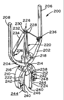
[0082] Referring now to Figures 22-27 there is shown exciser 200, a third
embodiment of
the present invention which is formed of elongate first and second halves 202
and 204, each
respectively having a handle portion 206, 208 and a jaw portion 210, 212.
First and second
halves 202 and 204 are pivotally joined together through rivets 214 to form a
basic structure
similar to an ordinary pair of pliers or clippers. Formed in first and second
halves 202 and
204 is central recess 216, in which is disposed barrel 218. Barre1218 has the
general form of
a parallelepiped having closed sides and open ends. Opposite sides of barrel
218 are
provided with holes through which rivets 214 extend, thereby securing barrel
218 to the rest
of exciser 200. Extending through the open ends of barre1218 are integral
tweezers or
forceps 220 comprising first and second flexible arms 222 and 224. Arms 222
and 224 are
fixed together at attached end 226 of tweezers 220. Fixed to attached end 226
are short rods
228 which are separated from and attached to each other through neck 230. Rods
228 extend
in directions parallel to the longitudinal axes of rivets 214.
[0083] Neck 230 extends through slot 232 centrally provided in elongate spring
steel strip
234, the opposite ends 236 of which are pivotally attached to first and second
exciser halves
202 and 204. Spring steel strip is plastically deformed at its center, and
retains and controls
CA 02460949 2004-03-16
WO 03/028563 PCT/US02/31037
longitudinal movement of integral tweezers or forceps 220 through the
engagement of rods
228 with the portions of strip 234 on opposite sides of slot 232.
[0084] First and second arms 222 and 224 of tweezers 220 are provided with
plastically
deformed portions 238 which, when tweezers 220 are longitudinally moved in the
direction
of arrow 252, causes the opposed free ends 240 of first and second arms 222
and 224 to move
towards each other and close. As discussed further hereinbelow, the closing
action of free
ends 240 of integral tweezers or forceps 220 capture the lesion to be excised,
and longitudinal
movement of tweezers 220 in the direction of arrow 252 pulls the lesion to be
excised away
from the skin.
[0085] Jaw portions 210 and 212 are each provided with opposed blades or
cutting edges
242 which, when the jaws are closed, move towards each other and, when the
jaws are fully
closed, abut each other. Thus, skin located outside perimeter P of lesion L to
be excised is
pinched between blades 242 and cut from the remainder of the skin thereby.
Blades 242, jaw
portions 210, 212, halves 202, 204 or indeed entire exciser 200 may be made of
surgical
stainless steel.
[0086] Near the free ends of jaw portions 210 and 212 are located opposed,
staple-
engaging portions having flat surfaces 244 to which are adhered first and
second separate
staple halves 246 and 248 which comprise staple 250, another embodiment of a
skin-closure
device in accordance with the present invention. When staple halves 246 and
248 are
separated or at least not fully engaged, staple 250 has an open condition.
First and second
staple halves 246 and 248 are, and thus staple 250 is, closed through
manipulation of exciser
220 which interlocks the staple halves to each other. With the staple halves
in this fully
engaged state, the staple has a closed condition.
[0087] The operation of exciser 200 is now discussed with reference to Figures
24-27. In
a first state shown in Figure 24, free ends 240 of the integral tweezers or
forceps capture
lesion L to be excised from skin S, and the lesion is pinched therebetween as
handle portions
206 and 208 are closed towards each other slightly.
[0088] In a second, sequential state shown in Figure 25, further movement of
handle
portions 206 and 208 towards each other causes spring steel strip 234 to flex
and its center to
move in the direction of arrow 252, which forces tweezers 220 in that
direction. Movement
tweezers 220 upward in the direction of arrow 252 brings deformed portions 238
of first and
second arm 222 and 224 into sliding engagement with the opening of barrel 218
and forces
free ends 240 of the first and second arms 222 and 224 closer together,
pinching lesion L as it
CA 02460949 2004-03-16
WO 03/028563 PCT/US02/31037
16
is pulled away from skin S. After tweezer free ends 240, and lesion L
therebetween, have
moved to a position within the jaws formed by portions 210 and 212 such that
lesion
perimeter P is past blades 242, staple halves 246 and 248 enter engagement
with the skin
outside of perimeter P and with each other in the manner disclosed further
hereinbelow.
[0089] In a third sequential state shown in Figure 26, handle portions 206 and
208 have
been brought further together, and tweezers have moved further in the
direction of arrow 252.
In this state, staple 250 is fully closed, and blades 242 are brought into
abutting engagement
with each other, severing lesion L from skin S below lesion perimeter P.
Although staple 250
may achieve its fully closed condition prior to actual engagement of blades
242 with skin S,
the closing of the staple and the excision of lesion L may alternatively occur
substantially
simultaneously.
[0090] In a fourth sequential state shown in Figure 27, exciser 200, with
excised lesion L
still captured between tweezer free ends 240, is removed from the patient,
staple 250 having
closed skin S below the excision site such that the dermis located on opposite
sides of the
excision site are in abutting contact and an elliptically-shaped closure wound
is formed as
described above. The adhesive, which holds staple halves 246 and 248 to their
respective flat
surfaces 244 of the staple-engaging portions at the free ends of the exciser
jaws, breaks free
upon slight release of handle portions 206, 208 which are urged away from each
other by
spring steel strip 232, and exciser 200 can then be freely removed, leaving
staple 250 behind.
As handle portions 206, 208 are more fully released, tweezers 220 move in a
direction
opposite to arrow 252, allowing free ends 240 to separate, freeing excised
lesion L.
[0091] Referring now to Figures 28-31 there is shown exciser 300, a fourth
embodiment
of a device according to the present invention, in a series of sequential
states of operation.
Exciser 300, like exciser 200 has a basic structure similar to that of an
ordinary pair of pliers
or clippers, and a common skin-closure device may be used with these exciser
embodiments.
[0092] Exciser 300 has a pair of elongate first and second halves 302 and 304,
each
respectively having handle portion 306, 308 and jaw portion 310, 312, halves
302 and 304
being pivotally joined together by pin 314. Rather than being provided with
integral tweezers
or forceps, as exciser 200 is, exciser 300 is used with separate, known
tweezers or forceps
320 as shown. Tweezers 320 are used to capture and pull lesion L away from the
skin S of
the patient prior to moving handle portions 306 and 308 towards each other to
close the skin-
closure device or staple, and excise lesion L. Alternatively, the lesion may
be captured and
CA 02460949 2004-03-16
WO 03/028563 PCT/US02/31037
17
pulled with a skin hook (not shown). Except for these differences, the
structure and operation
of exciser 300 are substantially identical to those of exciser 200.
[00931 Exemplary tweezers 320 have first and second arms 322 and 324 joined at
attached end 326. With the ends of jaw portions 310, 312 placed against skin S
and lesion L
placed loosely therebetween, tweezer free ends 340, which may be serrated,
grasp lesion L
which is then pulled away from skin S of the patient and into the jaws of
exciser 300. Once
the captured lesion has been pulled into jaw portions 310 and 312 to an extent
that lesion
perimeter P is above blades 342, handle portions 306 and 308 are squeezed
further together,
and staple halves 246 and 248 which comprise staple 250 are brought into
engagement with
the skin outside the outer perimeter of the lesion L and with each other, as
shown in
Figure 29.
[0094] In Figure 30, staple 250 is fully closed on skin S and blades 342 sever
lesion L
from skin S at a location outside lesion perimeter P, as described above. As
noted above,
although staple 250 may achieve its fully closed condition prior to actual
engagement of
blades 342 with skin S, the closing of the staple and the excision of lesion L
may alternatively
occur substantially simultaneously. The lesion held by tweezers 320 is then
removed from
the excision site. In Figure 31, the jaws of exciser 300 are separated,
causing the adhesive,
which held staple halves 246, 248 to flat surfaces 344 of the staple-engaging
portions of the
jaws, to break free. The resulting elliptically-shaped excision wound, in
which the dermis
located on opposite sides of the excision is held in abutting contact by
staple 250, is
substantially identical to that resulting from use of exciser 200.
[0095] Referring now to Figures 32 and 33, there are respectively shown
staples 250a and
250b, first and second embodiments of staple 250 which can be used with either
of above-
described excisers 200 and 300. Identical elements of staples 250a and 250b
are identified
with a common reference numeral, whereas corresponding elements of staples
250a and 250b
are identified alphanumerically with a common numeric portion an alphabetic
character (a or
b) which correlates with a particular embodiment staple 250a or 250b. Each
embodiment of
staple 250 comprises staple halves 246 and 248 which, in the figure, are
respectively shown
adhered to flat surfaces 244, 344 ofjaw portions 210, 310 and 212, 312 of
excisers 200, 300.
Those skilled in the art will recognize that this association between staple
halves and jaw flat
surfaces may be reversed. Staple halves 246, 248 may be made of surgical
stainless steel or a
suitable plastic material.
CA 02460949 2004-03-16
WO 03/028563 PCT/US02/31037
18
[0096] Each staple half 246 is provided with elongate flat central portion 360
extending
between legs 362 and 364. A suitable releasable adhesive 366, which is later
broken free
during removal of the exciser from the patient as described above, is provided
between the
outer planar surface of flat central portion 360 and the abutting surface 244,
344 of jaw
portion 210, 310.
[0097] Similarly, each staple half 248 is provided with elongate flat central
portion 370
extending between legs 372 and 374, staple half 248 being releasably adhered
to its mating
jaw surface 244, 344 by adhesive 366.
[0098] Pointed pins 368 extend from the inner planar sides of flat central
portions 360,
370, and when staple 250 is closed, the terminal ends of pins 368 of one
staple half abut the
interfacing inner surface of the other staple half. Further, with staple 250
closed, the pins
alternate along the staple length on the basis of which staple half they
extend from.
Moreover, each staple half 246, 248 is substantially symmetrical about the
center of its
central portion 360, 370, thereby allowing the staple halves to each be
oriented on flat
surfaces 244, 344 in either of two orientations 180 degrees apart; i.e., the
locations of legs
362 and 364 of staple half 246, or the locations of legs 372 and 374 of staple
half 248 may be
switched relative to the exciser.
[0099] Referring to Figure 32, the ends of legs 362a and 364a are provided
with barbs
376 which, when staple 250a is closed, are interconnected with barbs 378
provided at the
ends of legs 372a and 374a, the interconnecting barbs holding staple 250a in
its closed
condition. The interconnection of barbs 376 and 378 occurs as they slide past
each other,
resiliently deflecting at least one leg of each interconnecting pair, and
become hooked to each
other.
[0100] Referring to Figure 33, the legs 362b and 364b are substantially
tubular and
telescopically engage legs 372b and 374b, which are interference fitted
therein during closure
of staple 250b to maintain its closed condition. The engaging surfaces of legs
362b, 364b and
372b, 374b may be smooth, their sliding interference fit being substantially
as disclosed
above with respect to rod portions 128 and tube portions 130 of staple 122 of
second
embodiment exciser 100 (see Figure 15).
[0101] Staple halves 246b, 248b which are made of plastic may alternatively
have its legs
372b, 374b provided with ribs 380, as shown in Figure 33, which are compressed
as they are
forced into smooth-walled hollow legs 362b, 364b, the compression of ribs 380
providing a
secure interference fit between the interconnected legs. As shown in Figure
33, the interior
CA 02460949 2004-03-16
WO 03/028563 PCT/US02/31037
19
surfaces of hollow legs 362b and 364b may be also provided with recesses 382
into which
ribs 380 are received as legs 372b, 374b are forced therein, the interfitting
engagement of ribs
380 and recesses 382 holding staple 250b in its closed condition.
[0102] While the present invention has been described as having exemplary
structures and
methods, the present invention can be further modified within the spirit and
scope of this
disclosure. This application is therefore intended to cover any variations,
uses, or adaptations
of the invention using its general principles. Further, this application is
intended to cover
such departures from the present disclosure as come within known or customary
practice in
the art to which this invention pertains and which fall within the limits of
the appended
claims.