Note: Descriptions are shown in the official language in which they were submitted.
CA 02466037 2004-05-06
WO 03/039405 PCT/US02/36153
1
STENT DELIVERY DEVICE WITH EMBOLIC PROTECTION
BACKGROUND OF THE INVENTION
1. The Field of the Invention
The invention generally relates to the field of interventional cardiology.
More
specifically, the invention relates to interventional cardiology procedures
that require
the placing of a stent in a body lumen, such as a body lumen of a patient or
animal.
The present invention further relates to systems for providing embolic
protection
during placing of a stent in a body lumen.
2. The Relevant Technology
Human blood vessels often become occluded or blocked by plaque, thrombi,
other deposits, or material that reduce the blood carrying capacity of the
vessel.
Should the blockage occur at a critical place in the circulatory system,
serious and
permanent injury, and even death, can occur. To prevent this, some form of
medical
intervention is usually performed when significant occlusion is detected.
Several procedures are now used to open these stenosed or occluded blood
vessels in a patient caused by the deposit of plaque or other material on the
walls of
the blood vessel. Angioplasty, for example, is a widely known procedure
wherein an
inflatable balloon is introduced into the occluded region. The balloon is
inflated,
dilating the occlusion, and thereby increasing the intra-luminal diameter.
Another procedure is atherectomy. During atherectomy, a catheter is inserted
into a narrowed artery to remove the matter occluding or narrowing the artery,
i.e.,
fatty material. The catheter includes a rotating blade or cutter disposed in
the top
thereof. Also located at the tip are an aperture and a balloon disposed on the
opposite
side of the catheter tip from the aperture. As the tip is placed in close
proximity to the
fatty material, the balloon is inflated to force the aperture into contact
with the fatty
material. When the blade is rotated, portions of the fatty material are shaved
off and
retained with the interior lumen of the catheter. This process is repeated
until a
sufficient amount of fatty material is removed and substantially normal blood
flow is
resumed.
In another procedure, introducing a stent into the stenosed region to open the
lumen of the vessel treats stenosis within the artery or other blood vessel.
The stent
typically includes a substantially cylindrical tube or mesh sleeve made from
such
material as stainless steel or Nitinol. The design of the material permits the
diameter
CA 02466037 2004-05-06
WO 03/039405 PCT/US02/36153
2
of the stent to be radially expanded, while still providing sufficient
rigidity such that
the stent maintains its shape once it has been enlarged to a desired size.
To place a stent, many medical devices are typically used. Once access to the
inside of the arterial system is established, usually through the femoral
artery, a guide
catheter is inserted into the artery and the tip thereof is guided to a
position just
proximal to the stenosed region to be treated. This guide catheter serves the
purpose
of allowing other devices to rapidly be delivered to that position without
each being
carefully guided from the point of access, through the tortuous anatomy of the
arterial
system to the point of intervention.
Typically, a small diameter guidewire is then inserted through the guide
catheter and guided to the point distal to the stenosed region. When guidewire
access
to the lesion is established, and if there is sufficient cross sectional area
in the
narrowed part of the lesion, a stent, mounted on a delivery device, is
installed over the
guidewire. When correctly placed within the stenosed region, the stent will
then be
deployed, propping open the vessel at that point.
Various types of stents are used in these cases, but a common one requires
that
the stent be deployed from, or expanded from, a compressed state by a balloon
upon
which it is mounted. The balloon is inflated from the proximal end of the
delivery
device to a high pressure, which both opens the stenosis and embeds the stent
into the
inner lumen of the vessel at that point.
Once the guidewire is placed, the guidewire is used as a guide for all of the
other devices that are used in the procedure. These devices have an inner
lumen
through which the proximal end of the guidewire, which is outside of the body
of the
patient, is inserted. The device is then slid along the guidewire into the
body,
allowing the guidewire to guide the device to the required position in the
vascular
system. The process of sliding another device over the guidewire is commonly
known as an exchange.
Two basic types of devices facilitate exchanging of stent systems and dilation
balloons. The first type of device encloses a guidewire within an inner lumen
of the
device for the entire length of the device. The second type of device only
encloses the
guidewire for a small distal segment of the device, with the remainder of the
guidewire exiting from the inner lumen of the device through a side hole to
allow the
device and the guidewire to be side by side. In both cases, control of the
guidewire is
paramount during the exchange as the correct positioning of the device is
reliant upon
CA 02466037 2004-05-06
WO 03/039405 PCT/US02/36153
3
maintaining the position of the guidewire; this being difficult as at least a
section of
the guidewire is inaccessible due to it being enclosed in the inner lumen of
the device
being exchanged.
Providing a stent delivery device that reduces the complexity of an
interventional procedure would advance the art of stent delivery. Furthermore,
reducing the number of devices used to perform a stent implanting procedure
would
advance the art of stent delivery.
In addition, when these interventional procedures are performed, embolic
particles may break off, flow down-stream, and cause potential adverse events.
Devices are emerging that are designed to catch or filter these particles to
prevent
their down-stream flow, to occlude the vessel during the intervention, and
then
allowing the particles to be aspirated out before they may flow downstream.
Current technology for embolic protection devices requires that they be
delivered in a sheath distal to the point of intervention. This requires
crossing the
lesion with a large-diameter, relatively stiff device that is itself a
potential embolic
event that may occur before the embolic protection device is in place. The
sheath
must then be removed allowing the filter to be deployed in the vessel. After
the
device is deployed, balloons, stents, or other therapies of choice may be
exchanged
over the device to treat the area of interest. When the procedure is
completed, the
embolic protection device is captured by another catheter that is exchanged
over the
embolic protection device capturing any potential embolic material within it.
This
relatively complicated procedure adds complexity to providing stenting and
other
procedures.
The device and methods described herein are meant to overcome deficiencies
of the current devices allowing quicker, safer and easier protection and
stenting
procedures to be undertaken.
BRIEF SUMMARY OF THE INVENTION
Embodiments of the present invention can provide systems, methods, and
devices that combine the functionality of a guidewire, a stent delivery
device, a
3o dilation balloon, and an embolic protection device, or subset grouping
thereof, into a
single device insertable into a body lumen. In this manner, embodiments of the
present invention reduce the number of devices needed to perform a procedure,
decrease the time needed to perform the procedure, reduce the difficulty and
CA 02466037 2004-05-06
WO 03/039405 PCT/US02/36153
4
complexity of the procedure, thereby creating the potential for safer
procedures and
increased effectiveness to the patient.
In one embodiment, a delivery device includes a guide member having a distal
end and a proximal end. The guide member functions as a guide catheter, a
guidewire, and a stent delivery device. A dilation assembly is disposed at the
distal
end of the guide member with a stent preloaded upon the dilation assembly. The
distal end of the guide member is configured to apply a restraining force upon
the
dilation assembly to selectively maintain the dilation assembly and stent
within a
lumen of the delivery device. Associated with the distal end of the guide
member is a
1o restraining member or mechanism that can be operated to release the
restraining force
applied to the dilation assembly and stent, thereby allowing the dilation
assembly and
stent to be deployed from within the lumen. The restraining mechanism
cooperates
with an actuating assembly to deploy the dilation assembly and stent.
In one embodiment, the actuating assembly cooperates with a proximal end of
the guide member and includes an actuating member that extends from the
restraining
mechanism or member at a distal end of the delivery device to an actuating
element
disposed at the proximal end of the guide member. Thus, operation of the
actuating
element translates movement to the actuating member to release the restraining
mechanism or member and release the restraining force applied by the
restraining
mechanism or member, whether alone or in combination with the distal end of
the
guide member, upon the dilation assembly and/or the stent.
In operation, the delivery device is placed in position within a body lumen of
a
patient, with the dilation assembly and stent in a restrained position.
Operation of the
actuating assembly releases the dilation assembly and the stent from within
the guide
member. The guide member may be pulled proximally to allow the dilation
assembly
and stent to be entirely free of the guide member. Alternatively, a dilation
tube and/or
a positioning member connected to the dilation assembly may be advanced
distally to
deploy the dilation assembly and the stent. The stent may then be placed in
the
vasculature by inflating the dilation balloon associated with the dilation
assembly, for
3o example, through the dilation tube. After the stent is implanted, the
dilation assembly
is deflated and the delivery device can be removed from the patient.
According to another aspect of the present invention, the delivery device can
include an embolic protection device that is adapted to collect embolic
particles
released during the procedure. As the stent is implanted, the embolic
protection
CA 02466037 2004-05-06
WO 03/039405 PCT/US02/36153
device can filter the blood flowing past the lesion and prevent embolic
particles or
matter flowing downstream. In one configuration, the embolic protection device
is
mounted to a distal end of a guidewire associated with the delivery device.
The
embolic protection device can be. a filter assembly that includes a filter and
a filter
5 basket. The filter basket includes a plurality of struts that restrain the
filter during
insertion of the delivery device into the body lumen, while supporting and
deploying
the filter upon releasing a restraining force applied to the plurality of
struts to
maintain the filter assembly in a closed position during insertion of the
delivery
device. The structures used to apply the restraining force to the plurality of
struts can
1o be similar to the structures applying the restraining force to the dilation
assembly
and/or stent.
According to another aspect of one embodiment of the present invention, the
delivery device may cooperate with a capture mechanism or device for
retrieving the
filter assembly without removing the delivery device from the body. In one
embodiment, a distal end of the dilation assembly functions as the capture
mechanism. This distal end is adapted to optionally retain the filter assembly
during
insertion of the delivery device into a body lumen and subsequently capture at
least a
portion of the filter assembly following implanting of the stent associated
with the
delivery device. In another embodiment, a separate capture mechanism or device
can
be exchanged over the guide member and/or a guide wire to capture at least the
guide
member and/or the filter assembly. In still another configuration, a stent,
stent
delivery device, or balloon catheter can be preloaded upon the guidewire
and/or
dilation tube having a filter assembly disposed at a distal end thereof, and
substantially simultaneously dispose the stent, stent delivery device, or
balloon
catheter and associated filter assembly within a body lumen.
Thus, the delivery devices of the present invention allow protected
interventions to be accomplished with a single device insertion, without
requiring
exchanges, while still allowing guidewire access distal to the treatment
region
throughout the entire procedure.
These and other objects and features of the present invention will become
more fully apparent from the following description and appended claims, or may
be
learned by the practice of the invention as set forth hereinafter.
CA 02466037 2004-05-06
WO 03/039405 PCT/US02/36153
6
BRIEF DESCRIPTION OF THE DRAWINGS
To further clarify the above and other advantages and features of the present
invention, a more particular description of the invention will be rendered by
reference
to specific embodiments thereof that are illustrated in the appended drawings.
It is
appreciated that these drawings depict only typical embodiments of the
invention and
are therefore not to be considered limiting of its scope. The invention will
be
described and explained with additional specificity and detail through the use
of the
accompanying drawings in which:
Figure 1 illustrates a perspective view of an exemplary stent delivery device
in
1 o accordance with one aspect of the present invention;
Figure 2 illustrates a sectional side view of a distal end of the device of
Figure
l;
Figure 3 illustrates a sectional side view of the distal end of the device of
Figure 1 with a distal end in an unrestrained configuration;
Figures 4a and 4b illustrate a sectional side view of the distal end of the
device
of Figure 1 with a deployed dilation assembly;
Figure 5 illustrates a sectional side view of the distal end of the device of
Figure 1 with associated inflated dilation balloon and implanted stent;
Figure 6 illustrates a sectional side view of an exemplary proximal end of the
device of Figure 1 in accordance with another aspect of the present invention;
Figure 7 illustrates a plan view of a distal end of another embodiment of the
stent delivery device in accordance with another aspect of the present
invention;
Figure 8 illustrates a side view of the distal end of the stent delivery
device of
Figure 7 in accordance with one aspect of the present invention;
Figure 9 illustrates a perspective view of a distal end of another embodiment
of a stent delivery device in accordance with one aspect of the present
invention;
Figure 10 illustrates a perspective view of the distal end of the stent
delivery
device of Figure 9 with deployed dilation assembly in accordance with one
aspect of
the present invention;
Figure 11 illustrates a perspective view of another embodiment of a stent
delivery device of the present invention;
Figure 12 illustrates another perspective view of the distal end of the
delivery
device of Figure 11 before a restraining member is coupled to the delivery
device;
CA 02466037 2004-05-06
WO 03/039405 PCT/US02/36153
7
Figure 13 illustrates a perspective view of the distal end of the delivery
device
of Figure 11, illustrating the restraining member partially coupled to the
delivery
device;
Figure 14 illustrates a side view of another restraining mechanism usable with
the delivery device of Figure 11 in accordance with one aspect of the present
invention;
Figure 15 illustrates a perspective view of another embodiment of the stent
delivery device in accordance with one aspect of the present invention;
Figure 16 illustrates a perspective view of the distal end of the delivery
device
of Figure 15 before a restraining mechanism is coupled to the delivery device;
Figure 17 illustrates a side view of the delivery device of Figure 15
illustrating
the restraining member partially coupled to the delivery device;
Figure 18 illustrates a side view of the delivery device of Figure 15
illustrating
the restraining member partially coupled to the delivery device;
Figure 19 illustrates a side view of the delivery device of Figure 15
illustrating
the restraining member partially coupled to the delivery device;
Figure 20 illustrates a perspective view of another embodiment of a stent
delivery device in accordance with another aspect of the present invention;
Figure 21 illustrates a perspective view of another embodiment of a stent
delivery device in accordance with another aspect of the present invention;
Figure 22 illustrates a side view of the delivery device of Figure 21 before
the
restraining mechanism is coupled to the delivery device;
Figure 23 illustrates a side view of the delivery device of Figure 21
illustrating
the restraining member partially coupled to the delivery device;
Figure 24 illustrates a perspective view of the delivery device of Figure 21
having the restraining mechanism coupled to a distal end thereof;
Figure 25 illustrates a perspective view - of a proximal end of another
embodiment of a stent delivery device in accordance with another aspect of the
present invention;
Figure 26 illustrates a perspective view of a proximal end of yet another
embodiment of a stent delivery device in accordance with another aspect of the
present invention;
CA 02466037 2004-05-06
WO 03/039405 PCT/US02/36153
8
Figure 27 illustrates a perspective view of a proximal end of yet another
embodiment of a stent delivery device in accordance with another aspect of the
present invention;
Figure 28 illustrates another embodiment of the proximal end of another
embodiment of a stent delivery device in accordance with another aspect of the
present invention;
Figure 29 illustrates a sectional side view of another embodiment of a stent
delivery device of the present invention in accordance with another aspect of
the
present invention;
Figure 30 illustrates a sectional side view of the distal end of the stent
delivery
device of Figure 30 in accordance with another aspect of the present
invention;
Figure 31 illustrate sectional side views of another embodiment of a stent
delivery device in accordance with another aspect of the present invention;
Figure 32 illustrates a sectional side view of another embodiment of a stent
delivery device in accordance with another aspect of the present invention;
Figure 33 illustrates a sectional side view of another embodiment of a stent
delivery device in accordance with another aspect of the present invention;
Figure 34 illustrates a sectional side view of yet another embodiment of a
stent
delivery device in accordance with another aspect of the present invention;
Figure 35 illustrates a sectional side view of still another embodiment of a
stent delivery device in accordance with another aspect of the present
invention;
Figure 36 illustrates a sectional side view of another embodiment of a stent
delivery device in accordance with another aspect of the present invention;
Figure 37 illustrates a sectional side view of an embodiment of a stent
delivery
device that includes an embolic protection device in accordance with another
aspect
of the present invention;
Figure 38 illustrates a sectional side view of a distal end of the delivery
device
of Figure 37;
Figure 39 illustrates a sectional side view of a portion of the delivery
device of
Figure 37 with a filter assembly deployed in accordance with another aspect of
the
present invention;
Figure 40 illustrates a, sectional side view of a portion of the delivery
device of
Figure 37 with the filter assembly and the stent deployed in accordance with
another
aspect of the present invention;
CA 02466037 2004-05-06
WO 03/039405 PCT/US02/36153
9
Figure 41 illustrates a perspective view of a restraining mechanism for a
filter
assembly usable with the delivery device of Figure 37 in accordance with
another
aspect of the present invention;
Figure 42 illustrates a perspective view of a filter assembly usable with the
delivery device of Figure 37 in accordance with another aspect of the present
invention;
Figure 43 illustrates a perspective view of the embodiment of the filter
assembly of Figure 42 in accordance with another aspect of the present
invention;
Figure 44 illustrates a perspective partial sectional view of a distal end of
to another embodiment of a delivery device in accordance with another aspect
of the
present invention;
Figure 45 illustrates a perspective view of an embodiment of a capture
mechanism according to one aspect of the present invention; and
Figure 46 illustrates a perspective view of another embodiment of a capture
mechanism according to one aspect of the present invention.
Figure 47 illustrates a perspective view of another embodiment of a delivery
device having a dilation assembly which can be rapidly exchanged with a
capture
mechanism;
Figures 48-51 illustrate a perspective view of another embodiment of a
delivery device and a method of using a delivery device having embolic
protection
and a capture mechanism according to another aspect of the present invention;
and
Figures 52-54 illustrate another method of treating a body lumen using a
delivery device and separate capture mechanism according to another embodiment
of
the present invention.
DETAILED DESCRIPTION OF THE PREFERRED EMBODIMENTS
The present invention provides systems, methods, and devices that combine
the functionality of a guide catheter, a guidewire, a stent delivery device, a
dilation
balloon, and/or an embolic protection device, or a subset group of such
devices, into a
single device that is insertable into a body lumen. In this manner, the
present
invention reduces the number of devices needed to deliver and position a
stent,
providing the possibility of decreasing the time needed to perform procedures
and
reducing the difficulty and complexity associated with performing a procedure.
Further, embodiments of the present invention aid with decreasing the
possibility of
patient complications during and subsequent to the procedure.
CA 02466037 2009-04-16
Referring now to Figure 1, depicted is an exemplary embodiment of a delivery
device of the present invention, designated by reference number 10. As
illustrated,
delivery device 10 includes a guide member 12 having a distal end 14 and a
proximal
end 16. The term "guide member" can refer to any structure that is capable of
5 functioning as a guidewire that can be steered through the tortuous anatomy
of a
patient. It will be appreciated that guide member 12 can be hollow or
partially hollow
depending upon design considerations.
Extending between distal end 14 and proximal end 16 of guide member 12 is a
lumen 18 within which is disposed a dilation assembly 40 and a stent 42 (see
Figure
10 2). Distal end 14 of guide member 12 includes a tip 15 that is configured
for
percutaneous insertion into a body lumen, while proximal end 16 either
includes or is
adapted to cooperate with an actuating assembly 20 that is adapted to deploy
dilation
assembly 40 and/or stent 42.
Illustratively, guide member 12 can. have an outside diameter of between about
0.010
inches to about 0.650 inches and an inside diameter or diameter of lumen 18
from
about 0.004 inches to about 0.55 inches.
Additionally, guide member 12 can be fabricated from a variety of different
materials. For example, guide member 12 can be fabricated from Nitinol, steel,
metals, metal alloys, composites, plastic, polymers, synthetic materials, such
as, but
TM
not limited to, PEEK, Rydel, or combinations thereof.
Additionally, guide member 12 can have the configuration of a braid-
reinforced polymer tube or a rigid polymer tube. Furthermore, guide member 12
can
be covered with one or more coatings. For instance, and not by way of
limitation,
guide member 12 can include one or more coatings that improve lubricity,
reduce
platelet aggregation, or have anti-thrombogenic properties. In addition to the
above,
guide member 12 can include one or more hydrophilic coatings, heparinized
coatings,
Polytetrafluoroethylene (PTFE) coatings, silicone coatings, combinations
thereof, or
other coatings that may aid with positioning guide member 12 and/or preventing
damage to the body lumen.
Optionally, guide member 12 may include one or more cuts, slits, grooves, or
other structures, illustratively identified by numeral 17, that provide
flexibility to all
or a portion of guide member 12. Although reference is made to use of cuts,
slits, or
grooves to provide flexibility, it can be appreciated by one skilled in the
art that guide
member 12 or other portion of device 10 may have a lattice structure, i.e.,
portions of
CA 02466037 2004-05-06
WO 03/039405 PCT/US02/36153
I1
guide member 12 or device 10 removed therefrom, which provides flexibility to
a
portion of guide member 12 and/or other portion of device 10.
The cuts, slits, or grooves can be located at any location of guide member 12
and may have various pitches to allow or provide for different flexibilities.
These one
or more grooves, cuts or slits can' partially or completely extend through
portions of
guide member 12. Additionally, these grooves, cuts, or slits can have a
variety of
different configurations, such as but not limited to, straight, helical,
geometric,
combinations thereof, or various other configurations known to those skilled
in the
art, so long as those same provide flexibility to guide member 12. Further,
any
number of grooves, cuts, or slits can be included in guide member 12 and
optionally
portions of dilation assembly 40. For example, the more grooves, cuts, or
slits
included in guide member 12 or a portion of dilation assembly 40, the greater
the
flexibility of guide member 12, and hence delivery device 10. Similarly, the
depth of
each groove, cut, or slit can vary depending upon the desired flexibility. For
instance,
the deeper the groove, cut, or slit, the greater the flexibility of guide
member 12, and
hence delivery device 10. Furthermore, differences in the configuration of
each
groove, cut, or slit can affect the flexibility of guide member 12 and
therefore delivery
device 10. For instance, the steeper the sides of a particular groove, cut, or
slit, the
less flexibility provided to guide member 12 and/or delivery device 10.
Figure 1 depicts dilation assembly 40 and stent 42 (Figure 2) disposed at tip
15
of guide member 12. Dilation assembly 40 terminates in an atraumatic tip 48.
Dilation assembly 40 and stent 42 are retained at tip 15 of guide member 12 by
a
restraining mechanism or restraining member 25. In the embodiment of Figure 1,
an
actuating member 28 operates restraining member 25 and extends to an actuating
assembly 20 disposed at a proximal end of device 10. Actuating member 28
extends
to the proximal end of device 10 and is exposed to allow the restraint applied
by
restraining member 25 to be released as a clinician moves actuating member 28
in a
proximal direction. Alternatively, actuating member 28 can optionally extend
outside
guide member 12 to proximal end 16 of device 10.
Dilation assembly 40 is connected to a dilation tube 44 that extends along the
length of guide member 12. Dilation tube 44 is used to fill a dilation balloon
46 with
a fluid. The fluid may be introduced through a luer lock fitting 45 located at
proximal
end 16 of guide member 12. Dilation tube 44 may also be used, in some
embodiments, as a positioning member for deploying dilation assembly 40 and
stent
CA 02466037 2004-05-06
WO 03/039405 PCT/US02/36153
12
42. Additionally, dilation assembly 40 of device 10 is coupled by dilation
tube 44 to
actuating element 21. By sliding actuating element 21 with respect to proximal
end
16 of guide member 12, dilation assembly 40 is moved with respect to guide
member
12 and can be deployed from tip 15 of guide member 12. These and other
features of
the present invention will now be described in further detail.
With reference now to Figure 2, distal end 14 of guide member 12 includes
one or more struts 24 that are adapted to retain dilation assembly 40 and
stent 42
within lumen 18 until the same are to be deployed. Each strut 24 can be biased
to
extend outwardly to release dilation assembly 40 and stent 42. Although
reference is
made to each strut 24 being biased to extend outwardly, it can be understood
by one
skilled in the art that each strut 24 need not be biased to extend outwardly.
The one or more struts 24 can be formed using a variety of different
processes.
For instance, the processes can include, but not limited to, machining
processes
performed using a laser or conventional machining process, including, but not
limited
to, hydro-machining, grinding, end milling, slitting saws, abrasive saws,
electrical
discharge machines, combinations thereof, or other machining processes capable
of
creating slots or slits sufficient to form one or more struts 24. In the
embodiment of
Figure 2, each strut 24 can be formed integrally with guide member 12. In
other
embodiments, one or more of struts 24 are formed as part of a discrete strut
assembly
that is attached to guide member 12.
Surrounding struts 24 is restraining member 25. In the embodiment of Figure
2, restraining member 25 is a sleeve 26. Sleeve 26 is adapted to retain or
maintain
struts 24 in a restrained or closed configuration so that the combination of
sleeve 26
and struts 24 maintain dilation assembly 40 and stent 42 within lumen 18.
Sleeve 26
is adapted to cooperate with the exterior of guide member 12 so that sleeve 26
can be
displaced in a proximal direction to release struts 24. Since struts 24, in
this
exemplary configuration, are biased to extend outwardly, upon moving sleeve 26
in a
proximal direction, struts 24 extend outwardly to release dilation assembly 40
and
stent 42.
Sleeve 26 can be fabricated from various types of materials so long as sleeve
26 is capable of securely retaining struts 24. For instance, sleeve 26 can be
fabricated
from heat shrink synthetic material, including but not limited to, low-density
polyethylene (LDPE), polyethylene terphthalate (PET), Polytetrafluoroethylene
CA 02466037 2004-05-06
WO 03/039405 PCT/US02/36153
13
(PTFE), fluorinated ethylene propylene (FEP), polyethylene (PE), polyurethane
(PU),
silicone tubing, and other suitable polymers or synthetic materials.
Actuating member 28 extends from sleeve 26, travels along an exterior of
guide member 12, and passes through an aperture 30 in guide member 12.
Actuating
member 28 continues to travel within lumen 18 of guide member 12 until it
reaches
proximal end 16 of guide member 12. It will be appreciated that in other
embodiments, actuating member 28 may remain external to lumen 18 of guide
member 12.
Actuating member 28 can be fabricated from various materials and have
1o various configurations so long as it is capable of performing the function
of displacing
sleeve 26. For example, actuating member 28 can be fabricated from plastics,
polymers, metals, composites, alloys, synthetic materials, and combinations
thereof.
As shown in Figure 2, dilation assembly 40 includes a dilation balloon 46
mounted to
a dilation tube 44. Dilation tube 44 extends from distal end 14 of guide
member 12
toward proximal end 16 of guide member 12. Dilation tube 44 can include a
plurality
of holes 50. Each hole 50 and/or plurality of holes 50 in combination provide
a fluid
path to an interior 52 of dilation balloon 46. In this way, fluid may pass
along a
lumen 54 of dilation tube 44 to flow into dilation balloon 46. To restrict the
flow of
such fluid, atraumatic tip 48 seals the distal end of dilation tube 44. In
addition to
providing a fluid path to inflate dilation balloon 46, holes 50 provide a
fluid path to
deflate dilation balloon 46 or remove the fluid to deflate dilation balloon
46. Each
hole 50 can have various configurations so long as each hole 50 is capable of
allowing
fluid to pass therethrough.
Dilation tube 44, in one configuration, is an internal support for dilation
balloon 46 and stent 42. Dilation tube 44 can be fabricated from Nitinol,
steel,
metals, metal alloys, composites, plastic, and combinations thereof. Further,
dilation
tube 44 can be covered with a variety of different coatings, such as, but not
limited to,
one or more coatings to improve 'lubricity, anti-thrombogenic properties, and
reduce
platelet aggregation. Other coatings can include, but not limited to,
hydrophilic
coatings, heparinized coatings, Polytetrafluoroethylene (PTFE) coating,
silicone
coating, or combinations of the coatings described herein.
Dilation tube 44 may have a variety of different configurations and
embodiments. In another embodiment, dilation tube 44 includes a proximal end
where provision is made for connecting dilation tube 44 to an inflation device
with an
CA 02466037 2004-05-06
WO 03/039405 PCT/US02/36153
14
annular clamping device, such as a touhy-borst adaptor. Alternatively, as
shown in
Figure 1, a proximal end of dilation tube 44 has the form of a luer fitting,
whether the
male or female part of the luer fitting.
Mounted to a distal end of dilation tube 44 is an atraumatic tip 48.
Atraumatic
tip 48 is disposed within lumen '54 of dilation tube 44 and seals dilation
tube 44,
prevents fluid from escaping therefrom during inflation and deflation of
dilation
balloon 46, and provides a flexible tip that aids in positioning and steering
of delivery
device 10 through the tortuous anatomy of the patient. In the illustrative
embodiment,
dilation tube 44 extends to a distal end of dilation balloon 46 and atraumatic
tip 48 is
to disposed therein. Alternatively, dilation tube 44 can extend to a position
proximal to
the distal end of dilation balloon 46 and a portion of atraumatic tip 48 then
extends
from a distal end of dilation tube 44 to a position distal to the distal end
of dilation
balloon 46. Furthermore, in another alternate embodiment, dilation tube 44
terminates within a lumen formed in atraumatic tip 48.
Atraumatic tip 48 includes a core 56 that is surrounded by a flexible coil 58.
As shown, flexible coil 58 terminates at a distal end of tip 48 with an
atraumatic
portion, such as a solder ball or other mechanism for forming an atraumatic
distal end
of tip 48. More generally, atraumatic tip 48 can have a variety of other
configurations
so long as atraumatic tip is flexible and optionally shapeable. Furthermore,
atraumatic tip 48 may be radiopaque to allow steerable positioning of delivery
device
10 while allowing a physician or clinician to observe the location of tip 48
using
appropriate devices, such as a fluoroscopic device or X-ray device. Materials
that
facilitate or provide radiopacity may include, but not limited to, platinum,
alloys of
platinum, gold, or combinations thereof, metals, alloys, plastic, polymer,
synthetic
material, combinations thereof, or other materials that provide an appropriate
radiopaque signature, while capable of being shaped by a physician or
clinician.
Alternatively, tip 48 can be a polymer that is dipped or coated with an
appropriate
radiopaque material, such as, but not limited to, barium sulphate, bismuth
subcarbonate, titanium dioxide, or combinations thereof.
Referring now to Figure 3, depicted is distal end 14 of delivery device 10
upon
disposition of sleeve 26 in a proximal direction. In this illustrative
configuration,
because struts 24 are biased to extend outwardly, dilation assembly 40 and
stent 42
can be deployed from within lumen 18. Deploying of dilation assembly 40 and
stent
42 can occur as guide member 12 is displaced in a proximal direction, dilation
tube 44
CA 02466037 2004-05-06
WO 03/039405 PCT/US02/36153
is displaced in a distal direction, or a combination of proximal and distal
movements
of guide member 12 and dilation tube 44 respectively.
Referring now to Figure 4a, schematically depicted is delivery device 10 in a
deployed configuration where dilation assembly 40 and stent 42 have been
deployed
5 at a lesion 70 of a body lumen 72. Deployment of dilation assembly 40 and
stent 42
can be achieved through manipulating actuating assembly 20 (Figures 1 and 2).
Upon
positioning dilation balloon 46 and stent 42 to the desired position, such as
adjacent to
lesion 70, fluid can be introduced through lumen 54 of dilation tube 44 to
expand
dilation balloon 46 and therefore. deploy or force stent 42 into body lumen 72
and
i o surrounding lesion 70, as is illustrated in Figure 5.
Various configurations of stent 42 are known to those skilled in the art. For
example, an expandable stent may be used that automatically opens under the
pressure of dilation balloon 46. In another configuration, a self-expanding
stent can
be used, as illustrated in Figure 4b with dotted lines. The self-expanding
stent
15 automatically opens as the restraining force applied by struts 24 and/or
restraining
member 25 is removed and guide member 12 is moved proximal to the stent. In
this
case, the self-expanding stent surrounds dilation balloon 46, as illustrated
in Figure
4b, or alternatively, the stent can surround dilation tube 44 with dilation
balloon 46
being located proximal to the stent and still mounted to dilation tube 44, as
illustrated
by dotted lines referenced by numeral 46b. Various stents may be used with the
present invention, so long as the stent can be reduced in size to surround the
dilation
balloon and be disposed within guide member 12 of delivery device 10.
Referring now to Figure 6, depicted is an exemplary embodiment of actuating
assembly 20 that can be used to deploy dilation balloon 46 and stent 42.
Operating
actuating assembly 20 releases dilation assembly 40 and stent 42 from a
restrained
configuration at distal end 14 of guide member 12. More specifically, dilation
balloon 46 forming part of dilation assembly 40 can be deployed with stent 42
being
disposed substantially around dilation balloon 46.
As illustrated, actuating assembly 20 includes an actuating element 21 coupled
to a proximal end of dilation tube 44. Actuating element 21 includes a distal
end 74
configured to be mounted to and cooperate with proximal end 16 of guide member
12.
A proximal end 76 of actuating element 21 is attached to a proximal end of
dilation
tube 44, while a proximal end of actuating member 28 passes through a sealed
aperture 47 of actuating element 21. In this exemplary embodiment, the
proximal end
CA 02466037 2004-05-06
WO 03/039405 PCT/US02/36153
16
of dilation tube 44 includes a luer fitting 45 that allows various
complementary luer
fittings to be attached thereto. For instance, a syringe (not shown) can be
attached to
luer fitting 45 for introducing fluid to and removing fluid from dilation
balloon 46
(Figure 5) during inflation and deflation of dilation balloon 46. Although
reference is
made to use of luer fitting 45, it can be understood by one skilled in the art
that
various other configurations of fitting can be attached to or formed at the
proximal
end of dilation tube 44.
Actuating element 21 is adapted to be displaced in a distal direction to
deploy
dilation assembly 40 and stent 42. To aid with positioning actuating element
21,
distal end 74 can have a step configuration and include protrusions 78 that
mate with
complementary indentations 80 formed in proximal end 16 of guide member 12.
The
protrusions 78 and indentations 80 provide an indication of the relative
position of
dilation assembly 40 and stent 42 relative to distal end 14 of guide member
12.
Therefore, actuating element 21 and/or guide member 12 can include one or more
protrusions and indentations. As actuating element 21 is displaced in a distal
direction, protrusions 78 mate with indentations 80. To seal lumen 18 of guide
member 12, one or more seals 84 surround protrusions 78. Additionally, one or
more
seals (not shown) can surround dilation tube 44 and/or actuating member 28.
Illustratively, each seal can be one or more O-rings in one or more grooves,
one or
more O-rings, a gasket, or a viscous fluid seal.
When actuating element 21 is displaced in the distal direction, distal end 74
contacts a wall or stop 82 formed in guide member 12 that prevents further
displacement of actuating element 21 in the distal direction. Through this
configuration, actuating element 21 is prevented from excessive longitudinal
displacement in the distal direction. This stopping of the longitudinal
displacement of
actuating element 21 indicates that dilation balloon 46 and stent 42 are
deployed from
within lumen 18 of guide member 12 to the desired position for expanding or
implanting stent 42.
Although reference is made to one manner of indicating the particular location
of stent 42, one skilled in the art can identify a variety of different
embodiments. For
instance, a plurality of indentations and/or protrusions can be included
within
actuating element 21 and guide member 12 to control the distance which
actuating
element 21 and, consequently, stent 42 is displaced. In another configuration,
a wall
or stop formed in actuating element 21 can mate with the distal end of guide
member
CA 02466037 2004-05-06
WO 03/039405 PCT/US02/36153
17
12 to prevent excessive longitudinal displacement in the distal direction. In
still
another configuration, a combination of one or more walls or stops in
actuating
element 21 and guide member 12 can be used. In still another configuration,
distal
end 74 of actuating element 21 can be tapered and cooperate with a taper
formed in
proximal end 16 of guide member 12. The complementary tapers control the
longitudinal displacement of actuating element 21 relative to proximal end 16
of
guide member 12. In still other configurations, a combination of indentations,
protrusions, walls, stops, threads, or tapers can be used. Various other
manners are
known to control the distance traveled by actuating element 21 while
indicating the
i 0 position of stent 42.
In addition to the above, it can be appreciated that actuating element 21 can
include one or more elements, such that wall or stop 82 and indentations 80
are
formed in separate elements or members that are attached or coupled to
proximal end
16 of guide member 12. By so doing, actuating element 21 can be fabricated
separately from guide member 12, thereby reducing costs and expenses
associated
with fabricating proximal end 16 of guide member 12 in the desired
configuration.
Figures 7 through 24 illustrate alternative embodiments for restraining
mechanism 25. It will be appreciated that many features of the delivery
devices
depicted in Figures 7 through 24 are substantially similar in structure and
function as
for delivery device 10. Consequently, features and functions of one embodiment
of
the present invention are applicable to other embodiments of the present
invention.
Referring now to Figures 7 and 8, another illustrative embodiment of a
delivery device 100 of the present invention is depicted. As shown, a guide
member
112, which can be similar to the other guide members described herein, has a
distal
end 114, a proximal end (not shown), and a lumen 118 extending from distal end
114
to the proximal end. A tip 115 of guide member 112 includes a plurality of
struts 124,
such as two or more struts. Each strut 124 can be optionally biased so that a
distal
end of each strut 124 moves outwardly from a longitudinal axis of guide member
112
when each strut 124 is released by a restraining member 125. Although
reference is
made to each strut 124 being biased, one skilled in the art can appreciate
that one or
more of struts 124 can be biased.
As shown in Figure 8, at least one strut, designated by reference numeral
124a,
is biased toward the longitudinal axis of guide member 112. Disposed upon
strut
124a, as more clearly seen in Figure 7, is an atraumatic tip 148. This
atraumatic tip
CA 02466037 2004-05-06
WO 03/039405 PCT/US02/36153
18
148, either alone or in combination with strut 124a, may be shapeable by a
physician
or clinician before insertion into a body lumen. In this manner, the physician
or
clinician is able to configure tip 148 with an appropriate shape, such as, but
not
limited to a "J" shape, which enables guide member 112 to be guided through
the
tortuous anatomy of a patient. All or a portion of atraumatic tip 148 can be
fabricated
from platinum, platinum alloys, radiopaque materials, materials doped or
coated with
a radiopaque material, metals, alloys, plastic, polymer, synthetic material,
combinations thereof, or other materials that provide an appropriate
radiopaque
signature, while are capable of being shaped, whether alone or in combination
with
strut 124a, by a physician or clinician. In this configuration, a guidewire
with an
associated dilation assembly can be disposed within lumen 118, with a distal
end of
the guidewire optionally including a flexible atramnatic tip, since atraumatic
tip 148
can function as the atraumatic tip for delivery device 100.
To maintain struts 124 in a restrained position, i.e., not extending outwardly
from guide member 112, restraining member 125 surrounds struts 124. The
restraining member 125 and other restraining members or mechanisms described
herein are examples of means for applying a restraining force upon one or more
struts
or means for applying a restraining force upon a distal end of a guide member.
In this
embodiment, restraining member 125 can extend completely or partially from the
distal end to the proximal end of guide member 112. For example, restraining
member 125 can surround substantially only struts 124 or can have a
configuration
similar to those depicted in Figures 9-24.
In the configuration depicted in Figures 7 and 8, restraining member 125 or
means for applying a restraining force is a catheter 127 that applies a force
against
struts 124 to prevent struts 124 from extending outwardly or applies a force
against
struts 124 to maintain a dilation assembly 140 and a stent 142 in lumen 118.
Through
displacing guide member 112 with respect to catheter 127, or vice versa, the
force
applied to struts 124 is released and, in one configuration, the distal ends
of struts 124
are allowed to move outwardly to allow dilation assembly 140 and stent 142 to
be
3o deployed.
As mentioned above, catheter 127 can extend completely or partially the
length of the guide member. In another configuration, catheter 127 can be
replaced
with a sleeve or other structure that completely or partially extends toward
the
proximal end of guide member 112 from the distal end. These alternate
CA 02466037 2004-05-06
WO 03/039405 PCT/US02/36153
19
configurations are also means for applying a restraining force, as described
herein.
These restraining members or mechanisms can be radiopaque or include one or
more
radiopaque markers that aid with positioning the device. Furthermore, these
restraining members or mechanisms can be slidable relative to the guide member
using an actuating member and/or an actuating assembly disposed on an exterior
of
the guide member, within a lumen of the guide member, or partially within the
lumen
and partially on the exterior of the guide member. The actuating assembly may
be
similar in structure and function to actuating assembly 20 described in Figure
6 or any
other actuating assembly described herein. Therefore, systems, methods, and
devices
of the present invention can optionally use catheters, sleeves, bands, or
other
structures described herein interchangeably to perform the desired function of
restraining one or more struts or a distal end of the guide member.
Figures 9 and 10 depict another embodiment of a delivery device 200 of the
present invention. As illustrated, delivery device 200 includes a guide member
212
with a plurality of struts 224 disposed at a distal end 214 thereof. Struts
224 are
maintained in a restrained position using a restraining member 225. In this
embodiment, restraining member 225 is a sleeve 226 surrounding struts 224.
Sleeve
226 acts as a restraining member or mechanism that applies a force against the
struts
to prevent the struts from extending outwardly or to maintain the dilation
balloon
and/or stent within the lumen.
Struts 224, when in a restrained position, maintain dilation assembly 240 and
stent 242 within lumen 218 of guide member 212. Disposed within sleeve 226 or
between sleeve 226 and guide member 212 are one or more actuating members 228.
Actuating members 228, optionally forming part of the restraining mechanism or
member, are attached to guide member 212 at a location proximal to the
proximal end
of each strut 224, identified by letter A. Actuating members 228 extend
distally to the
distal end of sleeve 226 and subsequently extend proximally on the outside of
sleeve
226 to terminate at the proximal end (not shown) of device 200. Since one end
of
each actuating member 228 is located at the proximal end of sleeve 226,
whether
forming part of sleeve 226, attached to sleeve 226, attached to guide member
212, or
combinations thereof, displacing actuating member 228 in the proximal
direction
causes actuating member 228 to preferentially separate sleeve 226 into one or
more
portions 232, illustrated in dotted lines. By so doing, struts 224 are
released, as
illustrated in Figure 10.
CA 02466037 2004-05-06
WO 03/039405 PCT/US02/36153
To operate actuating members 228, a proximal end (not shown) of actuating
member 228 extends to a proximal end (not shown) of guide member 212, either
within or without lumen 218 of guide member 212. Actuating members 228 can
extend to an actuating element (not shown) of an actuating assembly, such as,
but not
5 limited to, the actuating assembly of Figure 6 and other actuating
assemblies
described herein and understood by one skilled in the art in light of the
teachings
contained herein. The actuating member 228 can be displaced in the proximal
direction relative to guide member 212. By so doing, the restraining force
applied by
sleeve 226 is released, struts 224 extend outwardly, and dilation assembly 240
and/or
1o stent 242 are deployed.
Sleeve 226 can be formed from a variety of different materials, so long as the
material is sufficiently strong to secure struts 224, while being configured
to
preferentially separate under the action of actuating members 228. For
example,
sleeve 226 can be fabricated from heat shrink synthetic material, including
but not
15 limited to, low-density polyethylene (LDPE), polyethylene terphthalate
(PET),
Polytetrafluoroethylene (PTFE), fluorinated ethylene propylene (FEP),
polyethylene
(PE), polyurethane (PU), or silicone tubing.
The one or more actuating members 228 can be formed from a variety of
different materials, so long as the material used is sufficiently strong to
allow an
20 actuating assembly, such as, but not limited to, those actuating assemblies
disclosed
herein, to displace actuating member 228 proximally without breaking the same.
For
example, actuating members 228 can be fabricated from plastics, polymers,
metals,
composites, alloys, synthetic materials, and combinations thereof.
Instead of using actuating members 228, embodiments of the present invention
can employ various other means to preferentially separate sleeve 226. For
example,
sleeve 226 can have dissolvable chemical bonds which dissolve due to a
chemical
reaction with the fluid in the body lumen within which the delivery device is
disposed, bonds that are broken through applying resistive heating,
ultrasonic, or radio
frequency energy to actuating members 228 and/or region of the body lumen
containing device 200, preferential tear or cut regions or zones where the
material has
a weaker strength than other regions or zones of the sleeve, or combinations
thereof.
Referring now to Figures 11 through 14, depicted is an embodiment of a
delivery device 300 having another embodiment of a restraining member or
mechanism 325. In this embodiment, restraining member 325 is in the form of a
CA 02466037 2004-05-06
WO 03/039405 PCT/US02/36153
21
sleeve 326 which is adapted to surround one or more struts 324 of a guide
member
312 and apply a restraining force against struts 324 to maintain struts 324 in
a
restrained configuration. Sleeve 326 includes a first side 364 and a second
side 366
with first and second sides 364, 366 being separated by an intermediate
portion 368.
Intermediate portion 368 surrounds guide member 312 in such a manner that
portions
of intermediate portion 368 contact, are juxtaposed to, are contiguous with,
or are
adjacent to one another. An actuating member 328 passes through such portions
of
intermediate portion 368 to secure sleeve 326 upon guide member 312. To
further aid
with applying a restraining force against struts 324, first side 364 and
second side 366
1 o are folded to attach to respective portions of outside surface of sleeve
326.
The process of forming the restraining member or mechanism of Figure 11 is
illustrated in Figures 12 and 13. With reference first to Figure 12, which
depicts
sleeve 326 in an open position before actuating member 328 is coupled thereto,
sleeve
326 can be directly formed on guide member 312 or can be formed on a separate
tubular member and subsequently attached or coupled to guide member 312.
Sleeve
326 is illustrated as having a generally polygonal configuration, however, one
skilled
in the art can appreciate that sleeve 326 can have various other
configurations so long
as it is capable of performing the functions described herein. In this
exemplary
configuration, sleeve 326 is coupled directly to guide member 312. First side
364 and
second side 366 of sleeve 326 are wrapped around at least a portion of guide
member
326, until a portion of intermediate portion 368 is in close proximity with
another
portion of intermediate portion 368, as illustrated in Figure 13.
Alternatively, a first
side 364 can contact second side 366 or be juxtaposed, contiguous, or adjacent
to
second side 366.
When the portions of intermediate portion 368 are in close proximity,
actuating member 328, or alternatively some other actuating member, is
stitched
through both portions of sleeve 326 to couple the portions of intermediate
portion
368, as shown in Figure 13. Once actuating member 328 is drawn substantially
straight or otherwise positioned through sleeve 326, first end 364 and second
end 366
are respectively folded to attach to respective outside surfaces of sleeve
326, as shown
in Figure 11.
As illustrated in Figure 14, in an alternate configuration, sleeve 326 can
include a plurality of apertures 360 on portions of intermediate portion 368
that
receive actuating member 328. In this manner, actuating member 328 can pass
CA 02466037 2004-05-06
WO 03/039405 PCT/US02/36153
22
through apertures 360 rather being stitched through sleeve 326. In another
embodiment, first end 364 of sleeve 326 can be coupled to second end 364 of
sleeve
326 without attaching first end 364 or second end 366 to the outside surface
of sleeve
326. In still another configuration, a portion of first end 364 can overlap a
portion of
second end 366, or vice versa. Alternatively, first end 364 and second end 366
contact each other but do not overlap. Similarly, first end 364 and second end
366
can be adjacent to one another, adjoining one another, contiguous to one
another, or
juxtaposed to one another.
To operate the restraining member or mechanism described in reference to
1o Figures 11-14, a proximal end of actuating member 328 extends to a proximal
end of
guide member 312, either within or without a lumen of the guide member 312.
Upon
displacing actuating member 328 in a proximal direction relative to guide
member
312, vice versa, or combination thereof, actuating member 328 is released from
being
disposed through at least a portion of sleeve 326. By so doing, the
restraining force
applied by sleeve 326 is released, struts 324 extend outwardly, and the
dilation
assembly and/or stent are deployed. A clinician or physician can initiate the
longitudinal motion of actuating member 328, either directly or through using
of an
actuating mechanism or device.
Sleeve 326 can be formed from a variety of different materials, so long as the
material is sufficiently strong to restrain one or more struts 324. For
example, sleeve
326 can be fabricated from various types of polymer or silicone films, such as
but not
limited to, heat shrink plastic, polymer, low-density polyethylene (LDPE),
polyethylene terphthalate (PET), Polytetrafluoroethylene (PTFE), fluorinated
ethylene
propylene (FEP), polyethylene (PE), polyurethane (PU), or silicone tubing.
Actuating member 328 can be formed from a variety of different materials, so
long as the material used is sufficiently strong to allow the actuating
assemblies
disclosed herein to displace actuating member 328 proximally without breaking
actuating member 328. For example, actuating member 328 can be fabricated from
plastics, polymers, metals, composites, alloys, synthetic materials,
combinations
thereof, or other material that is capable of performing the function of being
disposed
through sleeve 326 and capable of being withdrawn therefrom.
Referring now to Figures 15-19, illustrated is another embodiment of a
delivery device 400 having an alternate configuration of a restraining member
or
mechanism. This particular embodiment utilizes a restraining member or
mechanism
CA 02466037 2004-05-06
WO 03/039405 PCT/US02/36153
23
425 having a hinged configuration with an actuating member 438, optionally
forming
part of restraining member or mechanism 425, acting as the pin to maintain the
hinged
portions of the restraining member in a configuration that retains or
restrains a portion
of the guide member.
As shown in Figure 15, restraining member 425 is a sleeve 426 having a
plurality of channels 464a-464f that are adapted to receive actuating member
428.
Both a first side 466 and a second side 468 of sleeve 426 are formed with some
of
channels 464a-464f, i.e., channels 464a, 464c, and 464e on first side 466 and
channels
464b, 464d, and 464f on second side 468. By passing actuating member 428
through
1o channels 464a-464f in sequential order, so that actuating member 428 passes
through
a channel on first side 466 and subsequently a channel on second side 468,
first side
466 is coupled to second side 468 and sleeve 426 applies a restraining force
against
struts 424 of guide member 412.
An exemplary process of forming the restraining member or mechanism of
Figure 15 is illustrated in Figures 16-19. With reference first to Figure 16,
which
depicts sleeve 426 in an open position before actuating member 428 is coupled
thereto, sleeve 426 includes a number of extensions or tongues 460a-460f.
These
extensions 460a-460f are configured to form channels 464a-464f and surround a
tubular member or tube, such as, but not limited to, a guide member 412 within
which
actuating member 428 is located.
To attach sleeve 426 to guide member 412, sleeve 426 is positioned over the
desired portion of guide member 426. Actuating member 428 is placed in close
proximity to guide member 412, as shown in Figures 17-19. The ends of the
extensions 460a-460f are inserted between guide member 412 and actuating
member
428, as shown in Figure 18. Alternatively, extensions 460a-460f can be
partially
wrapped around guide member 412 and actuating member 428 placed into contact
with these partially wrapped extensions 460a-460f.
After the extensions 460a-460f are pulled tightly around guide member 412
and actuating member 428, an end of each extension 460a-460f is folded over
3o actuating member 428 to attach to the outer surface of sleeve 426, as shown
in Figures
15 and 19. In this manner, channels 464a-464f are formed and sleeve 426 is
configured with actuating member 428 to releasably restrain struts 424 of
guide
member 412.
CA 02466037 2004-05-06
WO 03/039405 PCT/US02/36153
24
Releasing the restraining force applied by sleeve 426, alone or in combination
with actuating member 428, is achieved through displacing actuating member 428
longitudinally with respect to guide member 412, vice versa, or combination
thereof.
Actuating member 428 is released from channels 464a-464f to allow the biasing
force
of struts 424 to extend the struts outwardly to deploy dilation assembly
and/or stent.
A clinician or physician can initiate the longitudinal motion of actuating
member 428,
either directly or through using of an actuating mechanism or device.
Referring now to Figure 20, depicted is another delivery device 500 having
another embodiment of a restraining member or mechanism 525 of the present
invention. The restraining member 525 includes a cord 529 forming a number of
hoops 564a-564n. One or more of hoops 564a-564n are adapted to receive an
actuating member 528, which is, optionally part of restraining member or
mechanism
525. The actuating member 528 is disposed within hoops 564a-564n so that cord
529
applies a restraining force against struts 524 of guide member 512. Actuating
member 528 can be removed from hoops 564a-564n to thereby allow struts 524 to
extend outwardly to deploy the dilation assembly and/or stent. Cord 529 may be
made from metallic wires, polymer actuating members, or other materials that
can be
manipulated to form hoops through which an actuating or securing member.
Optionally, cord 529 is adapted to expand outwardly either under the influence
of one or more struts or due to a biasing force applied or incorporated within
cord 520
by the configuration and/or material of the cord, the hoops, and/or the
restraining
member.
Cord 529 can be attached to guide member 512 and/or one or more of the
struts associated therewith through various attachment mechanisms. For
instance,
cord 529 can be attached to guide member and/or one or more of the struts
through
adhesives, mechanical fasteners, securing loops, or other manner that securely
attaches cord 529 to guide member 512 and/or one or more of struts 524.
Alternatively, cord 529 may be attached to actuating member 528 and be removed
when actuating member 528 is moved in a proximal direction. A clinician or
physician can initiate the longitudinal motion of actuating member 528, either
directly
or through using of an actuating mechanism or device.
Referring now to Figures 21-24, depicted is another delivery device 600
having another embodiment of a restraining member or mechanism 625 of the
present
invention. As illustrated, a guide member 612 includes a plurality of struts
624 that
CA 02466037 2004-05-06
WO 03/039405 PCT/US02/36153
are adapted to extend outwardly to enable deployment of the stent and dilation
balloon disposed within a lumen 618 of guide member 612. A restraining member
625 restrains struts 624. This restraining member 625, in one configuration,
is a
flexible member 627 configured with flaps 660 and 662. The flaps 660 and 662
5 extend between a gap 664 between the two adjacent struts 624a and 624b and
are
adapted to be pulled around struts 624 to compress stent (not shown) and
dilation
balloon (not shown) within lumen 618, as illustrated in Figure 23. These flaps
660
and 662 can be two separate members that are bonded or otherwise connected to
struts
624a and 624b or a single member that is coupled to struts 624a and 624b while
1o forming flaps 660 and 662.
When flaps 660 and 662 have been positioned to securely retain struts 624,
they are then stitched together at a location 666, identified in Figure 23,
with an
actuating member 628. This actuating member 628, optionally forming part of
the
restraining member or mechanism, extends the length of delivery device 600
toward
15 an actuating assembly, such as, but not limited to, the actuating assembly
described in
Figure 6 and other actuating assemblies known to those skilled in the art in
light of the
teachings contained herein. A clinician or physician can initiate longitudinal
motion
of actuating member 628 to release restraining member or mechanism 625, either
directly or through using of an actuating mechanism or device as known to
those
20 skilled in the art.
Following coupling of flaps 660 and 662 using actuating member 628, flaps
660 and 662 are folded back around struts 624 and the remainder of flaps 660
and
662, and then attached to struts 624, or other portion of guide member 612, as
illustrated in Figure 24. When actuating member 628 is displaced in a proximal
25 direction, flaps 660 and 662 are released and stent (not shown) and
dilation balloon
(not shown) are deployed as struts 624 extend outwardly.
Referring now to Figure 25, depicted is an illustrative embodiment of a
proximal end of a delivery device 700a. The features and structures discussed
with
other embodiments of the delivery device of the present invention apply to
delivery
3o device 700a.
As shown, a proximal end 716 of a guide member 712 terminates in a guide
member housing 722. This guide member housing 722 can be integrally formed
with
guide member 712 or alternatively be a separate member coupled, connected, or
attached to a proximal end of guide member 712. Proximal end 716 of guide
member
CA 02466037 2004-05-06
WO 03/039405 PCT/US02/36153
26
712 is coupled to an actuating element 721 of an actuating assembly 720. This
actuating element 721 slidably engages with guide member housing 722.
Manipulation of actuating element 721 effects the movement of dilation tube
744
upon which is mounted the dilation balloon (not shown). Actuating member 728
extends through an aperture 786 in actuating element 721 that is adapted with
a seal
(not shown) through which actuating member 728 can slide. In this manner,
aperture
786 and the seal (not shown) allow access for the operator to release or
displace the
restraining member (not shown) that restrains the one or more struts (not
shown)
disposed at distal end 714 of delivery device 700a. The seal can include a
polymer
1o gasket, such as, but not limited to, polyurethane, silicone rubber, or
other materials
that are capable of making a seal around actuating member 728 and allow the
actuating member 728 to slide therethrough while a fluid seal is maintained.
A dilation tube 744, optionally having a similar configuration to dilation
tube
44 of Figure 1, extends from a distal end 714 of guide member 712 through
guide
member housing 722 to terminate and be attached to proximal end 776 of
actuating
element 721. As depicted, proximal end 776 of actuating element 721 includes a
luer
fitting 745, which is adapted to cooperate with a complementary luer fitting
for
inflating and deflating a dilation balloon (not shown) disposed at distal end
714 of
guide member 712.
In this illustrative embodiment, an additional luer fitting 790 is formed in
or
coupled to actuating element 721. Luer fitting 790 is provided to infuse fluid
through
a lumen 718 of guide member 712, thereby allowing introduction of a contrast
media
in the blood flow around the vicinity of the device as it is advance in the
vasculature.
Referring now to Figure 26, an alternate configuration of delivery device 700a
is
depicted as illustrated delivery device 700b. In this configuration, the
engagement
between actuating element 721 and guide member housing 722 can be achieved
through complementary threads 792 formed in actuating element 721 and guide
member housing 722. These complementary threads 792 can be configured to allow
longitudinal movement of actuating element 721 relative to guide member
housing
722 through rotational motion of actuating element 721 or motion parallel to
the
longitudinal axis of guide member 712. By using threads 792, very precise
control of
the longitudinal movement of the dilation balloon (not shown) and stent (not
shown)
disposed at distal end 714 of guide member 712 can occur.
CA 02466037 2004-05-06
WO 03/039405 PCT/US02/36153
27
Although reference is made to using complementary threads, it can be
understood by one skilled in the art in light of the teaching contained herein
that
various other structures can be used to provide controllable longitudinal
movement of
actuating element 721 relative to guide member housing 722. For instance,
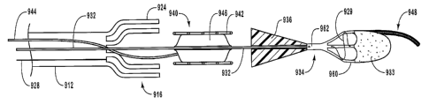
actuating
element 721 can include a key that mates with a key way formed in guide member
housing 722, or vice versa. Further, although reference is made to rotational
motion
and motion parallel to the longitudinal movement of the dilation balloon and
stent,
one skilled in the art can identify various other directions of motion that
can enable or
facilitate deployment of the dilation balloon and/or stent. For instance, the
motion of
actuating element can be at any angular orientation relative to the
longitudinal axis of
the guide member, whether or not such motion includes one or more revolutions
of
the actuating element relative to the guide member.
As depicted in Figure 27, another embodiment of delivery device 700c is
illustrated. To aid with moving actuating element 721 relative to guide member
housing 722, actuating element 721 and guide member housing 722 and/or guide
member 712 can include optional handles 796 and 798 respectively. These
handles
796 and 798 can optionally include gripping regions that are adapted to
cooperate
with one or more appendages of a user of the device. In another configuration,
each
handle 796 and 798 can have a substantially constant cross-section along their
lengths. In still other configurations, each handle 796 and 798 can have
variable
cross-sections along their lengths. Additionally, although a single luer
fitting 745 is
depicted in Figure 27, it can be understood by one skilled in the art that
delivery
device 700c can include one or more fittings to facilitate introduction of one
or more
fluids to an interior of delivery device or to a dilation balloon.
Figure 28 shows yet another embodiment of delivery device 700d in which
actuating element 721 includes a housing 760 that contains a rotatable gear
762
adapted to cooperate with complementary features or structures 770 formed in a
proximal end of guide member 712. The gear 762, with associated one or more
teeth,
features or structures 768, can be manipulated or rotated by an actuator 764
as a
clinician or other individual selects an actuator member 766 and rotates
actuator 764
to rotate gear 762. Optionally, actuator 764 has one or more teeth, features
or
structures that can cooperate with gear 762, such that rotational motion of
actuator
764 is translated to movement of gear 762.
CA 02466037 2004-05-06
WO 03/039405 PCT/US02/36153
28
As actuator 764 and hence gear 762 are rotated, the complementary features
770 of guide member 712 mate with the teeth, features or structures 768 of
gear 762
to move guide member 712 in a proximal and/or distal direction, dependent upon
the
rotational direction of actuator 764. By so doing, the dilation assembly and
the stent
can be deployed from a distal end (not shown) of device 700d.
In addition to moving or positioning guide member 712, actuating member
728 may also be operated through using a sliding switch 762 associated with
housing
760 and actuating element 721. The actuating member 728 is coupled to a leg
762
that is attached to switch 762, while sliding switch 762 is slidably coupled
to housing
760. The sliding translation of switch 762 moves actuating member 728 in the
respective direction to release a restraining force applied by the restraining
member or
mechanism (not shown) of device 700c. One skilled in the art can identify
various
other configurations of actuating element 721 in light of the teaching
contained
herein.
Referring now to Figures 29-37, depicted are various configurations of
alternative embodiments of a delivery device in accordance with the present
invention. The features and functions of other described delivery devices
apply to the
discussion of delivery devices 800a through 800g. Furthermore, it will be
appreciated
that the majority of features and functions described with respect to delivery
device
800a also apply to delivery devices 800b through 800g described further below.
The
delivery devices of Figures 29-37 illustrate various embodiments wherein a
delivery
device is adapted to be used with a guidewire. For ease of explanation, the
embodiments of Figures 29-37 do not include a restraining member or mechanism
that restrains the dilation assembly and stent inside the guide member.
However, it
will be appreciated that any restraining member or mechanism with any
actuating
assembly, as disclosed herein or understood by those of skill in the art, may
be
employed with devices 800a-800g.
As shown in Figure 29, delivery device 800a includes a guide member 812
having a proximal end 816 and a distal end 814, with a lumen 818 extending
from
distal end 814 toward proximal end 816. The distal end 814 can have a similar
configuration to the other guide member distal ends described herein. For
instance, a
restraining member (not shown) may be disposed at distal end 814 to cooperate
with
structures adapted to restrain a dilation assembly 840 and/or a stent 842.
Disposed at
proximal end 816 is a guide member housing 822 that cooperates with an
actuating
CA 02466037 2004-05-06
WO 03/039405 PCT/US02/36153
29
element 821 of an actuating assembly 820, in a similar manner to that
described with
respect to Figure 6.
Extending from an aperture 834 in a proximal end of actuating element 821
toward distal end 814 of guide member 812 is a guidewire 832. As shown best in
Figure 30, guidewire 832 cooperates with a dilation assembly 840 disposed at a
distal
end of guide member 812. In this illustrative configuration, dilation assembly
840
includes a tubular member 836 that cooperates with a dilation balloon 846
coupled or
attached thereto. The tubular member 836 can function as a positioning member
that
facilitates deployment of dilation assembly 840 and stent 842. The guidewire
832
io extends through tubular member 836 that allows dilation balloon 846, and a
stent 842
coupled to dilation balloon 846, to be moved along guidewire 832 when
necessary.
The internal diameter of a lumen of tubular member 836 is complementary to the
exterior diameter of guidewire 832.
Guidewire 832 terminates at a distal end with an atraumatic tip 848 that can
is include a core wire 856 wrapped with a coiled spring 858. The core wire 856
may be
an extension of the remainder of guidewire 832 or alternatively may be a
separate
member coupled or attached to the distal end of guidewire 832. In either case,
core
wire 856 can be made from the same or a different material than guidewire 832
and
may optionally be a solid member or a tubular member.
20 The dilation balloon 846 of dilation assembly 840 is inflated through a
dilation
tube 844 that extends from dilation balloon 846 to terminate at the proximal
end of
actuating element 821 with a luer fitting 845. An additional luer fitting 890
may be
provided attached to actuating element 821. It will be appreciated that luer
fitting 890
can perform substantially the same function as luer fitting 790.
25 The distal end of dilation tube 844 cooperates with an interior of dilation
balloon 846. The distal end of dilation tube 84 can be connected to tubular
member
836, dilation balloon 846, or to both tubular member 836 and dilation balloon
846.
The dilation tube 844 can be used to position dilation balloon 846 and/or
stent 842
during a procedure. Consequently, dilation tube 844 can have sufficient
strength to
3o enable distal movement of dilation tube 844 to translate to distal movement
of the
remainder of dilation assembly 840. Similarly, dilation tube 844 can have
sufficient
strength to enable proximal movement of dilation tube 844 to translate to
proximal
movement of the remainder of dilation assembly 840.
CA 02466037 2004-05-06
WO 03/039405 PCT/US02/36153
Delivery device 800a is configured so that guidewire 832 can be positioned in
a body lumen, and delivery device 800a can be removed from within the body
lumen
while retaining guidewire 832 at the desired position. As such, other
conventional,
interventional devices may then be used to complete the procedure. A device
may be
5 connected to the distal end of the guidewire such as, for example, a filter
assembly for
collecting embolic particles that are dislodged in the body vessel during a
stenting
operation, as will be discussed in greater detail hereinafter. Other devices
may be
exchanged over guidewire 832 as will be understood by those of skill in the
art.
Depicted in Figure 31 is another embodiment of a device 800b. The device
l0 800b includes a dilation assembly 840b that is adapted to cooperate with a
guidewire
832. As shown, dilation assembly 840b includes a tubular member 836 that
cooperates with an expandable dilation balloon 846. Disposed at a proximal end
of
tubular member 836 is a positioning member 838. The positioning member 838 is
coupled to a proximal end of tubular member 836 to facilitate transfer of
forces
1s applied to positioning member 838 to tubular member 836 to position
dilation
assembly 840. The positioning member 838 can be coupled or attached to tubular
member 836, dilation balloon 846, or both tubular member 836 and dilation
balloon
846, whether such coupling or attachment occurs at a proximal end, distal end,
or
other portion of tubular member 836 and/or dilation balloon 846 between the
20 respective proximal ends and distal ends thereof. Similarly, the coupling
or attaching
of positioning member 838 to one or both of tubular member 836 and dilation
balloon
846 can be to or upon internal and/or external surfaces of tubular member 836
and
dilation balloon 846. By so doing, positioning member 838 can be manipulated
by a
physician or clinician to position dilation assembly 840b in the desired
location to
25 dilate a stent (not shown) and/or lesion. For instance, positioning member
838 can be
used to slide dilation balloon 846 along guidewire 832.
As shown, positioning member 838 is separate from dilation tube 844.
Although reference is made to positioning member 838 being separate from
dilation
tube 844, it can be appreciated that positioning member 838 can be removably
3o disposed within dilation tube 844, while being capable of positioning
dilation balloon
846 in the desired location within the body lumen or vessel. For instance,. as
illustrated in dotted lines in Figure 31, extending from tubular member 836 or
formed
in tubular member 836 is a stop 837 that can cooperate with a distal end of a
positioning member 838 disposed within a lumen of dilation tube 844. By moving
the
CA 02466037 2004-05-06
WO 03/039405 PCT/US02/36153
31
positioning member in the distal direction, the distal end of the positioning
member
cooperates with stop 837 to move dilation assembly 840b in the distal
direction. To
move tubular member 836 in the proximal direction, a clinician or physician
can
move dilation tube 844 in a proximal direction. In another configuration, stop
837
can include a recess (not shown) that friction fits or otherwise cooperates
with the
distal end of the positioning member, such that the positioning member is
retained in
the recess with sufficient force that the positioning member can move the
tubular
member 836 in both proximal and distal directions.
The proximal end of guide member housing 822 as illustrated in Figure 31,
1o cooperates with an actuating assembly 821b, while a distal end 814 of guide
member
812 cooperates with a restraining member or mechanism (not shown) and is
adapted
to aid in applying a restraining force to dilation assembly 840b and/or stent
842.
Actuating element 821b is adapted to enable a clinician to operate delivery
device
800b to deliver stent 842, such as in a similar manner to the device described
in
Figure 29. For instance, positioning member 838 can be coupled to actuating
element
821b such that distal movement of actuating element 821b moves dilation
assembly
840b.
In addition, the proximal end of actuating element 821b includes an annular
clamping mechanism 862, such as, but not limited to, touhy-borst adaptor,
compressible polymer or silicone rubber gasket, or other clamping mechanisms
862
known to those skilled in the art in light of the teaching contained herein.
The annular
clamping mechanism 862 receives guidewire 832 and creates a mechanical
connection and a fluid seal between actuating element 821b and guidewire 832.
This
seal prevents fluid escaping from within lumen 818, while providing a
mechanism for
releasing delivery device 800b from guidewire 832 in the event that other
conventional, interventional devices are to be used without loosing the
vascular access
that is gained by the device as a whole. For example, by rotating annular
clamping
mechanism 862, the seal is broken and delivery device 800b can be removed from
guidewire 832. A similar clamp or other seal can cooperate with positioning
member
838 to prevent fluid escaping from within device 800b.
Figure 32 depicts another embodiment of delivery device 800c. In this
embodiment, tubular member 836 extends substantially between distal end 814
and
proximal end 816 of guide member 812. Tubular member 836 is adapted to receive
guidewire 832 therethrough. Thus, tubular member 836 extends from a distal end
of
CA 02466037 2004-05-06
WO 03/039405 PCT/US02/36153
32
dilation balloon 846 to a proximal end of guide member 812, such that the
proximal
end of tubular member 836 terminates at a point proximal to a proximal end of
dilation tube 844. Additionally, the proximal end of tubular member 836
cooperates
with a proximal end of guide member 812 and/or an actuating element 821c
disposed
at the proximal end of guide member 812.
Actuating element 821c includes a fixed portion 829 and a movable portion
831 slidably disposed with portion 829. The portion 829 can be integrally
formed
with a proximal end of guide member 812 or a separate member that is coupled
or
attached to the proximal end of guide member 812, where such coupling or
attaching
1o can be achieved by complementary threads, key and keyway configuration,
chemical
bonding, thermal bonding, or adhesives.
The portion 831 cooperates with portion 829 in sealing manner so that a fluid
entering an interior space defined by the interiors of portion 829 and a
portion of
portion 831 is prevented from exiting therefrom. This seal can be created by
one or
more sealing members 833 and/or between the tolerances associated with portion
829
and portion 831. Illustratively, sealing member 833 can be one or more O-rings
in
one or more grooves, one or more O-rings, gasket, or viscous fluid seal.
The portion 831 contains a support structure 823 extending across a distal end
thereof. The proximal end of tubular member 836 is fixedly attached to support
structure 823. Support structure 823 also includes an aperture 825 through
which
extends guidewire 832. Preferably, a seal 827 is disposed between and/or
within
aperture 825 and guidewire 832 to retain fluid inside guide member 812.
Consequently, upon depressing portion 831 in the direction of arrows A,
dilation
balloon 846 is deployed from within lumen 818 of guide member 812. Similarly,
upon moving portion 831 of actuating element 821c in the direction of arrows
B,
dilation balloon 846 is retracted into lumen 818 of guide member 812.
As depicted in Figure 33, another embodiment of delivery device 800d is
illustrated. In this embodiment, dilation balloon 846 is coupled or attached
directly to
guidewire 832. Consequently, positioning member 838 is connected to guidewire
832
3o and/or optionally dilation balloon 846 instead of tubular member 836.
Positioning
member 838 is manipulatable by a physician, clinician, or the like to position
dilation
assembly 840 in the desired location to dilate the stent and lesion.
Consequently, by
moving positioning member 838, dilation balloon 846 can be placed in the
position to
optionally pre-dilate the lesion and/or dilate the lesion during implanting of
stent 842.
CA 02466037 2004-05-06
WO 03/039405 PCT/US02/36153
33
Figure 34 illustrates another embodiment of delivery device 800e. In this
embodiment, positioning member 838 is connected to dilation balloon 846.
Positioning member 838 is manipulatable by a physician, clinician, or other
individual
to position dilation assembly 840 in the desired location to dilate the stent
and lesion.
Consequently, by moving positioning member 838, dilation balloon 846 can be
placed
in the position to optionally pre-dilate the lesion and/or dilate the lesion
during
implanting of a stent (not shown).
Guidewire 832 passes between dilation balloon 846 and stent 842. Although
not depicted for ease of explanation, guidewire 832 may have an atraumatic tip
1o attached or formed at a distal end thereof. The dilation balloon 846
includes an
integrally formed dilation tube 844 that extends from a distal end of dilation
balloon
846. The dilation balloon 846, as with other dilation balloons described
herein, can
have various configurations, such that dilation balloon 846 can having
substantially
constant cross-section along its length or alternatively have a variable cross-
section
along its length. Furthermore, the dilation balloons of the present invention
can be
formed from one or more separate dilation balloons, with associated one or
more
dilation tubes, which collectively provide the functionality of a single
dilation
balloon.
In addition, Figure 34 depicts a tip 864 disposed at distal end of guidewire
832. Tip 864 provides a transition between guidewire 832 and guide member 812
to
limit the potential of damaging the body lumen or vessel of the patient during
insertion and removal of delivery device 800e during a procedure. Various
types of
tips are known to those skilled in the art, such as, but not limited to, those
discussed
herein and others known to one skilled in the art in light of the teaching
contained
herein. For instance, tip 864 can have various configurations so long as the
configuration provides a transition between guidewire 832 and guide member 812
to
aid in preventing damage to the body lumen or vessel during insertion and
removal of
delivery device 800e. Further, tip 864 can be coupled or attached to guidewire
832
through various manners, such as, but not limited to, adhesives, mechanical
bonds,
thermally created bonds, being integrally formed therewith, or combinations
thereof.
As depicted in Figure 35, illustrated is another embodiment of delivery device
800f. In this embodiment, guidewire 832 passes between guide member 812 and
dilation assembly 840 to terminate distally of a distal end of guide member
812.
Although not depicted for ease of explanation, guidewire 832 may have an
atraumatic
CA 02466037 2004-05-06
WO 03/039405 PCT/US02/36153
34
tip attached or formed at a distal end thereof. Connected to dilation balloon
846 is a
positioning member 838, similar to those described herein. Positioning member
838
is manipulatable by a physician, clinician, or other individual to position
delivery
device 800f in the desired location to dilate the stent and lesion.
Consequently, by
moving positioning member 838, dilation balloon 846 can be placed in the
position to
optionally pre-dilate the lesion and/or dilate the lesion during implanting of
stent 842.
Delivery device 800f also includes a tip 864, similar in structure and
function to the
tip 864 shown and discussed with respect to Figure 34.
Figure 36 illustrates yet another embodiment of a delivery device of the
to present invention. As shown, a delivery device 800g includes a dilation
assembly
840g, which may be similar to other dilation assemblies described herein,
which is
disposed past a distal end 814 of guide member 812. Guide member 812 acts as a
positioning member to position dilation assembly 840g in the desired location
to
dilate the stent and lesion. Consequently, by moving guide member 812,
dilation
balloon 846 can be placed in the position to pre-dilate the lesion and/or
dilate the
lesion during implanting of a stent. Although not depicted for ease of
explanation,
guidewire 832 may have an atraumatic tip attached or formed at a distal end
thereof.
Figures 37 through 44 depict another aspect of the present invention. During a
procedure to dilate a lesion and/or implant a stent at a lesion, often emboli
becomes
dislodged and is carried downstream in the body vessel. To prevent the emboli
from
blocking even smaller body vessels further downstream, one or more embodiments
of
the present invention can include means for providing embolic protection. The
means
for providing embolic protection can be included in a delivery device having a
unitary
configuration where the delivery device and the means for provide embolic
protection, such as a filter device, can be inserted into a body lumen
substantially
simultaneously.
Referring to Figure 37, an exemplary delivery device 900 is depicted having
many of the same features and functionality of the delivery devices heretofore
described. Consequently, the descriptions of the various other delivery
devices
3o described herein apply to delivery device 900. As illustrated, delivery
device 900
includes a guide member 912 having a dilation assembly 940 and stent 942
disposed
therein. It will be appreciated that in the embodiment of Figure 37 and
subsequent
embodiments hereafter, a restraining member or mechanism, illustrated in
dotted
lines, may be disposed at distal end 914 of guide member 912 to restrain
dilation
CA 02466037 2004-05-06
WO 03/039405 PCT/US02/36153
assembly 940 and stent 942 adjacent or near distal end 914 of guide member 912
until
deployment of same is desired. It will be appreciated that any restraining
member or
mechanism may be employed as disclosed herein or understood by those of skill
in
the art. Furthermore, appropriate. structures may be employed for deploying
dilation
5 assembly 940 and stent 942 as described herein or understood by those
skilled in the
art.
With continued reference to Figure 37, delivery device 900 has a filter
assembly 931 disposed distally of guide member 912. Consistent with teachings
of
the present invention, delivery device 900 has a guidewire 932 disposed
through
1o dilation assembly 940 and optionally through filter assembly 931. In the
illustrated
configuration, guidewire 932 terminates at filter assembly 931, with the
filter
assembly 931 being coupled to a distal end of guidewire 932 and includes an
atraumatic tip, as will be described in more detail below.
Filter assembly 931 is adapted to provide embolic protection during use of
15 device 900. As depicted in Figures 37 and 38, filter assembly 931 has a low
profile to
facilitate insertion of the same with a body lumen. A transition member 936 is
disposed between filter assembly 931 and dilation assembly 940. The transition
member 936 is adapted to provide a transition between guide member 912 and
filter
assembly 931. This transition prevents damage to the body lumen within which
20 device 900 is disposed and prevents catching upon a wall or junction of one
or more
body lumens as device 900 is steered through the tortuous anatomy of a
patient. As
illustrated, transition member 936 includes a passageway 938 disposed
therethrough
for receiving guidewire 932. Passageway 938 can be adapted to securely retain
guidewire 932 therein or optionally removably receive guidewire 932.
Alternatively,
25 transition member 936 can include a hole through which guidewire 932 passes
or is
received. In still another configuration, transition member 936 includes a
hole
adapted to receive a distal end of guidewire 932, while a distal end of
transition
member 936 is formed or cooperates with filter assembly 931.
Figures 37 and 38 illustrate filter assembly 931 being restrained in
preparation
30 for deploying filter assembly 931, while Figures 39-41 depict filter
assembly 931
being deployed or activated. As shown in Figure 41, filter assembly 931
includes a
filter basket 934 and a filter 933. Before deployment, filter 933 can be
disposed
inside filter basket 934, surround filter basket 934, or a combination
thereof. The
filter 933 is adapted to capture embolic particles or material that may become
CA 02466037 2004-05-06
WO 03/039405 PCT/US02/36153
36
dislodged during a procedure associated with delivery device 900 or optionally
other
procedures when delivery device 900 is optionally slidably removed from
guidewire
932 and associated filter assembly 931. Consequently, filter 933 can
optionally float
within a body lumen upon being deployed, with a distal end of filter 933
floating in
the body lumen and the proximal end of filter 933 being coupled to filter
basket 934.
In another configuration, a distal end of filter 933 can be coupled to a
portion of filter
basket 934.
The filter 933 can be fabricated from a variety of different materials, such
as,
but not limited to, a woven or braided plastic or metallic mesh, a perforated
polymer
film, a Nitinol mesh, combinations thereof, or other material that is capable
of
capturing material within flowing blood, while allowing the blood to flow
through the
pores or apertures thereof. Generally, filter 933 can be fabricated from a
variety of
materials so long as filter 932 is capable of being packed within filter
basket 934, and
optionally float in the blood flow or stream passing through the body lumen
within
which it is inserted, and is bio-compatible.
Filter 933 can have a variety of differently sized pores ranging from about 50
microns to about 200 microns, from about 60 microns to about 180 microns, or
from
about 75 microns to about 150 microns. For instance, the pores can have a
variety of
different configurations, such as but not limited to circular, oval,
polygonal,
combinations thereof or other configurations known to one skilled in the art
in light of
the teaching contained herein. In one configuration, therefore, filter 933 can
include
pores that are differently sized and configured. Consequently, a major or
minor axis
of each pore can have a variety of different sizes ranging from about 50
microns to
about 200 microns, from about 60 microns to about 180 microns, or from about
75
microns to about 150 microns. Generally, the pore size can vary as needed, so
long as
the pores are sized so that the pores do not compromise blood flow through the
filter,
i.e., prevent blood flowing through the filter, and collect material that
could
potentially occlude smaller downstream vessels, potentially blocking blood
flow to
tissue or result in stroke or infarction.
In addition to the above, filter 933 can be coated with a hydrophilic coating,
a
heparinized coating, a Polytetrafluoroethylene (PTFE) coating, a silicone
coating,
combinations thereof, or various other coatings as know or desired by one
skilled in
the art in light of the teaching contained herein.
CA 02466037 2004-05-06
WO 03/039405 PCT/US02/36153
37
The filter basket 934 supports filter 933 following deployment of filter 933.
The filter basket 934 includes a plurality of struts 960 that extend from a
body 962.
Struts 960 of filter basket 934 are adapted to extend outwardly to position
filter 933
within the body lumen. A strut 960a of struts 960 can include an atraumatic
tip 948,
with struts 960a forming at least a portion of the core wire of atraumatic tip
948. This
strut may also be covered with a flexible coil 958. The body 962 of filter
basket 934
includes a hole 967 that is adapted to receive guidewire 932. Alternatively,
body 962
can include a passageway that is adapted to receive a distal end of guidewire
932.
The filter 933 can be attached to struts 960 of filter basket 934 in a variety
of
1 o ways. For instance, filter 933 can be attached through adhesives, solvent
bonding,
thermal bonding, mechanical connections, or combinations thereof. Further, the
distal
end of two or more struts 960 can include a hole through which strands of
filter media
932 can be passed and attached to struts 960. Alternatively, the strands can
be tied in
a knot, folded back upon filter 933, and affixed to filter 933. Various other
manners
exist of coupling or connecting filter 933 to filter basket 934.
Optionally, filter assembly 931 includes a number of radiopaque bands and/or
markers affixed to a variety of positions on filter assembly 931. For
instance, bands,
markers or other means for radiopacity can be included upon filter 933, filter
basket
934 and/or struts 960. In other configurations, the delivery device generally
includes
means for radiopacity at one or more locations or positions thereof to aid
with
viewing the position of the delivery device and the various elements and
components
thereof.
As illustrated, a restraining member or mechanism 925 restrains struts 960,
while another retraining member, shown in dotted lines, restrains a distal end
of guide
member 912. Optionally, restraining member or mechanism 925 restrains both the
distal end of guide member 912 and struts 960. Figures 37 and 38 depict
restraining
member or mechanism 925 restraining struts 960, while Figures 39-41 depict
struts
960 being released from restraining member or mechanism 925. In the exemplary
configuration of Figure 41, restraining member or mechanism 925 has a similar
configuration to restraining member or mechanism 525. Therefore, restraining
member or mechanism 925 includes a cord 929 forming a number of hoops, with
one
or more of the hoops being adapted to receive an actuating member 928, which
is
optionally part of restraining member or mechanism 925. The actuating member
928
is disposed within the hoops so that cord 929 applies a restraining force
against struts
CA 02466037 2004-05-06
WO 03/039405 PCT/US02/36153
38
960. Actuating member 928 can be removed from the hoops to thereby allow
struts
960 to extend outwardly to deploy filter 933. Cord 929 may be made from
metallic
wires, polymer actuating members, or other materials that can be manipulated
to form
hoops through which an actuating or securing member. Optionally, cord 929 is
adapted to expand outwardly either under the influence of one or more struts
or due to
a biasing force applied or incorporated within cord 929by the configuration
and/or
material of the cord, the hoops, and/or the restraining member.
Cord 929 can be attached to one or more struts 960 of filter assembly 931
through various attachment mechanisms. For instance, cord 929 can be attached
to
guide member and/or one or more of the struts through adhesives, mechanical
fasteners, securing loops, or other manner that securely attaches cord 929 to
one or
more of struts 960. Alternatively, cord 929 may be attached to actuating
member 928
and be removed when actuating member 928 is moved in a proximal direction. A
clinician or physician can initiate the longitudinal motion of actuating
member 928,
either directly or through using of an actuating mechanism or device. Although
reference is made to one particular embodiment of restraining member or
mechanism
925, one skilled in the art can appreciate that other restraining members or
mechanism
described herein can be used to restrain struts 960.
As shown, filter basket 934 includes one or more holes 970 that are adapted to
receive at least a portion of restraining member or mechanism 925. The holes
970 can
be disposed at various locations of filter assembly 931. For instance, and not
by way
of limitation, body 962 and each strut 960 can include one or more holes 970.
The
restraining member or mechanism 925 can be at least partially disposed through
one
or more of holes 970, with cord 929 or other portion of restraining member or
mechanism 925 being optionally releasably coupled to one of struts 960 or body
962
of filter basket 934. Moving actuating member 928 of restraining member or
mechanism 925 in a proximal direction causes struts 960 to move outwardly to
release
filter 933.
A proximal end (not shown) of restraining member or mechanism 925 or
actuating member 928 can be accessible by a clinician or physician to allow
the same
to operate restraining member or mechanism 925 to release the restraining
force
applied to struts 960. Optionally, the proximal end of restraining member or
mechanism 925 can cooperate with an actuating assembly that can be operated to
CA 02466037 2004-05-06
WO 03/039405 PCT/US02/36153
39
move restraining member or mechanism 925 as needed to release the restraining
force
applied by restraining member or mechanism 925.
In the illustrative configuration of Figures 37-41, actuating member 928 of
restraining member or mechanism 925 can be moved in a proximal direction with
sufficient movement and force to be removed from engagement with the hoops of
cord 929. By breaking the coupling or engagement between actuating member 928
and cord 929, struts 960 are allowed to expand or move outwardly to deploy
filter
933. Following deploying filter 933, an actuating assembly (not shown) can be
manipulated to deploy dilation assembly 940 and stent 942 from guide member
912,
1o in a similar manner to that described herein, and as illustrated in Figure
40.
Therefore, two actuating assemblies can be used, one to release restraining
member or
mechanism 925 and one to release dilation assembly 940 and stent 942.
It will be appreciated that restraining member or mechanism 925 is but one
means for restraining struts 960 of filter basket 934. Other configurations
may be
employed, such as, but not limited to, the restraining configurations or means
for
restraining described in Figures 2-24. For instance, struts 960 of filter
basket 934 can
be restrained in the same manner as the strut associated with the guide member
of the
present invention.
Turning to Figure 40, depicted is delivery device 900 with filter assembly 931
deployed and dilation assembly 940 and stent 942 deployed from guide member
912.
Deploying of dilation assembly 940 and stent 942 can be achieved in a similar
manner
to that described with respect to other dilation assemblies and stents
discussed herein.
Similarly, manipulating restraining member or mechanism 925 to release struts
960
and deploy filter 933 can deploy filter assembly 931. Through moving guide
member
912 relative to guidewire 932, vice versa, or combinations thereof, dilation
assembly
940 and stent 942 can be released from within guide member 912.
Figures 42 and 43 illustrate another embodiment of a filter assembly 1031.
Filter assembly 1031 has another embodiment of a mechanism for restraining
struts
1060. This particular configuration of struts 1060 illustrates that struts
1060 can be
coupled to or attached to a distal end of a guidewire 932 or transition member
936
(Figure 37). The length of struts 1060 can vary based upon the particular
configuration of guide member 1012.
A restraining mechanism 1064 maintains struts 1060 in a restrained position as
shown in Figure 43. In this embodiment, restraining mechanism 1064 includes a
CA 02466037 2004-05-06
WO 03/039405 PCT/US02/36153
tubular member 1062 attached to each strut 1060 and a restraining or actuating
member 1025 disposed therein. Although reference is made to tubular member
1062
being attached to each strut 1060, it can be understood that one or more
tubular
members 1062 can be attached to each strut 1060 and/or fewer than each strut
1060
5 includes tubular member 1062.
Each tubular member 1062 is adapted to receive restraining or actuating
member 1025. As shown in Figure 43, when struts 1060 are restrained, tubular
members 1062 are aligned to receive restraining or actuating member 1025. That
is,
each tubular member 1062 is staggered on adjacent struts 1060 with respect to
other
1o tubular members 1062, such that tubular members 1062 line up from the
proximal end
to the distal end of filter assembly 1031. Restraining or actuating member
1025 is
then disposed through the series of tubular members 1062 to restrain struts
1060 and
prevents them from extending outwardly, as illustrated in Figure 43.
Restraining or actuating member 1025 extends from filter assembly 1031 into
15 a lumen of guidewire 1032 to terminate at a proximal end of guide member
1012 and
optionally extend beyond the proximal end of guide member 1012. Alternatively,
restraining or actuating member 1025 can extend proximally from filter
assembly
1031 to exit through an aperture 1069, depicted in dotted lines, before
terminating at
the proximal end of guide member 1012 and optionally extend beyond the
proximal
20 end of guide member 1012. In this latter configuration, restraining or
actuating
member 1025 can be disposed externally to guide member 1012 or partially
externally
to guide member 1012 as it extends to the proximal end of guide member 1012
and
optionally extend beyond the proximal end of guide member 1012. It will be
appreciated that a clinician or physician can manipulate restraining member or
25 mechanism 1064 to release the restraining force applied by restraining
member or
mechanism 1064. Alternatively, restraining member or mechanism 1064 can be
optionally operated by an actuating assembly similar to that described herein,
such as,
but not limited to, the actuating assembly described with respect to Figure 6,
or any
other actuating assembly known by those of skill in the art.
30 Each tubular member 1062 coupled to struts 1060 can be fabricated from a
metal, a plastic, polymer, a polymer, a synthetic materials, whether or not
the material
is the same as that forming guide member 1012. In one embodiment, each tubular
member 1062 is a polymer tube, such as a polyimide or polyurethane tube that
is fixed
to respective struts 1060 with adhesive. In another configuration, each
tubular
CA 02466037 2004-05-06
WO 03/039405 PCT/US02/36153
41
member 1062 is a metallic cut tube that may be attached to respective struts
1060 with
and adhesive or solder. In still another configuration, each strut 1060
includes an
aperture through which actuating member 1025 passes to restrain struts 1060
and
prevents the same from extending outwardly.
Referring now to Figure 44 is an exemplary configuration of another filter
assembly according to another aspect of the present invention. The features of
functions of filter assembly 1131 are applicable to other filter assemblies of
the
present invention, and vice versa. Furthermore, the discussion related to the
one or
more other struts of filter assembly 1131 is applicable also to the struts
associated
1 o with the guide members of the delivery devices of the present invention.
As depicted in Figure 44, filter assembly 1131 includes a body 1162 and one
or more struts 1160. Coupled to one or more struts 1160 is a filter 1133.
Extending
from body 1162 through filter 1133 is an atraumatic tip 1148, with associated
coil
1158. For ease of explanation, the restraining member or mechanism associated
with
filter assembly 1131 is not shown, however, it will be understood that any of
the
restraining members or mechanisms described herein can be used to apply a
restraining force to one or more struts of filter assembly 1131.
Struts 1160 extend from a body 1162 of filter assembly 1131. Although
reference is made herein to struts 1160 being integrally formed with body
1162, it can
be appreciated that struts 1160 can be separate members coupled to body 1162.
Further, struts 1160 can be integrally formed with guidewire 1132 or separate
members coupled to guidewire 1132.
Each strut 1160 includes a distal portion 1162, a proximal portion 1166, and
an intermediate portion 1164 disposed between distal portion 1162 and proximal
portion 1166. Struts 1160 may attach to filter 1133 on the exterior of filter
1133, on
the interior of filter 1133, along the edge of filter 1133, through filter
1133, or
combinations of one or more of the proceeding. To provide additional surface
area to
connect each strut 1160 to filter 1133, each strut 1160 can be configured so
that distal
portion 1162 has a cross-sectional dimension larger than intermediate portion
1164.
Stated another way, distal portion 1162 can have a larger surface area than
intermediate portion 1164. The large cross-sectional area provided by the
cross-
sectional dimension of distal portion 1162 provides large area for bonding
each strut
1160 to filter 1133. In this configuration, a strong bond is created between
each strut
1160 and filter 1133.
CA 02466037 2004-05-06
WO 03/039405 PCT/US02/36153
42
Similarly, each strut 1160 can be configured so that proximal portion 1166 has
a cross-sectional dimension larger than intermediate portion 1164, while
optionally
having a similar, larger, or smaller cross-sectional dimension than distal
portion 1162.
By having a large cross-sectional dimension and hence large surface area, each
strut
1160 can apply a greater biasing force to extend strut 1160 outwardly to
deploy filter
1133.
By varying the cross-sectional dimensions of distal portion 1162, intermediate
portion 1164, and/or proximal portion 1166, the degree of bias exerted by each
strut
1160 to move distal portion 1162 toward the wall of a blood vessel can be
varied.
1o The biasing force can also be changed through optionally varying the length
of each
strut 1160 and/or changing the curvature of each strut 1160.
Although reference is made herein to each strut 1160 having the above-
referenced configurations, one skilled in the art can appreciate that one or
more of
struts 1160 can be configured as described above. Further, each strut 1160 can
optionally be configured differently so that each strut 1160 can have similar
or
dissimilar biasing forces compared to others struts 1160 of the same delivery
device.
Through varying the biasing forces, the delivery device can be used for a
variety of
different procedures or blood vessel configurations.
Struts 1160 can be formed from Nitinol, stainless steel, metals, alloys,
composites, plastics, polymers, synthetic materials, or combinations thereof.
Each
strut 1160 can have a generally straight distal portion 1162, proximal portion
1166,
and/or intermediate portion 1164. Alternatively, each strut 1160 can have a
generally
curved distal portion 1162, proximal portion 1166, and/or intermediate portion
1164.
In still another configuration, each strut 1160 can have a combination of one
or more
straight and/or one or more curved portions.
Coupled to body 1162, such as within a lumen or hole 1137, is an atraumatic
tip 1148. The atraumatic tip 1148 can include a core wire 1156 and a flexible
coil
1158 disposed thereon. Core wire 156 passes through an aperture 1170 in a
distal end
of filter 1133. Alternatively, core wire 1156 passes through one or more pores
formed
in filter 1133. To secure filter 1133 to atraumatic tip 1148, a securing coil
1186
surrounds a portion of coil 1158 and the distal end of filter 1133. Although
this is one
manner to connect filter 1133 to atraumatic tip 1148, one skilled in the art
can identify
various other manners to connect filter 1133 to atraumatic tip 1148. For
instance, the
distal end of filter 1133 can be bonded to atraumatic tip 1148 using
adhesives,
CA 02466037 2004-05-06
WO 03/039405 PCT/US02/36153
43
mechanical fasteners, crimping, seals, friction fit, press fit, or other
manners to
connect filter 1133 to atraumatic tip 1148. In another configuration, filter
1133 is not
connected to atraumatic tip 1148 but can slide along a portion of atraumatic
tip 1148.
Turning now to Figures 45 and 46, two exemplary embodiments of capturing
device or mechanism capable of being used to capture the filter of the filter
assembly
are depicted. After the filter is deployed, it is desirable to capture the
filter after the
stenting operation has taken place. More specifically, it is desirable to
capture the
embolic particles that may have been captured by the filter and remove the
same.
Figure 45 illustrates a capture device 1200 according to one aspect of the
present
invention. Capture device 1200 includes a capture catheter 1202. As shown,
capture
catheter 1202 includes a capturing portion 1204 and a positioning member 1206
connected or attached to capturing portion 1204. Capturing portion 1204 has a
distal
end 1208 and a proximal end 1210. Capturing portion 1204 includes a lumen 1212
extending from distal end 1208 to terminate at an aperture 1214 at proximal
end 1210
thereof. The distal end 1208 optionally includes one or more radiopaque
markers or
bands 1216, only one being shown. Similarly, a proximal end of positioning
member
1206 can include one or more radiopaque marker or bands 1216. More generally,
capture device and any of the delivery devices and guidewires of the present
invention
can include one or more radiopaque indicators, whether such indicators are
marker,
bands, studs, or other radiopaque display elements.
Lumen 1212 is configured to receive a guidewire with attached filter assembly
(not shown) of a delivery device. In one embodiment, lumen 1212 can include a
stop
member 1218, depicted in dotted lines, with a hole 1220 there through. A
guidewire,
as represented by dotted lines identified by reference numeral 1232, passes
through
hole 1220 of stop member 1218. The guidewire 1232 can have various
configurations, such as, but not limited to those described herein and others
known to
those skilled in the art.
Stop member 1218 prevents a filter assembly disposed at a distal end of
guidewire 1232 to pass through hole 1220 once capture catheter 1202 has
received
within lumen 1212 the filter assembly associated with guidewire 1232
sufficiently
that the filter of the filter assembly is at least closed to prevent escape of
embolic
material. In one configuration, the filter assembly and associated filter are
completely
enclosed by capture portion 1204 of capture device 1200. In other
configurations, the
filter assembly and/or filter are partially enclosed by capture portion 1204
of capture
CA 02466037 2004-05-06
WO 03/039405 PCT/US02/36153
44
device 1200. One skilled in the art can identify various other configurations
of stop
member 1218, so long as stop facilitates completely or partially capturing the
filter
assembly and/or the filter associated with guidewire 1232.
Positioning member 1206 is attached to capture catheter 1202 and can be used
to move capture catheter 1202 along guidewire 1232, whether such movement is
caused by moving catheter 1202 relative to guidewire 1232, guidewire 1232
relative
to catheter 1202, or combination thereof. Positioning member 1206 has
sufficient
stiffness that application of a force at a proximal end 1224 can be
transferred to
longitudinal motion of capturing portion 1204 of capture catheter 1202. In one
1o configuration, positioning member 1206 is a solid member, while in another
configuration positioning member 1206 is partially or completely hollow.
Positioning
member 1206 can be fabricated from a polymer, a plastic, polymer, a synthetic
material, a metal, an alloy, combinations thereof, or other material that can
be used for
medical devices and has the needed stiffness.
As illustrated in Figure 46, an alternate embodiment of a capture device 1300
is illustrated. As shown, capture device 1300 has a form of a tubular member,
whether such tubular member is completely hollow or partially hollow along its
length. The capture device 1300 includes a capturing portion 1304 disposed at
a
distal end 1308. A lumen 1312 extends between distal end 1308 and a location
proximal of distal end 1308 to terminate at an aperture 1326. In one
embodiment, the
location of aperture 1326 and the proximal end of lumen 1312 coincide; such
that
lumen 1312 extends from proximal end 1310 to distal end 1308 of capture device
1300. Aperture 1326 is adapted to receive a guidewire 1332, in a similar
manner to
aperture 1214 of Figure 45. Lumen 1312 is configured to receive a filter
assembly of
a delivery device (not shown). More generally, lumen 1312 completely or
partially
receives a filter assembly and/or filter associated with guidewire 1322.
Capturing portion 1304 is configured to prevent passage of filter assembly of
the delivery device. In this exemplary configuration, the length of lumen 1312
is
optionally configured to prevent capturing portion 1304 from being advanced
further
over the filter assembly and/or the filter thereof than is required. In other
configurations, lumen 1312 can be advanced over the filter assembly and/or the
filter
more than is required to capture the same. In another configuration, lumen
1312 can
include a stop member similar to stop member 1218 discussed herein.
Furthermore,
CA 02466037 2004-05-06
WO 03/039405 PCT/US02/36153
capturing portion 1304 can optionally include one or more radiopaque markers
similar
to markers 1216 disposed at and/or between a distal end and a proximal end
thereof.
Turning now to Figure 47, depicted is another embodiment of the delivery
device of the present invention. In this exemplary configuration, a delivery
device
5 1400 includes a filter assembly 1431 mounted to a guidewire 1432. The filter
assembly 1431 and guidewire 1432 cooperate with a dilation assembly 1440,
which
can have an over-the-wire configuration or a rapid exchange configuration,
i.e.,
guidewire 1432 is disposed within a dilation tube 1444 of dilation assembly
1440
along substantially the entire length of dilation tube 1444 or along a
substantially
10 short portion of dilation tube 1444, respectively. Optionally, a tip 1464,
illustrated in
dotted lines, is coupled or connected to guidewire 1432. The tip 1464 can have
a
similar configuration to tip 864 so that tip 1464 provides a transition
between
guidewire 1432 and a tubular member 1436 of device 1400 when filter assembly
1431
is disposed within tubular member 1436.
15 The filter assembly 1431 can have a similar configuration to the other
filter
assemblies described herein. Therefore, filter assembly 1431 includes one or
more
struts (not shown) that are restrained by a restraining member 1425. This
restraining
member 1425 can optionally include an actuating member or the restraining
member
1425 can extend to a proximal end 1416 of device 1400. The restraining member
or
20 mechanism 1425, including an optionally associated actuating member, can be
manipulated by a physician or clinician to release the filter basket of filter
assembly
1431. Alternatively, an actuating element and/or assembly can be used to
manipulate
restraining member or mechanism 1425, including an optionally associated
actuating
member. The actuating element or assembly can have one of the varieties of
25 configurations described herein and such others as known by one skilled in
the art in
light of the teaching contained herein. Generally, restraining member or
mechanism
1425 can have similar configurations to the other restraining members or
mechanisms
described herein.
The dilation assembly 1440 includes a tubular member 1436 having a
30 proximal end 1438 and a distal end 1439. Optionally disposed at proximal
end 1438
is a luer fitting 1445 that is adapted to cooperate with a dilation tube 1444
that is in
fluid communication with a dilation balloon 1446 mounted to tubular member
1436.
As may be appreciated, other fittings can be disposed at proximal end 1438 of
device
1400. In other configuration, tubular member 1436 terminates proximal to a
proximal
CA 02466037 2004-05-06
WO 03/039405 PCT/US02/36153
46
end of a dilation balloon 1446 of dilation assembly 1440, while dilation
balloon 1446
cooperates with an inflation tube or dilation tube, as described herein with
respect to
other dilation tubes.
The distal end 1439 is optionally adapted to cooperate with filter assembly
1431 and receive filter assembly 1431 therein. Alternatively, stent delivery
device
1400 can be any type of over-the-wire or rapid-exchange stent delivery device
known
to those skilled in the art, whether or not such device includes a distal end
adapted to
receive or otherwise cooperate with a filter assembly. In the illustrated
configuration,
distal end 1439 can be either integrally formed with tubular member 1436 or
to alternatively be a separate member coupled or attached to a distal end of a
tubular
member having or formable to the desired configuration described herein. In
the
latter case, the separate member can be solvent bonded, melt flow bonded, or
adhered
to tubular member 1436.
In one configuration, filter assembly 1431 is disposed within distal end 1439
1s of tubular member 1436 during insertion of device 1400. Alternatively,
filter
assembly 1431 can be disposed, at least partially, distal to distal end 1439
during
insertion of device 1400. In this manner, distal end 1439 can protect filter
assembly
1431 as device 1400 is advanced through the tortuous anatomy of a body lumen.
In
another configuration, as illustrated in Figure 47, filter assembly 1431 can
be
20 positioned distal to distal end 1439 of tubular member 1436.
In addition to distal end 1439 being adapted to receive filter assembly 1431
before deployment of filter assembly 1431, distal end 1439 is optionally
adapted to
capture at least a portion of filter assembly 1431 following deployment of a
stent
1442. For instance, a lumen of distal end 1439 can be configured to receive
and
25 cooperate with filter assembly 1431, with associated filter basket 1434 and
filter 1433
(Figure 48). The distal end 1439 can be configured as a rigid end, a
substantially rigid
end, a flexible end, or a substantially flexible end, so long as distal end
1439 is
adapted to cooperate with filter assembly 1431. For instance, distal end 1439
can
have an outer diameter that is equal to or less than the outside diameter of
device 1400
30 about stent 1432 in an unexpanded state. In another configuration, distal
end 1439
can include one or more struts similar to the struts described herein, where
the struts
expand to a diameter that is equal to or less than the outside diameter of
device 1400
about stent 1432 in an unexpanded state. In still another configuration,
distal end
1439 includes one or more flexible portions, such as between adjacent struts
of one or
CA 02466037 2004-05-06
WO 03/039405 PCT/US02/36153
47
more struts, which can be used to capture filter assembly 1431. In still
another
configuration, distal end 1439 is not adapted to receive filter assembly 1431,
but a
separate capture mechanism, such as but not limited to capture devices 1200
and 1300
may be used to capture at least a portion of filter assembly 1431.
Illustrated in Figures 48-51 is one manner by which device 1400 can be used
to deploy a stent and subsequently capture filter assembly 1431. As
illustrated,
delivery device 1400 is inserted into a body lumen 1472 until dilation
assembly 1440
and stent 1442 are disposed in close proximity to a lesion 1470.
Following positioning of dilation assembly 1440 and stent 1442, guidewire
1432 can be advanced to deploy filter assembly 1431, as illustrated in Figure
48.
Furthermore, a restraining or actuating member (not shown), but similar to
those
described herein, can be activated to deploy filter 1433 as filter basket 1434
expands.
It will be appreciated that various mechanisms may be used to restrain filter
assembly
1431 before deployment as described herein or as understood by those of skill
in the
art.
Once filter basket 1434 has expanded to position filter 1433 within body
lumen 1472, introducing fluid to dilation balloon 1446 along a dilation tube
1444
expands dilation balloon 1446 to implant stent 1442 into body lumen 1472, as
depicted in Figure 49. As dilation balloon 1446 forces stent 1442 into contact
with
lesion 1470 and a wall of body lumen 1472, embolic particles and material may
become dislodge and float downstream from lesion 1470. The filter assembly
1431
collects such dislodged embolic particles and materials and prevents the same
from
floating further downstream.
Upon stent 1442 being implanted into body lumen 1472 and the embolic
particles and materials being collected by filter assembly 1431, dilation
balloon 1446
can be deflated, as depicted in Figure 50. Once deflated, guidewire 1432 may
be
proximally withdrawn, thereby allowing capture of filter assembly 1431 by
distal end
1439 of tubular member 1436, as illustrated in Figure 51. In another
embodiment,
tubular member 1436 can be moved in a distal direction and/or guidewire 1432
moved in a proximal direction, a combination thereof, or vice versa, to
capture filter
assembly 1431. Although Figure 51 depicts filter assembly 1431 being partially
received at distal end 1439, one skilled in the art can appreciate that
substantially all
of filter assembly 1431, with associated filter basket 1434 and filter 1433,
can be
received within a lumen of tubular member 1436.
CA 02466037 2004-05-06
WO 03/039405 PCT/US02/36153
48
Thus, delivery device 1400 provides the possibility of a method of treating a
vessel within the body while including distal protection that has not
previously been
possible with available devices. That is, a dilation assembly 1440 may be
preloaded
onto a guidewire 1432 having filter assembly 1431 coupled thereto, and
inserted into
a body lumen as a single unitary delivery device 1400
Although reference is made to tubular member 1436 including a modified
distal end 1439 that is adapted to capture filter assembly 1431, one skilled
in the art
can appreciate that another capture mechanism can be used to capture filter
assembly
1431. For instance, in another configuration, such as illustrated in Figures
52-54, a
separate capture mechanism or capture catheter can be used to capture filter
assembly
1431 and/or the remainder of delivery device 1400. For instance, once a
procedure
has been completed, dilation assembly 1440 can be removed from guidewire 1632
once stent 1442 is imbedded or otherwise implanted in lumen 1472, as
illustrated in
Figure 52. Following removal of dilation assembly 1440, as illustrated in
Figure 53,
an appropriate capture mechanism 1450 being exchanged over guidewire 1432,
whether an over-the-wire exchange or rapid exchange. The capture mechanism
1450
can surround at least a portion of filter assembly 1431 as capture mechanism
1450
moves distally over filter assembly 1431, as illustrated in Figure 54.
Alternatively,
guidewire 1432 may be drawn proximally to draw filter assembly 1431 into
capture
mechanism 1450, or a combination of proximal and distal movements of capture
mechanism 1450 and/or guidewire 1432. After filter assembly 1431 is captured,
the
entire system may then be removed from the patient's body, completing the
procedure. Consequently, this embodiment requires the need for the exchange of
dilation assembly 1440 for a capture mechanism 1450 that is adapted to
retrieve the
filter assembly 1431 providing embolic protection.
In addition to the above, it can be understood that the filter assemblies of
the
present invention can be used in association with any type of stent, stent
delivery
device, balloon catheter, or other medical device to be disposed within a body
lumen
and that could be preload or exchanged upon a guidewire and/or dilation tube
as
described herein. This is the case, whether such devices are capable of being
used in
a rapid exchange or over-the-wire configuration. For instance, and with
respect again
to Figure 47, a filter device formed from filter assembly 1431 coupled or
cooperating
with guidewire 1432 and/or dilation tube 1444, can be preloaded with a stent,
stent
delivery device, or balloon catheter by a clinician or physician outside of
the body
CA 02466037 2004-05-06
WO 03/039405 PCT/US02/36153
49
lumen. It can be appreciated that any combination of filter assembly,
guidewire,
and/or dilation tube described herein can be deemed a filter device according
to the
embodiments of the present invention.
The stent, stent delivery device, or balloon catheter cooperating with the
filter
device can include a distal end that is adapted or not adapted to receive the
filter
assembly. Once the stent, stent delivery device, or balloon catheter is
coupled to the
filter device, the combination of devices can inserted into the body lumen and
steered
to the appropriate location within the body lumen. The filter assembly 1431
can be
operated in a similar manner to that described herein, as can the dilation
balloon and
stent.
Generally, therefore, embodiments of the present invention can provide
systems, methods, and devices that combine the functionality of a guidewire, a
stent
delivery device, a dilation balloon, an embolic protection device, or subset
grouping
thereof, into a single device insertable into a body lumen. In this manner,
embodiments of the present invention reduce the number of devices needed to
perform a procedure, decrease the time needed to perform the procedure, reduce
the
difficulty and complexity of the procedure, thereby creating the potential for
safer
procedures and increased effectiveness to the patient.
Portions of the various delivery devices and associated dilation assemblies,
stents, guide members, actuator assemblies, guidewires, filter assemblies, and
other
elements of the present invention can be used interchangeably one with
another.
Therefore, descriptions of one delivery device and associated components
and/or
elements is also applicable to other delivery devices described herein and
such other
devices as known by one skilled in the art in light of the disclosure herein.
The present invention may be embodied in other specific forms without
departing from its spirit or essential characteristics. The described
embodiments are
to be considered in all respects only as illustrative and not restrictive. The
scope of
the invention is, therefore, indicated by the appended claims rather than by
the
foregoing description. All changes which come within the meaning and range of
3o equivalency of the claims are to be embraced within their scope.