Note: Descriptions are shown in the official language in which they were submitted.
CA 02474378 2004-07-23
WO 03/065904 PCT/US02/41018
METHOD AND APPARATUS FOR ATTACHING CONNECTIVE
TISSUES TO BONE USING A KNOTLESS SUTURE ANCHORING
DEVICE
Background of the Invention
This invention relates generally to methods and apparatus for attaching
soft tissue to bone, and more particularly to anchors and methods for securing
connective tissue, such as ligaments or tendons, to bone. The invention has
particular application to arthroscopic surgical techniques for reattaching the
rotator cuff to the humeral head, in order to repair the rotator cuff.
It is an increasingly common problem for tendons and other soft,
connective tissues to tear or to detach from associated bone. One such type of
tear or detachment is a °'rotator cuff° tear, wherein the
supraspinatus tendon
separates from the humerus, causing pain and loss of ability to elevate and
externally rotate the arm. Complete separation can occur if the shoulder is
subjected to gross trauma, but typically, the tear begins as a small lesion,
especially in older patients.
Today, the typical method for repairing a torn rotator cuff is surgical,
through a large incision. This approach is presently taken in almost 99% of
rotator cuff repair cases. There are two types of open surgical approaches for
repair of the rotator cuff, one known as the "classic open" and the other as
the
"mini-open".
The classic open approach requires a large incision and complete
detachment of the deltoid muscle from the acromion to facilitate exposure. The
cuff is debrided to ensure suture attachment to viable tissue and to create a
reasonable edge approximation. In addition, the Numeral head is abraded or
notched at the proposed soft tissue to bone reattachment point, as healing is
enhanced on a raw bone surface. A series of small diameter holes, referred to
as
"transosseous tunnels", are "punched" through the bone laterally from the
abraded
or notched surface to a point on the outside surface of the greater
tuberosity,
CA 02474378 2004-07-23
WO 03/065904 PCT/US02/41018
commonly a distance of 2 to 3 cm. Finally, the cuff is sutured and secured to
the
bone by pulling the suture ends through the transosseous tunnels and tying
them
together using the bone between two successive tunnels as a bridge, after
which
the deltoid muscle must be surgically reattached to the acromion. Because of
this
maneuver, the deltoid requires postoperative protection, thus retarding
rehabilitation and possibly resulting in residual weakness. Complete
rehabilitation takes approximately 9 to 12 months.
The mini-open technique, which represents the current growing trend and
the majority of all surgical repair procedures, differs from the classic
approach by
gaining access through a smaller incision and splitting rather than detaching
the
deltoid. Additionally, this procedure is typically performed in conjunction
with
arthroscopic acromial decompression. Once the deltoid is split, it is
retracted to
expose the rotator cuff tear. As before, the cuff is debrided, the humeral
head is
abraded, and the so-called °'transosseous tunnels'°, are
'°punched" through the
bone or suture anchors are inserted. Following the suturing of the rotator
cuff to
the humeral head, the split deltoid is surgically repaired.
Although the above described surgical techniques are the current standard
of care for rotator cuff repair, they are associated with a great deal of
patient
discomfort and a lengthy recovery time, ranging from at least four months to
one
year or more. It is the above-described manipulation of the deltoid muscle
together with the large skin incision that causes the maj ority of patient
discomfort
and an increased recovery time.
Less invasive arthroscopic techniques are beginning to be developed in an
effort to address the shortcomings of open surgical repair. Working through
small trocar portals that minimize disruption of the deltoid muscle, a few
surgeons have been able to reattach the rotator cuff using various bone anchor
and
suture configurations. The rotator cuff is sutured intracorporeally and an
anchor
is driven into bone at a location appropriate for repair. Rather than thread
the
suture through transosseous tunnels, which are difficult or impossible to
create
arthroscopically using current techniques, the repair is completed by tying
the
cuff down against bone using the anchor and suture. Early results of less
invasive
2
CA 02474378 2004-07-23
WO 03/065904 PCT/US02/41018
techniques are encouraging, with a substantial reduction in both patient
recovery
time and discomfort.
Unfortunately, the skill level required to facilitate an entirely arthroscopic
repair of the rotator cuff is inordinately high. Intracorporeal suturing is
clumsy
and time consuming, and only the simplest stitch patterns can be utilized.
Extracorporeal knot tying is somewhat less difficult, but the tightness of the
knots
is difficult to judge, and the tension cannot later be adjusted. Also, because
of the
use of bone anchors to provide a suture fixation point in the bone, the knots
that
secure the soft tissues to the anchor by necessity leave the knot bundle on
top of
the soft tissues. In the case of rotator cuff repair, this means that the knot
bundle
is left in the shoulder capsule where it can be felt by the patient
postoperatively
when the patient exercises the shoulder joint. So, knots tied arthroscopically
are
difficult to achieve, impossible to adjust, and are located in less than
optimal
areas of the shoulder. Suture tension is also impossible to measure and adjust
once the knot has been fixed. Consequently, because of the technical
difficulty of
the procedure, presently less than 1 % of all rotator cuff procedures is of
the
arthroscopic type, and is considered investigational in nature.
Other difficulties with arthroscopic rotator cuff repair techniques are
shortcomings related to currently available suture anchors. Suture eyelets in
bone
anchors available today, which, like the eye of a needle, are threaded with
the
thread or suture, are small in radius, and can cause the suture to fail at the
eyelet
when the anchor is placed under high tensile loads.
There are various bone anchor designs available for use by an orthopedic
surgeon for attachment of soft tissues to bone. The basic commonality between
the designs is that they create an attachment point in the bone for a suture
that
may then be passed through the soft tissues and tied, thereby immobilizing the
soft tissue. This attachment point may be accomplished by different means.
Screws are known for creating such attachments, but suffer from a number of
disadvantages, including their tendency to loosen over time, requiring a
second
procedure to later remove them, and their requirement for a relatively flat
attachment geometry.
3
CA 02474378 2004-07-23
WO 03/065904 PCT/US02/41018
Another approach is to utilize the difference in density in the cortical bone
(the tough, dense outer layer of bone) and the cancellous bone (the less
dense,
airy and somewhat vascular interior of the bone). There is a clear demarcation
between the cortical bone and cancellous bone, where the cortical bone
presents a
kind of hard shell over the less dense cancellous bone. The aspect ratio of
the
anchor is such that it typically has a longer axis and a shorter axis and
usually is
pre-threaded with a suture. These designs use a hole in the cortical bone
through
which an anchor is inserted. The hole is drilled such that the shorter axis of
the
anchor will fit through the diameter of the hole, with the longer axis of the
anchor
being parallel to the axis of the drilled hole. After deployment into the
cancellous
bone, the anchor is rotated 90' so that the long axis is aligned
perpendicularly to
the axis of the hole. The suture is pulled, and the anchor is seated up
against the
inside surface of the cortical layer of bone. Due to the mismatch in the
dimensions of the long axis of the anchor and the diameter of the hole, the
anchor
cannot be retracted proximally from the hole, thus providing resistance to
pull-
out. These anchors still suffer from the aforementioned problem of eyelet
design
that stresses the sutures.
Still other prior art approaches have attempted to use a "pop rivet"
approach. This type of design requires a hole in the cortical bone into which
a
split shaft is inserted. The split shaft is hollow, and has a tapered plug
leading
into its inner lumen. The tapered plug is extended out through the top of the
shaft, and when the plug is retracted into the inner lumen, the tapered
portion
causes the split shaft to be flared outwardly, ostensibly locking the device
into the
bone.
Other methods of securing soft tissue to bone are known in the prior art,
but are not presently considered to be feasible for shoulder repair
procedures,
because of physicians' reluctance to leave anything but a suture in the
capsule
area of the shoulder. The reason for this is that staples, tacks, and the like
could
possibly fall out and cause injury during movement. As a result of this
constraint,
the attachment point often must be located at a less than ideal position.
Also, the
tacks or staples require a substantial hole in the soft tissue, and make it
difficult
4
CA 02474378 2004-07-23
WO 03/065904 PCT/US02/41018
for the surgeon to precisely locate the soft tissue relative to the bone.
As previously discussed, any of the anchor points for sutures mentioned
above require that a length of suture be passed through an eyelet fashioned in
the
anchor and then looped through the soft tissues and tied down to complete the
securement. Much skill is required, however, to both place the sutures in the
soft
tissues, and to tie knots while working through a trocar under endoscopic
visualization.
There have been attempts to solve some of the problems that exist in
current anchor designs. One such approach is disclosed in U.S. Patent No.
5,324,308 to Pierce. In this patent, there is disclosed a suture anchor that
incorporates both proximal and distal wedge blocks each having inclined mating
faces. The distal wedge block has two suture thread holes at its base through
which a length of suture may be threaded. The assembly may be placed in a
drilled hole in the bone, and when tension is placed on the suture, the distal
wedge block is caused to ride up against the proximal wedge block, expanding
the projected area within the drilled hole, and locking the anchor into the
bone.
This approach is a useful method for creating an anchor point for the suture,
but
does not in any way address the problem of tying knots in the suture to fix
the
soft tissue to the bone.
The problem of placing sutures in soft tissues and tying knots in an
endoscopic environment is well known, and there have been attempts to address
the problem and to simplify the process of suture fixation. One such approach
is
disclosed in U.S. Patent No. 5,383,905 to Golds, et al. The patent describes a
device for securing a suture loop about bodily tissue that includes a bead
member
having a longitudinal bore and an anchor member adapted to be slidably
inserted
within the bore of the bead member. The anchor member includes at least two
axial compressible sections, which define a passageway to receive two end
portions of a suture loop. The axial sections collapse radially inwardly upon
insertion of the anchor member within the bore of the bead member to securely
wedge the suture end portions received within the passageway.
Although the Golds et al. patent approach utilizes a wedge-shaped
5
CA 02474378 2004-07-23
WO 03/065904 PCT/US02/41018
member to lock the sutures in place, the suture legs are passing through the
bore
of the bead only one time, in a proximal to distal direction, and are locked
by the
collapsing of the wedge, which creates interference on the longitudinal bore
of
the anchor member. Also, no provision is made in this design for attachment of
sutures to bone. The design is primarily suited for locking a suture loop,
such as
is used for ligation or approximation of soft tissues.
An approach that includes bone attachment is described in U.S. Patent No.
5,5S4,S35 to Greenfield. In this patent, a two-part device for attaching soft
tissue
to bone is shown. A bone anchor portion is screwed into a hole in the bone,
and
is disposed to accept a plug that has been adapted to receive sutures. In one
embodiment, the suture plug is configured so that when it is forced into its
receptacle in the bone anchor portion, sutures that have been passed through
an
eyelet in the plug are trapped by friction between the wall of the anchor
portion
and the body of the plug portion.
Although there is some merit to this approach for eliminating the need for
knots in the attachment of sutures to bone, there exists a problem with not
being
able to properly set the tension in the sutures. The user is required to pull
on the
sutures until appropriate tension is achieved, and then to set the plug
portion into
the bone anchor portion. This action increases the tension in the sutures, and
may
garrot the soft tissues or increase the tension in the sutures beyond the
tensile
strength of the material, causing the sutures to break. In addition, the
minimal
surface area provided by this anchor design for pinching or locking the
sutures in
place will abrade or damage the suture so that the suture's ability to resist
load
will be greatly compromised.
A disclosure that incorporates bone attachment and eliminates knot tying
is set forth in U.S. Patent No. 5,702,397 to Goble et al. One embodiment, in
particular, is shown in Fig. 23 of that patent and includes a bone anchor that
has a
threaded body with an inner cavity. The cavity is open to one end of the
threaded
body, and joins two lumens that run out to the other end of the threaded body.
Within the cavity is disposed a gear, journaled on an axle. A length of suture
is
threaded through one lumen, around the gear, and out through the other lumen.
A
6
CA 02474378 2004-07-23
WO 03/065904 PCT/US02/41018
ball is disposed within the cavity to ride against a tapered race and
ostensibly lock
the suture in place. What is not clear from the patent disclosure is how the
force
D shown as the tension in the suture would lock the ball into the race.
Although
this embodiment purports to be a self locking anchor adapted for use in blind
holes for fixing sutures into bone, the construct shown is complicated, and
does
not appear to be adequate to reliably fixate the suture.
What is needed, therefore, is a new approach for repairing the rotator cuff
or fixing other soft tissues to bone, wherein: suture tension can be adjusted
and
possibly measured, the suture anchor resides completely below the cortical
bone
surface, there is no requirement for the surgeon to tie a knot to attach the
suture to
the bone anchor, and wherein the procedure associated with the new approach is
better for the patient, saves time, is uncomplicated to use, and easily taught
to
practitioners having skill in the art.
Summary of the Invention
The present invention solves the problems outlined above by providing
innovative bone anchor and connective techniques which permit a suture
attachment which lies entirely beneath the cortical bone surface. In the
present
state of the art, the sutures which are passed through the tissues to be
attached to
bone typically are threaded through a small eyelet incorporated into the head
of
the anchor and then secured by tying knots in the sutures. Endoscopic knot
tying
is an arduous and technically demanding task. Therefore, the present invention
discloses devices and methods for securing sutures to a bone anchor without
the
requirement of knot tying.
In accordance with one embodiment of the present invention, a knotless
suture anchor apparatus for anchoring a length of suture with respect thereto
is
provided. The apparatus includes an anchor body having a proximal end, a
distal
end, and a lumen opening at the proximal end such that a length of suture may
be
introduced into the lumen from the proximal end. A plurality of suture-locking
elements are located within the anchor body lumen and are each movable
7
CA 02474378 2004-07-23
WO 03/065904 PCT/US02/41018
therewithin from respective first positions to second positions. When in their
first
positions the locking elements together define a generally uniform cross-
section
axial passage that is sized to permit axial movement of the length of suture
therethrough. When displaced to their second positions, the cross-section of
the
axial passage converts to be irregular and therefore substantially restricts
axial
movement of the length of suture therethrough.
The axial passage may be located generally in the center of the lumen,
wherein the suture-locking elements each move toward the center of the lumen
from their first to their second positions. In a preferred embodiment, the
suture-
locking elements are substantially C-shaped and each surrounds and defines
approximately three-quarters of the axial passage. At least one of suture-
locking
elements desirably moves in a different direction than the others from their
respective first to their second positions. There are preferably at least four
suture-
locking elements that are stacked axially and arranged to move radially within
the
lumen, and wherein adjacent suture-locking elements move in opposite
directions. A pair of suture-locking plugs may be provided that contact
different
suture-locking elements. The locking plugs are axially displaceable within the
lumen and cam the suture-locking elements in opposite directions from their
first
to their second positions.
In a further aspect of the present invention, a knotless suture anchor
apparatus for anchoring a length of suture with respect to a body cavity
comprises
an anchor body and a plurality of suture-locking elements. The anchor body is
sized to fit within the body cavity and has a proximal end, a distal end, and
a
lumen opening at the proximal end such that a length of suture may be
introduced
therein. The locking elements are radially movable within the lumen of the
anchor body from respective first positions to second positions. In their
first
positions, the locking elements together define a least one axial passage
sized to
permit axial movement of the length of suture therethrough. In their second
positions, the locking elements reduce the size of the passage so as to, clamp
the
length of suture therein and substantially restrict axial movement of the
length of
suture therethrough.
8
CA 02474378 2004-07-23
WO 03/065904 PCT/US02/41018
Preferably, the axial passage is centered in the lumen and the suture-
locking elements each move radially toward the center of the lumen from their
first to their second positions. The locking elements may be C-shaped, each
surrounding approximately three-quarters of the axial passage. Desirably, at
least
one of the suture-locking elements moves in a different direction than the
others.
Furthermore, a pair of suture-locking plugs may be provided that, when axially
displaced within the lumen, contact different suture-locking elements and move
them in different directions. Each suture-locking plug has a first cross-
sectional
size and is attached to an actuation rod having a smaller cross-section, the
actuation rod extending through the anchor body and to a proximal end of the
apparatus to permit external manipulation of the suture-locking plug. The
actuation rod may be separated from the suture-locking plug at a point of
tensile
weakness.
Alternatively, each suture-locking element has an aperture that is offset
from the center of the lumen and at least one cavity around an external edge.
Alternating suture-locking elements have apertures that are offset in opposite
directions and partially aligned to permit passage of the smaller sized
actuation
rod. The length of suture passes between the cavities and the inner wall of
the
anchor body. Axially displacing the actuation rod pulls the larger locking
plug
into the partially aligned apertures so as to radially displace the locking
elements
and clamp the length of suture against the inner wall of the anchor body.
In accordance with a further aspect of the invention, a method of securing
soft tissue with respect to a body cavity without knots is provided. The
method
includes a step of passing a length of suture through soft tissue so that a
loop of
suture material is embedded in the soft tissue resulting in two free ends. An
anchor body is provided having an open proximal end and a lumen. A plurality
of suture-locking elements located within the anchor body lumen are each
movable within the lumen from respective first positions to second positions.
In
their first positions, the locking elements together define a generally
uniform
cross-section axial passage sized to permit axial movement of the length of
suture
therethrough. In their second positions, the locking elements convert the
cross-
9
CA 02474378 2004-07-23
WO 03/065904 PCT/US02/41018
section portion of the axial passage to be irregular and therefore
substantially
restrict axial movement of the length of suture therethrough. The method
includes passing the two free ends of the length of suture into the lumen of
the
anchor body through the open proximal end and through the passage with the
suture-locking elements in their first positions. The two free ends extend out
of
the lumen through the open proximal end. The anchor body is fixed with respect
to a body cavity, and the loop of suture material is tightened by pulling on
one or
both of the two free ends of the length of suture. Finally, two free ends of
the
length of suture are fastened with respect to the anchor body without knots by
displacing the suture-locking elements to their second positions.
The soft tissue may be a tendon, and the body cavity is formed in a bone.
More particularly, the tendon is the rotator cuff tendon, and the bone is the
humeral head.
The method may further include providing a suture-locking plug that is
axially displaceable within the lumen so as to contact at least some of the
suture-
locking elements and move them from their first to their second positions.
Desirably, at least one of the suture-locking elements moves in the opposite
direction to the others from their first to their second positions, and the
method
includes axial displacement of the suture-locking plug to move the suture-
locking
elements in opposite directions.
The present invention also provides a method of securing soft tissue with
respect to a body cavity without knots. The method includes passing a length
of
suture through soft tissue so that a loop of suture material is embedded in
the soft
tissue resulting in two free ends. An anchor body having an open proximal end
and a lumen is provided. The two free ends of length of suture are passed into
a
generally axially uniform passage in the lumen of the anchor body through the
open proximal end and wrapped around a pulley at a distal end. The two free
ends extend through the passage and back out of lumen through the open
proximal end such that there are four strands within the anchor body. The
anchor
body is fixed with respect to a body cavity, and the loop of suture material
is
tightened by pulling one or both of the two free ends of the length of suture
that
CA 02474378 2004-07-23
WO 03/065904 PCT/US02/41018
extend out of the proximal end of the anchor body. Finally, the two free ends
of
the length of suture are fastened with respect to the anchor body without
knots by
displacing a series of suture-locking elements within the anchor body to
reduce
the size of the passage and desirably convert it from uniform to irregular.
In the described method, the soft tissue may be a tendon and the body
cavity may be formed in bone. In a particular preferred operation, the tendon
is
the rotator cuff tendon, and the bone is the numeral head. The step of fixing
the
anchor body with respect to the body cavity may include forming a body cavity,
passing the anchor body therein, and radially extending an anchoring member.
In
a preferred embodiment, the anchoring member is located adjacent a proximal
end of the anchor body and interferes with the cortical layer of the bone to
prevent proximal removal of the anchor body from the cavity. The method may
include providing a suture-locking plug movable within the lumen from a first
position to a second position that causes displacement of the locking elements
and compression of the two free ends of the length of suture. A proximal
actuation rod that extends out of the lumen from the proximal end of the
anchor
body may be coupled to the suture-locking plug, wherein the method includes
displacing the actuation rod in the proximal direction with respect to the
anchor
body, and desirably severing the actuation rod from the suture-locking plug
after
the step of compressing the suture.
Now, it is to be understood that the above described invention is
particularly suited to locking sutures that have been passed through soft
tissues
and are to be anchored to bone. The creation of an anchor point within the
bone
is outside the scope of this invention, although many alternative methods of
anchoring suture to bone are contemplated. For example, some currently
preferred methods are discussed in U.S. Patent Application Serial No.
09/616,802, entitled Method & Apparatus for Attaching Connective Tissues to
Bone Using a Suture Anchoring Device, filed on July 14, 2000, and U.S. Patent
Application Serial No. 09/876,260, entitled Method ~z Apparatus for Attaching
Connective Tissues to Bone Using a Cortical Bone Anchoring Device, filed on
June 6, 2001. The referenced applications are commonly assigned with the
11
CA 02474378 2004-07-23
WO 03/065904 PCT/US02/41018
present application, and are expressly incorporated by reference in their
entirety
herein. Other prior art anchors, such as screws, moly bolts, and pop rivets
may be
adapted for use with the present invention as well.
The invention, together with additional features and advantages thereof,
may best be understood by reference to the following description taken in
conjunction with the accompanying illustrative drawing.
Brief Description of the Drawings
Fig. lA is a partial sectional view through the left shoulder of a human as
seen from the front showing the use of a minimally invasive soft tissue to
bone
attachment system, or suture anchor system, of the present invention;
Fig. 1B is an enlarged sectional view taken within the circle denoted 1B in
Fig. 1 A;
Figs. 1C-1F are enlarged sectional views of several steps in the use of the
suture anchor system of Fig, lA to reattach a rotator cuff tendon;
Fig. 2 is a perspective exploded view of a combined suture-locking portion
and bone anchor structure in a distal end of an exemplary suture anchor system
of
the present invention;
Fig. 3 is a partially assembled elevational view of the distal end of the
suture
anchor system of Fig. 2;
Fig. 4 is a plan view of the distal end of the suture anchor system of Fig. 2
in an assembled state, ready for use in the operational step of Fig. 1B;
Fig. 5 is a partial longitudinal sectional view as seen in elevation of the
distal
end of the assembled suture anchor system of Fig. 4;
Fig. SA is an end elevational view of the suture locking portion of the system
of Fig. 5 taken along line SA-SA;
Fig. SB is a transverse sectional view of the suture locking portion of the
system of Fig. 5 taken along line SB-SB, showing movable suture-locking
elements
disposed within an anchor body;
Fig. 6 is an end elevational view of two of the suture-locking elements seen
12
CA 02474378 2004-07-23
WO 03/065904 PCT/US02/41018
in Fig. SB isolated to better illustrate their cooperative shapes;
Fig. 7A is a perspective view of several internal components of the suture
locking portion of the system of Fig. 2, specifically illustrating a pair of
suture-
locking plugs arranged for axial movement to the outside of a plurality of
inter-
engaging suture-locking elements shown in first positions, and four strands of
suture
slidable within a lumen defined by the suture-locking elements when in their
first
positions;
Fig. 7B is a perspective view of the suture-locking portion components of
Fig. 7A after axial displacement of the suture-locking plugs which forces the
suture-
locking elements into second positions, thus reducing the size of the lumen
defined
therein and clamping the strands of suture;
Figs. 7C and 7D are longitudinal sectional views of the suture locking
portion of the system of Fig. 2 taken along the corresponding section lines in
Figs.
7A and 7B;
Fig. 8 is a partial longitudinal sectional view of the assembled suture anchor
system similar to Fig. 5, and illustrates deployment of abone anchoring member
and
tightening of the strands of suture within the system; the figure also
illustrates the
suture-locking plugs and elements in their positions as shown in Fig. 7A;
Fig. 9 is a view similar to Fig. 8 after axial displacement of the suture-
locking plugs have forced the suture-locking elements into their second
positions,
as was seen in Fig. 7B, thus clamping the strands of suture therein;
Fig. 9A is a transverse sectional view through the suture-locking portion of
the system of Fig. 9 taken along line 9A-9A;
Fig. 10 is a partial longitudinal sectional view through an alternative suture-
locking portion and bone anchor structure in a distal end of an exemplary soft
tissue
to bone attachment system of the present invention;
Fig. 1 lA is an end elevational view of the bone anchor structure of Fig. 10
taken along line 11-11;
Fig. 11 B is a plan view of a single bone anchor member of the bone anchor
structure seen in Fig. 10;
Figs. 12A and 12B are transverse sectional views through the alternative
13
CA 02474378 2004-07-23
WO 03/065904 PCT/US02/41018
suture-locking portion seen in Fig. 10 and taken along line 12-12,
respectively
illustrating suture-locking elements in their first or undeployed positions
and their
second or deployed positions.
Description of the Preferred Embodiment
The present invention provides an improved knotless suture anchor
apparatus for anchoring a length of suture with respect to a body cavity. In
the
exemplary embodiment described herein, the apparatus is used to anchor a
length
of suture to a bone structure, specifically the Numeral bone of the human
shoulder. The length of suture is desirably looped through soft tissue, such
as a
rotator cuff tendon, to approximate and fix the soft tissue with respect to
the body
cavity (e.g., bone structure). It should be understood, however, that the
suture
anchor apparatus may be utilized to secure a length of suture to body cavities
other than in a bone structure, and may even be used to anchor the suture
outside
of a body cavity, or merely to a predetermined location within the body. In
this
regard, the preferred apparatus includes an anchor body within which the
length
of suture may be anchored without knots. If the anchor body is to be implanted
within the body cavity, structure on its exterior or coupled therewith may
also be
provided for securing the anchor body therein. In a preferred embodiment, the
anchor body is positioned within a pre-formed cylindrical cavity in a bone
structure, and a bone anchor is deployed at one end of the anchor body to hold
it
within the cavity.
As mentioned, the present invention is particularly well-suited for
repairing rotator cuff injuries by re-attaching the rotator cuff tendon to the
outside
of the Numeral head. The invention permits minimally invasive surgeries on
such
injuries and greatly facilitates rapid and secure fixation of the rotator cuff
tendon
to the Numeral head. It should be understood that the same principles
described
herein apply to the repair of other injuries in which soft tissue is to be re-
attached
to a bone structure.
Figs. lA-1F are cross-sectional views through the left shoulder of a
14
CA 02474378 2004-07-23
WO 03/065904 PCT/US02/41018
human as viewed from the front and illustrate the use of an exemplary soft
tissue
to bone attachment system, or suture anchor system 20, for repairing a rotator
cuff tendon injury. The rotator cuff tendon 22 is shown in its natural
position
overlying the bulbous Numeral head 24 of the humerus bone 26. In rotator cuff
injuries, the tendon 22 partially or completely separates from its attachment
point
to the Numeral head 24, which point of attachment is typically located along
an
angled shelf, the greater tuberosity 28. In minimally invasive surgeries to
repair
the rotator cuff injury, the surgeon threads one or more sutures through the
rotator
cuff tendon 22 and anchors them to the greater tuberosity 28. The suture
anchor
system 20 of the present invention facilitates this latter step of anchoring
the
sutures to the greater tuberosity 28.
With reference first to Fig. lA, a generally tubular trocar 30 provides a
conduit through the soft tissue of the shoulder for passage of the suture
anchor
system 20 of the present invention. Per convention, the trocar has a proximal
end
outside of the patient that the surgeon manipulates, and a distal probe or end
that
enters the body and through which the surgery is performed. Typically, the
surgeon makes an incision or stab wound through the outer dermal layers of
sufficient size to permit passage of the trocar 30 through the skin and the
deltoid
muscle, into proximity with the Numeral head 24. Various trocars and
techniques
for creating the approach passageway are known and may be utilized with the
present invention. In addition, more than one incision and conduit may be
necessary to perform the several suturing and anchoring steps.
After establishing one or more direct conduits to the Numeral head 24, the
surgeon passes a length of suture through the soft tissue of the rotator cuff
tendon
22 so that a loop 32 of suture material is embedded therein, as seen in Fig.
1B.
The two free ends 34a, 34b of the length of suture are withdrawn from the
patient
and coupled to the suture anchor system 20. The specifics of this coupling and
subsequent manipulation of the two free ends of the suture will be described
more
fully below. For the purpose of explaining the exemplary method of use, it is
sufficient to understand that the two free ends 34a, 34b pass into a lumen at
the
distal end of the suture anchor system 20 and, after being looped around
suture
CA 02474378 2004-07-23
WO 03/065904 PCT/US02/41018
anchoring structure, extend through the lumen in a proximal direction to a
proximal end of the system to enable fixation or pulling of the suture ends.
Therefore, the two free ends 34a, 34b are shown at the top of Fig. 1B
projecting
from a proximal end of the system 20. The system 20 further includes a
plurality
of concentrically disposed cannulas or tubes as shown that perform the
knotless
suture anchoring operation. The interrelationship and functioning of these
tubes
will also be more fully explained below.
The exemplary suture anchor system 20 as illustrated is particularly
suitable for anchoring a suture to a body cavity, specifically the hurneral
head 24
as shown. When anchoring sutures to such a bone structure, a conventional
technique is to first form a blind hole or cavity 40 through the cortical
layer 42
and into the soft cancellous matter 44, as seen in Figs. 1B and 1C. The
surgeon
then positions a suture anchor 46 within the cavity 40 and secures it therein
to
prevent removal from the cavity.
The suture anchor 46 performs two functions: anchoring itself within the
body cavity and anchoring the sutures therein. In the embodiment as
illustrated
in Figs. 1C and 1D, the former function is accomplished using an expandable
anchoring member 48 located at the proximal end of the suture anchor 46. The
anchoring member 48 functions like a toggle bolt used in ceiling fixtures, and
specifically expands to a larger dimension in the cavity 40 beyond the hard
cortical bone 42. Fig. 1D shows the anchoring member 48 after having been
radially expanded from proximal movement of the suture anchor 46 (compare to
the axial location of the suture anchor in Fig. 1 C). In this manner, the
suture
anchor 46 is prevented from being removed from the cavity 40 once the
anchoring member 48 is deployed.
The present invention illustrates a particular anchoring member 48,
although any similar expedient will work. For example, a different toggle-like
anchoring member may be used such as shown in co-pending application Serial
Number 09/876, 488 filed on March 2, 2001, expressly incorporated by reference
herein. Alternatively, an anchoring structure that expands into contact with
the
cancellous matter 44 or a body resembling a screw may also be used. In short,
16
CA 02474378 2004-07-23
WO 03/065904 PCT/US02/41018
the present invention is not considered to be limited by the particular
anchoring
structure that secures the suture locking portion to the bone or other body
cavity.
The second function of the suture anchor 46 is the anchoring or fixation of
the suture with respect to the suture anchor itself, without the use of knots.
Desirably, the particular manner of anchoring the suture with respect to the
suture
anchor 46 permits easy adjustment of the length of suture between the suture
anchor 46 and the loop 32 formed in the soft tissue prior to anchoring the
suture.
This adjustment allows the surgeon to establish the proper tension in the
length of
suture for effective repair of the soft tissue, and reattachment of the
rotator cuff
tendon 22 in the illustrated embodiment. So, for example, Fig. 1D also
illustrates
the two free ends 34a, 34b of the length of suture having been pulled taught
prior
to securing within the suture anchor 46 (see comparison with Fig. 1 C).
Fig. lE shows the fully deployed suture anchor 46 after the free ends 34a,
34b have been placed in tension and locked within the suture anchor. The step
of
1 S locking the length of suture within the suture anchor 46 is desirably
accomplished
by proximal displacement of a pair of suture-locking plugs, which are
connected
to actuation rods or pull wires. The movement arrows 49 indicate this
displacement, and the specifics of the locking structure will become clear
below.
Importantly, and as also explained below, the present invention enables the
length
of suture to be anchored without altering the proper tension.
Although not shown, the remaining steps in the procedure involve
withdrawing the concentric tubes from the surgical site as seen in Fig. 1F and
severing the free ends 34a', 34b' close to the suture anchor 46. It should be
noted
that no portion of the suture anchor 46 or sutures 34a', 34b' projects above
the
outer surface of the humeral head 24, and in addition no knots are left to
irntate
the patient.
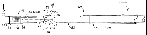
Figs. 1-6 are various views illustrating a distal end of the exemplary
suture anchor system 20 of the present invention. The several components of
the
system are seen exploded in Figure 2 and can be grouped as the suture anchor
46,
the bone anchoring member 48, and a delivery system 50. For purpose of
orientation, the right side will be referenced as the proximal side and the
left side
17
CA 02474378 2004-07-23
WO 03/065904 PCT/US02/41018
as the distal side. Prior to a detailed discussion of the suture anchor 46 and
anchoring member 48, several concentrically disposed tubes comprising the
delivery system 50 will be described.
An inner delivery tube 52 slides within an introducer tube 56 that has a
shoulder 54. The introducer tube 56 may include a valve (not shown) on a
proximal end to prevent fluid leakage therefrom. Alternatively, such a fluid
leakage valve may be provided on the proximal end of the trocar 30 seen in
Fig.
lA. The concentric tubes 52, 56 of the suture anchor system 20 are relatively
axially movable to deploy the suture anchor 46. Various means are known to
relatively displace concentric tubes a predetermined distance and/or with a
predetermined displacement force. For example, the concentric tubes may extend
out of the trocar 30 to an actuation device in the form of concentric syringe
bodies/finger tabs. Alternatively, the concentric tubes may be attached to
relatively movable parts in a gun-type handle, and actuated by triggers or
other
such levers. It is to be understood therefore that the present invention is
not
limited by the particular actuation device on its proximal end, and no further
description in this regard will be provided.
The suture anchor 46 includes a generally tubular anchor body 60, two
series of suture-locking elements 62a, 62b, a distal end cap 64, a proximal
end
cap 66, and a pair of suture-locking plugs 68a, 68b. As seen, each of the
suture-
locking plugs 68a, 68b has an actuation rod 70 removably attached to a
proximal
end, that extends proximally within the delivery tube 52 and eventually
projects
from the proximal end of the suture anchor system 20, as seen in Fig. lE.
The components of the suture anchor 46 are shown assembled in Fig. 3,
and again in Fig. 4 as assembled with the other components of the suture
anchor
system 20. The end caps 64, 66 have stepped extensions that fit closely within
the inner diameter of the tubular anchor body 60 such that the outer surfaces
of
these three elements define a smooth outer cylinder. The thus assembled anchor
body 60 and end caps 64, 66 define a tube having a lumen (not numbered)
opening at proximal and distal ends. The end caps 64, 66 axially retain the
suture-locking elements 62a, 62b within the anchor body 60. As will be
18
CA 02474378 2004-07-23
WO 03/065904 PCT/US02/41018
described below, these elements 62a, 62b cooperate to secure a length of
suture
within the anchor 46 by relative radial movement, and are stacked closely yet
with negligible compression so as to enable relative movement. The close
stacking of these elements 62a, 62b and presence of the end caps 64, 66
prevent
relative axial movement therebetween. As seen in Fig. 2, two diametrically
opposed bores 71 in the distal end cap 64 receive and align the locking plugs
68,
while two smaller diametrically opposed bores 73 in the proximal end cap 66
receive and align the actuation rods 70.
The bone anchoring member 48 is seen in perspective in Fig. 2, and in
elevation in Fig. 3. As mentioned above, the tubular anchoring member 48 is
exemplary only, and other structures may be utilized. For instance, the
anchoring
member 48 illustrated is a separate element disconnected from the suture
anchor
46. Alternatively, an anchoring member that is formed integrally with, or
connected to, the suture anchor 46 may be used.
The bone anchoring member 48 includes a pair of wings 72a, 72b that are
connected by a pair of deformable strips 74. The wings 72a, 72b are shown in
their undeployed, unexpended states in Figs. 2 and 3, wherein they, along with
the strips 74, define something of a U-shape in elevation. As will be seen
below,
in particular with reference to Fig. 8, the wings 72a, 72b are ultimately
deployed
outwardly with respect to each other such that the strips 74 assume a
relatively
linear shape, aligned with a proximal surface of the wings. This outward
deployment is indicated by the arrows 76 in Fig. 3. A pair of stop surfaces 78
ultimately contact and limit this outward deployment, as seen in Fig. 8.
The exemplary bone anchoring member 48 is located between the suture
anchor 46 and the delivery tube 52. In the undeployed state, as seen in Figs.
3
and 4, the U-shaped proximal surface of the member 48 conforms and is
rotationally fixed with respect to a blunt distal tip 80 of the delivery tube
52. As
seen in Fig. 4, corners of the distal stop surfaces 78 contact the proximal
end cap
66 of the suture anchor 46. The bone anchoring member 48 defines a lumen 82
therethrough, as seen in Fig. 2. The actuation rods 70 pass through the hollow
suture anchor 46, through the lumen 82, and through a passage 84 in the
delivery
19
CA 02474378 2004-07-23
WO 03/065904 PCT/US02/41018
tube 52, to the proximal end of the trocar 30 (Fig. lA). As will be clear
shortly,
tension on the actuation rods 70 maintains the suture anchor 46 and bone
anchoring member 48 together and held against the blunt distal tip 80 of the
delivery tube 52, as in Fig. 4. Even after removal of the delivery tube 52 and
actuation rods 70, as seen in Fig. 1F, the suture anchor 46 and bone anchoring
member 48 remain held together under the tension of the two free ends 34a, 34b
of the length of suture.
Fig. 4 shows the suture loop 32 extending transversely from within an
axial slot 86 of the delivery tube 52. As seen in Fig. 2, both the
intermediate tube
54 and introducer tube 56 are also provided with axial slots 88, 90,
respectively.
The slots 86, 88, 90 align and permit lateral passage of the two free ends
34a, 34b
of the length of suture into the passage 84 in the delivery tube 52, and from
there
through the bone anchoring member lumen 82 and into the suture anchor lumen
to be anchored.
. Now, referring back to Fig. 1B, there is shown the entrance of the two free
ends 34a, 34b of the length of suture into the aligned slots in the suture
anchor
system 20. The loop 32 is first embedded in the rotator cuff tendon 22 and
then
the two free ends 34a, 34b can be withdrawn from the body and inserted into
the
system 20. The suture anchor 46 is then fixed in the humeral head 24 and the
suture anchored therewithin. The aligned slots 86, 88, 90 (Fig. 2) in the
system
20 allow the concentric tubes 52, 54, 56 to help in securing the rotator cuff
tendon
22 to the humeral head 24 and then be easily removed.
With reference now to Fig. 5, the two free ends 34a, 34b of the length of
suture pass in a distal direction through the bone-anchoring member lumen 82
and into the lumen of the suture anchor 46. The two ends 34a, 34b pass
completely through the suture anchor 46 and loop around a cross member 92 in
the distal end cap 64 (see Figs. 2 and SA). After looping around the cross
member 92, the free ends 34a, 34b return in a proximal direction back through
the
lumen of the suture anchor 46 and the bone anchoring member lumen 82. Within
the lumen of the suture anchor 46 there are four separate strands of the two
free
ends 34a, 34b of the length of suture, as can be seen in the cross-section of
Fig.
CA 02474378 2004-07-23
WO 03/065904 PCT/US02/41018
SB. At the location of the aligned slots 86, 88, 90, the four strands separate
and
the two free ends 34a', 34b' continue in a proximal direction to the proximal
end
of the system 20.
The structure and function of the suture-locking elements 62a, 62b will
now be described with reference to Figs. 5, 6, 7A, and 7B. Fig. SB shows the
shape of one of the first series of suture-locking elements 62a overlying one
of
the second series of suture-locking elements 62b, and both surrounding the
four
strands of the two free ends 34a, 34b of the length of suture. The elements
62a,
62b are shown in their undeployed, first positions. The interior edges of the
suture-locking elements 62a, 62b define a generally round passage 94 within
which the strands of the length of suture are constrained. The passage 94 has
a
generally uniform axial cross-section, meaning that the majority of the cross-
section of the internal passage is relatively smooth axially. Therefore, the
shape
of the passage 94 is generally as seen in Figs. SB and 6 along the entire
axial
stack of elements 62a, 62b.
There are advantageously more than four total elements 62a, 62b for a
minimum of suture clamping, and preferably there are at least ten. With four
elements 62, two on each side moving in opposite directions, adequate
frictional
interference with the length of suture is created. Of course, the greater
number of
elements 62 increases the frictional resistance to suture pull-through, and
concurrently the clamping force can be reduced. Moreover, although alternating
elements 62a, 62b moving 180 ° to each other are shown, more than two
differently oriented elements can be used that move in different yet not
necessarily opposite directions. For instance, three series of elements that
move
in directions that are oriented 120° with respect to each other can be
used.
Each suture-locking element 62a, 62b is substantially C-shaped and
surrounds and defines (at its level within the anchor body lumen) at least
three-
quarters of the axial passage 94. With reference to Fig. 6, each element 62a
(and
each element 62b) has a central bridge portion 96 and a pair of arcuate arms
98a,
98b. The passage 94 is defined by the inner edges of the bridge portion 96 and
arms 98a, 98b, and the profile is generally round except for an inward bulge
100
21
CA 02474378 2004-07-23
WO 03/065904 PCT/US02/41018
at the bridge portion. Each of the arms 98a, 98b terminates at points that are
spaced apart a distance corresponding to a circumferential arc around the
passage
94 of less than 45 °, and preferably about 30°.
The elements 62a, 62b in the two series are stacked in an alternating
fashion, so that each element in the first series of elements 62a is
surrounded by
two elements in the second series of elements 62b, and visa versa (except, of
course, for those on the stack ends). Moreover, the alternating elements 62a,
62b
are oppositely oriented 180° about the axis such that the spaced-apart
ends of
each element 62a align with the inward bulge 100 at the bridge portion 96 of
each
adjacent element 62b. This can be seen in Fig. 6 at the top and bottom.
Because
there is a space between the ends of the arms 98a, 98b, a slight non-
uniformity in
the inner wall of the passage 94 is created. That is, there is a gap between
each
two adjacent bulges 100 of the first series of elements 62a. However, because
the
bulges 100 are relatively closely spaced and aligned, the passage 94 can still
be
considered generally uniform in axial cross-section. Yndeed, Fig. 6
illustrates an
arc ~ on one side of the two series of elements 62a, 62b that corresponds to
the
portion of the passage 94 on that side that is entirely uniform (i.e., smooth)
when
the elements are in their first positions.
The outer edge of each of the first series of elements 62a is generally
round, and in combination with the outer edges of the second series of
elements
62b, defines a cylinder that fits closely within the tubular anchor body 60,
as seen
in Fig. 5B. Each of the elements 62a, 62b defines a substantially semi-
circular
cavity 102 in its outer edge at the location of the bridge portion 96. As seen
in
Figs. SB and 6, the cavities 102 in the first series of elements 62a are
diametrically opposed from the cavities in the second series of elements 62b.
The
series of aligned cavities 102 on both sides of the suture anchor 46 creates
tunnels
through which an actuation rod 70 passes, when the suture anchor 46 is
assembled, and prior to actuation thereof. This is seen in Fig. SB which
illustrates the relative sizes of the actuation rods 70 and the larger
diameter
suture-locking plugs 68a, 68b. With reference to Fig. 5, it will be noted that
in
the undeployed state the suture-locking plugs 68a, 68b are located just distal
to
22
CA 02474378 2004-07-23
WO 03/065904 PCT/US02/41018
the tubular anchor body 60 and enclosed suture-locking elements 62a, 62b.
Figs. 7A and 7B show the suture-locking elements 62a, 62b and suture-
locking plugs 68a, 68b isolated to better illustrate their interaction and the
advantageous mechanism for anchoring one or more lengths of suture without
knots. Fig. 7A shows the elements 62a, 62b in their undeployed relationship,
as
previously illustrated in Figs. 5-6, while Fig. 7B shows the deployed state.
Fig.
7C is a cross-section through the bridge portions 96 of each element 62a, 62b
in
their first positions.
Deployment involves axial movement of the suture-locking plugs 68a,
68b in the direction of arrows 110 which causes radial movement of the suture-
locking elements 62a, 62b. Each suture-locking plug 68a, 68b has a proximal
taper 112 that initially resides adjacent the distal-most suture-locking
element 62a
or 62b. Proximal movement in the direction of arrows 110 of the actuation rods
70 pulls the tapers 112 and then the suture-locking plugs 68a, 68b into the
aligned
series of cavities 102 defined on the outer edges of the suture-locking
elements
62a, 62b. As can be seen from Fig. SB, forcing the larger diameter suture-
locking
plugs 68a, 68b into the aligned cavities 102 in turn cams each of the suture-
locking elements 62a, 62b radially inward. In particular, the first series of
suture-
locking elements 62a moves in an opposite direction to the second series of
suture-locking elements 62b, both moving toward the center of the anchor body
lumen.
Radially inward movement of the suture-locking elements 62a, 62b from
first positions to second positions converts the cross-section of the axial
passage
94 from generally uniform to irregular, and therefore substantially restricts
axial
movement of the lengths of suture 34a, 34b that are disposed therein. The
irregularity can be seen in the cross-section of Fig. 7D and generally
comprises
alternating misaligned bulges 100 or "teeth" that compress the lengths of
suture
34a, 34b from opposite sides. Because the bulges 100 are misaligned, the
effect
is an irregular compression of the lengths of suture 34a, 34b that creates
significantly more frictional resistance to suture pull-through, than if the
bulges
were aligned.
23
CA 02474378 2004-07-23
WO 03/065904 PCT/US02/41018
Another way to state the clamping effect is that the suture-locking
elements 62a, 62b are initially disposed in first positions that together
define the
axial passage 94 sized to permit axial movement of the lengths of suture 34a,
34b
therethrough. Axial movement of the suture-locking plugs 68a, 68b into the
~ tunnels created by the cavities 102 cams the elements 62a, 62b inward toward
the
center of the anchor body lumen into second positions that, taken as an
aggregate,
reduce the size of the passage 94. The reduced passage 94 clamps the lengths
of
suture 34a, 34b therein and substantially restricts their axial movement
therethrough.
The suture-locking elements 62a, 62b are dimensioned to compress or
"crush" the length of suture in the lumen 94 and interfere with its axial
movement
therethrough. The amount of interference may be measured by the amount of pull
force necessary to move the suture once the elements 62a, 62b are in their
second
positions. Desirably, the pull force is in a range that would exceed the LISP
(United States Pharmacopeia) Standard knot pull strength (LJSP 24) of the
suture
used. In the specific case of #2 braided polyester suture, this knot pull
strength is
approximately 3.5 Kgf. In practice, however, the knot pull strength of
commercially available #2 braided polyester sutures approaches 14 Kgf.
The particular structure and arrangement of the suture-locking elements
62a, 62b may differ from that shown. For instance, the elements may not be
oriented in radial planes and be displaced radially, but instead may be angled
and
be displaced at an angle. Or, the elements may be arranged to rotate in one or
more directions upon axial translation of the locking plugs 68a, 68b, thus
creating
the meshing teeth, so to speak, that grip the suture strands. Also, there may
be
only one series of elements that displace in one direction, thus crushing the
suture
strands against the inner wall of the tubular anchor body 60 or against a
fixed
structure within. Those of skill in the art will therefore understand that the
elements 62a, 62b disclosed are exemplary only, and others are contemplated.
The materials used in the system 20 are surgical grade metals or polymers.
For example, the implantable suture anchor 46 and bone anchoring element 48
may be made of a biocompatible polymer such as polyethylene or a metal such as
24
CA 02474378 2004-07-23
WO 03/065904 PCT/US02/41018
titanium. The suture locking elements 62a, 62b are desirably metal, although
certain hard plastics or polycarbonates may be used. The materials of the
devices
used to implant the anchor 46, such as insertion tubes 52, 56, need not be as
durable as the implantable materials. The anchors may also be fabricated from
bio-absorbable materials commonly used for implantation such as polyglycolide
(PGA), polylactide (PLA), homopolymer of 1-lactide (LPLA), or other bio-
absorbable materials known in the art.
In use of the system 20, the various components as described above are
first procured and assembled. The surgeon creates the operating ports
necessary
in the dermal layers and forms the body cavity 40 in the humeral head 24 as
seen
in figures lA and 1B. The hole 40 has been drilled in the bone at the location
chosen by the surgeon for anchor fixation. The delivery system 50 is inserted
through one of the operating ports, and the shoulder 54 of the introducer tube
56
is positioned within the hole 40. By pushing on the deployment tube 52, the
anchor 46 is forced out of the introducer tube 46 and down into the hole 40.
The
shoulder 54 of the introducer tube 46 ensures that the anchor 46 is delivered
into
the hole 40 below the hard outer layer of cortical bone 42 so that the
anchoring
member 48 can bear upon the cortical bone 42.
Figs. 8 and 9 further illustrate the suture-locking function of the present
invention along longitudinal sections, and also show the entire bone anchoring
and suture-tightening aspects. In Fig. 8, the suture loop 32 can be considered
to
be embedded in soft tissue, and thus relatively securely positioned. The bone
anchoring member 48 has been deployed such that its flat proximal surface
abuts
the inside wall of a body cavity, such as the inside wall of the hard cortical
bone
42 of the humeral head 24, as previously described.
The suture anchor system 20 including the delivery tube 52 remains in
place held against the bone anchoring member 48 by the locking plugs 68a, 68b
and the tension in the actuation rods 70. Because the locking plugs 68a, 68b
remain in their distal position, the suture-locking elements 62a, 62b are un-
deployed in their first positions and the lengths of suture 34a, 34b are free
to slide
within the passage 94.
CA 02474378 2004-07-23
WO 03/065904 PCT/US02/41018
At this stage, the surgeon adjusts the tension in the lengths of suture 34a,
34b by pulling on the free ends 34a', 34b', or pulling on one end while
holding
one fixed, in the direction of arrow 120 in Fig. ~. Adjustment of the length
of the
suture between the suture anchor 46 and the loop 32 is very important to
ensure
proper fixation of the rotator cuff tendon 22 with respect to the humeral head
24.
If the suture is pulled too tightly, the rotator cuff tendon 22 may be unduly
stressed, and the loop 32 may even pull free from the tendon. On the other
hand,
if the suture is too loose, the goal of reattaching the tendon 22 in its
proper
location will be compromised.
As mentioned above, the lengths of suture 34a, 34b wrap around the cross
member 92 (see Fig. SA) which acts as a pulley of sorts and permits the
sutures to
freely slide therepast. The result of pulling on the free ends 34a°,
34b' is to pull
the portions between the system 20 and the loop 32 taught. This is also
depicted
in Fig. lI~. The particular tension established in the sutures 34a, 34b
depends on
the patient characteristics, the type of soft tissue being reattached, and
surgeon
judgement.
After adjusting the tension of the sutures 34a, 34b, the actuation rods 70
are displaced in a proximal direction, as indicated at 122 in Fig. 9. As
described
above, this step causes the suture-locking elements 62a, 62b to cam inward and
reduce the size of the passage, clamping the sutures 34a, 34b in between an
irregular pattern of "teeth." The cross sectional view of Fig. 9A shows the
resulting clamped configuration of the sutures 34a, 34b.
One advantage provided by the present invention is the ability to tighten a
suture loop embedded within soft tissue to a predetermined tension, and then
lock
the suture within a suture anchor without even slightly altering that tension.
Importantly, because the suture-locking elements 62a, 62b are displaced
radially,
they do not urge the sutures 34a, 34b to migrate axially within the tubular
anchor
body 60, and therefore do not change the length on either side of the cross
member 92. This ensures that the proper tension established between the suture
anchor 46 and the loop 32 embedded in the soft tissue does not change.
Subsequently, the actuation rods 70 are detached from the suture-locking
26
CA 02474378 2004-07-23
WO 03/065904 PCT/US02/41018
plugs 68a, 68b by further pulling in the direction of arrows 122, thus causing
a
point of weakness to sever. The point of weakness is not shown, but typically
comprises a narrow neck or frangible point on each rod 70 disposed just
proximal
to, or within a bore of, the corresponding locking plug 68a or 68b. At this
stage,
the concentric tubes 52, 54, 56 can be removed from the operation site and the
sutures 34a, 34b severed close to the bore 82 of the bone anchoring member 48.
After any further post-procedure steps, the site of the operation can then be
closed.
The distal end of an alternative bone anchoring and suture locking system
130 is shown in Figs. 11-12B and includes a tubular anchor body 132 housing a
plurality of identical suture-locking elements 134. Fig. 11 also shows the two
free ends of a length of suture 136 extending through the anchor body 132 on
one
side and wrapping around at a distal loops 138 to continue through the body on
the opposite side. The cross-sectional view in Fig. 12A illustrates the
location of
the four strands of the suture 136. Although not shown, the distal loops 138
wrap
around a cross member or pulley fixed with respect to the anchor body 132, as
in
the earlier-described embodiment.
A suture-locking plug 140 attached to an actuation rod 142 is initially
located at the distal end of the stack of suture-locking elements 134. The
actuation rod 142 passes through a partially aligned series of central
apertures 144
in the suture-locking elements 134, as seen in Fig. 12A. In this regard, there
are a
plurality of configurations of suture-locking elements 134 that differ only in
the
location of the central aperture 144. Desirably, there is a single shape of
element
134 having a central aperture 144 that is offset from but overlapping the
central
axis. Adjacent elements 134 are oriented in opposite directions so that their
apertures 144 are offset in opposite directions. The aligned portions of the
apertures 144 are large enough for passage of the actuation rod 142, though
smaller than the locking plug 140.
In use, the suture-locking elements 134 are initially in first positions as
seen in Fig. 12A. The outer edge of each element 134 has two diametrically-
opposed cavities 146 that are sized to receive two of the suture strands 136.
The
27
CA 02474378 2004-07-23
WO 03/065904 PCT/US02/41018
cavities 146 form axially-uniform tunnels with the inner wall of the tubular
anchor body 132 that permit the free ends of the sutures to pass easily
therethrough, and thus facilitate the suture tensioning step as explained
above.
Unlike the earlier embodiment, the tunnels have the an entirely uniform cross-
section along their axial length, as seen in Fig. 12A.
Proximal displacement of the actuation rod 142 and attached locking plug
140 forces a taper at the leading or proximal end of the locking plug into the
partially aligned apertures 144, thus caroming alternating elements 134 in
opposite radial directions into second positions. That is, the locking plug
140 lies
on the central axis of the system, and thus each offset aperture 144 is forced
toward the axis as well. The size of each aperture 144 is just large enough to
permit passage of the locking plug 140, and thus the final configuration as
seen in
Fig. 12B has the apertures 144 aligned concentrically about the locking plug
along the central axis.
Fig. 12B also shows the clamping of the lengths of suture by the outward
movement of each suture-locking element 134. Because only every other one of
the elements 134 clamps each pair of suture strands, there is again an
irregular
passage created. That is, each pair of two strands of suture is compressed
against
the inner wall of the tubular anchor body 132 by a series of spaced apart
edges of
the cavities 146 of every second element 134. The tunnels formed by the
cavities
146 and the inner wall of the tubular anchor body 132 are thus reduced in size
and
rendered non-uniform.
Figures 11-11B also show an alternative bone anchoring structure. Rather
than a single bone anchoring member, such as member 48 seen in Fig. 2, the
system 130 has a plurality of relatively thin bone anchoring members 150 that
are
stacked axially together. This bone anchoring system is described in co-
pending
application serial number 09/876,260 filed June 6, 2001, which has already
been
expressly incorporated by reference herein. Each member 150 has a V-shape
prior to deployment, as seen in Fig. 11, and has a generally oval outer
profile as
seen in plan view in Fig. 11B. Two off center apertures 152 in each member 150
permit passage of the strands of suture. A central aperture 154 permits
passage of
28
CA 02474378 2004-07-23
WO 03/065904 PCT/US02/41018
the actuation rod 142.
Accordingly, although an exemplary embodiment of the invention has
been shown and described, it is to be understood that all the terms used
herein are
descriptive rather than limiting, and that many changes, modifications, and
substitutions may be made by one having ordinary skill in the art without
departing from the spirit and scope of the invention. In particular, it is
noted that
the procedures, while oriented toward the arthroscopic repair of the rotator
cuff,
are applicable to the repair of any body location wherein it is desired to
attach or
reattach soft tissue to bone, particularly using an arthroscopic procedure.
29