Note: Descriptions are shown in the official language in which they were submitted.
CA 02477220 2004-08-19
SUTURE ANCHOR AND APPROXIMATING DEVICE
Jeffrey E. Yeung, Teresa T. Yeung
FIELD OF INVENTION
This invention relates to suture anchors and minimally invasive methods for
delivering and fastening suture within tissue.
BACKGROUND
Suture anchors have been developed for anchoring sutures in endoscopic or
arthroscopic surgery through single sided access. Most prior art suture
anchors are
delivered from a lumen of a needle or a tubular device. Prior art include US
patent
4,235,238 by H. Ogiu et al., issued on Nov. 25, 1980, US patent 4,741,330 by
J.
Hayhurst, issued on May 3, 1988, US patent 4,669,473 by W. Richards et al.,
issued on
June 2, 1987, US patent 5,800,445 by K. Ratcliff et al., issued on September
1, 1998, US
patent 5,041,129 by J. Hayhurst et al., issued on August 20, 1991, US patent
5,845,645 by
P. Bonutti, issued on Dec. 8, 1998, US patent RE36,974, reissued on Nov. 28,
2000, and
US patent 6,312,448 by P. Bonutti, issued on Nov. 6, 2001. Since the anchors
reside
within the lumen of the delivery device, the size of the needles or tubular
members is
correspondingly larger, making tissue penetration more difficult and
traumatic.
Several prior art anchors reside outside and around a needle. For delivery, a
push rod
is used to push along one side of the suture anchor, sliding along the needle
into the
tissue. A suture connected at the opposite side of the push rod is used to
pull the anchor
as the anchor is being pushed by the push rod. A series of patents by P.
Bonutti, US
patent 5,814,072, issued on Sep. 29, 1998, US patent 5,948,002, issued on Sep.
7, 1999,
US patent 6,033,430, issued on Mar. 7, 2000 and US patent application
publication
number US2001/0002440, publication date: May 31, 2001, proposed the push and
pull
method to pivot the anchor within tissue. Pivoting of an anchor within tissue
is classified
as partial-thickness suture fastening. To facilitate instant pivoting, the
suture is connected
close to both distal and proximal ends of the anchor to provide favorable
leverage for
anchor rotation. Figure 1 depicts the prior art 235, which has completed the
rotation
within tissue. The suture 122 is looped near or at both ends of the anchor
235, as
depicted in the prior art patents. For favorable leverage, the strands of
suture 122
1
CA 02477220 2004-08-19
connected to the anchor 235 are widely spaced apart. As tension is applied to
the suture
122, the strands of suture 122 spread open, as indicated by the shaded area
236, opening
or pushing out the tissue 130 along the path of anchor 235 entry. Especially
within soft
tissue, the widely spaced sutures 122 wedge open the tissue directly above the
anchor
235. As a result, the pullout strength of the anchor 235 is likely to be low.
The probable
mode of failure is likely to be anchor 235 pullout, as depicted in Figure 2,
rather than
suture 122 breakage. While the widely spaced suture 122 provides favorable
leverage for
rapid rotation, it appears to sacrifice the strength of tissue anchoring.
Another prior art suture anchor, US patent 5,626,614 by C. Hart, issued on May
6,
1997, also resides outside and around a needle. Hart's invention is designed
for fastening
or proximating tissues separated by two distinct walls, such as the stomach
and
abdominal walls, using full-thickness fastening. Unfortunately, most tissue
within the
body adheres to adjacent tissue with no clear separation, space or cavity.
Therefore, full-
thickness anchor pivoting to fasten or proximate two tissues has limited use.
SUMMARY OF INVENTION
Organs and/or tissues, especially in urology, virtually adhere to each other.
This
invention is capable of anchoring a suture in either partial- or full-tissue
thickness
fastening, without the cumbersome manipulations of the suture or delivery
device as
described in prior art. In addition, in certain embodiments, the suture anchor
contains a
platform designed to improve anchoring strength within tissue.
More particularly, in accordance with one embodiment, there is provided a
suture
anchor deployable with a suture anchor delivery device having a needle, the
suture anchor
comprising a suture anchor body having a longitudinal axis and formed of an
elastic
material, said suture anchor body having a straightened position and a curved
position, a
passage extending through said suture anchor along said longitudinal axis,
said passage
sized and configured for the needle of the suture anchor delivery device to
pass
therethrough, a suture opening sized and configured to received a suture, said
suture
opening passing through a portion of said suture anchor, a guiding platform
attached to
one side of said suture anchor body, wherein said suture body is resiliently
straightened
into said straightened position when the needle of said suture anchor delivery
device is
located within said passage, and wherein when the needle is removed, said
suture anchor
body resumes said curved position.
. 2
CA 02477220 2004-08-19
In accordance with a further embodiment of the invention, there is provided a
suture anchor deployable with a suture anchor delivery device having a needle,
the suture
anchor comprising a suture anchor body having a longitudinal axis and formed
of an
elastic material, said suture anchor body having a straightened position and a
curved
position, a passage extending through said suture anchor along said
longitudinal axis, said
passage sized and configured for the needle of the suture anchor delivery
device to pass
therethrough, a suture opening sized and configured to received a suture, said
suture
opening passing through a portion of said suture anchor, a guiding fin
attached to one side
of said suture anchor body, wherein said suture anchor body is resiliently
straightened
into said straightened position when the needle of said suture anchor delivery
device is
located within said passage, and wherein when the needle is removed, said
suture anchor
body resumes said curved position.
In accordance with yet another embodiment of the invention, there is provided
a
suture anchor deployable with a suture anchor delivery device having a needle,
the suture
anchor comprising a suture anchor body having a longitudinal axis, a passage
extending
through said suture anchor along said longitudinal axis, said passage sized
and configured
for the needle of the suture anchor delivery device to pass therethrough, a
suture opening
sized and configured to receive a suture, said suture opening passing through
a portion of
said suture anchor, a guiding fin attached to a first side of said suture
anchor body, a
guiding platform attached a second side of said suture anchor body.
In another embodiment of the invention, there is provided a suture anchor
delivery
device, comprising a needle, a first sleeve located around said needle and a
second sleeve
located around said needle and said first sleeve.
A curved anchor made with elastic material contains a lumen for the needle. A
fin
protrudes from one side and a platform covers the opposite side of the anchor.
The fin is
on the concave side and at the proximal end, while the platform is on the
convex side of
the curved anchor. A suture passes through an opening in the platform, loops
around the
concave side of the anchor, and exits through another opening in the platform.
As a
result, both strands of the suture can be pulled from the convex side of the
anchor.
The suture anchor is resiliently straightened by a rigid needle inserted
through the
lumen of the anchor. The needle contains a widened portion or a step to
prevent the
anchor from sliding up the needle. The needle is used to deliver the anchor by
puncturing
3
CA 02477220 2004-08-19
into tissue. At a proper depth, the needle can then be withdrawn. The
protruded fin is
tapered for tissue insertion, but behaves as a tissue snagging barb, hooking
onto the tissue
and resisting pullout. As a result, the needle withdrawal strips the anchor
off the needle,
and at the same time deploys the anchor within the tissue at the proper depth.
The anchor resumes the elastic curvature within the tissue after withdrawal of
the
rigid needle. The fin at the proximal end of the concave curvature is
laterally pressed into
the adjacent tissue, while the central portion of the convex curvature
connecting to the
suture is pushed in the opposite direction further away from the fin. In
essence, curvature
resumption within tissue increases the distance between the fin and the
openings for the
suture, as the fin is pressed laterally into the tissue. When the strands of
suture are pulled
on the convex side of the anchor, the curved anchor begins to rotate within
tissue from a
vertical, or inserting position, to a horizontal, or fastening position. The
platform is also
repositioned from vertical to horizontal to greatly resist pullout during
tissue fastening
and repair.
Multiple anchors can be linked by a suture and delivered in series into
tissue. When
the suture is pulled , the anchors draw close to each other to shorten or
approximate the
pierced tissue.
REFERENCE NUMBER
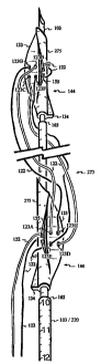
100 Intervertebral disc 122 Suture
101 Urethra 123 Opening for suture
102 Urethropelvic ligament 35 125 Suture knot
103 Stepped or smooth needle 126 Cortical bone
104 Lumen of suture anchor 127 Bladder
109 Plunger 128 Nucleus pulposus
111 Disc compressor 130 Soft tissue
112 Bladder neck 40 131 Lateral wall of urethra
113 Mucosa 132 Rectum
114 Vagina 133 Platform of anchor
115 Pubic symphysis 134 Fin of anchor
117 Urine 138 Tendon or ligament
118 Cancellous bone 45 144 Suture ancllor
119 Annular contact surface 150 Lumen of urethra
4
CA 02477220 2004-08-19
151 Posterior wall of urethra 30 247 Outer tube
152 Anterior wall of urethra 248 Side window
153 Needle indentation 249 Sharp edge
154 Catheter 250 Suture cutting device
155 Bend stop 251 External sphincter
156 Gap of bend stop 35 252 Internal sphincter
157 Incision 253 Cardinal ligament
159 Handle of positioning device 254 Sacrouterine ligament
160 Lifting hand piece 255 Fascia
161 Uterus 256 Ovary
163 Uterus positioning tool 40 257 Round ligament
164 Suture attachment 258 Fallopion tube
165 Step of trocar or needle 259 Grasping device
171 Distal round end 260 Guide arm
172 Shaft of positioning device 261 Pointer
185 Trocar guide 45 262 Glide track
188 Psoas major muscle 263 Endoscope
196 Retractor 264 Suture gripping device
220 Sleeve of trocar or needle 265 Flap
221 Grippers on the sleeve 266 Cone
235 Prior art suture anchor 50 267 Loop
236 Area of suture spread 268 Scar tissue
237 One-way grip 269 Lumen oP needle
238 Suture passage of the grip 270 Collagen bundles
239 Suture lock 271 Cervix
240 Cone hole 55 272 Adipose tissue
241 Gripper 273 Approximating device
245 Knot pusher 274 Retropubic space
246 Inner tube 275 Body of anchor
BRIEF DESCRIPTION OF THE DRAWINGS
60 Figure 1 depicts the tissue 130 opening above the prior art anchor 235,
caused by
spreading 236 of the sutures 122 as tension is applied.
5
CA 02477220 2004-08-19
Figure 2 depicts prior art anchor 235 pullout as a probable result of the
tissue 130
opening directly above the prior art anchor 235.
Figure 3 depicts a suture anchor 144 with an elastically curved body 275,
lumen 104,
a fin 134, a relatively flat platform 133 and two openings 123 for a suture
122.
Figure 4 depicts the elastic body 275 being resiliently straightened by a
trocar or
needle 103 inserted through the lumen 104 of the anchor 144.
Figure 5 depicts the resiliently straightened anchor 144 resting on a step 165
of the
needle 103.
Figure 6 shows a side view of the anchor 144 with the stepped needle 103. The
distal
tip of the anchor 144 is beveled. The platform 133 and fin 134 are tapered for
tissue
penetration.
Figure 7 depicts the top view of the anchor 144 with suture 122 exiting from
openings
123 on the elliptical platform 133 tapered at both distal and proximal ends.
Figure 8 shows the bottom view of the anchor 144, indicating the tapered
distal tip,
and looping of the suture 122 under the anchor 144 to distribute suture 122
tension.
Figure 9 depicts the rotational direction of the curved suture anchor 144
within tissue,
as tension is applied to suture 122.
Figure 10 depicts penetration of the stepped needle 103 loaded with the suture
anchor
144 into soft tissue 130.
Figure 11 depicts the anchor 144 resuming the curved configuration and
pressing the
fin 134 laterally into the tissue 130 after the withdrawal of the stepped
needle 103.
Figure 12 depicts tension applied to the suture 122 pulling on the curved
anchor 144
and driving the fin 134 further laterally.
Figure 13 depicts the tension driven rotation of the anchor 144, orienting the
large and
relatively flat platform 133 from a vertical to a horizontal position to
resist anchor 144
pullout.
Figure 14 indicates a normal, well-supported bladder 127 in dashed lines and a
descended bladder 127 with a widened bladder neck 112 in solid lines.
Figure 15 shows a failed lumen 100 closure and hypermobility under stress with
the
urethropelvic ligament 102 pulling the lateral walls 131 of the poorly
supported urethra
101.
6
CA 02477220 2004-08-19
Figure 16 indicates a mid-longitudinal view of Figure 15 and urine 1171eakage
during stress with urethropelvic ligaments pulling perpendicularly above and
below the
plane of the page.
Figure 17 shows a prior art procedure for treating urinary incontinence
through a large
incision 157 for passing sutures 122 and pulling the vagina 114 forward to
support or
compress the posterior wall of the ureth'ra 101.
Figure 18 depicts a section of the surgically corrected urethra 101 with
sutures 122
pulling the vaginal 114 tissue to support and gently compress the urethral
posterior wall
151.
Figure 19 indicates lumen 150 closure of the surgically corrected urethra 101
under
stress, with urethropelvic ligaments 102 pulling the lateral walls 131 of the
supported
urethra 101.
Figure 20 shows a small incision 157 for inserting the stepped needle 103 with
the
suture anchor 144 into the vaginal wall.
Figure 21 depicts the urethral posterior wall 151 supported by sutures 122 and
anchors 144 within the vagina 114.
Figure 22 indicates a proximal end of a suture anchor 144 with an elliptical
lumen
104, sized and configured to fit over a stepped needle 103 with an elliptical
cross-section.
Figure 23 shows a lengthened fin 134, sized and configured to fit into an
indentation
153 on a stepped needle 103.
Figure 24 depicts a uterine 161 prolapse.
Figure 25 depicts a repositioned uterus 161 pierced with the stepped needle
103
through a small incision 157.
Figure 26 depicts uterus 161 fastening with sutures 122 and anchors 144. The
suture
122 is knotted 125 onto the ligament or fascia on the abdominal wall.
Figure 27 depicts penetration of the stepped needle 103 with the suture anchor
144
through a torn ligament 138 into decorticated bone 118.
Figure 28 depicts the suture anchor 144 resuming some of the curved
configuration
within the bone 118 after being dislodged from the withdrawn stepped needle
103.
Figure 29 depicts suture 122 tension driving the fin 134 further laterally
into the bone
118.
7
CA 02477220 2004-08-19
Figure 30 depicts another anchor 114 delivered by the stepped needle 103
througli the
torn ligament 138 into cancellous bone 118.
Figure 31 depicts a suture knot 125 tied to fasten the torn ligament 138 onto
the bone.
Figure 32 shows a bend stop 155 with a closed gap 156 beneath the platform 133
to
prevent excessive anchor 144 bending under significant suture 122 tension.
Figure 33 shows a stepped needle 103 resiliently straightening the anchor 144
with
the bend stop 155. In the straightened position, the gap 156 is open.
Figure 34 depicts a side view of the straightened anchor 144 with an open gap
156
beneath the platform 133.
Figure 35 indicates a bottom view of the straightened anchor 144 showing bend
stops
155 with open gaps 156 beneath the platform 133.
Figure 36 shows a straight anchor 144 with a large fin 134 and a tapered
proximal
end.
Figure 37 shows a side view of the straight anchor 144, as shown. in Figure
36, with
dimensions Wi, Li and L2.
Figure 38 shows another straight anchor 144 with elevated suture openings 123.
Figure 39 shows a side view of the anchor 144 with elevated suture openings
123, as
shown in Figure 38, with dimensions W2, L, and L2.
Figure 40 depicts a curved suture anchor 144 with a protruded suture
attachment 164,
a fin 134 and a small platform 133.
Figure 41 depicts another curved suture anchor 144 with the protruded suture
attachment 164 but without a platform.
Figure 42 shows a curved suture anchor 144 without a fin.
Figure 43 depicts a curved suture anchor 144 with a platform 133 on the
concave side
of the curvature. The fin 134 is made blunt.
Figure 44 shows the suture anchor 144 of Figure 43 resiliently straightened by
a
needle 103 with a sliding sleeve 220.
Figure 45 depicts penetration of the stepped needle 103 with the sleeve 220 to
deliver
a suture anchor 144 through a bulging intervertebral disc 100.
Figure 46 depicts pushing of the anchor 144 by the sliding sleeve 220 to expel
the
suture anchor 144 beyond the distal edge of the disc 100.
8
CA 02477220 2004-08-19
Figure 47 depicts a disc compressor 111 with two openings 123 for a suture 122
and a
cylindrical or blunt region 119 to compress the disc 100.
Figure 48 depicts bulge compression by fastening the disc compressor 111 with
a
suture 122 secured by the anchor 144 outside the disc 100.
Figure 49 depicts portions of two anchors 144 connected by a suture 122 to
form an
approximating device 273 for tightening or shortening tissue.
Figure 50 shows a double-stepped 165 needle 103 resiliently straightening two
anchors 144 with a suture 122 arrangement similar to Figure 49.
Figure 51 indicates deployment of the anchors 144 within tissue after
withdrawal of
the needle 103.
Figure 52 shows orientation of the suture 122 designed to resist sliding
through holes
123B and 123G when the anchor 144 is in a vertical or inserting position.
Figure 53 depicts anchors 144 pivoting within tissue as the suture 122 is
pulled.
Figure 54 shows anchor 144 insertion into tissue 130 with the needle 103, as
the
initial step for deploying the approximating device 273.
Figure 55 indicates partial withdrawal of the needle 103 to deploy the distal
anchor
144 within tissue 130.
Figure 56 depicts proximal anchor 144 insertion by pushing the sleeve 220, and
distal
anchor 144 pivoting by pulling on the suture 122.
Figure 57 shows complete insertion of the proximal anchor 144 into the tissue
130 by
pushing the sleeve 220 and pulling suture 122.
Figure 58 indicates withdrawal of the needle and curvature resumption of the
proximal anchor 144 within tissue 130.
Figure 59 depicts composition of a suture lock 239 with sutures 122 passing
through a
cone 266 over a one-way grip 237 with individual grippers 241.
Figure 60 shows the lock 239 assembly with the suture 122 fastened between the
cone
266 and grippers 241. A plunger 109 is used to advance the suture lock 239.
Figure 61 indicates pulling on the sutures 122 and pushing on the plunger 109
against
and lock 239 to draw the anchors 144 together as an approximating device 273
within
tissue.
Figure 62 depicts knot 125 tying within tissue using a knot pusher 245.
9
CA 02477220 2004-08-19
Figure 63 sliows an inner tube 246 containing a channel opening from the
distal end
to a side window 248.
Figure 64 shows an outer tube 247 also containing a channel opening from the
distal
end to a side window 248.
Figure 65 depicts a suture cutter 250 assembled by fitting the inner tube 246
into the
outer tube 247 with overlapping side windows 248.
Figure 66 indicates threading a pair of sutures 122 through the distal
opening, out the
overlapping side windows 248 of the inner tube 246 and outer 247 tube.
Figure 67 shows a mid-longitudinal view of the suture cutter 250 with sharp
edges
249 at the side windows 248.
Figure 68 depicts cutting of the suture 122 by the sharp edges 249 as the
outer tube
247 slides over the inner tube 246.
Figure 69 shows a mid-longitudinal view of Figure 68.
Figure 70 depicts suture 122 cutting with the cutter 250 after knots 125 are
tied.
Figure 71 indicates retraction of an incision 157 to expose a scarred 268
external
sphineter 251, a common cause of anal incontinence.
Figure 72 shows cutting of the sphincter 251 in a prior art surgical
procedure.
Figure 73 depicts overlapping and suturing the external sphincter 251 to
tighten the
internal sphincteric muscle 252.
Figure 74 shows a lumen 269 in the needle 103 for delivering radiopaque,
echogenic
or other tracing agent to guide needle 103 insertion.
Figure 75 shows tightening of the scarred 268 external sphincter 251 with
multiple
approximating devices 273.
Figure 76 depicts a flexible needle 103 with a tapered tip, as a sewing
needle, for
delivering the approximating device 273.
Figure 77 depicts rotational advancement of the flexible needle 103 between
collagen
bundles 270 of tendon or ligament 138.
Figure 78 depicts a lumen 269 in the rotational needle 103 for delivering
radiopaque,
echogenic or other tracing agent to guide needle 103 insertion.
Figure 79 indicates a cross-sectional view of the uterine 161 supportive
structure,
cardinal 253 and sacrouterine 254 ligaments, and fascia 255.
CA 02477220 2004-08-19
Figure 80 shows insertions of multiple approximating devices 273 into cardinal
253
and sacrouterine 254 ligaments supporting the uterus 161.
Figure 81 indicates the ascendant cervix 271 as the result of sutures 122
tightening to
plicate the cardinal 253 and sacrouterine 254 ligaments.
Figure 82 depicts partial insertion of the proximal anchor 144 of the
approximating
device 273 into tissue 130 by advancing the sleeve 220.
Figure 83 shows a prior art suture-gripping device 264 with flaps 265 biased
against
the upward tensile force applied to the suture 122.
Figure 84 indicates the suture gripping device 264 and plunger 109 positioned
to
tighten the anchors 144 of the approximating device 273 after withdrawal of
the needle.
Figure 85 depicts fastening of the approximating device 273 by tying knots 125
beneath the suture-gripping device 264.
Figure 86 shows a guide 185 to direct needle 103 insertion along a track 262,
with an
extendible arm 260 and a pointer 261 to indicate the destination of the needle
103.
Figure 87 depicts needle 103 insertion through the vaginal 114 wall, lateral
to the
urethra 101 into fascia 255 and adipose tissue 272.
Figure 88 indicates support of the posterior urethral wall 151 by the anchors
144 of
the approximating devices 273.
Figure 89 also shows support of the posterior urethral wall 151 by tightening
or
restricting between fascia covering the anterior urethral wall 152 and the
vaginal 114.
Figure 90 depicts double approximating devices 273 loaded on a single needle
103.
Figure 91 shows fastening of the double approximating devices 273 after
insertion of
a single needle 103.
Figure 92 shows an inner sleeve 220 for deploying the distal anchor 144 and an
outer
sleeve 220 for deploying the proximal anchor 144 from the needle 103.
Figure 93 depicts the proximal end of a platform 133 tapered over the proximal
end of
the body 275 to facilitate pivoting and rotation within tissue.
Figure 94 shows the side view of the tapered platform 133 over the proxinial
end of
the body 275 supported by a shape-matching step 165.
DETAILED DESCRIPTION OF THE EMBODIMENTS
11
CA 02477220 2004-08-19
A curved anchor 144 is made with elastic material containing a longitudinal
lumen or
passage 104, a fin 134 at or near the proximal end, and a relatively flat
platform 133 on
the convex side of the curvature with two openings 123 for a suture 122, as
shown in
Figure 3. Through the openings 123 on the platform 133, the suture 122 is
looped around
the concave side of the curved anchor 144 for tension distribution. Figure 4
depicts a
relatively rigid trocar or needle 103 inserted through the lumen 104 to
resiliently
straighten the elastic anchor 144. The needle 103 is marked with measuring
units, visible
under endoscope, to indicate depth of needle 103 penetration into tissue. The
distal
portion of the needle 103 is sized and configured to fit into the lumen 104 of
the anchor
144. To prevent the anchor 144 from sliding up the needle 103 during tissue
penetration,
the cross-sectional diameter of the needle 103 is not uniform. A step 165 on
the needle
103, as shown in Figures 4 and 5, blocks the anchor 144 from sliding upward,
over the
needle 103. Figure 5 depicts the proximal end of the resiliently straightened
anchor 144
resting on the step 165 of the needle 103, with the fin 134 protruding over or
above the
step 165. In essence, the elastic suture anchor 144 has a curved position and
a
straightened position.
Figure 6 depicts a side view of the curved anchor 144 straightened by the
rigid
stepped needle 103. The distal tips of the anchor 144, platform 133 and fin
134 are
tapered and/or beveled to accommodate tissue penetration. The proximal end of
the fin
134 is designed to resist anchor 144 pull out during withdrawal of the stepped
needle 103.
Figure 7 depicts the top view of the anchor 144 with an elliptical platform
133 tapered at
both distal and proximal ends. The tapered distal end of the platform 133 is
designed for
tissue penetration spearheaded by the stepped needle 103. Figure 8 depicts the
bottom
view with tapered distal ends of the anchor 144 and the fin 134 for ease of
tissue
penetration. The suture 122 passes through the openings 123 on the platform
133 and
loops under the straightened anchor 144 to distribute tension of the suture
122. Since the
suture 122 is not tied to the anchor 144, the suture 122 can slide freely,
even after the
anchor 144 is fastened within tissue. A sliding suture 144 can be useful,
sometimes
essential in tissue reattachment or other surgical manipulations.
The fin 134 serves as a reversed barb or a snag, favoring tissue penetration
but
resisting anchor 144 pullout. The anchor 144 is delivered by tissue piercing
with the
1 12
CA 02477220 2004-08-19
stepped needle 103, as shown in Figure 5. The depth of anchor 144 insertion is
known by
the measuring units on the stepped needle 103, as shown in Figures 4 and 5. As
the
stepped needle 103 is withdrawn, the barb-like fin 134 catches, hooks or snags
onto the
surround tissue, allowing the anchor 144 to slide off the withdrawn stepped
needle 103.
The anchor 144 remains in the tissue with the suture 122 attached. In essence,
the anchor
144 is delivered in the tissue simply by inserting and withdrawing the stepped
needle 103.
Driven by suture 122 tension, the delivered anchor 144 is designed to rotate
and
fasten within tissue. After withdrawal of the stepped needle 103, the anchor
144 resumes
the curved configuration, laterally pressing the pointed proximal end of the
fin 134 into
the tissue. Three points curved anchor 144: the suture openings 123 on top of
the
platform 133, the fin 134 and the distal end of the anchor 144, form a
triangle. In
essence, the lateral separation between the protruded fin 134 and the suture
122
connecting points or openings 123 increases with resumption of the anchor 144
curvature.
The distance, W, between the suture openings 123 and the proximal end of the
fin 134, as
shown in Figure 9, provides initial rotational torque, when tension is applied
to the suture
122 by the surgeon. The tapered proximal end of the platform 133 is shaped for
lateral
tissue penetration when the anchor 144 is pulled by the suture 122. The curved
arrow in
Figure 9 indicates the rotational direction of the anchor 144 within the
tissue from vertical
to near horizontal, about 90 , as a direct response to suture 122 tension,
shown as a
straight arrow. The fin 134 guides, spearheads and/or prevents the anchor 144
from
twisting during rotation or pivoting within tissue, repositioning the platform
133 from
being parallel with the suture 122, as shown in Figure 5, to being near
perpendicular with
the suture 122 for maximum anchoring power. Anchor 144 rotation within the
tissue may
also be favored if L, is longer than L2, where Li is the distance between the
proximal end
of the anchor 144 to suture openings 123, and L2 is the distance between the
distal end of
the anchor 144 to suture openings 123. However, depending on the size and
shape of the
platform 133, if Li is significantly longer than L2, the anchor 144 may over
rotate, beyond
90 . As a result, the suture 122 would no longer be perpendicular to the
platform 133,
and the anchoring strength could possibly weaken.
Partial thickness suturing is common in open surgery, and rotation of the
curved
anchor 144 within the tissue allows the surgeon to obtain partial thickness
suturing in
13
CA 02477220 2004-08-19
endoscopic, arthroseopic or laparoscopic procedures. The curved suture anchor
144 is
designed for: (1) elastically straightening with the stepped needle 103, (2)
tissue
penetration with tapered distal portions, (3) dislodging with the barb-like
fin 134, (4)
curvature resumption following needle 103 withdrawal, (5) rotation within the
tissue
driven by suture 122 tension, and (6) anchoring strength with the large
platform 133.
Figure 10 depicts penetration of the stepped needle 103 loaded with the suture
anchor
144 into soft tissue 130. A scale on the stepped needle 103 visible to the
surgeon
measures the depth of anchor 144 insertion. The fin 134 of the anchor 144
protrudes
outwardly, catching the tissue 130 and preventing the anchor 144 from pulling
out as the
stepped needle 103 is withdrawn. In essence, withdrawal of the stepped needle
103
dislodges or strips off the anchor 144, allowing the suture anchor 144 to
remain at or near
the intended depth of insertion. Figure 11 depicts resumption of the curved
configuration
of the anchor 144 after withdrawal of the stepped needle 103. The curvature
also
provides compression on the fin 134, embedding the fin 134 laterally into
tissue 130.
Figure 12 depicts tension applied to the suture 122 to pull and rotate the
anchor 144 from
an insertion or vertical position to an anchoring or horizontal position. The
initial lateral
mobility is favored by (1) the curvature of the suture anchor 144, and (2)
protrusion of the
fin 134. During rotation, twisting of the anchor 144 along the longitudinal
axis is
prevented by the fin 134 and the platform 133 as both laterally penetrate into
tissue 130.
Figure 13 depicts further tension applied to the suture 122, orienting the
platform 133 to
nearly perpendicular to the suture 122 under tension. With the large surface
area of the
platform 133 pressing against the tissue 130, the suture 122 is secured with
good
anchoring strength for surgical repair. The rotation of the anchor 144 within
the tissue
provides partial thickness suturing with endoscopic, arthroscopic or
laparoscopic
capability.
It is widely believed that most of the urinary incontinence in women is
related to a
descended position of the bladder 127, the funneling of the bladder neck 112
and/or
diminished posterior 151 urethral support. The dashed line of Figure 14
indicates the
normal position and the solid line depicts a descended position of the bladder
127 with its
funnel-shaped bladder neck 112. Figure 15 shows a failed lumen 100 closure and
hypermobility under stress with the urethropelvic ligaments 102 pulling the
lateral walls
1 14
CA 02477220 2004-08-19
131 of the poorly supported urethra 101. Figure 16 shows the mid-sagittal view
of Figure
15 during stress, with urethropelvic ligaments pulling perpendicularly above
and below
the plane of the page. Figure 16 also indicates that the section of poorly
supported
posterior wall 151 withdraws from mucosal 113 coaptation, leading to urine 117
leakage.
Numerous existing surgical procedures are designed to treat urinary
incontinence.
The traditional surgical treatment for urinary incontinence is to add
backboard support to
the urethral posterior wall 151, usually by repositioning the vagina 114 with
sutures 122.
Figure 17 indicates the pre-surgical position of the vagina 114 with a dotted
line, and that
of the urethra 101 and bladder with dashed lines. Figure 17 also shows a large
incision
157 required for repositioning and suturing both the vagina 114 and urethra
101 toward
the abdominal wall. The post-surgical positions of the vagina 114 and
backboard-
supported urethra 101 are depicted with solid lines. The sutures 122 are
knotted 125 to
fascia or ligament on the abdominal wall. Figure 18 indicates a section of the
backboard-
supported posterior wall 151. This significantly invasive procedure provides
the
backboard support needed for lumen 150 closure during stress with concurrent
pulling of
the urethropelvic ligaments 102 to prevent urine leakage, as shown in Figure
19.
Through a much smaller incision 157, the suture anchor 144 system can provide
similar backboard support to the posterior wall 151 of the urethra 101. A
catheter 154 is
introduced through the urethra 101 into the bladder 127. The descended bladder
127,
depicted in dotted lines, is lifted by the pressure against the wall of the
vagina 144.
Through the vagina 114, the surgeon can also feel the catheter 154 within the
urethra 101
to guide the needle/anchor 103/144 insertion lateral to the urethra 101, as
shown in Figure
20, into the vaginal 114 wall. As the stepped needle 103 is withdrawn, the fin
134 hooks
onto the vaginal 114 tissue, stripping the anchor 144 off the withdrawing
needle 103.
The method of guiding the needle 103 with the surgeon's finger is currently
being used
with the Stamey needle, a prior art device, for repairing stress urinary
incontinence.
Unlike the Stamey needle, the needle/anchor 103/144 system does not require
passing the
suture 122 back and forth from the vagina 114 cavity to the abdominal wall.
Furthermore, the suture 122 introduced by the Stamey needle is exposed within
the
vagina, which increases the risk of infection. The suture anchor 144 on the
other hand,
can be deployed within the vaginal 114 wall, as partial thickness suturing in
open surgery.
1 15
CA 02477220 2004-08-19
The suture anchor 144 can also be delivered and deployed in the vaginal 114
cavity, as
full thickness suturing. Figure 21 depicts four suture anchors 144 fastened
within the
anterior vaginal 114 wall, providing backboard support to the posterior wall
151 of the
urethra 101. The sutures 122 from the anchors 144 are knotted to fascia or
ligament,
similar to Figure 17, but requiring only a much smaller incision 157. The
orientation of
the anchor 144 within tissue can be significant. For example, the anchors 144
deployed
perpendicular to the urethra 101, as depicted in Figure 21, may provide a more
firm
backboard support than the anchors 144 deployed parallel to the urethra 101.
To prevent twisting between the anchor 144 and needle 103, the lumen 104 of
the
anchor 144 can be made non-round, elliptical for example, as shown in Figure
22, with
the stepped needle 103 sized and configured to fit the lumen 104. Figure 23
shows an
extended fin 134 sized and configured to fit into an indentation 153 on the
stepped needle
103. Similarly, an extended portion from the stepped needle 103 can fit into
an
indentation in the anchor 144 to prevent the anchor 144 from spinning on the
stepped
needle 103.
Figure 24 depicts a patient with uterine 161 prolapse, a common problem in
women.
Uterine 161 prolapse is normally surgically treated with hysterectomy, removal
of the
uterus 161, either through vaginal or abdominal incision. The following
procedure is
ideally used in conjunction with the ligament-tightening procedure described
in Figures
80 and 81. Figure 25 depicts lifting and repositioning of the uterus 161 with
a uterine
tool 163 containing a blunt distal end 171 , a shaft 172, a handle 159 and a
lift 160. The
stepped needle 103 with the suture anchor 144 is then inserted through a small
incision
157, guided by an endoscope, into the repositioned uterus 161. As the stepped
needle 103
is withdrawn, the fin 134 hooks onto the uterine 161 tissue, dislodging the
anchor 144
from the withdrawn needle 103. The needle 103 and anchor 144 insertion
procedure is
repeated, and the sutures 122 are knotted 125 on the fascia or a ligament on
the
abdominal wall, as shown in Figure 26, similar to the suture 122 tying for
correcting
urinary incontinence. Other supporting structures, such as the round ligament
and broad
ligament of the uterus, may also be suitable for fastening the suture 122 to
and supporting
the repositioned uterus 161.
16
CA 02477220 2004-08-19
The suture anchor 144 can also be used in orthopaedic repairs. Figure 27
depicts
penetration of the stepped needle 103 and anchor 144 through a torn ligament
138 into
freshly decorticated cancellous bone 118. The stepped needle 103 also contains
a sleeve
220, freely sliding over the stepped needle 103. The position of the ligament
138 can be
manipulated and maintained with grippers 221 on the distal end of the sleeve
220, as the
stepped needle 103 is withdrawn. During needle 103 withdrawal, the fin 134
acts as a
barb, hooking onto the cancellous bone 118, and stripping the anchor 144 off
the
withdrawing needle 103. Figure 28 depicts curvature resumption of the suture
anchor
144 within the porous cancellous bone 118 after having slid off the withdrawn
stepped
needle 103. Figure 29 depicts tension applied to the suture 122, pulling on
the curved
anchor 144 and driving the fin 134 further laterally. The platform 133 of the
anchor 144
provides a large surface area to press against the bone 118 and resist pull
out. Figure 30
depicts another anchor 114 delivered by the stepped needle 103 through the
torn ligament
138 into the cancellous bone 118. The stepped needle 103 is then withdrawn
with the
second anchor 114 also fastened within bone 118. Figure 31 depicts suture knot
125
tying to fasten the torn ligament 138 onto the bone. In arthroscopic surgery,
slip knots
125 are most frequently tied and delivered to the surgical site with a knot
125 pushing
device. The fastened ligament 138 will eventually heal and reattach onto the
cancellous
bone 118. In essence, the sutures 122 and anchors 114 are merely used to
maintain the
position of the torn ligament 138; reattachment and healing occur naturally
with the
surgically inflicted bleeding bone 118. Therefore, both the anchors 144 and
sutures 122
can be made with biodegradable materials to prevent device migration with
time.
The anchoring strength of the suture anchor 144 can be further improved. The
anchor
144 reaches full anchoring strength as the anchor 144 forms almost a T-
configuration or
is perpendicular with the suture 122, as shown in Figure 13. With excessive
tension on
the suture 122, the elastic anchor 144 may curve further, or even fold into a
V-
configuration. As a result, the anchoring strength would greatly decrease. To
prevent the
anchor 144 from excessive bending or folding, bend stops 155 can be added
along both
sides of the anchor 144 to increase rigidity and anchoring strength of the
anchor 144.
Figure 32 depicts the bend stop 155 with a gap or V-groove 156 beneath the
platform
133. When the suture anchor 144 is in the curved configuration, the gap 156 is
closed to
17
CA 02477220 2004-08-19
resist further bending of the anchor 144, as depicted in Figure 32. As the
elastic anchor
144 is resiliently straightened by the stepped needle 103, the gap 156 is
opened, as shown
in Figure 33. Figure 34 depicts the side view of the resiliently straightened
anchor 144,
showing the open gap 156 of the bend stop 155 beneath the platform 133. Figure
35
depicts the bottom or belly view of the resiliently straightened anchor 144,
showing the
bilateral bend stops 155 and open gaps 156. The bend stops 155 are designed
and
positioned to limit or resist excessive anchor 144 bending to maximize
anchoring
strength.
A straight and rigid anchor 144 with the fin 134 can also rotate within tissue
by
utilizing the tension applied to the suture 122. As mentioned, the curvature
of the anchor
144, as shown in Figure 9, increases the distance, W, to provide additional
torque for
lateral rotation. For a rigid anchor 144, as shown in Figure 36, a larger and
more
protruded fin 134 may adequately provide torque for the anchor 144 rotation
within the
tissue. Figure 37 depicts the side view of the rigid anchor 144 showing a
distance, WI,
measured from the proximal tip of the fin 134 to the suture opening 123. The
distance,
W i, provides the initial rotational torque as tension is applied to the
suture 122 by the
surgeon. By elevating the suture openings 123 from a protrusion, a rigid
anchor 144,
shown in Figure 38 with side view in Figure 39, provides an even greater
distance, W2,
for greater initial rotational torque. The fin 134 can be made pointed or
angled, as shown
in Figures 36 to 39 to facilitate lateral tissue penetration and anchor 144
rotation.
Rotation of the anchor 144 within tissue is also favored when L> > L2, where
LI is the
distance between the proximal tip of the fin 134 and the suture openings 123,
and L2 is
the distance between the distal end of the anchor 144 and the suture openings
123. The
tapered proximal ends, as shown in Figures 36 and 38, also help to facilitate
lateral
insertion into tissue during anchors 144 rotation.
Several derivatives may provide adequate anchoring strength for the suture
122.
Figure 40 depicts a suture attachment 164 without threading through the
platform 133.
For light duty suture 122 anchoring , the platform 133 may not be necessary.
Figure 41
shows an anchor 144 with the fin 134 but without a platform. Figure 42 shows a
curved
anchor 144 without a fin. With a curvature built into the anchor 144, it may
be sufficient
18
CA 02477220 2004-08-19
to provide initial torque to rotate the anchor 144 within tissue when tension
is applied to
the suture 122.
The suture anchor 144 may also be used for full thickness anchoring. Figure 43
depicts a curved suture anchor 144 with a platform 133 on the concave side of
the
curvature. The fin 134 is made blunt to avoid damage to adjacent tissue. The
anchor 144
is loaded onto the stepped needle 103 with a sleeve 220 capable of sliding
over the
stepped needle 103, as shown in Figure 44. The sleeve 220 is similar to that
shown in
Figure 28 for holding and manipulating tissue. For full thickness suture 122
anchoring,
the sleeve 220 can also be used to push the anchor 144 off the stepped needle
103 and
deploy the anchor 144 outside the tissue. The protruded fin 134 can provide an
additional
function, as a contact point for the sleeve 220. Figure 45 depicts a cross
section of a
bulging L4-5 intervertebral disc 100 located between psoas major muscles 188.
Under
fluoroscopic guidance or other means, the stepped needle 103 carrying the
anchor 144, as
shown in Figure 44, is delivered through a small posteriolateral incision,
into the bulging
annulus and nucleus pulposus 128, as shown in Figure 45. The advancement of
the
stepped needle 103 stops as the distal tip of the stepped needle 103 exits the
disc 100.
The sliding sleeve 220 is used to push and expel the anchor 144 with the
attached suture
122 out of the disc 100. Especially with a radiopaque coating on the anchor
144, it is
possible to see the orientation of the anchor 144. When tension is applied to
the suture
122, the platform 133 of the anchor 144 is likely to conform and press against
the outer
surface of the disc 100, as shown in Figure 46. Otherwise, the orientation of
the anchor
144 can be corrected by advancing the distal tip of the sleeve 220 to
manipulate the
anclior 144 and pull on the suture 122 until the suture anchor 144 is properly
positioned.
Both the stepped needle 103 and sleeve 220 are withdrawn after proper
deployment of the
anchor 144.
Figure 47 depicts a curved disc compressor 111 with two openings 123 for the
suture
122 and a round or blunt annular compressing region 119. Figure 48 depicts
knot 125
tying and bulge compression of the fastened disc compressor 111. The suture
122 is
secured with full thickness anchoring by the anchor 144 and compressor 111.
The bulge
is compressed and fastened to alleviate pain from nerve impingement.
1 19
CA 02477220 2004-08-19
Two suture anchors 144 with unique suture 122 arrangement between them can be
loaded in series on a stepped needle 103 to be deployed within tissue. As the
suture 122
is pulled by the surgeon, the anchors 144 draw close to each other, pulling in
or
approximating the inserted tissue. Figure 49 depicts portions of two anchors
144
connected by a suture 122 through holes 123A, 123B, 123C, 123D, 123E, 123F,
123G
then 123H. Proximal ends of the suture 122 are threaded through a plunger 109.
The
holes 123B, 123C, 123F and 123G are angled to facilitate sliding of the suture
122 after
anchor 144 rotation. The suture 122 between the holes 123D and 123E forms a
stationary
loop beneath the proximal anchor 144. As the suture 122 is being pulled and
the plunger
109 is being pushed against the proximal anchor 144, the strands of suture 122
will slide
from 123F to 123G and from 123C to 123B. With the stationary loop beneath the
proximal anchor 144, the anchors 144 will draw close to each other to
approximate,
compress or plicate (fold) the inserted tissue. The distal and proximal suture
anchors 144
with the suture 122 form an approximating device 273 designed for minimally
invasive
use.
Two resiliently straightened anchors 144 are loaded in series on a double-
stepped 165
needle 103, as indicated in Figure 50. Similar to Figure 49, the suture 122 is
threaded
through holes 123A, 123B, 123C, 123D, 123E, 123F, 123G then 123H. For
clarification, the suture 122 from holes 123A to 123D is white and from holes
123E to
123H is black. Both white and black sutures 122 are slack to clarify points of
origin.
The distal end of the proximal anchor 144 is tapered for lateral tissue
penetration. The
lumen 104 of the distal anchor 144 is smaller than the lumen 104 of the
proximal anchor
144, each corresponding to the sizes of the distal and proximal steps 165 of
the needle
103. The distance between the steps 165 can be pre-set or fixed to deliver the
anchors
144.
As the fins 134 of the distal and proximal anchors 144 snag into tissue, the
needle 103
is withdrawn to deposit both anchors 144 with the connecting suture 122, as
shown in
Figure 51. Both anchors 144 resume their curved configuration. In vertical or
insertion
position, the angled suture holes 123B and 123G of the distal anchor 144 are
designed to
resist suture 122 sliding and to favor pivoting of the distal anchor 144, as
shown in Figure
52. The rotation of the distal anchor 144 creates tension on the suture 122
connecting
CA 02477220 2004-08-19
holes 123C to 123D and 123F to 123E, as shown in Figures 49 and 53. The
tension of
the sutures 122 lifts the proximal anchor 144 by the loop beneath holes 123D
to 123E, as
shown in Figures 53 and 49. As a result, the proximal anchor 144 also rotates,
laterally
pressing the pointed distal end into the tissue, with the fin 134 behaving
like a rudder to
direct rotation.
The proximal anchor 144 can also be inserted by a sliding sleeve 220, rather
than by
the stationary second step 165 of the needle 103. Figure 54 shows a stepped
needle 103
insertion to deliver the distal anchor 144 into the tissue 130. As the tissue
130 is snagged
by the fin 134, partial withdrawal of the needle 103 deposits the distal
anchor 144 within
tissue 130, as indicated in Figure 55. The proximal anchor 144 is delivered by
pushing the
sleeve 220 and pulling the suture 122, as shown in Figure 56. Suture 122
pulling also
initiates pivoting of the distal anchor 144. Figure 57 shows complete
insertion of the
proximal anchor 144 into the tissue 130. The needle 103 is then withdrawn to
deposit the
proximal anchor 144, as shown in Figure 58, to complete the installation of
the
approximating device 273.
The approximating device 273 can be tightened and maintained under tension. A
one-way suture lock 239 prevents backsliding during tying and allows further
tightening
of the suture 122 to fasten the approximating device 273. Figure 59 depicts
the
composition of a suture lock 239 with a pair of sutures 122 passing through a
hole 240 of
a cone 266 into a loop 267 of an one-way grip 237 with individual grippers
241, then
threaded through a passage 238 at the proximal end of the grip 237. The suture
122
passed through the loop 267 helps to direct the one-way grip 237 into the cone
266. The
passage 238 of the grip 237 provides a foundation for suture knot 125 tying.
The loop
267 and passage 238 also keep the pair of sutures 122 apart to obtain maximum
locking
strength within the cone 266. The cylindrical grippers 241 are arranged in
angle, layers,
sized and configured to fit within the cone 266. Each layer of the grippers
241 are
tapered, narrow at the top and widened at the base, biased against backsliding
of the
suture 122 but allowing further suture 122 tightening. Figure 60 shows the
lock 239
assembly with the pair of sutures 122 fastened between the cone 266 and biased
grippers
241. The pair of sutures 122 is inserted into a plunger 109. The plunger 109
is bilaterally
tapered at the distal end, as shown in Figure 60, for pushing against the
proximal end of
21
CA 02477220 2004-08-19
the one-way grip 237 without interfering with the pulling of the suture 122 to
tighten the
approximating device, as shown in Figure 61. As an optional procedure,
slipknots 125
can be tied then delivered by a knot pusher 245 onto the proximal end of the
one-way grip
237, as shown in Figure 62.
Cutting the excess suture 122 beneath the tissue helps to conceal the entire
approximating device 273, which may be advantageous since exposure of the non-
degradable suture 122 can promote infection. A suture 122 cutting device 250
contains
an inner tube 246 and outer tube 247. Figure 63 shows a channel open from the
distal
end of the inner tube 246 to a side window 248 of the suture cutter 250.
Figure 64 shows
the outer tube 247 also containing a side window 248. The inner tube 246 is
tightly fitted
inside the outer tube 247 with overlapping side windows 248, as shown in
Figure 65, to
form the suture cutter 250. The suture cutter 250 is a relatively thin tubular
device. The
excess suture 122 is threaded through the distal opening and out the
overlapping side
windows 248 of the inner tube 246 and outer tube 247, as shown in Figure 66.
By
straightening and holding onto the proximal ends of the excess suture 122, the
cutter 250
can slide along the suture 122 into tissue through the entry punctured by
needle 103 and
anchors 144. Figure 67 shows a mid-longitudinal view of the suture cutter 250
with sharp
edges 249 at the side windows 248. As the outer tube 247 slides against the
inner tube
246 or vice versa, the sharp edges 249 behave like scissors, cutting the
sutures 122
extending out of the side windows 248, as shown in Figure 68. Figure 69 shows
a mid-
longitudinal view of suture 122 cutting by sliding the outer 247 and inner
tube 246
against each other. Figure 70 depicts suture 122 cutting with the device 250
after knot
125 tying. The cutter 250 is then withdrawn from tissue. As a result, all
components are
concealed within the tissue to complete the installation of the minimally
invasive
approximating device.
One of the most common causes of anal incontinence is scarring of the external
sphincter from childbirth. The scarred tissue 268 of the external sphincter
251 can be
revealed beneath adipose tissue 272 with retractors 196 opening a semi-
circular incision
between the vagina 114 and the rectum 132, as shown in Figure 71. Currently,
the
scarred sphincter 251 is cut, as shown in Figure 72. Then the scarred tissue
268 is
overlapped, sutured and knotted 125 to tighten around the internal sphincter
252 beneath,
22
CA 02477220 2004-08-19
as indicated in Figure 73. The tightness of the sphincteric repair is judged
by the feel of
the surgeon's finger. After surgical repair of the sphincter 251, painful
defecation is
inevitable. Infection is also common.
Sphincter 251 repair can be minimally invasive using the approximating devices
273.
To guide the needle 103 into the proper location, radiopaque, echogenic or
other tracing
agents can be injected througli a lumen 269, as shown in Figure 74, as the
needle 103
advances into the body. Within the loosely packed adipose tissue 272, the
injected
tracing agent is likely to diffuse quickly. However, within highly structured
and relatively
dense tissue, such as muscle, tendon, ligament or organ, diffusion of the
tracing agent is
limited, so it might be possible to indicate the shape of the tissue, an
important criterion
for verifying the target site for suture 122 anchoring.
The muscular external sphincter 251 encircles the rectum 132 beneath the
adipose
tissue 272, as shown in Figures 71 and 75. With guidance, the needle 103 is
laterally
inserted between the vagina 114 and rectum 132 to bridge both sides of the
loose external
sphincter 251. The needle 103 can be made with a slight curvature for
puncturing
through skin and adipose tissue 272, then into both sides of the loose
sphincter 251. The
anchors 144 can be inserted with the procedures similar to Figures 54 to 58,
positioning
the pair of anchors 144 into opposite sides of the loose sphincter 251. Figure
75 depicts
tightening of the external sphincter 251 by pulling the suture 122 and pushing
the plunger
109 against the proximal end of the suture lock 239 at the same time, as shown
in Figure
61. As a result, the approximating device 273 restricts and narrows the
circular external
sphincter 251 by taking up the scarred 268 and loose tissue, as shown in
Figure 75. The
sutures 122 can then be knotted 125 and cut beneath the skin, as shown in
Figures 62, 70
and 75. The suture 122, anchors 144 and lock 239 can be made with
biodegradable
materials. Oozing from the sphincteric 251 muscle traumatized by insertions of
needles
103 and suture anchors 144 can initiate permanent tissue adhesion, holding and
keeping
the sphincter 251 in the approximated position even after degradation of the
suture 122
and the anchors 144.
The tips of most surgical needles are designed to cut as well as puncture into
tissue.
On the other hand, for delivering the approximating device 273 along a slender
tissue, a
tip without cutting edges, similar to a sewing needle shown in Figure 76, is
preferred.
23
CA 02477220 2004-08-19
The tip with non-cutting edges is more likely to advance within a tissue with
longitudinally oriented fibers, especially accompany with rotation during
advancement.
The slender tissue can be a tendon or a ligament with collagen bundles 270
formed
lengthwise along the tissue. Figure 77 depicts the needle 103 with non-cutting
edges
being advanced along a ligament 138 using rotational motion to drill and split
a path
between collagen bundles 270. The needle 103 can also be made with flexible or
shape
memory material, such as nickel-titanium alloy, to conform within the tendon
or ligament
138. When the appropriate depth of the needle 103 is reached, both the distal
and
proximal anchors 144 can then be individually delivered with sleeves 220. To
guide the
rotational needle 103 into tissue, radiopaque, echogenic or other tracing
agents can also
be injected through a lumen 269, as shown in Figure 78.
Uterine prolapse is commonly caused by sagging ligaments. The current
treatment is
hysterectomy. Figure 79 indicates a cross-sectional view of uterine 161
supports. The
cardinal ligament 253 provides for lateral support, sacrouterine ligament 254
for posterior
support and fascia 255 for anterior support to the uterus 161.
Similar to the hysterectomy procedure through the vagina 114 under general
anesthesia, the muscles and ligaments are relaxed. The uterus 161 is pulled
down from
the vagina 114 by a grasping device 259 to expose the cardinal 253 and
sacrouterine 254
ligaments, as shown in Figure 80, with ovaries 256, fallopian tubes 258 and
round
ligaments 257 within the abdomen. Using various guiding and insertion
techniques, the
needle 103 is advanced along the ligament 253 or 254 to deliver the anchors
144, as
shown in Figure 80. The sutures 122 are loaded with suture locks 239 and
plungers 109.
The approximating devices 273 are then individually tightened by advancing the
plungers
109 against the suture locks 239, while the sutures 122 are being pulled to
plicate and
shorten the ligament 253 and/or 254, as shown in Figure 81. In essence, the
ligament 253
and/or 254 is folded, crinkled or bunched together under the tension of the
approximating devices 273. As a result, the cervix 271 and the entire uterus
161 are lifted
by the shortened cardinal 253 and/or sacrouterine 2541igaments.
The shortened ligament can be permanently maintained to uphold the uterus 161.
As
the ligament 253 and/or 254 are traumatized by insertions of needles 103 and
anchors
144, oozing from the traumatized tissue can initiate tissue adhesion to hold
and keep the
24
CA 02477220 2004-08-19
ligament 253 and/or 254 in the plicated position even after degradation of the
suture 122
and the anchors 144. The plicated ligament 253 and/or 254 also undergo tissue
remodeling, including collagen crosslinking, which may also result in
permanent
shortening of the ligament 253 and/or 254.
A modified procedure and a suture-gripping device are designed for fastening
an
anchor 144 within thin tissue. Figure 82 depicts partial insertion of the
proximal anchor
144 of the approximating device 273 into a thin tissue 130. Figure 83 shows a
prior art
suture-gripping device 264, with jutted flaps 265 biting and resisting upward
slippage of
the suture 122. The suture-gripping device 264 loaded on the suture 122 is
followed by
the plunger 109, as indicated in Figure 84. The needle 103 and sleeve 220 are
then
withdrawn from tissue 130. Similar to the procedure depicted in Figure 61, the
sutures
122 are pulled, and the plunger 109 is pushed against the suture gripping
device 264 to
draw the proximal anchor 144 into the tissue 130 and tighten the approximating
device
273. Then, knots 125 are tied beneath the gripping device 264 to secure the
sutures 122,
as shown in Figure 85.
Accuracy of needle 103 insertion of the approximating device 273 can be
improved
with a guide 185, as shown in Figure 86. The guide 185 contains a track 262
for the
needle 103 to slide along, an extendible arm 260 to align with the needle 103,
and a
pointer 261 to indicate the target site. In addition, measuring units on the
arm 260
indicate depth of needle 103 penetration.
As mentioned, the traditional surgical treatment for urinary incontinence is
to provide
backboard support to the urethral posterior wall 151 by pulling the vagina 114
forward
with sutures 122. The sutures 122 are then fastened onto the fascia or
ligament in the
abdominal wall, as indicated in Figures 17 and 18. The approximating device
273 can
provide similar backboard support to the posterior wall 151 without any
incision 157.
Figure 87 depicts the vagina 114 is dilated with a retractor 196. The needle
103 is
inserted through the anterior wall of the retracted vagina 114, lateral to the
bladder neck
112, through the fascia 255 or ligament into adipose tissue 272 above the
pubic
symphysis 115. The distal anchor 144 is then deployed within the adipose
tissue 272 and
the proximal anchor 155 within the vaginal 114 wall with the suture-gripping
device 264.
The approximating device 273 is then tightened by pulling the suture 122 and
pushing the
CA 02477220 2004-08-19
plunger 109. The tightness of the plication can be seen through the urethra
101 with an
endoscope 263. The suture 122 is then knotted 125 and cut, as shown in Figures
85 and
88. Figure 88 shows a minimally invasive approach to supporting the posterior-
urethral
wall 151 of the urethra 101 by pulling the vaginal 114 wall forward with
approximating
devices 273. As mentioned, trauma from insertion of needles 103 and anchors
144 can
lead to tissue adhesion, providing permanent posterior wall 151 support even
after
degradation of the suture 122, anchor 144 and gripping device 264.
It may also be possible to tighten the bladder neck 112 and restrict the
sphincteric
region of the urethra 101 without involving the ligament or fascia 255 in the
abdominal
wall. The needle 103 can be inserted lateral to the bladder neck 112 or the
urethra 101,
into the retropubic space 274, area between the pubic symphysis 115 and
bladder/urethra
127/101, to deliver the distal anchor 144. The proximal anchors 144 are
deployed as
mentioned within the vaginal 114 wall. As the approximating devices 273 are
tightened,
the bladder neck 112 as well as the urethra 101 are sandwiched between the
anterior 152
fascia and the vagina 114, as shown in Figure 89, to tighten the bladder neck
112 and
treat sphincteric deficiency.
The most difficult step in installing the approximating device 273 is probably
the
guidance oi'the needle 103 safcly and accurately into tissuc. 'I'o maximire
thc bcnefit
from the effort of needle 103 insertion, multiple pairs of approximating
devices 273 can
be loaded or passed along the needle 103, as shown in Figure 90. With only a
single
needle 103 insertion, the approximating strength is greatly enhanced with
multiple
devices 273 installed, as shown in Figure 91.
The dynamics of anchor 144 pivoting or rotation responding to suture 122
tension is
especially significant within thin tissue 130. From observation within
transparent gel
wax, the initial movement of a crude prototype anchor 144 responding to suture
122
tension was in both pullout and lateral rotational directions. A similar
result was obtained
in meat. The suture 122 was not truly fastened until the prototype anchor 144
had rotated
from the insertion position to fastening or perpendicular position. Before the
fastened
position was achieved, the suture 122 could be pulled with some resistance.
The pivotal
or rotational efficiency of the anchor 144 can probably be described by the
pullout
distance of the pulled suture 122. In an experiment using pork and the crude
prototype
26
CA 02477220 2004-08-19
anchor 144, the pullout distance was about one and half lengths of the
prototype anchor
144 before the anchor 144 was secured. Within thin tissue, the anchor 144
would be
pulled out before reaching the fastened position. With modifications to the
crude
prototype anchor 144, rotational efficiency can be significantly improved.
The needle 103 can also contain an inner and outer sleeves 220. The sleeves
220 are
stacked over each other, and both sleeves 220 capable of sliding over the
needle 103, as
shown in Figure 92. The lumen 104 of the distal anchor 144 fits over the
distal portion of
the needle 103, but too small to fit over the inner sleeve 220. The slightly
larger lumen
104 of the proximal anchor 144 fits over the inner sleeve 220, but too small
to fit over the
outer sleeve 220. In essence, the inner sleeve 220 supports the distal anchor
144 and the
outer sleeve 220 supports the proximal anchor 144, with both sleeves 220 and
anchors
144 fit over the needle 103. Spearheading by the needle 103, the anchors 144
and sleeves
220 are punctured into tissue. Within a proper depth into the tissue, the
inner sleeve 220
is held stationary while the needle 103 is partially withdrawn to disengage
and deploy the
distal anchor 144. Similarly, the outer sleeve 220 is held stationary while
the needle 103
is fully withdrawn to deploy the proximal anchor 144.
The fin 134 can extend beyond the length of the body 275 and be made pointed
to
spearhead and expedite the rotation of the suture anchor 144, as shown in
Figure 93. The
side view of the pointed and extended fin 134 is more evident in Figure 94.
The
sliarpened fin 134 helps lateral penetration into tissue 130. Extension of the
fin 134
lengthens LI favors and expedites lateral rotation of the anchor 144. Even
though Li is
significantly lengthened, the suture holes 123 are still at or near the center
of the platform
133 to prevent excessive rotation after reaching the fastened position.
Anchor 144 rotation begins with lateral tissue 130 penetration of the fin 134,
followed
by the proximal end of the body 275, then the platform 134 of the anchor 144.
To ease
tissue 130 penetration and expedite rotation, the proximal portion of the
platform 133 is
tapered and curved toward the fin 134, as shown in Figures 93 and 94. As the
anchor 144
rotates, the curved platform 133 follows the fin 134 and smoothly lodges into
the tissue
130. The tapered proximal end of the anchor 144 is supported by a shape-
matching step
165 on the needle 103, as shown in Figure 94. The shape-matching contact
between the
27
CA 02477220 2004-08-19
anchor 144 and the step 165 also helps to minimize spinning of the anchor 144
around the
delivering needle 103.
Location of the elastic curvature of the anchor 144 can also affect the
rotational
efficiency. The curvature near the proximal end of the anchor 144 is more
likely to have
better rotational efficiency than the efficiency of the curvature situated
near the distal end
of the anchor 144.
A wide range of materials can be used to fabricate the suture anchor 144.
Biocompatible polymers, such as polypropylene, polyethylene, poly-ether-ether-
ketone,
acetal resin, polysulfone and polycarbonate, are possible candidates. For
biodegradable
capability, the anchor 144 can be made with polylactate, polyglycolic, poly-
lactide-co-
glycolide, polycaprolactone, trimethylene carbonate or combinations of these
materials.
Many of these degradable polymers are in US FDA approved products. Other
degradable
polymers, such as polydioxanone, polyanhydride, trimethylene carbonate, poly-
beta-
hydroxybutyrate, polyhydroxyvalerate, poly-gama-ethyl-glutamate, poly-DTH-
iminocarbonate, poly-bisphenol-A-iminocarbonate, poly-ortho-ester,
polycyanoacrylate
and polyphosphazene can also be used. Nickel-titanium alloy, spring-tempered
stainless
steel, titanium, stainless steel or other metallic material provides strength
and durability.
"1'he anchor 144 can also be coated with biocompatible polymers, such as
polyurethane, polytetrafluoroethylene, silicon, ultra high molecular weight
polyethylene
or other material. For additional biological and surgical benefits, the anchor
144 can also
be coated with lubricants, growth factors, nutrients, buffering agents,
collagen,
hydroxyapatite, analgesics, sealants, blood clotting agents, antibiotics,
radiopaque or
echogenic agents. All materials should be able to withstand sterilization by
gamma,
electron beam, autoclave, ETO, plasma or UV light to prevent infection.
The stepped needle 103 and sleeve 220 can be made with stainless steel,
titanium,
nickel titanium other metal or alloy. The stepped needle 103 and sleeve 220
can be
coated with lubricant, blood clotting, radiopaque or echogenic agents. For
hard-to-reach
surgical sites, the stepped needle 103 can be made curved to gain
accessibility for the
surgeon. To accommodate the curvature of the stepped needle 103, the sleeve
220 can
also be made with elastic material, such as nickel titanium, polypropylene,
polyethylene
or other flexible material. The stepped needle 103 and sleeve 220 can also be
coated with
lubricant, antibiotic, radiopaque or echogenic agents.
1 28
CA 02477220 2004-08-19
The suture 122 can be permanent or biodegradable, braided or monofilament. The
suture 122 can also be metallic for strength and durability.
In summary, the anchor 144 is designed for partial thickness or full thickness
suture
122 anchoring and is delivered with the stepped needle 103. Deployment oCthe
anchor
144 can be as simple as inserting and withdrawing the stepped needle 103 in
and from
tissue. The sleeve 220 sliding over the stepped or a smooth needle 103 can be
helpful in
deploying the anchor 144 and manipulating tissue. The curvature of the anchor
144
promotes initial anchor 144 rotation within tissue when tension is applied to
the suture
122. The fin 134 is designed to (1) dislodge the anchor 144, (2) enhance
initial rotation
of the anchor 144, and (3) stabilize the anchor 144 during rotation. The
platform 133,
especially fortified with bend stops 155, is designed to increase the
anchoring strength
within tissue. When multiple anchors 144 are delivered in series into tissue,
as the suture
122 is pulled, the anchors 144 draw close to each other to plicate or
approximate the
pierced tissue.
It is to be understood that the present invention is by no means limited to
the
particular constructions disclosed herein and/or shown in the drawings, but
also includes
any other modification, changes or equivalents within the scope of the claims.
Many
features have been listed with particular configurations, curvatures, options,
and
embodiments. Any one or more of the features described may be added to or
combined
with any of the other embodiments or other standard devices to create
alternate
combinations and embodiments.
It should be clear to one skilled in the art that the current embodiments,
materials,
constructions, methods, tissues or incision sites are not the only uses for
which the
invention may be used. It has been foreseen that the anchor 144 and the
stepped needle
103 can be applied in other surgical and non-surgical purposes. Different
materials,
constructions, methods or designs for the anchor 144, stepped needle 103 or
the sleeve
220 can be substituted and used. Nothing in the preceding description should
be taken to
limit the scope of the present invention. The full scope of the invention is
to be
determined by the appended claims.
29