Note: Descriptions are shown in the official language in which they were submitted.
CA 02479349 2004-09-15
t;ngusn translation of International cation No. PCTIEP03/02285
~.. ney Docket: 29791-20043.00
Express Mail Label No. EV301223095US
BIOPSY DEVICE AND BIOPSY NEEDLE MODULE THAT CAN BE INSERTED INTO THE
BIOPSY DEVICE
Technical field
The invention relates to a biopsy device for tissue removal in the form of a
manual component
with at least one elastic force-actuated clamping cradle for a biopsy needle
unit, which features
an outer hollow needle with a distally sharpened cutting blade and a hollow
biopsy needle
mounted in the interior of the hollow needle with a tissue sample removal
chamber provided in its
distal end region, wherein the outer hollow needle is slidably seated relative
to the hollow biopsy
needle, as well as with a pressure source connectable to the hollow biopsy
needle. A biopsy _
needle module suitable for operation of the biopsy device is also described.
State of the art
DE 40 41 614 C1 discloses a suction biopsy device, which is designed as a
manual device and
possesses a vacuum source and a biopsy cannula connector, which can be rotated
by means of
a rotation drive connected via a flexible shaft. A biopsy cannula designed as
a hollow cannula
can be connected to the biopsy cannula connector, and said biopsy cannula
preferably features a
circumferential, sharpened cutting edge at its distal end, along the hollow
channel of which a
vacuum can be applied by means of the vacuum source, which is designed as a
piston-cylinder
unit, as soon as the hollow cannula has been positioned at a specific
intracorporal tissue location.
WO 96/28097 discloses a similar vacuum-supported biopsy device which, although
it does not
provide for a rotatable hollow cannula, does feature a syringe piston
arrangement for generating
a vacuum and disposed inside a manual device.
DE 100 34 297 Al describes, in contrast to the preceding suction biopsy
devices with only one
hollow needle, a tissue removal endoscopy instrument possessing a biopsy
needle arrangement,
which features a circumferentially sharpened hollow needle at its distal end
and a hollow biopsy
needle disposed inside the hollow needle, wherein the internally disposed
biopsy needle features
at its distal end a depression for removal of tissue samples. A suction
instrument for generating a
vacuum is disposed on the proximal end of the hollow biopsy needle. Tissue is
removed by
pushing the biopsy needle arrangement in a shared position into a tissue
region to be examined,
wherein the biopsy needle features a distal tip, which slightly protrudes from
the hollow needle at
its distal end, so as to facilitate penetration of the biopsy needle
arrangement into the tissue, on
CA 02479349 2004-09-15
t l
the one hand, and to prevent penetration of tissue into the interior of the
hollow needle, on the
other. When the biopsy needle arrangement is suitably positioned inside the
tissue, the hollow
needle is pulled a predetermined distance in a proximal direction, wherein the
internally disposed
biopsy cannula remains in its position and the depression is exposed. The
vacuum applied along
the biopsy needle results in an active lowering and/or pulling in of
surrounding tissue fragments
into the depression. When the distal end of the hollow needle with its
sharpened distal end is
pushed forward over the biopsy needle in controlled manner, a tissue fragment
is severed and
enclosed within the depression in the biopsy needle. When the entire biopsy
needle arrangement
is pulled back, the severed tissue sample is removed from the body for
examination purposes.
The entire tissue removal process described above is performed in such a way
that the needle
movements and the vacuum application can be performed manually, individually
and separate
from one another.
In contrast, the biopsy needle arrangement described in WO 98/25522 permits a
relative motion,
actuated by elastic force, between the internally disposed hollow biopsy
needle and the outer
hollow needle enclosing the biopsy needle. In this case, the biopsy needle is
also positioned at
the distal end of the sharpened distal tip of the hollow needle, wherein a
vacuum source is
provided to supply a targeted vacuum through the hollow biopsy needle and into
the region of its
depression and supports the tissue intake process. The process of positioning
the biopsy needle
relative to and, ultimately, inside the tissue region to be examined is
exclusively manual. This
type of positioning, especially when examining hard tissue regions, produces
only unsatisfactory
biopsy results.
Similar vacuum-supported tissue removal devices are, moreover, disclosed by GB
2 018 601 A
and EP 0 890 399 Al. In these cases, however, the vacuum sources, as well as
other control
units needed to guide the biopsy needle in a controlled manner, are designed
and featured as
external auxiliary units to be connected to the biopsy needle arrangement.
US 2001/0011156 Al also describes a vacuum-supported biopsy device comprising
a compactly
designed hand-held device, the housing of which contains all drive elements
necessary for the
needle drive of the biopsy needle arrangement. However, a vacuum source
separate from the
hand-held device is provided which is connectable by means of a corresponding
supply line to the
needle arrangement inside the hand-held device at a suitable connection point.
2
CA 02479349 2004-09-15
Portrayal of the invention
Commencing with a biopsy needle arrangement according to WO 98/25522, which is
viewed as
the most relevant state of the art, the underlying objective of the invention
is to further develop a
biopsy device for tissue removal, which is designed in the form of a hand-held
device and
features at least one elastic force-actuated clamping and shooting device in
the form of a
clamping cradle for a biopsy needle unit which features an outer hollow needle
with a distally
sharpened cutting blade as well as a hollow biopsy needle seated inside the
hollow needle with a
tissue sample removal chamber at its distal end region, wherein the outer
hollow needle is
slidably seated relative to the hollow biopsy needle, and which features a
vacuum source
connectable with the hollow biopsy needle, in such a way that ease of
operation of the biopsy
device is optimized to such an extent that improved examination of tumors can
be guaranteed, in
that the size and structure of the tissue sample removable with the biopsy
device is such that it -
provides a pathologist with an excellent basis for further histological
examination. In addition, an
objective of the invention is to improve the tissue removal process itself.
Specifically, this means
that the needle movements of the biopsy needle unit required for the tissue
severing process and
the generation of pressure for targeted development of a vacuum must be
precisely matched to
one another. An essentially manual matching of needle movements to vacuum
generation, as in
the case of WO 98/25522, is to be avoided.
In the interest of improving ease of operation, the biopsy device should also
feature as a compact
a design as possible and, as a hand-held device, should allow for easily
moveable single-hand
operation, if necessary, so that a single operator can perform the tissue
removal process with one
hand. In the same vein, the biopsy device should be designed as an
autonomously operating
hand-held instrument, the operation of which does not require any external
control or supply units
that would require connecting lines connected to the hand-held device. This
applies, in particular,
to the avoidance of a connecting line to an external vacuum source and/or
power supply.
Moreover, the pressure source with which the generation of a vacuum is
preferably to be
achieved should be designed to be as simple as possible and should operate
reliably. If possible,
the removal of tissue samples should occur in such a way that the user, in
most cases a
pathologist, can be provided with a non-drilled and undamaged tissue sample
for evaluation.
Finally, the biopsy device should be inexpensive and should allow for a cost-
efficient solution with
respect to replaceable biopsy needles, which are to be viewed as disposable
material.
3
CA 02479349 2011-10-18
87180-53
According to one aspect of the present invention, there is provided a
biopsy device for tissue collection in the form of a handpiece with at least
one
spring-loadable clamping and launch device in the form of a clamping carriage
for
a biopsy needle unit, comprising an outer hollow needle with a scarfed cutting
blade on the distal side and a hollow biopsy needle on bearings inside the
outer
hollow needle, with a tissue sample collection area provided in its distal end
area,
whereby the outer hollow needle relative to the hollow biopsy needle is on
sliding
bearings, and with a pressure source, which is combinable with the hollow
biopsy
needle, wherein the handpiece comprises an enclosure, inside of which the
following components are permanently integrated: at least a first and second
drive
unit, the clamping carriage, which can be linked with the first drive unit in
such a
fashion that the clamping carriage can be placed in a clamped state and is
lockable in this state, wherein the enclosure comprises at least one enclosure
cover, and wherein the following components are detachably integratable in the
inside of the enclosure in an open position of the enclosure cover, the biopsy
needle unit, which is on bearings in a biopsy needle holder, which can be
linked at
least with the clamping carriage, whereby the distal needle areas of the outer
hollow needle and the hollow biopsy needle for tissue collection extend
outside
the enclosure, and the pressure source, which in the proximal area of the
hollow
biopsy needle is connected gastight with same via at least one connecting
conduit
and which is linked with the second drive unit to produce a pressure level,
whereby the connecting conduit runs at least partially inside the enclosure.
According to another aspect of the present invention, there is
provided a biopsy needle module for implementation in such a biopsy device,
which comprises the following components: a biopsy needle unit integrated in a
biopsy needle holder, which comprises an outer hollow needle with a cutting
blade
scarfed on the distal side and a hollow biopsy needle on bearings inside the
outer
hollow needle, with a tissue sample collection area provided at its distal end
area,
4
CA 02479349 2011-10-18
87180-53
whereby when turned the outer hollow needle slides relative to the inner
hollow
needle, a connecting conduit and a pressure source, whereby the connecting
conduit the hollow biopsy needle is connected gastight with the pressure
source.
According to still another aspect of the present invention, there is
provided biopsy device, comprising: a needle carrier configured to be
replaceable;
and a biopsy needle having a cutting element, the biopsy needle with the
cutting
element being movably carried by the needle carrier; the biopsy needle having
a
sample chamber and the cutting element having a cutting blade, the cutting
element
being extendable, to move the cutting blade over the sample chamber, and
retractable to move the cutting blade from the sample chamber; a drive unit
configured to receive the needle carrier, the drive unit having a drive member
positioned to engage the cutting element, the drive unit having a needle-
shooting
mechanism with a cocked configuration and a released configuration; the drive
unit
having a fixed stop and being configured such that when the drive member moves
in a first direction the cutting element is extended relative to the biopsy
needle
carrier and when the drive member moves further in the first direction, the
cutting
element is forced against the fixed stop, thereby causing the needle carrier
to be
driven against the needle-shooting mechanism and changing the needle-shooting
mechanism to the cocked configuration, and thereafter, the drive unit retracts
the
cutting element from the fixed stop; and the drive unit having a user-
actuatable
control to release the needle-shooting mechanism causing the needle-shooting
mechanism to shoot the needle carrier and the biopsy needle together with the
cutting element a specified distance.
Yet another aspect of the present invention provides a use of such a
biopsy device for tissue collection.
According to a further aspect of the present invention, there is
provided a biopsy needle module suitable for use in a biopsy device described
above, which comprises the following components: a biopsy needle unit
integrated
in a biopsy needle holder, which comprises an outer hollow needle with a
cutting
blade scarfed on the distal side and a hollow biopsy needle on bearings inside
the
outer hollow needle, with a tissue sample collection area provided at its
distal end
area, whereby when turned the outer hollow needle slides relative to the inner
4a
CA 02479349 2011-10-18
87180-53
hollow needle, a connecting conduit and a pressure source, whereby the
connecting conduit the hollow biopsy needle is connected gastight with the
pressure source.
According to a further aspect, the present invention relates to a
biopsy device for tissue collection in the nature of a handpiece with at least
one
clamping and closing device that is acted on by a spring force and is in the
form of
a clamping carriage for a biopsy needle unit, which has an outer hollow needle
with a distally bevelled cutting blade, and a hollow biopsy needle mounted in
the
inside of the outer hollow needle and having, provided at its distal end area,
a
tissue sample collection space mounted in a biopsy needle carrier which is
brought into operative connection at least with the clamping carriage, wherein
the
distal needle areas of the outer hollow needle and of the hollow biopsy needle
for
tissue collection protrude from the housing, and wherein the handpiece has a
housing with a housing cover, wherein components are securely integrated into
the housing interior in the open position, wherein a pressure source is
provided
which is connected to the hollow biopsy needle, and at least a first and
second
drive unit are integrated into the housing interior, wherein the clamping
carriage is
brought into operative connection with the first drive unit in such a way that
the
clamping carriage is transferred into a clamped state and can be locked in
this
state, and wherein a drive means mounted on the outer hollow needle is brought
into operative connection with the first drive unit, by which the outer hollow
needle
is set in rotation about the longitudinal direction of the needle, as a result
of which
the outer hollow needle shifts relative to the longitudinal axis of the hollow
biopsy
needle, and the pressure source, which in the proximal area of the hollow
biopsy
needle is connected to the latter in a gas-tight manner via at least one
connecting
line and is brought into operative connection with the second drive unit in
order to
generate a pressure level, wherein the connecting line runs at least partially
inside
the housing.
According to a further aspect, the present invention relates to a
biopsy device for tissue collection, comprising: a housing containing a power
source; and a removable element, comprising a biopsy needle module, a pressure
source and a hollow connecting element, the biopsy needle module having a
4b
CA 02479349 2011-10-18
87180-53
biopsy needle, a cutting sleeve and a biopsy needle carrier to which the
biopsy
needle and the cutting sleeve are mounted, wherein the removable element is
configured for insertion into the housing with both the pressure source and
the
biopsy needle carrier being contained within the housing and with the pressure
source and the biopsy needle carrier being spaced apart within the housing,
and
the hollow connecting element coupled as a fluid coupling between the biopsy
needle module and the pressure source; wherein the housing includes a lower
housing segment, a housing lid coupled to the lower housing segment, a first
end
lid and a second end lid, each of the first end lid and the second end lid
being
connected to the lower housing segment, wherein the second end lid comprises a
first U-shaped opening and a second U-shaped opening, wherein the first U-
shaped opening is configured to receive a first portion of the removable
element
and the second U-shaped opening is configured to receive a second portion of
the
removable element, and wherein a third portion of the removable element is
located between the first U-shaped opening and the second U-shaped opening
external to the housing; and wherein the first end lid comprises a third U-
shaped
opening at the top thereof, and the third U-shaped opening is configured to
receive a portion of the removable element.
A biopsy device for tissue removal in the form of a manual component with at
least
one elastic force-actuated clamping and shooting device in the form of a
clamping
cradle for a biopsy needle unit, which features an outer hollow needle with a
distally sharpened cutting blade and a hollow needle mounted in the interior
of the
hollow needle with a tissue sample removal chamber provided in its distal end
region, wherein the outer hollow needle is slidably seated relative to the
hollow
biopsy needle, as well as with a pressure source connectable to the hollow
biopsy
needle, is characterized, according to an embodiment of the invention, by the
fact
that the hand-held unit features a housing in which at least two drive
elements as
well as the clamping and shooting device in the form of a clamping cradle are
securely and detachably integrated. The two drive elements and the clamping
cradle are designed and disposed inside the housing in such a way that the
biopsy
needle unit seated in a biopsy needle carrier and a pressure source connected
to
the biopsy needle unit can be implemented in the interior of the housing, and
can
actively engage the components disposed therein and mentioned above in a
4c
CA 02479349 2011-10-18
87180-53
suitable manner. The biopsy needle unit, with its hollow biopsy needle, is
connected in a gas-tight manner to the pressure source via a connecting line
and
represents a self-contained biopsy needle module which, for reasons of
sterility,
must be viewed as a disposable article.
On the one hand, the biopsy needle carrier serves as a mechanical receptacle
structure for the biopsy needles, of which at least the outer hollow needle,
which
will be described in detail below, is moveable, by means of a spindle
mechanism,
by rotation around the longitudinal axis of the needle and along the hollow
biopsy
needle. On the other hand, the biopsy needles are jointly detachably connected
to
the clamping cradle through the biopsy needle carrier, through which the
process
of shooting both biopsy needles into a tissue region to be examined is
performed.
To this end, the biopsy needle carrier features a suitable coupling structure,
which
can be inserted into a corresponding counter-coupling structure on the
clamping
cradle.
4d
CA 02479349 2010-01-06
77619-32
In an embodiment, the biopsy needle carrier may feature a housing module, open
at one
end, through the open end of which the biopsy needles can be securely but
detachably integrated
into the biopsy needle carrier. In addition, the design of the biopsy needle
carrier, with its open
end being open, permits a drive element attached to the outer circumference of
the outer hollow
needle to mechanically engage a gear component, which is attached to the drive
shaft of one of
the drive units. As a result of this kinematic active connection existing
between the outer hollow
needle and the drive unit, the outer hollow needle can brought into rotation,
which results in said
outer hollow needle being shifted relative to the needle longitudinal axis of
the hollow biopsy
needle, which is firmly attached in the needle longitudinal direction of the
biopsy needle carrier. It
is precisely this kinematic mode of action that also triggers the clamping of
the clamping cradle, in
that, once a mechanical limit stop is reached on the outer hollow needle,
which is longitudinally
moveable relative to the hollow biopsy needle, the biopsy needle carrier and
the biopsy needle
arrangement are shifted in the clamping direction, together with the clamping
cradle, until the
clamped position is attained.
As a result, two functions can be served using only one driving mechanism,
i.e., clamping the
clamping cradle and triggering the motion of the biopsy needles, which is not
limited to the
relative longitudinal displacement of both biopsy needles, but rather, as will
be demonstrated
below, optionally includes additional rotational movements around the needle's
longitudinal axis.
In addition to the drive unit mentioned above, the sole function of the second
drive unit is the
targeted generation of a pressure level within the hollow biopsy needle and
the tissue sample
removal chamber connected to it Depending on the respective procedure being
performed with
the biopsy device, the pressure level represents either an overpressure or a
vacuum, which can
be generated and adjusted in a targeted manner using the pressure source.
Using such a biopsy device, it might be possible to perform a fully
autonomous tissue sample removal process which, moreover, can be performed by
a physician in
connection with single-handed operation. All procedures needed to remove a
tissue sample take
place automatically, i.e., without additional manual support, and can each be
triggered by
individual keystroke verifications on the biopsy device itself.
The individual steps for complete tissue removal are accomplished by the
biopsy device
in the following manner.
CA 02479349 2010-01-06
77619-32
Placement of the biopsy needle unit and the clamping cradle into a starting
position. This
first step is a form of reset function.
Placement of the clamping cradle into a tensioned state. _
Triggering of a shot, by means of which the biopsy needle unit is distally
shot into a tissue
region to be examined.
- Automatic development of a vacuum, which can be applied by the pressure
source,
through the connecting line, along the hollow biopsy needle, and into the
tissue removal chamber.
Tissue severing process, in which the outer hollow needle is shifted
proximally and, at the.
same time, the tissue removal chamber is released under vacuum conditions,
which results in
surrounding tissue material being sucked into the tissue removal chamber and
being severed
from the remaining tissue by the cutting action along the longitudinal edges
laterally bordering the
tissue removal chamber and configured as cutting edges, wherein the severing
process is
additionally supported by a periodic distally and proximally directed change
in motion of the
hollow biopsy cannula, so that, finally, the partially severed tissue sample,
which has been
sucked into the tissue sample removal chamber, is completely severed by the
outer hollow
needle being pushed distally forward.
Tissue sample removal process, which takes place outside the body, and in
which the
outer hollow needle proximally releases the tissue sample removal chamber, at
least in part, and,
due to application of overpressure through the hollow biopsy needle,
especially in the lower
region of the tissue sample removal chamber, severing of the tissue sample is
brought about, as
a result of which the tissue sample is easily removable from the tissue sample
removal chamber.
The procedures described above for careful tissue sample removal can be
reliably and safely
performed using the biopsy device of the invention. Of particular significance
is the fact that the
biopsy device is completely independent of external devices supporting the
tissue removal
process, while at the same providing a high degree of ease of operation, thus
easily allowing for
single-handed operation. The biopsy device will now be explained in detail
while making
reference to the exemplary embodiments described below.
The biopsy device is especially characterized in some embodiments by the
instrument panel to be
operated by a treating physician, which is provided in an exterior side wall
of the housing of the
biopsy device and preferably features only three control keypads,wtiich are
installed in an
especially clear manner and can be operated completely without error. Thus,
for example, light
signal fields are assigned to each control keypad, which provide the physician
with information on
the current operability of the individual control keypads and, furthermore,
ensure a predetermined
6
CA 02479349 2010-01-06
77619-32
completion of functions in accordance with the process described above.
Functions that are to be
performed with special care, such as the clamping of the clamping cradle or
the operation of the
tissue sample removal process, are equipped with a time delay feature, so that
they cannot be
triggered inadvertently. The biopsy device is characterized by these and many
other special features, as can be deduced from the following discussion, in
which reference is
made to the following exemplary embodiments.
Brief description of the invention
In the following, embodiments of the invention are described in exemplary
fashion, but without limiting the
general concept of the invention, on the basis of exemplary embodiments and
while making reference to
the drawings
Fig. 1 depicts a biopsy device with an opened housing lid (perspective view).
Fig. 2 depicts a hand-held device with parts of the biopsy device arranged
therein (without
housing base and lid) and interchangeable biopsy unit (highlighted in a
perspective view)
Fig. 3 depicts a longitudinal section A-A through the biopsy needle shown in
Fig. 1
Fig. 3a depicts a longitudinal section A-A through the biopsy needle shown in
Fig. 1 (as in
Fig.'3), proximal portion (enlarged).
Fig. 3b depicts an enlargement of segment A in Fig. 3a.
Fig. 3c depicts an enlargement of segment B in Fig. 3a.
Fig. 4 depicts a cross-section A-A in Fig. 3 (left section of housing).
Fig. 5 depicts a cross-section B-B in Fig. 3 (right section of housing).
Fig. 6 depicts the right housing end lid (interior) with integrated
microswitch.
Fig. 7 depicts the front side of the control panel.
Fig. 8a depicts the base block in the x-axis, viewed from the front
(perspective view).
Fig. 8b depicts the base block in the x-axis, viewed from behind (perspective
view).
Fig. 9a depicts the hand-held unit with the units of the biopsy device
attached to the housing,
without the housing lid and base and in the non tensioned state.
Fig. 9b depicts a section A -A through the unit of Fig. 9a.
Fig. 10a depicts the depiction of Fig. 9a, but with the clamping cradle in its
tensioned position.
Fig. 10b depicts the depiction of Fig. 9b, but in the tensioned position and
in the locked
position.
Fig. 11a depicts the biopsy needle tip in a side view.
Fig. 11b depicts a longitudinal section through Fig. 11a (sample removal
chamber opened).
Fig. 11c depicts the depiction of Fig. 11 b, but with (sample removal chamber
half-opened).
7
CA 02479349 2004-09-15
Fig. 11d depicts the depiction of Fig. 11b (sample removal chamber closed by
means of
cutting sleeve.
Fig. 11e depicts section A-A in Fig. 11a.
Fig. 11 f depicts section B-B in Fig. 11 a.
Fig. 11 g depicts an enlargement of the cut edge at A.
Fig. 12 depicts the biopsy needle carrier with biopsy needle/cutting sleeve
pressed and
plastic component pressed in (from below, rotated by approx. 90 ,
perspective view).
Fig. 12a depicts a section through the longitudinal axis of the proximal
portion of the biopsy
needle (enlarged).
Fig. 12b depicts section B-B through the multiple edge of the biopsy carrier
in the rotated
state; left limit stop. _
Fig. 12c depicts section B-B as depicted in Fig. 12b, but with multiple edge
in its centered
position.
Fig. 12d depicts section B-B as depicted in Fig. 12b, but pivoted; right limit
stop.
Fig. 12e depicts section A-A through needle deformation zone 0 of the biopsy
needle and
the cutting sleeve.
Fig. 12f depicts the distal side of the biopsy needle with sample removal
chamber and
cutting sleeve in basic position, corresponding to the position of the
multiple edge in Fig. 12c.
Fig. 12g depicts the biopsy needle as depicted in Fig. 12f, with pivoting of
the sample
removal chamber to the right and advanced cutting sleeve, corresponding
to the position of the multiple edge as depicted in Fig. 12d.
Fig. 12h depicts the biopsy needle as depicted in Fig. 12f, with pivoting of
the sample
removal chamber and retracting cutting sleeve corresponding to the
rotation of the multiple edge as depicted in Fig. 12b.
Fig. 13 depicts the vacuum-pressure device, installation and drive (viewed
from behind,
perspective view).
Fig. 14a depicts the vacuum-pressure device, with piston mounted on the
syringe base
(starting position for vacuum generation and end position for pressure
generation, partial section).
Fig. 14b depicts the vacuum-pressure device with retracted piston; end
position of the
vacuum stroke of the piston (partial section).
Fig. 14c depicts the position of the piston following clearing of the
ventilation hole; pressure
balancing position (partial section).
8
CA 02479349 2004-09-15
Fig. 14d depicts section A-A through the threaded spindle in Fig. 14c.
Fig. 15 depicts the base block and biopsy needle/cutting sleeve, prepared for
being
loaded with photocells and microswitches for measurement of actual
values.
Methods of executing the invention, commercial applicability
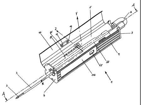
In the exemplary embodiment depicted in Fig. 1, all components necessary for
the completion of
a vacuum biopsy are integrated into the interior space of the housing of a
hand-held unit 1, so
that no cables or lines from the housing of the hand-held device to other
external supply units are
necessary.
The hand-held unit I thus represents a complete vacuum biopsy device, which is
freely moveable
in all directions. The distal portion of the hollow biopsy needle 2 and the
outer hollow needle 3,
which surrounds it coaxially and is referred to in the following as the
cutting sleeve, protrude from
the distal housing lid 6. Said cutting sleeve is used to remove and/or
completely sever the tissue
sample. In most instances, a coaxial cannula, which is not depicted, is
inserted into the tissue,
into which this segment of the biopsy needle 2 with cutting sleeve 3 is
introduced and is thus
positioned in front of the tissue to be examined. A connecting element 4, such
as a transparent,
flexible tube, which connects the pressure source disposed in parallel to the
biopsy needle or the
vacuum pressure-generating device 5 with the internal hollow space of the
biopsy needle 2 in a
gas-tight manner, is disposed outside the right proximal housing lid 7. The
hollow connecting
element 4 lies in direct proximity to the housing lid 7.The biopsy needle 2
with cutting sleeve 3
and additional elements, disposed in a biopsy needle carrier 37, forms,
together with the
connecting element 4 and the vacuum pressure-generating device 5, a biopsy
needle module 20
that is easily removed in an upward direction and easily inserted, hereinafter
referred to as the
removable element, which can be replaced as needed (Fig. 2). The housing lid
10 is opened for
this purpose. As Fig. 2, in particular, shows, the biopsy device can be
divided into parts that are
firmly attached to the housing (disinfected parts) and a removable element 20
(sterile part). While
the parts that are firmly attached to the housing are only disinfected, the
removable element 20 is
delivered in sterile packaging and is replaced as necessary, particularly with
each new patient. As
will be explained in detail later on, steps have been taken to ensure that the
disinfected part is not
contaminated with tissue fluid during use.
9
CA 02479349 2004-09-15
In the exemplary embodiment described below, the vacuum pressure-generating
device 5 is
arranged in parallel to the biopsy needle unit. Within the scope of the
invention, however, the
vacuum pressure-generating device can also be arranged in a prone position in
the axle of the
biopsy needle or the hand-held unit. Furthermore, it does not require a
separate connecting
element if, for example, it is placed directly onto the end of the biopsy
needle. In this case, the
connecting element is to be viewed as a suitable flange connection, such as in
the form of a Luer
lock.
A lower housing segment 9 and a housing lid. 10 hinged in the housing end
lids, together with a
locking latch 11, are positioned between the left and right housing end lids
6, 7. The lower
housing segment 9 is clamped between the housing end lids 6, 7 and/or
connected to a base
block 8 by means of tension rods or screws, some of which are screwed into the
base block B.
-
The housing lid 10 is hinged to an axle secured in the housing end lids 6, 7.
The housing lid 10 is
closed prior to operation of the biopsy device, with the inside contour of the
housing lid
corresponding to the outside contour of the biopsy needle carrier 37, which
will be described in
detail later on. The base block 8, which is firmly connected to the lower
housing segment by
means, for example, of fixing elements and/or a screw connection, is disposed
at approximately
the center of the interior space of the housing. The drive elements for the
vacuum pressure-
generating device 5, the cutting sleeve 3, and the clamping device for the
clamping cradle 28,
onto which the biopsy needle carrier 37 is mounted, are connected to the base
block 8. The base
block 8 extends from the center of the housing to the left, and a plate joined
to it cover the drives
and serves as a support for the control board, which is arranged a protected
manner inside or
below the cover 46. In addition, the base block 8 features a holder 36, open
at the top, for both
the biopsy needle and cutting sleeve, as well as an additional insertion
element 62 for the
vacuum pressure-generating device.
To identify the position of the individual elements, as well as the position
of the individual parts,
especially in the interior space of the housing, a system of coordinates was
drawn in Fig. 1,
wherein the center point of the coordinates of the system lies at the center
of the base block 8
(Fig. 1). Accordingly, in the following description movement in the direction
of the x-axis is
considered left (distal) and movement away from the x-axis is considered right
(proximal). For the
remaining coordinates, movement in the direction of the y axis is considered
upward, movement
away from the y axis downward, movement in the direction of the z axis
backward, and
movement away from the z axis forward (Fig. 1). Therefore, the system of
coordinates divides the
interior space of the housing and the remaining references into left and
right, front and back, and
--- - -- ----------
CA 02479349 2004-09-15
top and bottom. To facilitate understanding, these rules were modified for
depicting angled
rotational movement of the biopsy needle, with rotation around the common
longitudinal axis of
the biopsy needle and the cutting sleeve being depicted as movement to the
left (i.e., toward the
front) and right (toward the back).
With reference to these rules, the common drive mechanisms 106 for the
clamping device and
the cutting sleeve are located in approximately the lower, front, left portion
of the interior space of
the housing, and the drive mechanism for the vacuum pressure-generating device
5 in the upper,
rear, left portion of the housing. The power supply for the drive motors and
the remaining
electronic components, such as the control and/or monitoring elements, are
located in the lower,
right portion; batteries or a storage battery 111, such as a 7.2 V lithium ion
battery, 1Ah, are
preferably used for this purpose. The front, right, upper interior space of
the housing located
above the battery space is used primarily for the clamping cradle 28 and
locking element (Fig. 5),
which is connected to a block 26, which is part of the base block 8. The
battery space is sealed at
the top by a divider plate 114.
In the uppermost, front portion of the interior space of the housing, an
insertable and removable
biopsy needle carrier 37 is arranged in the U-shaped insertion holder 36, open
to the top, of the
base block 8 and in the upward-pointing bracket 40 disposed on both sides of
the clamping cradle
28, a biopsy needle/cutting sleeve unit with drive components being rotatably
supported in said
biopsy needle carrier, which extends along virtually the entire length of the
hand-held unit. As
described later on, the biopsy needle carrier is longitudinally displaceable
by means of the
clamping cradle. This means that in the non-tensioned state the left face of
the biopsy needle
carrier 37 almost rests against the housing end lid 6 and, in the tensioned
state, the right face
rests against the right housing end lid 7. "Virtually the entire length"
signifies that the biopsy
needle carrier is shortened by at least the amount of interior housing space
required for the
clamping process. If the clamping path of the clamping cradle is, for example,
20 mm, the biopsy
needle carrier must be displaceable by at least this amount. In general, the
clamping path ranges
from 15 to 25 mm, depending on the biopsy needle used. Consequently, it is
advantageous to
design the interior space to include the largest possible clamping path, plus
a few mm.
The clamping device (right, at front) itself comprises a clamping cradle 28
traveling on a pin 30,
the pin being screwed into the block 26 of the base block 8. The pin 30 is
proximally encircled by
a spiral spring 31. Another short spiral spring 124 is disposed on the pin 30
on the distal side of
the clamping cradle. One side of this short spiral spring rests on the block
26, while the other side
11
CA 02479349 2004-09-15
rests on an inner lip 122 on the distal side of the clamping cradle. The
spiral spring 31 rests on
the opposite side (proximal side) of the lip of the clamping cradle. The
locking mechanism (see,
in particular, fig. 9b and 10b) of the clamping cradle is secured to the block
26. The vacuum
pressure-generating device 5 and parts of the drive are arranged in the upper,
rear, right interior
space of the housing; the drive motor for the reduction gear for the vacuum
pressure-generating
device is located in the left, lower, rear region of the interior space of the
housing.
The housing lid, the lower housing segment, the housing end lid and the base
block are
preferably made of aluminum.
As described earlier, the hand-held unit 1 comprises a housing, which consists
of a lower housing
segment 9 with lateral walls of different heights, a housing lid 10, matched
to the lower housing -
segment, with longitudinally displaceable locking mechanism 11, and the two
housing end lids 6,
7.The lower housing segment is connected to the two housing end lids by means
of tension rods
or screws made, for example, of iron, some of which are screwed directly into
the base block 8.
The housing is approx. 200 mm in length, the housing end lids feature a
section cross-section of
approx. 40 x 40 mm (Fig. 2). The housing lid 10 pivots around an axle 104
secured in the housing
end lids 6, 7; the holes in the housing end lids are used for this purpose.
The nose 12 of the
locking mechanism 11 can be inserted into the depression 45 in the base block
8 to lock the
housing lid. The left housing end lid 6 features, in its upper front portion,
a U-shaped opening 13,
which is open at the top, for the forward-protruding portion of.the biopsy
needle/cutting sleeve 2,
3 and the guide roller 81 disposed thereon. The guide roller 81, which is
placed onto a coaxial
cannula when said cannula is used, also prevents tissue fluid from penetrating
into the housing.
The rear housing end lid 7 features two U-shaped openings 15, 16, which are
open at the top.
The hole 15 corresponds to the hole 13; it accepts the end of the round-
profile plastic component
47 placed onto the hollow biopsy needle. A nozzle 63 of the vacuum pressure-
generating device
is placed into the hold 16 (Fig. 2). Another plastic component 112 inserted
into the plastic part 47
features a peg 17, which is used to connect the connecting element 4 with the
outflow nozzle 64
of the vacuum pressure-generating device. The interior hollow space of the
biopsy needle is
continuously connected with the hollow space of the piston/cylinder
arrangement and the hollow
space of the vacuum pressure-generating device by means of the connecting
element 4, which is
also hollow. The connections are designed in such a way that air cannot
penetrate into the
system from the outside, nor can air escape to the outside when there is
overpressure; in other
words, the points of connection are designed to be airtight. The system,
designed in this manner,
causes the sealing element 76 to be pulled against the biopsy needle 2 when a
vacuum is applied
12
CA 02479349 2004-09-15
to the interior of the biopsy needle, which substantially improves sealing
action, but does not
negatively affect the rotational movement of the cutting sleeve relative to
the biopsy needle, but, if
suitably designed, does rotate the biopsy needle until the rotation is stopped
by a limiting device.
As Fig. 6, in particular, shows, a microswitch 18 is integrated into the lower
end of the hole 16 in
the housing end lid 7, and its switching pin 19 protrudes into the hole.
As soon as the nozzle 63 of the vacuum pressure-generating device is inserted
into the hole and
the housing lid is closed, the switching pin 19 of the microswitch 18 is
pressed downward and the
microswitch 18 releases the current supply. The terminals for connecting a
charging device can
be installed into the holes 97, 98 in the housing end lid.
A surface 113 for the control panel (Fig. 7) with control and monitoring
elements is provided on
the front side of the lower housing segment 9.
The control panel 57 to be attached to the housing is designed as an
independent component
which, for example, is glued to the surface 113 of the lower housing segment
9. This control
panel 57 is connected to other electronic components disposed in the housing,
as well as with the
power supply, by means of cables. Of the electriclelectronic components
connected to the control
panel, the circuit board disposed in the space 39 located beneath the cover 46
is especially worth
mentioning. A programmable microprocessor as well as other electronic
components are
disposed on the circuit board. The microprocessor is used to control the semi-
automatic process
control described later on. The control panel contains, in particular,
switches to operate the
biopsy device and diode to control the operating process. The control key 88
for mechanically
triggering the clamped clamping cradle protrudes from a depression 65 in the
lower housing
segment and somewhat presses out the control panel disposed above it, so that
the control key
can easily be felt through the foil of the control panel.
In designing the control and monitoring elements, steps were taken to ensure
that a distinction is
drawn between the clamping process of the clamping cradle and the triggering
of the clamping
cradle, on the one hand, and, on the other hand, between said clamping process
and
performance of the biopsy, such as severing of the sample and, in particular,
sample removal by
means of ejection of the sample.
13
CA 02479349 2004-09-15
Accordingly, the control key 88 (actuator) for the clamping cradle was placed
to the right, while
the clamping key 90 that triggers clamping of the clamping cradle was placed
to the left. The
program key 89 for completing the biopsy is centered. The control lights for
reset, completion of
the biopsy and ejection of the sample when the sample removal chamber is
opened are also
centered. When the program key 89 is pressed following insertion of the
removable element 20
and following closing and locking of the housing lid, as well as automatic
setting of the basic
position, two functions are activated, namely sample removal and sample
ejection.
Following insertion of the removable elements and closing of the lid, the
yellow reset diode 91 is
briefly illuminated and then flashes during setting of the basic position; the
reset diode is
extinguished following setting of the basic position. The sample removal diode
92 (green) and the
clamping diode (yellow) are illuminated and indicate that the operator can
activate one of the two
functions. If he presses the clamping key 90, the clamping cradle 28 is
brought into clamping
position and locked in this position. To prevent the clamping key from being
pressed
inadvertently, it is equipped with a delay circuit of about 1.2 seconds. The
yellow clamping diode
blinks during the clamping process. Following completion of the clamping
process, the locking
diode (green) blinks. The device, i.e., the biopsy needle, is then ready to be
shot into the tissue to
be examined and is triggered by means of the control key 88. Following the
shot into the tissue,
the locking diode is extinguished and the clamping diode (yellow) and the
sample removal diode
(green) are illuminated. Both functions (clamping or sample removal) can now
be activated. When
the program key 89 is pressed, the biopsy process is performed automatically,
as explained later
on. However, the clamping process could also be activated again. When the
biopsy process
(sample removal) is activated, it takes place automatically. Following
completion of the process,
the flashing green sample removal diode is extinguished and the yellow
ejection diode is
illuminated. When the program key is pressed again, the automated sample
removal process is
performed. Following completion of the process, the flashing ejection diode is
extinguished and
the yellow reset diode is illuminated, which means that the removable element
20 can be
removed, or that it can be automatically prepared for removing an additional
sale by pressing the
program key. This is followed by process as described above, i.e., either
clamping or sample
removal. For the event that the program key 89 is pressed for sample removal
(to eject the
sample), a delay circuit is provided that prevents ejection from occurring if
the program key is
touched inadvertently before the needle has been removed.
14
..................................
CA 02479349 2004-09-15
The battery charge diode 96 indicates the charging condition of the battery or
storage battery. As
described earlier, the diodes are wired in such a way that the diode flashes
during completion of
the specific process that was activated, and that the diode for the ensuing
process is illuminated
following completion of the process. If two options are available, both
subsequent diodes are
illuminated. In this case, the operator may select the option of his choice.
The colors of the
diodes are selected in such a way that procedures in the tissue are indicated
by a green light,
while external procedures are indicated by a yellow light. Delay circuits
(e.g., 1.2-1.5 seconds)
are provided for the functions or clamping and sample removal, so as to ensure
that the process
is activated deliberately. The mode of action and control options are
discussed in greater detail
during the description of the process sequence. Symbols (pictograms) on the
board symbolize
the individual processes. A perspective view of the base block 8 (as seen from
the front in the
direction of the x axis) is shown in Fig. 8a, while Fig. 8b depicts the base
block 8 from behind in
the x-axis (both are perspective views). The base block 8, when viewed in a
longitudinal direction,
can be divided into two halves; the front section is used to secure the common
drive for the
cutting sleeve and the clamping cradle and, in its front portion, to support
the biopsy needle
carrier (Fig. 8a); the rear section is used to secure the drive for the vacuum
pressure-generating
device as well as the support for the distal side of the vacuum pressure-
generating device (Fig.
8b). A central electronics circuit board is disposed between the two drive
motors 21, 58, below
the center rib 87, in the space 39 beneath. The base block 8 features, in its
left, front portion, a U-
shaped space 24, in which a toothed roller 23 driven by the geared motor 21 is
installed. To this
end, the drive shaft of the geared motor is supported and/or inserted in an
opening in the wall 25
of the base block 8. The toothed roller 23 is mounted onto the drive shaft and
is attached to it and
secured against rotation and displacement by means of a screw. On the other
side, the toothed
roller 23 is supported in the wall 22 of the base block 8. A DC motor with a
rotation speed of
approx. 11000 RPM is used as the drive motor. A planetary gear with high gear
reduction is
installed downstream from the DC motor, with the toothed roller 23 mounted on
its drive shaft.
Molded to the wall 22 and pointing to the right is another block 26, which
both accepts the
pivoting double handle 33 for the locking mechanism and serves to secure the
pin 30 guiding the
clamping cradle 28. The pins 30 are screwed into the threaded bore 29. During
the clamping
process, the clamping cradle 28 slides to the right on the divider plate 114
disposed below it.
During the clamping process, the spiral spring 31 disposed on the threaded pin
30 is
compressed. One end of the spiral spring rests against an end piece 32 of the
threaded pin or
directly on the housing end lid 7; the other end of the spiral spring, which
protrudes into a blind
hole in the clamping cradle, rests against ship resting on a lip 122 of the
guide hole 115. The
threaded pin 30, secured to the housing end lid 7 at one end and to the block
26 at the other,
CA 02479349 2004-09-15
carries at its distal end a short spiral spring 124, which also rests, on its
proximal side, against
another shim 125 resting against the circumferential lip 122 in a coaxial
blind hole 129 opposite
the hole 115. Both spiral springs have the same diameters, and the diameters
of the distal and
proximal bore 129, 115 in the clamping cradle and the distal bore 128 in the
block 26 are such
that the spiral springs can be easily inserted. All bores are coaxial to the
pin 30. The threaded pin
30 features a band 123 at the same axial distance to the circumferential lip
in the blind hole of the
cradle. In its starting position (resting position), the clamping cradle 28 is
held in resting position
by slightly loaded springs 31, 124 over the shims, as depicted in Fig. 3a and
3c. The shims rest
against both the corresponding side of the band and the lip, and are
vertically disposed. Thus, if
the cradle is deflected to the right or left, the respective spring will
attempt to return the clamping
cradle to its starting position; in a manner of speaking, the clamping cradle
is "swimming." The
clamping cradle 28 slides on the divider plate 114, in particular, and is
prevented from rotating by _
said cradle and by the side wall. An arm 99 of the double-armed handle 33 of
the locking device
engages a groove 27 of the clamping cradle 28 (Fig. 9a and 10a). The locking
device integrated
into the block 26 of the base block 8 consists of a double-armed handle 33,
which pivots around a
vertical axis (seen in the y axis) by means of a compression spring 34. The
axis 35, a vertically
disposed pin, is secured in the bores 38 of the base block. In the resting
state, the part 99 of the
double-armed handle lies in the groove 27 of the clamping cradle; the
compressed spring 34 acts
on the part 100 of the handle and presses the locking key 88 outward (toward
the front). The
locking key is easily felt in the control panel, which is pushed slightly
outward at this point after
clamping. As soon as the part 99 of the double-armed handle can lock into the
depression 82 in
the clamping cradle, the control key 88 is pushed outward. As a result of the
locking of the handle
part 99, the clamping cradle is locked in its the clamping state and can be
triggered, if needed, by
pressing the control key 88. As the clamping cradle is advantageously made of
plastic, it has
proven to be advantageous to place a metal part 83 into the depression so as
not to damage the
plastic, as the double-armed handle is also made of metal. In contrast to the
removable element
20, the hand-held unit with replaceable insert is reused several times. The
clamping path
corresponds to the depth of penetration of the biopsy needle into the tissue.
Consequently, the
length of the handle 99 also corresponds to the clamping path. As the depth of
penetration
generally ranges between 15 and 25 mm, the same hand-held unit can be used for
various
depths of penetration by suitably designing the handle 99 and modifying the
settings in the control
unit accordingly.
16
CA 02479349 2004-09-15
The clamping cradle 28, which is adjacent to the block 26, is disposed at the
same height as the
block 26, and has approximately the same profile as the block 26. The clamping
cradle features
two brackets 40 on its upper side. The upward-facing surface 41 of the
clamping cradle, the
upward-facing surface 44 of the block 26, and the upward-facing surface of the
extension 42 of
the base block 8 together form a flat support surface for the lower sliding
surface 43 of the biopsy
needle carrier 37 to be mounted (see Fig. 2). The biopsy needle carrier is
made of plastic. When
the clamping cradle is shifted from its non-tensioned resting state (Fig. 9a)
to its clamped state
(Fig. 10a), i.e., to the right, the biopsy needle carrier 37 held by the
brackets 40 slides across the
surface 42 and 44. It is also conceivable that the sliding surfaces are not
flat, as in the exemplary
embodiment, but feature uniquely structured sliding surfaces; what is
important is that the biopsy
needle carrier 37 can slide easily and in a straight line on the sliding
surface, and that, once the
control key 88 has been triggered, the biopsy needle can penetrate into the
tissue, the tumor, in a
straight line, For this reason, the upper outside contour of the biopsy needle
carrier is also
shaped to conform to the inside contour of the housing lid and features only a
small amount of
play to the housing lid, so as to prevent upward deflection of the biopsy
needle, which is also
advantageous during the clamping process.
Above the U-shaped space 24 for the toothed roller 23, at the level of the
sliding surface 42, the
base block 8 has a U-shaped holding device 36, which is open to the top, for
inserting the biopsy
needle/blade sheath, among other things. The primary function of this holding
device is that of a
radial thrust bearing, i.e., it supports the drive part that is connected to
the blade sheath, namely
the gear 74 or the plastic disk 78, in order to bring the clamping carriage
into its clamped position
by means of the drive device 106. On the distal side, the holding device also
serves as a stop for
the collar 127 in the execution of the back-and-forth movement and the
associated angular-
rotation movement.
A further U-shaped insertion element 62 is provided in the rear, upper part of
the base block; the
free end 61 (distal end) of the threaded spindle of the vacuum- and pressure-
generating device,
the end protruding from the syringe body, is inserted into the insertion
element. The insertion
element is embodied as a conduit, in which the threaded spindle 53 slides. In
the upper, central
region of the base block, a fastening device is provided for a disk that is
received by the recess
45; the latch 12 of the locking bar 11 of the housing lid is pushed into the
fastening device. A
cover 46, which is disposed on the base block 8 and faces left, separates the
space for the drive
motors and the inserted plate from the upper, left portion of the housing
interior, which primarily
serves in seating the replaceable biopsy-needle carrier 37, including the
biopsy needle and the
17
CA 02479349 2004-09-15
blade sheath. The cover 46 protects the electrical gear motors and the plate
from contamination.
The plate for the electronic components lies between the drive motors, and
beneath the center rib
in the space 39. Fig. 2 illustrates the biopsy-needle carrier 37, which can be
inserted into the
brackets 40 of the clamping carriage 28 with the biopsy needle 2 and the blade
sheath 3, as well
as further parts.
The hollow, circular biopsy needle 2 has a needle tip 70, which the specimen-
collection chamber
71 adjoins (Figs. 11a - 11f). The biopsy needle 2 having a round cross-section
is surrounded
coaxially by a blade sheath 3, also having a round cross-section, and having
at its left end, which
faces the specimen-collection chamber, a blade 72. In an especially preferred
embodiment, after
the biopsy needle has been inserted (with the specimen-collection chamber
being closed) and the
specimen-collection chamber has been opened, and the needle has performed a
repeated back- _
and-forth movement that is superimposed simultaneously by a predetermined,
limited angular-
rotational movement of the biopsy needle about its longitudinal axis, the
blade serves in cutting
out the specimen and holding it in the closed specimen-collection chamber, as
will be explained
in detail below. The distal blade of the blade sheath is preferably disposed
perpendicular to the
longitudinal axis of the biopsy needle and the blade sheath. The severing
procedure is preferably
effected through the rotation and simultaneous longitudinal displacement of
the blade sheath by
the threaded-spindle drive. It is also conceivable for the blade sheath not to
execute a
continuous movement, but for it to move in increments or to vibrate, i.e., the
traveling part is
moved forward and back by short distances. As can especially be seen from the
cross-sectional
representation in Fig. 11f, the longitudinal edges 68 of the specimen-
collection chamber are
located above the center point of the cross-section - in other words, the
specimen-collection
chamber extends beyond the Z-axis by about 15 - 30 . To improve the entrance
of solid, hard
tissue into the specimen-collection chamber, the longitudinal edges have a
blade. This blade at
the longitudinal edges is created through the reduction of the wall thickness
from above such that
the width b' at the cutting edge corresponds to the width b of the diameter of
a lower-lying blade-
sheath-tube, i.e., the wall thickness is reduced in the upper part and
utilized to embody the cutting
edge (Fig. 11f and enlarged view in Fig. 11g).
At the other, proximal end of the blade sheath, which faces away from the
blade 72, a threaded-
spindle sheath 73 is secured to a gear 74 that is disposed at the end face of
the threaded-spindle
sheath. The threaded-spindle sheath is disposed with the gear on the blade
sheath so as to be
fixed against rotation and displacement. A threaded-spindle nut 75 that is
pressed securely into
the biopsy-needle carrier 37 cooperates with the threaded spindle. The gear 74
is to the left, that
18
CA 02479349 2004-09-15
is, in front of the beginning of the spindle sheath. When the threaded-spindle
sheath is rotated by
the gear 74, the blade sheath is rotated and displaced longitudinally over the
biopsy needle 2.
On the distal side of the gear 74, a tubular piece 126 having the collar 127
is permanently
connected to the threaded spindle. The tubular piece is inserted into the
holding device 36, with
the collar 127 being located on the distal side in front of the holding
device. The length of the
tubular piece 126 approximately corresponds to the clamping path; the wall
thickness of the
holding device 36 must additionally be considered here (Figs. 3a and 3b). In
the initial position of
the device (closed specimen-collection chamber), the collar 127 travels to the
left, to the distal
side, whereas it comes to rest against the holding device 36 (distal side)
after the specimen-
collection chamber has been opened. As the spindle sheath continues to rotate
with the blade
device, that is, in the attempt to open the specimen-collection chamber wider,
the clamping _
carriage is pulled toward the block 26, counter to the effect of the short
coil spring, because the
collar 127 rests against the holding device 36 on the distal side.
Consequently, as will be
described further below, the biopsy needle can be set into a back-and-forth
movement that is
superimposed by a limited angular-rotational movement of the biopsy needle to
both sides. This
angular-rotational movement is effected by the attempt of the blade sheath to
carry the biopsy
needle along in the rotation; the needle, however, is prevented from rotating
past a
predetermined angular rotation, as can be seen particularly in Figs. 12b
through 12d.
The gear 74 at the left end of the threaded spindle meshes with the toothed
roller 23 after the
biopsy-needle carrier has been inserted into the brackets 40. To allow the
biopsy-needle carrier
37 to be inserted into the brackets of the clamping carriage when the carriage
is not clamped
(Fig. 2), the biopsy-needle carrier has two planar, parallel recesses 77 (Fig.
2). When the sliding
surface of the biopsy-needle carrier 37 is placed onto the surfaces 41, 42 and
44, the biopsy
needle is simultaneously inserted into the holding device 36 of the base block
8. On the left side
of the gear, a slightly conical plastic disk 78 can be incorporated in order
to improve the rotating
capacity of the spindle drive, especially if the holding device 36 is serving
as a support for
clamping the clamping carriage. When the biopsy-needle carrier is inserted
correctly, the carrier
slides to the right, with the sliding surface 43, across the surfaces 42 and
41 as the clamping
carriage is clamped. Because the specimen-collection chamber is closed after
the biopsy-needle
carrier has been inserted, the gear 74 rests against the holding device 36. If
the toothed roller 23
is driven further in the same direction, the threaded-spindle drive screws the
clamping carriage to
the right, by way of the biopsy-needle carrier, until it is latched; in the
process, the biopsy needle
is retracted, while the blade sheath remains in its position. The blade sheath
protrudes past the
19
CA 02479349 2004-09-15
tip of the biopsy needle after the latching procedure. Therefore, after the
clamping carriage has
been latched, the blade sheath is rotated back into the initial position
(opposite direction of
rotation); the gear 74 is displaced from the left to the right in the toothed
roller. After the clamping
carriage has been unlatched, the biopsy needle and the blade sheath with the
gear slide to the
left again with the biopsy-needle carrier. Now the blade sheath can be
displaced to the right
again in order to open the specimen-collection chamber until the collar 127
comes into contact.
The function of the "floating" seating of the clamping carriage in connection
with the controllable
drive motor and the tubular piece 126 connected to the blade sheath and having
the collar 127 is
explained in greater detail in connection with the biopsy procedure.
A sealing element 76 produces a connection between the right end of the blade
sheath and the
hollow biopsy needle that permits rotation, but is airtight, so that air
cannot enter between the _
biopsy needle and the blade sheath surrounding it coaxially, and air cannot
exit under
overpressure conditions. The-sealing element 76 comprises a plastic hose that
is pulled over the
proximal end of the blade sheath. The inside diameter is selected such that it
rests lightly against
the outside diameter of the biopsy needle. When a vacuum is generated in the
interior of the
biopsy needle, and thus between the biopsy needle (outside) and the blade
sheath (inside), the
elastic plastic hose is pulled against the outside diameter of the biopsy
needle. Provided that the
biopsy needle is rotated relative to the blade sheath, the hose can serve as a
restoring element
(restoring spring). For rotating the biopsy needle slightly by means of the
blade sheath, the
biopsy needle is slightly deformed in the region of the sealing element 76, so
it is oval-shaped at
the deformed point 0 (Fig. 12f). When the blade sheath rotates, the biopsy
needle is carried
along by the deformation 0 until the rotation of the needle is limited by a
stop (Figs. 12b through
12d).
This angular-rotational movement of the biopsy needle simultaneously effects
the pivoting of the
sharpened longitudinal edges of the biopsy-needle space to both sides about
the longitudinal axis
of the biopsy needle. Because this angular-rotational movement is effected by
the same drive
and occurs simultaneously with the back-and-forth movement of the biopsy
needle, the cutting
edges of the specimen-collection chamber sever the tissue, in the manner of a
driven knife, both
longitudinally in the X-axis and with an angular offset, so the tissue, which
is under pressure
(external and/or internal pressure) reliably enters the open specimen-
collection chamber. Fig. 12f
illustrates the specimen-collection chamber in the neutral initial position
after opening; Fig. 12g
shows the position following an angular rotation to the right by the angle a,
and the simultaneous
retraction of the biopsy needle by the distance X, (about 2 mm) to the
proximal side; Fig 12h
CA 02479349 2004-09-15
shows the position of the biopsy needle during a rotation to the left by the
angle R, and the
simultaneous movement of the biopsy needle to the distal side by the distance
X2 (about 2 mm).
The movement of the cutting edges of the specimen-collection chamber or the
biopsy needle
ensures that the tissue will be severed at the longitudinal edges, regardless
of the tissue
structure. The described movement of the biopsy needle, and therefore of the
sharpened
longitudinal edges of the specimen-collection chamber, also ensures that the
severed piece of
tissue will enter the specimen-collection chamber, even if the pressure that
is normally exerted is
absent.
A round, hollow plastic part 47 is placed onto the right end of the biopsy
needle 2 in a frictional,
airtight connection. At its left end, the plastic part 47 has a bearing
element 49, which is pressed
into the biopsy-needle carrier; at its right end, which protrudes from the
handpiece, a further
plastic part 112 is provided. This part can rotate relative to the plastic
part 47 and the biopsy
needle 2. An O-ring is inserted between the biopsy needle and the plastic part
112 to assure a
seal. At its right end, the plastic part has a tappet 17, onto which the
connecting element 4 is
pushed to form an airtight connection. Also disposed at the right end
protruding out of the biopsy-
needle carrier and the housing is a knurled knob 80, which can be rotated to
adjust the position of
the specimen-collection chamber radially without altering the position of the
blade sheath. Only a
single rotation of the specimen-collection chamber is associated with a
rotation of the biopsy
needle. The plastic part 47 is pressed, with the biopsy needle, the blade
sheath, the bearing
element 49 and the threaded-spindle nut 75, into the biopsy-needle carrier. By
way of the
bearing element 49 and its narrow guide in the blade sheath, the biopsy needle
is seated to rotate
in the biopsy-needle carrier and in the blade sheath, and to be displaced with
the biopsy-needle
carrier along the longitudinal axis. As explained above, the blade sheath can
be rotated axially
relative to the biopsy needle.
To the right of the bearing element 49, a polygonal member 50 is disposed on
the plastic part 47.
The polygonal member can be clamped to latch with the biopsy-needle carrier
37, so the
specimen-collection chamber of the biopsy needle can be brought into and held
in the position
that is most favorable for the biopsy collection by means of the knurled knob
80. During the
rotation, the two legs 39 of the biopsy-needle carrier, which comprises an
elastic plastic, are
spread by the corners of the polygonal member until the surfaces of the
polygonal member are
nearly perpendicular to the legs 39 again, and the polygonal member is latched
again (Fig. 12c).
The polygonal member is then adjusted by a predetermined increment. If the
polygonal member
21
CA 02479349 2004-09-15
is hexagonal, the rotational distance is 60 ; if more rotational increments
are desired, a polygonal
member having 8, 10, etc., sides should be selected accordingly.
As can be seen particularly in Figs. 12b through 12f, the biopsy-needle
carrier has two legs 39,
which are connected to one another by a cap element 116. In the plastic
carrier, the polygonal
member 50 of the plastic part is seated to be latched: The legs 39 connected
to the elastic
fastening element are first spread apart during a rotation in order to return
to their initial position
due to the elasticity. If the inscribed circle diameter S selected for the
polygonal member is
smaller than the distance A (clear width) of the two legs from one another,
the biopsy needle can
rotate slightly to both sides about its axis, by a predetermined angle (a or
(3) (Figs. 12b and 12d -
Fig. 12c illustrates the center position). The legs 39 of the biopsy-needle
carrier are not spread
here; on the contrary, they prevent the biopsy needle from rotating by a
larger angle, because the -
drive is configured such that the blade sheath can be rotated further, but the
resistance of the leg
limit is greater than the drive moment. The corners of the polygonal member
impact the legs 39,
and prevent a further rotation, because the torque acting on the biopsy needle
does not suffice to
spread the two legs. Because the plastic part 47 with the polygonal member is
permanently
connected to the needle, and the blade sheath was pushed onto the deformed
region 0 of the
biopsy needle when the specimen-collection chamber was opened, and the sealing
element 76
enters a frictional connection with the outside of the biopsy needle when the
specimen-collection
chamber is open, when the blade sheath is driven in a respective direction of
rotation, the biopsy
needle also rotates about its axis due to this frictional connection until the
stop effected by the
polygonal member prevents a further rotation in the absence of a greater
torque.
Because the vacuum that dominates in this phase pulls the elastic sealing
element more strongly
against the outside surface of the biopsy needle, the sealing element
facilitates the rotation of the
blade sheath relative to the biopsy needle, on the one hand; on the other
hand, the sealing
element acts as a restoring element if lightly touched. This limited
rotational movement is
understood to be an angular-rotational movement. The biopsy needle, which can
rotate to a
limited extent, ceases its angular-rotational movement due to the change in
the direction of
rotation, and returns to its initial position because of the twisted sealing
element, then is rotated in
the other direction counter to the effect of the elastic sealing element.
Generally, about one
rotation of the gear 74 in each direction (about one rotation from the zero
position) suffices for the
limited angular-rotational movement in connection with the back-and-forth
movement, as
described above. When the gear rotates, the biopsy needle is displaced from
the zero position by
22
CA 02479349 2004-09-15
about 2 mm to the left or right, and is simultaneously moved about the
longitudinal axis by the
angle a or P. Generally, this movement is repeated about five times in each
direction.
As shown particularly in Fig. 12, the blade sheath surrounding the biopsy
needle coaxially is
connected to the biopsy-needle carrier 37 by way of the threaded-spindle nut
75. The threaded-
spindle sheath 73 is seated so as to rotate in the threaded-spindle nut 75. A
rotation of the gear
74 by the drive motor of the toothed roller 23 causes the biopsy-needle
carrier and the clamping
carriage to be moved to the right as soon as the gear 74 comes into contact
with the holding
device 36. When the gear occupies a position within the length of the toothed
roller 74, that is,
provided that the gear is free and does not rest against the holding device or
the threaded-spindle
nut 75, the blade sheath can be adjusted alone, for example after the biopsy
needle has been
clamped, in order to align the needle tip and the blade sheath for returning
the blade sheath to _
the initial position, or for opening and closing the specimen-collection
chamber.
When the specimen-collection chamber is opened, the blade sheath is pushed
over the slightly
deformed region 0 of the biopsy needle. In this position, as the blade sheath
continues to rotate,
the biopsy needle is carried along in the direction of rotation by a
predetermined angle; because
the connection between the blade sheath and the biopsy needle only permits the
transmission of
a predetermined torque, however, the rotational movement of the biopsy needle
comes to a halt
when the corresponding corners of the polygonal member come into contact with
the legs of the
biopsy-needle carrier (Figs. 12b and 12d). The tubular piece 126 with the
collar 127, which is
mounted to the distal side of the gear, and cooperates with the holding device
36, serves in
setting the needle into a brief vibrating movement (back-and-forth movement),
in cooperation with
the control, and in simultaneously setting the biopsy needle into an
alternating angular-rotational
movement, as described above.
Because the vibrating (back-and-forth) movement is effected by the drive for
the blade sheath 3,
the connection of the blade sheath to the deformed point "0" of the biopsy
needle and the
configuration of the polygonal member 50 and the biopsy-needle carrier
simultaneously effect a
limited rotation of the biopsy needle in both directions of rotation
(depending on the direction of
rotation), which is superimposed over the back-and-forth movement of the
biopsy needle. These
two combined movements sever any type of tissue with the cutting edges of the
specimen-
collection chamber. The tissue, even if it is tough or has inclusions, can be
reliably pulled or
inserted into the specimen-collection chamber with or without pressure. The
aforementioned
23
CA 02479349 2004-09-15
vibrating (back-and-forth) movement of the biopsy needle, which also effects
the angular-
rotational movement, is described below:
When the toothed roller is driven, the gear 74 opens the specimen-collection
chamber until the
collar rests against the distal side of the holding device 36. If the same
direction of rotation is
maintained, and the gear no longer rests against the threaded-spindle nut, the
further rotation
causes the clamping carriage to be pulled over the biopsy-needle carrier
toward the block 26,
counter to the effect of the short coil spring, because the contact of the
collar 127 with the distal
side of the holding device 36 makes it impossible for the blade sheath to be
opened further. The
clamping path or the movement path (X, or X2) is about 2 mm, or corresponds to
about one
rotation of the blade sheath.
When the gear 74 stops against the threaded-spindle nut, the direction of
rotation of the motor is
reversed and, with the support of the short coil spring, the clamping carriage
returns to its initial
position (resting position), and the biopsy needle is rotated back into its
zero position. Because a
detector counts the motor rpm, and the actual values are stored in a
programmable
microprocessor, corresponding preset commands can be used to change the
direction of rotation
of the motor, so the clamping carriage is pulled toward the block again or
retracted after being
released. The continuous reversal of the motor's direction of rotation, as
specified in advance,
works in conjunction with the clamping and release of the carriage to effect a
back-and-forth
movement of the biopsy needle that is superimposed with a limited angular-
rotational movement
of the biopsy needle to both sides, depending on the direction of rotation.
Five back-and-forth
movements typically suffice to assure a good specimen collection, even of
tough tissue or tissue
having inclusions, such as calcification. The back-and-forth movement of the
biopsy needle, in
conjunction with the limited angular-rotational movement of the needle, and
the sharpened cutting
edges, severs the tissue, which has been pulled to the specimen-collection
chamber by a
vacuum, for example, at the side edges in order to permit or greatly
facilitate the entrance of the
specimen into the specimen-collection chamber, even if the specimen comprises
tough tissue.
The described movement of the biopsy needle, and thus of the sharpened
longitudinal edges of
the specimen-collection chamber, permits a superior severing of the tissue
after or during the
opening of the specimen-collection chamber. The same good tissue-severing
action can be
attained if this vibrating movement or angular-rotational movement is
performed during the
opening process, that is, when the blade sheath is retracted.
24
CA 02479349 2004-09-15
Figs. 12g and 12f illustrate this superimposed movement of the biopsy-needle
space in detail, in
the phases of left and right rotation as well as the zero position. Fig. 12g
and others depict the
initial position: The biopsy-needle space is open, the blade sheath is
retracted by about 2 mm
past the proximal edge of the specimen-collection chamber, and the polygonal
member is in the
neutral position (Fig. 12c). In the representations of Figs. 12h and 12g, the
biopsy needle is
retracted into the blade sheath, and the needle is simultaneously pivoted by
the angle a. Figs.
12f and 12d show the other direction of rotation, and the rotation by the
angle P. The proximal
edge of the biopsy-needle space is displaced by about 2 mm toward the distal
side relative to the
initial position, and is simultaneously pivoted by the angle (3 in the other
direction. Figs. 12g, 12f
and 12b through 12d illustrate a cycle that is repeated several times -
typically five times - under
the control of the microprocessor. This back-and-forth movement and the
limited angular-
rotational movement can be effected by electrical elements that are connected
to the needle or
the clamping carriage.
Figs. 11 a through 11 g illustrate details about the specimen-collection
chamber and the
embodiment of the biopsy-needle tip. Approximately 25% of the cross-section of
the specimen-
collection chamber 71 adjoining the needle tip 70 is open to the top. For
example, if the biopsy
needle has an outside diameter of 3.3 mm, the height H of the specimen-
collection chamber is
about 2.3 mm. The specimen-collection chamber is approximately between 15 and
25 mm long.
Adjoining the chamber is the hollow space of the biopsy needle. At the
transition, that is, at the
proximal end of the specimen-collection chamber, the cross-section of the
hollow space of the
biopsy needle is closed by between 50% and 75% by a narrowed region, such as a
stopper 79
(Figs. 11b through 1le). The height of the stopper is selected such that the
stopper extends
downward past the recess for the specimen-collection chamber.
As can be seen in Fig. 11e, an opening F is provided in the floor of the
specimen-collection
chamber; if the inside diameter of the needle is 3.0 mm, the height of the
opening is 0.6 mm. The
vacuum is intended to pull the tissue specimen into the specimen-collection
chamber as the
chamber is continuously opened, and bring the specimen into contact with the
wall of the
chamber. If an overpressure is present in the biopsy-needle hollow space, the
narrowed region,
the stopper, increases the pressure. The stopper is about 10 mm long, and is
glued or welded
into the hollow space. The use of laser welding has revealed that it is
advantageous-to construct
the left side of the stopper by removing material at the end face to make it
short, namely about 2
mm long. Consequently, in this region at the end face, the tube of the biopsy
needle is welded
completely to the end face of the stopper, and is airtight at the end face.
The stopper can also be
CA 02479349 2004-09-15
shorter, provided that the same effect is attained. The stopper can also be
replaced by a lip or a
latch of approximately the same height. It is critical that the narrowed
region be embodied to
allow the vacuum in the specimen-collection chamber to be effective from the
floor, so that the
specimen sticks to the wall of the chamber as the blade sheath is closed, that
is, during the
cutting process, and does not change its position. It has also proven
advantageous to provide
additional securing means on the specimen-collection wall. Sucking the
specimen into the
specimen-collection chamber from below results in a high filling capacity of
the specimen-
collection chamber, on the one hand, and the configuration of the chamber, in
particular, allows
the specimen to be secured well to the wall, on the other hand. For this
reason, it is crucial that
the lateral severing of the tissue through the described movement of the
sharpened longitudinal
edges of the specimen-collection chamber ensures that the tissue reaches the
chamber floor.
Because the blade sheath severs the specimen on the outside of the biopsy
needle, the process
of suctioning the specimen in the inside is also maintained during a severing
process, if possible.
The outside location of the blade sheath, and the fact that the tissue sticks
to the inside floor of
the specimen-collection chamber, prevent the specimen from being rotated or
twisted by the
rotating longitudinal movement of the blade sheath during the closing process.
The quality of the
specimen is greatly improved over systems that employ a twisting effect. The
pathologist obtains
a starting material whose cross-section corresponds to the section in the
tissue, not a twisted or
deformed mixture of tissues. Ejecting the specimen under pressure is a
reliable method of
depositing it. The stopper 79 permits this. In addition, the specimen-
collection chamber is
completely cleaned, so if the biopsy is repeated, there is no mixing of tissue
specimens (residual
particles).
Because the vacuum-generating device is simultaneously used as a pressure-
generating device,
the entire hollow space, particularly the biopsy needle, is cleaned during the
ejection process.
For normal tissue, it suffices to use the wall thickness of the biopsy-needle
tube, which is about
0.15 mm, as a lateral cutting edge. For hard and/or tight tissue, the filling
capacity achieved in
the specimen-collection chamber solely through vacuum suction is insufficient,
because the
tissue at the side edges is not adequately severed. Through the embodiment of
the long sides of
the specimen-collection chamber 71 as cutting edges 68, as can be seen
particularly in Figs. 11 g
and 11f, the superimposing of a repeated back-and-forth movement and a
limited, repeated
angular-rotational movement of the biopsy needle with the specimen-collection
chamber to the
left and right (as described), as well as the pressure effect of, for example,
an internal vacuum,
for example with the ultrasonic head, the specimen tissue is severed
longitudinally, so the
26
CA 02479349 2004-09-15
specimen travels to the floor of the specimen-collection chamber far better
because of the
vacuum or external pressure. The exertion of an external pressure or an
internal vacuum in the
biopsy-needle hollow space can reinforce the cutting effect of the
longitudinal edges. Severing
the long-side cutting surfaces of the tissue specimen to be removed through
the movement of the
needle attains a superior filling capacity, even with hard and/or leathery
tissue or tissue having
inclusions. This method provides adequate tissue material for testing. The
cutting edge at the
long side of the specimen-collection device is formed by the milling off of
the partial piece (T1)
from the wall thickness (Fig. 11 g). The diameter (ra) of the outside contour
of the biopsy-needle
tube is retained, while the inside diameter (ri) of the inside contour changes
over to the outside
contour, via a perpendicular wall, through the milling out of the partial
piece (T1) (Fig. 11 g).
Fig. 11d shows the closed specimen-collection chamber, in which the collected
specimen is
located. During the severing process, it has proven advantageous to move the
blade sheath 78
out of its end position, shown in Fig. 11d, and about 2 mm in the distal
direction, and then retract
the blade sheath by these 2 mm into its end position. This shearing effect
reliably severs fibers
that may not yet be completely severed, which further increases the quality of
the specimen.
The biopsy needle described here operates with a vacuum that is generated
internally. The back-
and-forth movement combined with the superimposed, limited angular-rotational
movement of the
biopsy needle, and thus of the blades of the specimen-collection chamber, also
produces
superior results in biopsy needles without a vacuum or if the vacuum fails,
especially if, for
example, an external pressure is exerted onto the tissue in the biopsy hollow
needle, such as
through ultrasound, instead of the internal vacuum. Using only the angular-
rotational movement,
superimposed with the back-and-forth movement of the needle in conjunction
with the long edges
of the specimen-collection chamber embodied as blades, however, considerably
improves the
tissue-severing process and facilitates the entry of the tissue specimen to be
cut out into the
specimen-collection chamber.
It must also be pointed out that the elastic sealing element is not required
as a restoring element
in every case; merely reversing the direction of rotation can effect the
restoring action.
Fig. 13 shows the drive and the installation of the vacuum- and pressure-
generating device 5
(view from behind, that is, along the Z-axis; housing lid and lower housing
part omitted).
27
CA 02479349 2004-09-15
in the upper, rear, right region, the vacuum- and pressure-generating device 5
is embodied as a
plunger/cylinder unit 69. It comprises a syringe body 52 having a threaded
spindle 53 inside it; at
its end facing the bottom 51 of the syringe, a plunger 54 with sealing
elements - a standard
feature of syringes - is secured to the spindle (Figs. 14a through 14d).
At the end of the syringe body 52 facing the base block 8, a threaded-spindle
nut 48 having a
gear 55 at its circumference is disposed on the threaded spindle. The threaded-
spindle nut has
one or more thread turns. The threaded spindle 53 cooperates with the threaded-
spindle nut 48.
The spindle has a pitch of about 5 mm per turn, so with each rotation by means
of the spindle
drive, the plunger moves by a precisely defined distance out of the syringe
body, that is, away
from the bottom 51 of the syringe, or toward the bottom of the syringe,
depending on the direction
of rotation. The gear rim 55 disposed at the circumference of the threaded-
spindle nut meshes -
with the drive pinion 56, which is secured to the power takeoff shaft of the
DC gear motor 58.
The power takeoff shaft of the DC gear motor 58 is seated in the base block 8;
for this purpose,
the power takeoff shaft is inserted into the transverse plate 59 of the base
block. When the DC
gear motor 58 is activated, the plunger is moved toward the bottom of the
syringe, or in the
direction of the base block 8, depending on the direction of rotation. A DC
motor having a high
rpm is used as the drive motor; a planetary gear having a high step-down is
disposed
downstream of the DC motor. The motor corresponds to the motor described above
for the
clamping device. Hence, a counting device, comprising a two-armed blade wheel
131 and a
photocell that is mounted to the motor side, is likewise secured to the distal
side of the DC gear
motor. The counting device is connected to the programmable microprocessor, so
the function of
the vacuum- and pressure-generating device can be controlled with the rpm:
After an initial value
has been ascertained, the functions can be called up with programmable or
programmed
specifications.
The plunger 54 is embodied in a known manner as a syringe plunger. The plastic
syringe body, a
cylinder with a floor, is transparent. To prevent the threaded spindle 53 from
rotating when the
threaded-spindle nut is driven, the two oppositely located surfaces 60 of the
threaded spindle are
planar (Fig. 14d). The threaded spindle is inserted into the insertion element
by its free end.
The spacing of the threaded-spindle surfaces corresponds to the width of the U-
shaped insertion
element 62 of the base block 8. Only a small amount of play exists between the
U-shaped cross-
section of the insertion element and the spindle surfaces on both sides. The
threaded-spindle nut
is supported on the base block.
28
CA 02479349 2004-09-15
To prevent the syringe body 52 from sliding out when the threaded-spindle nut
rotates, the
contact surface of the base block 8 has a slightly conical shape at the
bottom.
The connector 63 of the syringe body 52 is inserted into the passageway 16 of
the housing end
lid 7 such that the syringe body is held in an approximately horizontal
position.
To make the rotation of the threaded spindle smooth, the threaded-spindle nut
with the gear rim
has an approximately 1.5-mm thick phase 66 (Fig. 14c) on the side facing the
base block.
Because the surface of the rib 59 on the base block 8, which cooperates with
the phase 66 of the
threaded-spindle nut 48, is inclined from top to bottom, the vacuum- and
pressure-generating
device is pulled down during operation. To generate a sufficient vacuum of
about 200 hph in the
specimen-collection chamber, if the biopsy-needle length is about 250 mm and
the inside
diameter of the biopsy hollow needle is between 3 and 5 mm, a 20-ml syringe
body having a
length of about 90 mm is used. To also be able to use the syringe body to
generate pressure, a
ventilation bore 67 having an approximately 1.5-mm diameter is provided after
about' of the
length, corresponding to the stroke for generating the vacuum (position
according to Fig. 11 b).
The ventilation bore can also be embodied as an oblong slot. When the vacuum
is no longer
required, and the syringe plunger is moved past the ventilation bore 67 (Fig.
14c), the supply of
air (atmospheric pressure) via the ventilation bore 67 reduces the vacuum that
was previously
built up in the biopsy hollow needle. If the direction of rotation of the gear
motor is then reversed,
the depression of the plunger (toward the bottom of the syringe) causes the
vacuum- and
pressure-generating device to effect the buildup of an overpressure in the
system, so the
specimen is ejected after the specimen-collection chamber is opened. To
prevent tissue fluid
from escaping during the brief opening of the ventilation bore 67, the
ventilation bore may be
covered with an air-permeable sponge (not shown), for example. A delay circuit
that is integrated
into the control mechanism prevents the tissue specimen from being ejected
through the
inadvertent depression of the programming key 89; the delay circuit only
initiates the procedure
after the key has been held down for about 1.2 to 1.5 seconds. The specimen
cannot be ejected
until the biopsy needle has been removed from the tissue. Moreover, the
compressed air not
only cleans the specimen-collection chamber, but particularly the interior of
the biopsy needle as
well. The stopper that narrows the needle hollow space impedes or completely
prevents the
entry of tissue segments into the biopsy-needle hollow space. The narrowing of
the needle
hollow space by the stopper 79 increases the pressure at the specimen-
collection chamber, and
therefore improves the ejection of the specimen when the specimen-collection
chamber is only
half-open. In the use of the vacuum biopsy device, it is advisable to use a
specially designed
29
CA 02479349 2004-09-15
coaxial cannula that is tailored to the specifications and requirements of the
device. The cannula
must contain corresponding devices that prevent or preclude the entrance of
air and the exit of
tissue fluid; on the other hand, it should be able to be inserted easily into
the tissue.
The operation of the biopsy device is explained in detail below.
The following segments of the procedure are performed extensively
automatically after being
initiated:
a) starting and setting the initial position;
b) clamping the biopsy needle and injecting it into the tissue;
c) cutting the specimen out of the tissue (specimen collection); and
d) removing the specimen after the closed biopsy needle has been removed from
the tissue.
a) Starting and Setting the Initial Position
The removable insertion element 20, comprising the vacuum- and pressure-
generating device,
the elastic connecting element and the biopsy-needle carrier with the needle
and the blade
sheath, and further elements connected thereto, and a guide roller 81 that is
placed onto the
needle are delivered in sterile packaging. The removable elements (Fig. 2) are
held by an
insertion aid that is removed after the elements have been inserted into the
handpiece. This
insertion aid 104 has two grips for grasping the top, and brackets 108 for
holding the biopsy-
needle carrier 37 and the vacuum- and pressure-generating device. To secure
the vacuum- and
pressure-generating device in its position (parallel to the biopsy-needle
carrier), a pin 110
provided on the bracket-holding device is inserted into the ventilation bore.
The plunger 54 in the syringe body 52 is raised slightly (1 to 2 mm) from the
bottom of the
syringe, and the specimen-collection chamber 71 of the biopsy needle 2 is
opened to allow visual
checking of the specimen-collection chamber prior to insertion. After the
housing lid 10 has been
opened, the biopsy-needle carrier, including the biopsy needle 2, the cutting
device 3 and other
connected parts, such as the vacuum and pressure-generating device 5 connected
to the
connecting element 4, are inserted into the connecting elements provided on
the handpiece (Fig.
2). During the insertion process, it must be ensured that the gear 74 meshes
with the teeth of the
toothed roller 23. The blade sheath is inserted from above into the U-shaped
holding device 36;
at the same time, the brackets 40 of the clamping carriage are guided into the
recesses 77 of the
carrier element, and the guide roller 81 is inserted into the passageway 13,
so it surrounds the
CA 02479349 2004-09-15
flanks 101 and 102 of the housing end lid 6. The blade sheath can be
longitudinally displaced in
the guide roller, and is seated to rotate freely; the guide roller itself,
however, can no longer be
displaced relative to the blade sheath following insertion into the housing
end lid. On the one
hand, the vacuum and pressure-generating device is then inserted by the free
end 61 into the
insertion element 62 of the base block 8, which is open to the top, and into
the U-shaped
passageway 16, which is open to the top, by the connector 63, on the other
hand. The connector
63 lies above the switching pin 19. Because the insertion element on the side
of the base block
has a clear width that just permits the insertion of the threaded spindle
provided on both sides
with surfaces 60, the threaded spindle is held, fixed against relative
rotation, in the insertion
element. The gear rim 55 of the threaded-spindle nut 48 engages the drive
pinion 56 of the gear
motor after the insertion. The spacing between the base block and the housing
end lid 7 is
maintained to allow space for the syringe body 52 with the threaded-spindle
nut 48 placed on the -
syringe body. The unit formed by the syringe body and the gear placed on it is
held such that it
cannot be axially displaced.
After the insertion, the vacuum and pressure-generating device lies parallel
to the biopsy-needle
carrier and the connecting element 4 describes a curve of about 180 . It
should be noted that the
insertion takes place when the clamping carriage is not clamped. This means
that the gear 74
engages the right end of the toothed roller when the specimen-collection
chamber is open (Fig.
3). After proper insertion, the housing lid can be closed.
The described insertion aid can be employed to facilitate the insertion
process. The insertion can
also take place without the insertion aid, however. When the housing lid is
closed, the connector
63 is pressed down, thereby actuating the microswitch by way of the switching
pin 19 installed in
the housing end lid. This activates the electrical system, which is indicated
by the blinking reset
diode (yellow) 91 on the front of the handpiece. The reset diode blinks
yellow, which means that
the positioning procedure for the individual elements, i.e., the insertion
procedure, has not ended
yet; the DC gear motor 21 must first close the specimen-collection chamber 71
with the blade
sheath 3 (the specimen-collection chamber was partially open during the
insertion). This is
effected by the rotation of the threaded sheath connected to the blade sheath.
The blade sheath
moves to the left until the gear 74 comes to rest near the inside of the
holding device 36. After
the specimen-collection chamber has been closed, the plastic disk 78 rests
against the holding
device 36 (inside). During this process, or before or after it, the DC gear
motor 58 brings the
syringe plunger 54 into contact with the bottom 51 of the syringe. In this
phase, the counters of
the microprocessor for the movement of the biopsy needlelblade sheath unit and
the vacuum and
31
CA 02479349 2004-09-15
pressure-generating device are set at zero. From this initial position, the
programmed
movements are executed by way of the counting devices disposed on the two
motors. After the
initial positions for the vacuum and pressure-generating device and the biopsy
needle/blade
sheath unit have been reached, the clamping diode 94 and the specimen-
collection diode 92
illuminate (yellow and green, respectively), and the reset diode goes out.
b) Clamping the Biopsy Needle and Injecting the Biopsy Needle into the Tissue
In this phase, the operator must decide whether to initiate the clamping of
the clamping carriage,
or to collect a further tissue specimen after a first specimen has been
collected, for example.
When collecting a first tissue specimen, the operator depresses the clamping
key 90. This
initiates the clamping of the clamping carriage; the clamping diode blinks
yellow, and the -
specimen-collection diode (green) 92 goes out. The depression of the clamping
key (because of
the delay circuit, the key must be held down for about 1.2 to 1.5 seconds)
effects the supply of
current to the electrical DC gear motor 21, which drives the toothed roller
23. The gear 74 that
meshes with the toothed roller 23 rotates the spindle shaft, and
simultaneously the blade sheath
3 connected thereto.
Because the spindle nut 75 is pressed into the biopsy-needle carrier 37, and
the gear 74 is
supported against the holding device 36 by the plastic disk 78, the holding
device being
permanently connected to the housing by the base block 8, the rotation of the
threaded-spindle
sheath 73 causes the biopsy-needle carrier to move to the right. At the same
time, the biopsy
needle 2 connected to the biopsy-needle carrier via the bearing element 49 is
carried along,
which causes the tip of the biopsy needle to move into the blade sheath. The
biopsy-needle
carrier 37 is displaced to the right by way of the recess/bracket connection
of the clamping
carriage, and counter to the effect of the coil spring 31, until the spring 34
presses the lever 33 of
the latching element into the recess 82 of the clamping carriage. The clamping
carriage is locked
in this position. The gear motor receives the control command that the locked
position has been
reached, for example by way of a photocell that is recessed into the sliding
surface of the cover
plate and cooperates with the retracted biopsy-needle carrier, or by way of
the microprocessor,
which compares the actual rpm to the entered desired number, which was
preprogrammed. The
direction of rotation of the motor is reversed after the desired value has
been attained, and the
blade sheath is rotated back to the right by the distance that the blade
sheath traveled past the
biopsy-needle tip due to the displacement of the clamping carriage and the
biopsy needle. At the
end of this step, the blade sheath completely closes the specimen-collection
chamber (Fig. 11d),
32
CA 02479349 2004-09-15
as at the beginning of the clamping process. The locking diode 95 illuminates
green; the
clamping diode 94 stops blinking. To reduce the frictional force between the
gear and the support
element during the clamping process, an additional plastic disk 78 is disposed
between the gear
74 and the holding device 36, for example.
Now the biopsy needle of the biopsy device is inserted into a coaxial cannula
that has been
positioned in advance, for example. The proximal end of the positioned coaxial
cannula has a
seal that is dimensioned such that it seals the space between the blade sheath
and the cannula,
but permits the biopsy needle to be inserted, easily with the blade sheath.
The sealing ring
prevents air from being sucked in from the outside via the space between the
cannula and the
blade sheath. Likewise, the sealing ring prevents the escape of fluid
(cytological material) after
the biopsy needle has been inserted or injected. Thus, it is virtually
impossible to contaminate _
the disinfected handpiece, on the one hand; on the other hand, the flank 101
of the sterile guide
roller 81 prevents the handpiece from becoming contaminated from the cannula.
The tip of.the
biopsy needle is guided up to the swelling through the removal of the mandrel
in the coaxial
cannula and, after being positioned correctly, is injected into the swelling.
The injection is initiated by the depression of the actuating key 88. The
depression of the key
causes the clamping carriage to be released due to the pivoting of the two-
armed lever 33 about
the shaft 35. The clamping carriage is thrown to the left by the spring
action. The initiation of the
injection and the new needle position are reported to the microprocessor, for
example by an
integrated photocell. The specimen-collection diode illuminates green, and the
clamping diode
illuminates yellow.
C) Cutting the Sample out of the Tissue
A further depression of the programming key 89 initiates the specimen-
collection procedure; the
specimen-collection diode 92 blinks green. First, the DC gear motor 58 of the
vacuum and
pressure-generating device is activated. The plunger of the vacuum and
pressure-generating
device is moved in the direction of the base block, i.e., away from the bottom
of the syringe, until
it reaches a position shortly before the ventilation bore 67 is uncovered
(Fig. 14b). The vacuum is
generated in the system. After the end position has been attained, the system
activates the
motor 21, and the gear/spindle drive opens the blade sheath, which seals the
specimen-collection
chamber. During the opening process, the tissue and possible cytological fluid
(cytological
material) are supposed to be sucked or pressed into the specimen-collection
chamber due to the
33
CA 02479349 2004-09-15
vacuum that dominates in the system, or an externally exerted pressure.
Cytological fluid is
sucked into the biopsy-needle hollow space through the vacuum, among other
things, and flows
into the vacuum and pressure-generating device.
It has proven advantageous that the stopper (79) diverts the vacuum primarily
toward the lower
region, the lower side, of the specimen-collection chamber, and the stopper 79
impedes or
prevents the penetration of tissue into the biopsy hollow needle. After the
specimen-collection
chamber has been completely opened, or during the opening process, the biopsy
needle is
moved briefly back and forth about five times in a region of approximately 2
mm. In a preferred
embodiment, the biopsy needle and thus the specimen-collection chamber
simultaneously
perform an angular-rotational movement about the longitudinal axis. This
movement is effected
when the microprocessor issues the drive motor 21 the command to open the
specimen- _
collection chamber wider when the chamber is already completely open; this is
impossible,
because the collar 127 prevents a further displacement of the blade sheath to
the right.
The connection between the threaded spindle/threaded-spindle nut and the
biopsy-needle carrier
element causes the clamping carriage to be displaced to the distal side by
about 2 mm, thereby
compressing the short coil spring. After a predetermined rpm that corresponds
to the distance of
2 mm has been attained, the microprocessor control system reverses the
direction of rotation of
the drive motor. The coil spring and the motor return the clamping carriage to
its initial position.
This action reverses the drive motor again, and the clamping carriage is again
pulled counter to
the effect of the short coil spring; after the clamping path has been
traversed, the reversal is
effected, and so on. Because of this back-and-forth movement, and the
associated angular
rotation of the biopsy needle, the tissue is severed by the longitudinal edges
of the specimen-
collection chamber, and the specimen is reliably inserted into the specimen-
collection chamber,
even if the tissue is tough. The procedure can be repeated an arbitrary number
of times,
depending on the programming. Generally, five cycles suffice to control the
sharpened long sides
of the specimen-collection chamber to sever the lateral tissue of the tissue
specimen, even if the
tissue is hard or has inclusions, and to insert the specimen, easily and
completely, into the
specimen-collection chamber, for example using a vacuum.
It is emphasized here that, in the simplest case, the severing of the tissue
can also be performed
exclusively through the back-and-forth movement of the biopsy needle,
especially if an external
pressure is exerted, for example through ultrasound. The additional rotational
movement about
34
CA 02479349 2004-09-15
the needle's longitudinal axis can optionally be effected by the corresponding
provision of the
necessary measures explained above, and advantageously supports the tissue-
severing process.
Following the advantageous combined movement of the biopsy needle, as
described above, the
gear motor 21 is reversed and the specimen-collection chamber 39 is closed
through the rotation
of the blade sheath, with the cutting edge 72 of the blade sheath 3 severing
the tissue during the
closing process. Of course, a corresponding structural modification or a
corresponding control
and additional elements can also effect the back-and-forth movement or the
angular-rotational
movement of the biopsy needle for severing the lateral specimen edges, even as
the blade
sheath is opening. During the closing process, the blade sheath is advanced
past its closed
position by about 2 mm in the direction of the needle tip. This reliably
severs the tissue fibers.
Afterward, the blade sheath is retracted by 2 mm into the closed position.
The microprocessor, in which the desired values are stored, effects the
control of the processes;
the microprocessor compares these data to the measured data (counting data),
and controls the
processes accordingly. The special configuration of the specimen-collection
chamber, and the
possible generation of a vacuum, hold the tissue specimen in the specimen-
collection chamber
without rotating, so the blade sheath 3 surrounding the outside of the biopsy
needle and rotating
as it is longitudinally displaced does not rotate or twist the tissue
specimen, as described above.
After the specimen-collection chamber has been closed, the DC gear motor for
the vacuum
pressure-generating device 5 is activated. First the plunger 54 is retracted
until it uncovers the
ventilation bore (Fig. 11c). After the vacuum in the system has been
eliminated, the plunger
advances toward the bottom of the syringe until the ventilation bore is closed
again, in order to
prevent bodily fluid (cytological fluid) from leaking out. This brief opening
of the ventilation bore is
in a range of fractions of a second to avoid the entry of fluid into the
handpiece. To prevent fluid
from entering the handpiece via the ventilation bore or bores for safety
reasons, the bores can
additionally be covered with air-permeable material, so the interior of the
handpiece is not
contaminated. The specimen-collection diode 92 stops blinking. The ejection
diode 93
illuminates yellow. The biopsy needle with the closed collection chamber is
pulled out of the
cannula.
CA 02479349 2004-09-15
d) Removing the Specimen after the Biopsy Needle Has Been Removed from the
Tissue
After the biopsy needle has been removed from the tissue and a container has
been prepared for
receiving the tissue specimen and the tissue fluid, the programming key 89 is
depressed again
and the ejection diode 93 begins to blink. For safety reasons, the delay
circuit stipulates that the
programming key must be depressed for about 1.2 to 1.5 seconds before the
procedure is
initiated. First, the gear motor 21 of the blade sheath is actuated in order
to open the specimen-
collection chamber about halfway. Afterward, the DC gear motor 58 of the
vacuum- and
pressure-generating device is activated. The direction of rotation of the DC
gear motor 58 is
maintained, and the threaded spindle 53 moves, with the plunger, in the
direction of the bottom of
the syringe, so an overpressure now exists in the system. The plunger is
advanced toward the
bottom of the plunger, and the drive motor 58 is deactivated. The gear motor
21 retracts the
blade sheath further over the specimen-collection chamber once the plunger has
reached the
bottom of the plunger.
Because of the overpressure that has built up in the system, the specimen is
pushed out under
pressure into a prepared laboratory container when the specimen-collection
container is only half-
open. At the same time, the hollow space of the vacuum- and pressure-
generating device, the
biopsy needle and the specimen-collection chamber is cleared of tissue
particles and fluid. The
specimen is ejected when the specimen-collection chamber is about half-open
because this
assures the ejection of the tissue specimen - it does not fall back into the
specimen-collection
chamber due to a premature loss of the overpressure. The narrowing of the
biopsy-needle hollow
space by the stopper 79, which impedes or prevents the entrance of tissue into
the biopsy-needle
hollow space, has proven especially advantageous in specimen collection,
because the narrowed
cross-section increases the ejection pressure. The best ejection results were
obtained with a
half-open specimen-collection chamber, that is, when the blade sheath exposed
half of the axial
length of the specimen-collection chamber. The overpressure also pushes tissue
fluid out of the
specimen-collection chamber, and cleans it.
After the specimen-collection chamber has been completely opened, and the
removal and
cleaning have been performed, the ejection diode goes out. The reset diode 91
illuminates
yellow. Provided that no further specimens are to be collected, the housing
lid is opened and the
removable element 20 is removed. When the housing lid 10 is opened, the
microswitch 18
deactivates the system. If, however, a further specimen is to be collected
from the same area of
36
CA 02479349 2004-09-15
tissue, the operator depresses the programming key 89, and the reset diode 91
begins to blink.
The vacuum- and pressure-generating device, as well as the blade sheath,
returns to the initial
position.
After the procedure has been completed, the reset diode 91 goes out and the
specimen-collection
diode and the clamping diode illuminate (green and yellow, respectively). Now
the operator must
decide whether he wants to collect only one further tissue specimen from the
same injection site,
in which case he depresses the programming key 89, or wants to create a new
injection site by
clamping the biopsy needle, in which case he depresses the clamping key 90.
Depending on his
selection, the further process steps are performed in the order described
above. The procedure
can be repeated as many times as desired. After the specimen has been ejected,
the operator
need only decide whether he wants to collect a further specimen, or end the
specimen collection
and open the housing lid.
If it is necessary to collect the specimen at a site of the swelling that is
not located directly above
or at the specimen-collection chamber following the injection - for example,
it is located to the
side - the knurled knob 80 can be used to rotate the position of the specimen-
collection chamber
71. To allow the operator to verify the radial position of the specimen-
collection chamber, the
knurled knob is provided with a marking in the form of a notch 119, which
faces up when the
opening of the specimen-collection chamber faces up. In the respectively set
position, the biopsy
needle is fixed in place by the surfaces of the polygonal member 50 and the
elastic forces in the
carrier part. The specimen-collection procedure is the same as described
above.
After the biopsy has been completed, the lid is unlatched and the replaceable
element 20
(vacuum- and pressure-generating device, biopsy needle/blade device with all
elements attached
thereto) is lifted up and out. To make it impossible to open the housing when
the clamping
carriage is clamped, a fastening panel 84 is disposed on the biopsy-needle
carrier. In the
clamped state, the panel rests against the left end face 85 of the closing
device. The closing
device, which can be displaced in the X-axis, can no longer be moved to the
left into the open
position, and therefore the latch 12 can no longer be removed from the recess
45. Conversely,
the housing lid cannot be closed if the removable element has been inserted
into the pre-clamped
clamping carriage, because the fastening panel prevents the bar from being
inserted into the
space provided for it. The surface 85 of the bar impacts the fastening panel.
The battery-
charging diode 96 is shut off as soon as the housing lid is opened. When the
lid is closed and the
37
CA 02479349 2004-09-15
insertion element 20 is inserted, the battery-charging diode indicates whether
enough power is
available.
In principle, it is conceivable to control all of the steps for collecting a
specimen and clamping the
carriage, etc., by manually activating and deactivating the two gear motors
individually. It is
advantageous, however, to combine individual steps of the procedure and
perform them
automatically, so only the initiation of the sequence is effected by the
actuation of a switch. As
described above, this semi-automatic method has proven especially
advantageous.
In principle, there are two conceivable methods for ascertaining the actual
values for the
comparison with the desired values. One method is based on measuring the
longitudinal
displacement of the threaded spindle during insertion or withdrawal, and
measuring the axial -
displacement of the blade sheath or the biopsy-needle carrier. Photocells or
microswitches are
provided inside the housing, particularly on the extension of the base block
8, for detecting these
changes. A positioning finger 103 is additionally provided on the blade sheath
for photocell
measurement of the changes, while the free end 61 of the threaded spindle of
the vacuum- and
pressure-generating device, the end protruding from the plunger unit, can be
used as a
measurement point. In the use of the front edge of the biopsy-needle carrier
as a measurement
point with a photocell, there is no need for an additional positioning finger.
The recessed
photocells are covered with a suitable transparent material to prevent
possible contamination.
The positioning finger 103 extends through a slot in the biopsy-needle holder.
Recesses 107 are
provided at corresponding locations on the extension 46 of the base block 8.
Photocells or
microswitches are installed into these recesses, and cooperate with the free
end 61 of the
plunger spindle, with the positioning finger, or the edge 120 of the biopsy-
needle carrier (Fig. 15).
These signals (actual values) are processed in the electronic components, and
form the control
signals.
The other system is based on measuring the rpm of the DC gear motors, which
are converted
into units of length; this is particularly advantageous when the gear motors
effect the changes. In
this instance, a detector is mounted to the shaft of the DC motor, and
cooperates with a photocell
mounted to the housing of the DC motor. This detector comprises a two-armed
blade wheel 131
and a photocell that is connected to the motor (Fig. 3). These detectors on
the two drive motors
supply the counting pulses for the photocells, which transmit them further to
the programmable
microprocessor, which compares these ascertained data to the stored
specifications and triggers
the control pulses accordingly. Because the DC motors operate with an rpm of
about 10,000 to
38
CA 02479349 2004-09-15
12,000, as a function of load, and the downstream planetary gear that is
disposed on the power
takeoff side and cooperates with the spindle drive significantly reduces the
rpm, this permits a
precise longitudinal control. The longitudinal displacement by the spindle
drives is a consistently
identical amount, proportional to the number of drive revolutions, and the
number of revolutions is
therefore sufficient as a control signal for the precision of the longitudinal
displacement. For
precisely determining the position of the blade sheath 3 and the plunger 54 at
the beginning, that
is, after the removable element has been inserted and the housing lid 10 has
been closed, the
DC gear motor 58 rotates the plunger 54 until it stops against the bottom of
the syringe, and the
DC gear motor 21 brings the blade-sheath drive to the zero position by
bringing the gear 74 to a
stop against the threaded-spindle nut 75 (the threaded-spindle nut 75 runs
onto the gear 74).
From this zero position, the individual steps are then controlled by comparing
the settings and
actual values. The necessary cables from the measurement detector to the
electronic
components are accommodated in the housing, as is the plate with the
electronic components
(compartment 39. If necessary, it is also possible to combine the two
described control systems.
39
CA 02479349 2004-09-15
Parts List
1 Handpiece 34 Compression spring
2 Biopsy needle 35 Shaft
3 Blade sheath 36 Holding device
4 Connecting element 37 Biopsy-needle carrier
Vacuum/pressure-generating device 38 Bores
6 Housing end lid (left) 39 Legs
7 Housing end lid (right) 40 Brackets
8 Base block 41 Surface of clamping carriage
9 Lower housing part 42 Surface extension
Housing lid 43 Sliding surface
11 Locking bar 44 Surface of block 26
12 Latch 45 Recess
13 Passageway 46 Cover
14 Bore 47 Plastic part
Passageway 48 Threaded-spindle nut
16 Passageway 49 Bearing element
17 Tappet 50 Polygonal member
18 Microswitch 51 Bottom of syringe
19 Switching pin 52 Syringe body
Removable element 53 Threaded spindle
21 DC gear motor 54 Plunger
22 Wall 55 Gear (Gear rim)
23 Toothed roller 56 Drive pinion
24 U-shaped space 57 Operating panel
Wall 58 DC gear motor
26 Block 59 Transverse plate
27 Groove 60 Surfaces
28 Clamping carriage 61 Free end
29 Threaded bore 62 Insertion element
Pin 63 Connector
31 Coil spring 64 Discharge connector
32 End piece 65 Recess
33 Articulated lever 66 Phase
CA 02479349 2004-09-15
67 Ventilation bore 102 Flank of guide roller - right
68 Blade (longitudinal edges) 103 Positioning finger
69 Plunger/cylinder unit 104 Shaft
70 Needle tip 105 Drive device (vacuum)
71 Specimen-collection chamber 106 Drive device (biopsy needle,
72 Blade clamping device)
73 Threaded-spindle sheath 107 Recesses
74 Gear 108 Brackets
75 Threaded-spindle nut 109 Insertion aid
76 Sealing element 110 Pin
77 Recesses 111 Accumulator battery
78 Plastic disk 112 Plastic part
79 Stopper 113 Surface
80 Knurled knob 114 Separating plate
81 Guide roller 115 Guide bore
82 Recess 116 Cap element
83 Metal component 117 Holding pieces
84 Fastening panel 118 N.N.
85 End face 119 Notch
86 120 N.N.
87 Center rib 121 Disk insert
88 Actuating key 122 Lip
89 Programming key 123 Collar
90 Clamping key 124 Short coil spring
91 Reset diode 125 Disk insert
92 Specimen-collection diode 126 Tubular part
93 Ejection diode 127 Collar
94 Clamping diode 128 Distal bore in carriage
95 Locking diode 129 Proximal-side bore in block 26
96 Battery-charging diode 130 Photocell
97 Passageway 131 Blade wheel
98 Passageway 0 Deformed point
99 Arm of two-armed lever S Inscribed circle diameter
100 Part of lever A Clear spacing
101 Flank of guide roller - left X Proximal displacement
1
41
CA 02479349 2004-09-15
X Distal displacement
Rotational angle - right
Rotational angle - left
H Height of specimen-collection
chamber
F Height of opening
T Partial piece
r Outside diameter
r Inside diameter
42